Embed Size (px)
Citation preview
Dr.KN Subramanian M.Ch Orth., FRCS (Tr & Orth),
CCT Orth(UK) Consultant Orthopaedic Surgeon,
Special interest: Orthopaedic Sports Injury, Shoulder and Knee Surgery,
SPARSH Hospital for Advanced Surgeries, Infantry Road, Bangalore
BONE SCHOOL POST GRADUATE TEACHING 04/03/2012
MENISCUS TEARS
• Anatomy • Function • Epidemiology • Diagnosis • Outline of management
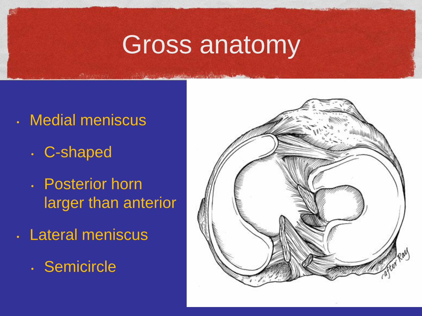
Gross anatomy
• Medial meniscus
• C-shaped
• Posterior horn
larger than anterior
• Lateral meniscus
• Semicircle
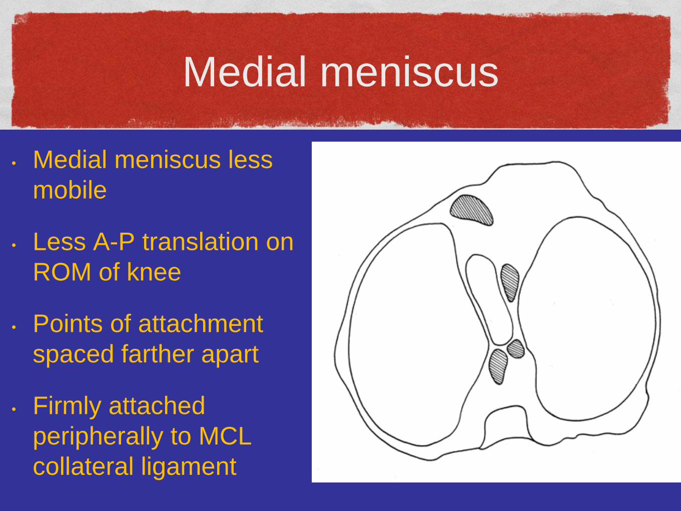
Medial meniscus
• Medial meniscus less
mobile
• Less A-P translation on
ROM of knee
• Points of attachment
spaced farther apart
• Firmly attached
peripherally to MCL
collateral ligament
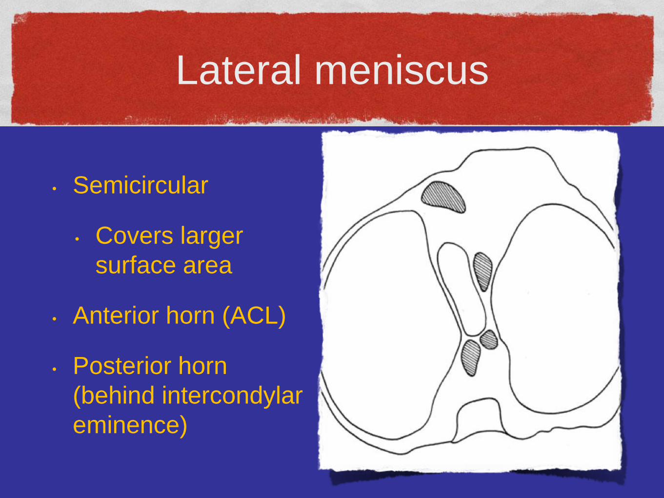
Lateral meniscus
• Semicircular
• Covers larger
surface area
• Anterior horn (ACL)
• Posterior horn
(behind intercondylar
eminence)
Lateral meniscus
• Posterior horn
• Meniscofemoral ligaments
• Posterior horn to lateral aspect of medial
femoral condyle
• Ligament of Humphrey (anterior PCL)
• Ligament of Wrisberg (posterior PCL)
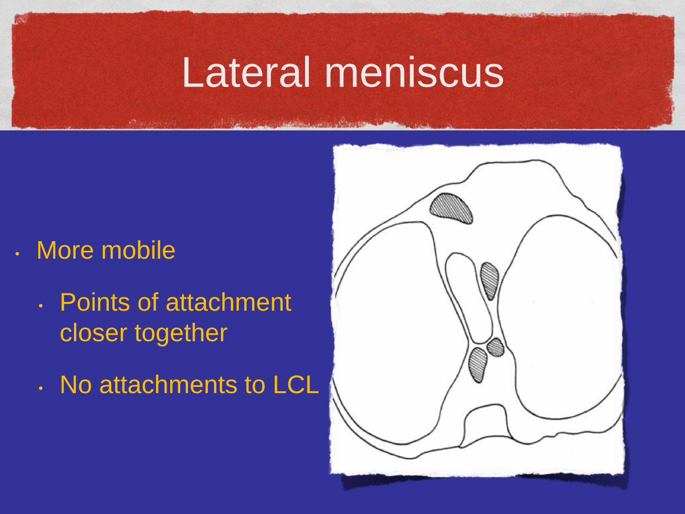
Lateral meniscus
• More mobile
• Points of attachment
closer together
• No attachments to LCL
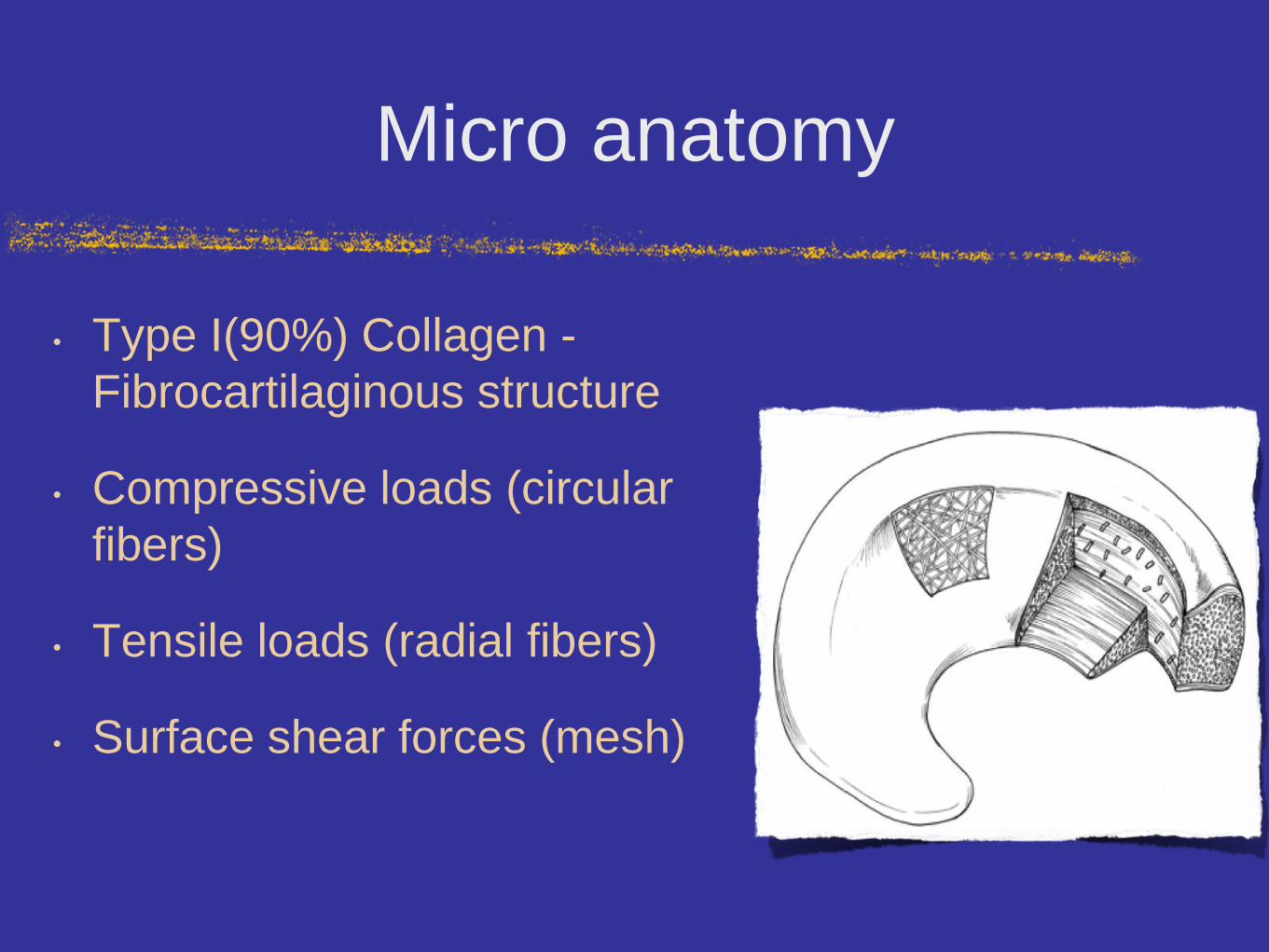
Micro anatomy
• Type I(90%) Collagen -
Fibrocartilaginous structure
• Compressive loads (circular
fibers)
• Tensile loads (radial fibers)
• Surface shear forces (mesh)
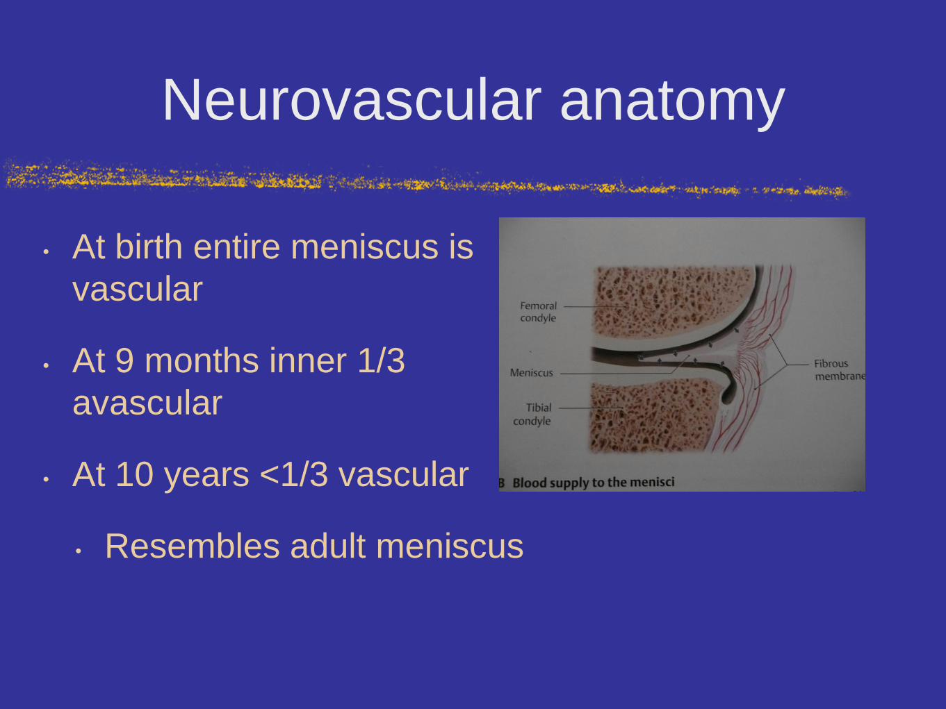
Neurovascular anatomy
• At birth entire meniscus is
vascular
• At 9 months inner 1/3
avascular
• At 10 years <1/3 vascular
• Resembles adult meniscus
Function of meniscus
• Load sharing & shock absorption
• Increased joint congruity
• Static stability
• Propioception
• Hoop stress
Biomechanics
• Total medial meniscectomy
• 50-70% reduction in contact area & 100%
increase in contact stress
• Total lateral meniscectomy
• 40-50% reduction in contact area & 200-300%
increase in contact stress
Epidemiology
• 60-70 per 100,000
• Male 3: female 1
• Male (21-30 yo) & female (11-20yo)
• Degenerative tears in 4-6th decades
Epidemiology
• 1/3 associated with ACL injury
• Acute ACL injury
• Lateral meniscus
• Chronic ACL deficient knee
• Medial meniscus
• Tibial plateau fracture
Physical exam
• Effusion
• Quads atrophy
• ROM
• Joint line tenderness (sensitive)
• Collateral & cruciate ligaments
Special tests
• McMurray & Apley tests
• Adjunct to diagnosis
• Meniscal tear
• Effusion, joint line tenderness, mechanical
symptoms & +ve McMurray test
Mcmurray test
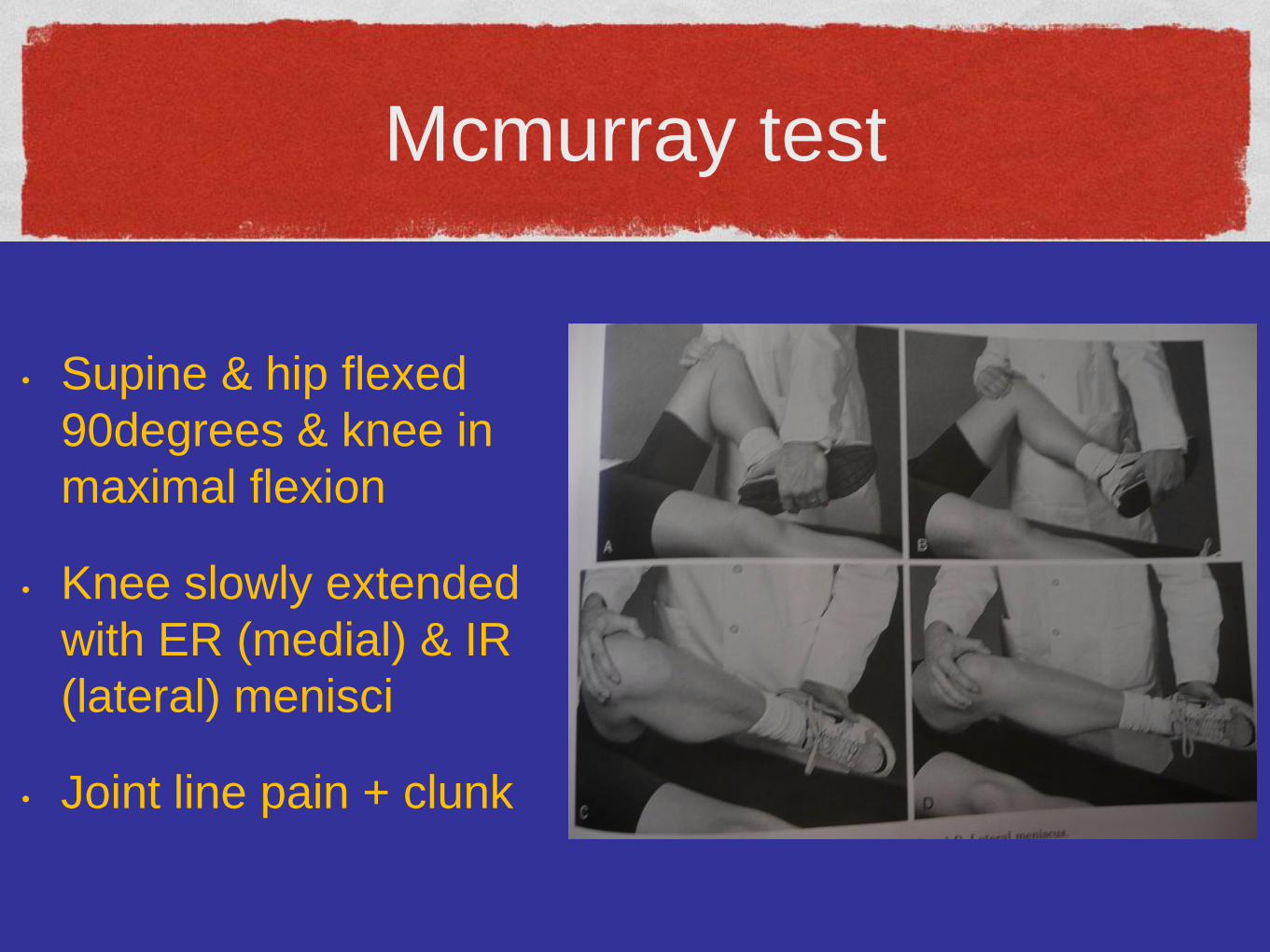
• Supine & hip flexed
90degrees & knee in
maximal flexion
• Knee slowly extended
with ER (medial) & IR
(lateral) menisci
• Joint line pain + clunk
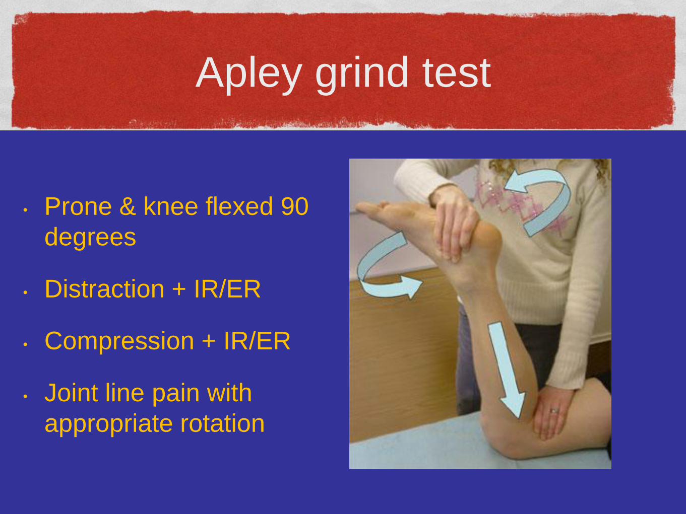
Apley grind test
• Prone & knee flexed 90
degrees
• Distraction + IR/ER
• Compression + IR/ER
• Joint line pain with
appropriate rotation
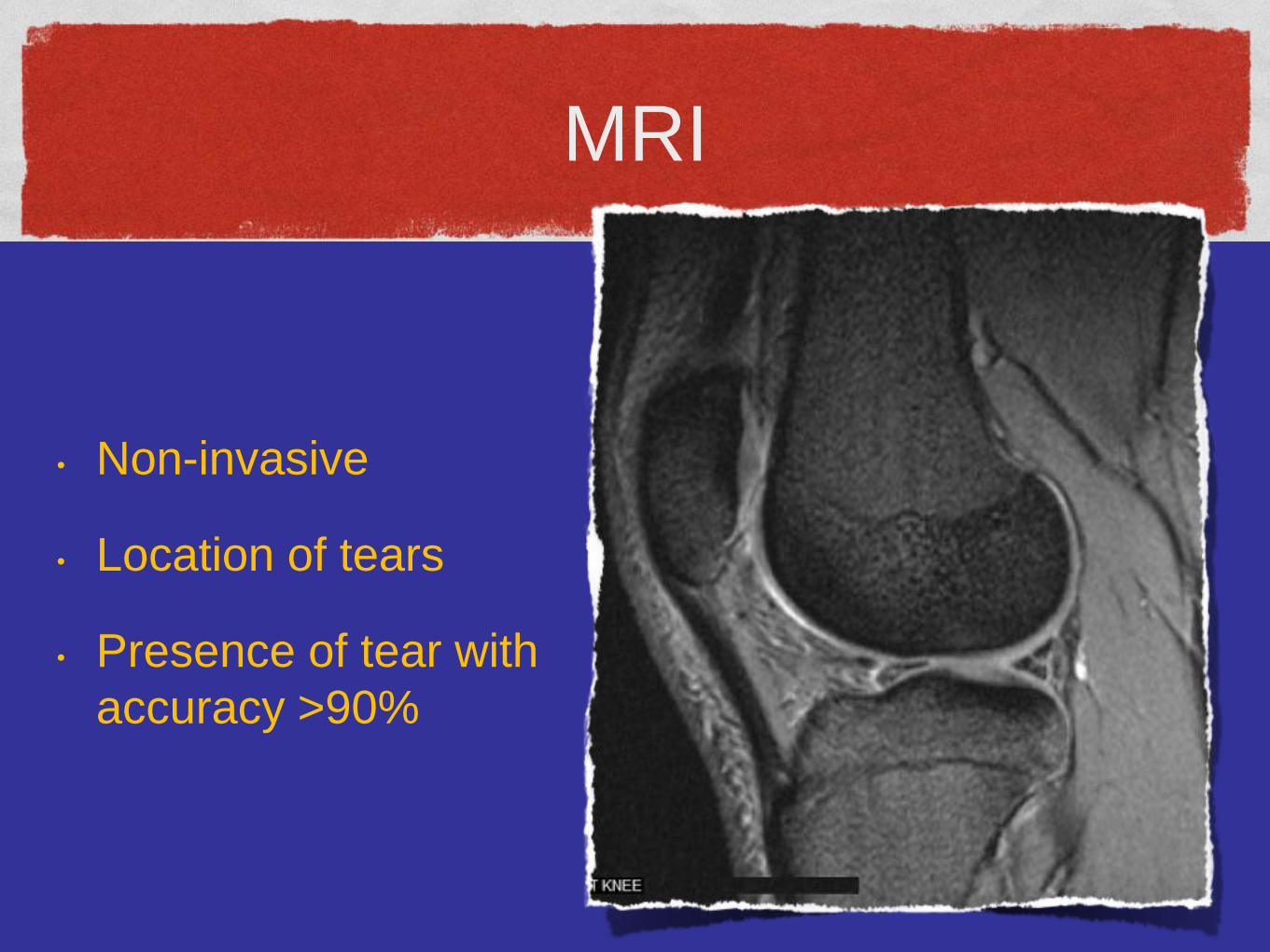
MRI
• Non-invasive
• Location of tears
• Presence of tear with
accuracy >90%
Classification
• Tear in Red zone/Red-White Zone/White Zone
• Unstable Tear/Stable Tear
• Vertical longitudinal, oblique, complex,
transverse (radial), and horizontal
• 80% oblique or vertical longitudinal
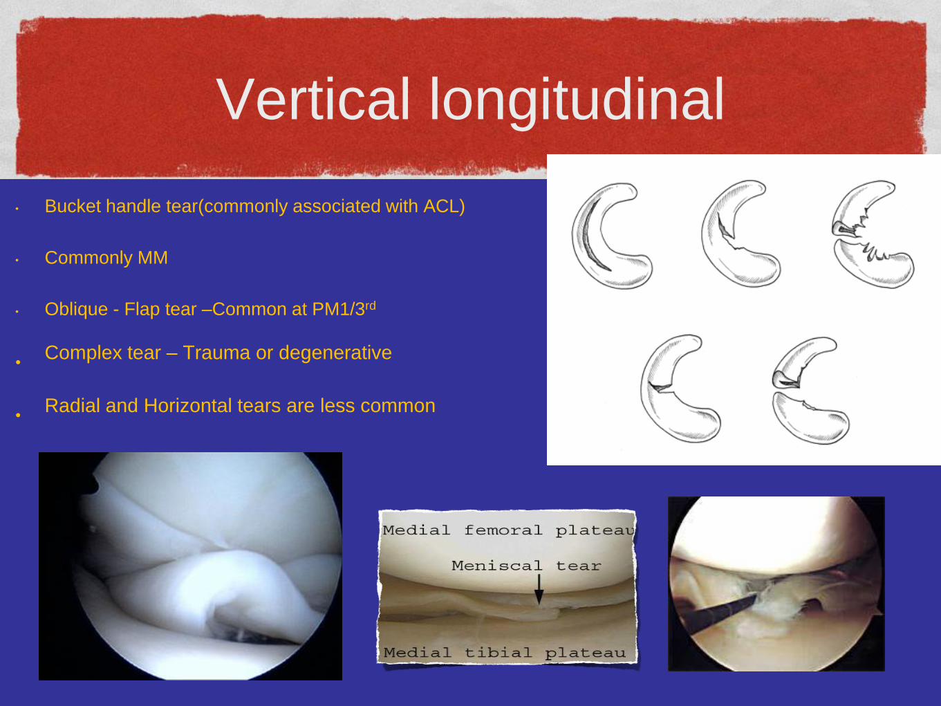
Vertical longitudinal
• Bucket handle tear(commonly associated with ACL)
• Commonly MM
• Oblique - Flap tear –Common at PM1/3rd
•Complex tear – Trauma or degenerative
•Radial and Horizontal tears are less common
Treatment
• Non-operative tx
• Operative tx
• Total vs partial meniscectomy
• Meniscal repair
Non-op tx
• Initial tx symptomatic
• Protected weight bearing
• Modified activity
• Ice
• NSAIDs
Meniscal repair
indications
• Red Zone tears – Bucket handle tears
• Patient Age, characteristic
• Tear characteristics – Reducible tears
Timing of repair
• No firm data on acute vs chronic repair
• Felt that acute repair fares better
• Chronic tears may deform & degenerate
• Difficult anatomic repair
• Location & appearance more impt factors
Age
• No specific age limit
• Most authors suggest partial meniscectomy age >45
• Successful repairs
Ideal repair
• Vertical longitudinal tear of lateral meniscus
• Peripheral zone (red-red)
• ACL injury
• ACL reconstruction
Meniscectomy
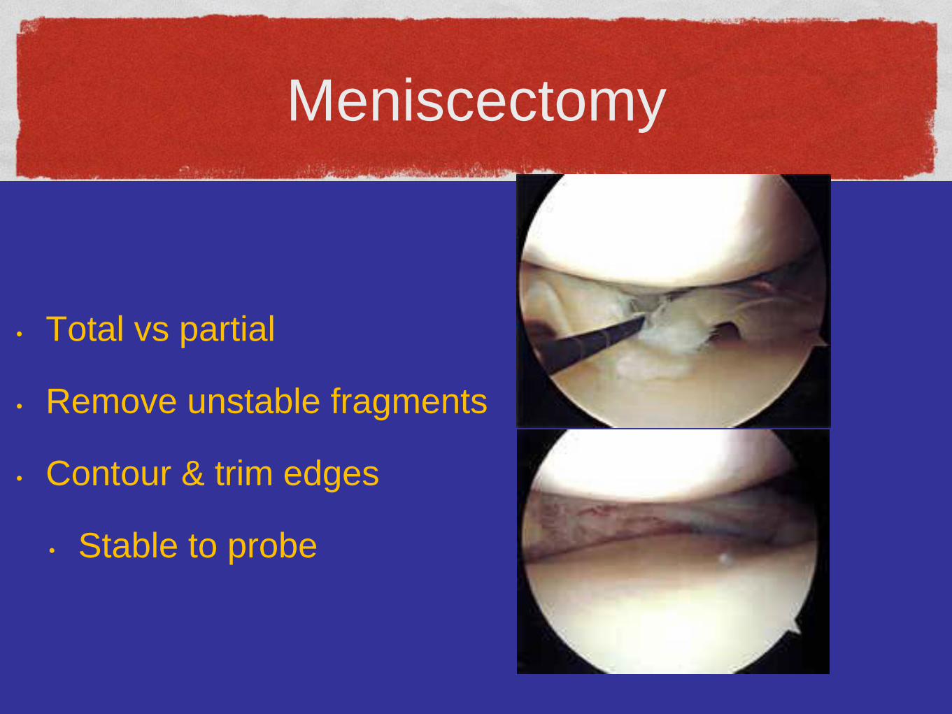
• Total vs partial
• Remove unstable fragments
• Contour & trim edges
• Stable to probe
Meniscal repair
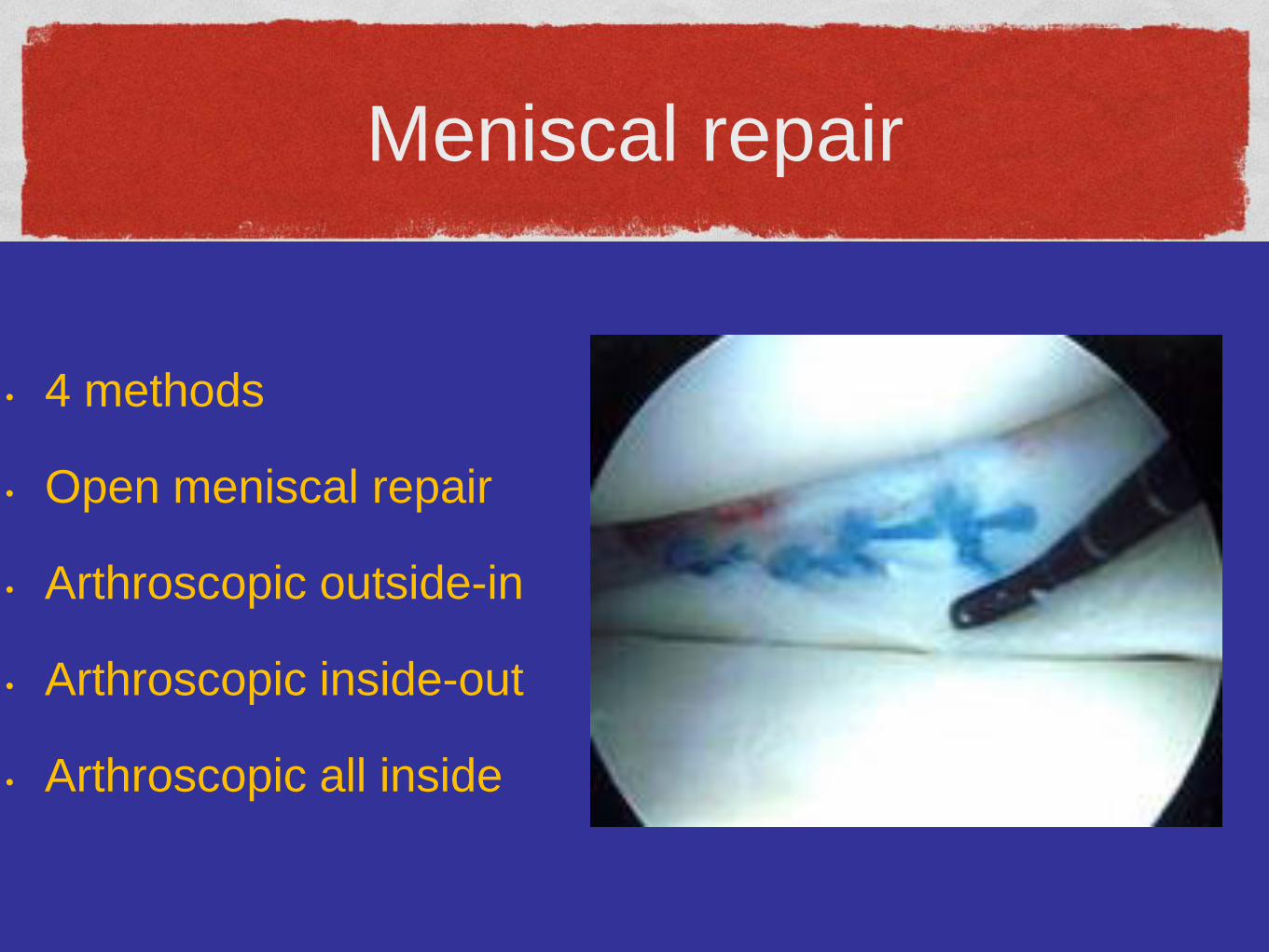
• 4 methods
• Open meniscal repair
• Arthroscopic outside-in
• Arthroscopic inside-out
• Arthroscopic all inside
Open meniscal repair
• Limited indications
• Knee dislocation and multi-ligament
reconstruction
• Longitudinal incision over joint line
• Vertical sutures from capsule into meniscus
Inside-out repair
• Versatile & user friendly
• Excellent healing rates in literature
• Suture passer thru central to peripheral & tied
over capsule
Outside-in repaire
• Minimize risk to peroneal nerve during LM repair
• Tight medial compartment
• Small incisions perpendicular to joint line
• Pass suture and tie knot inside joint
All inside repair
• Least # incisions
• Protect neurovascular structures
• Variety of techniques
• Bioabsorbable anchors and suture tied inside
joint
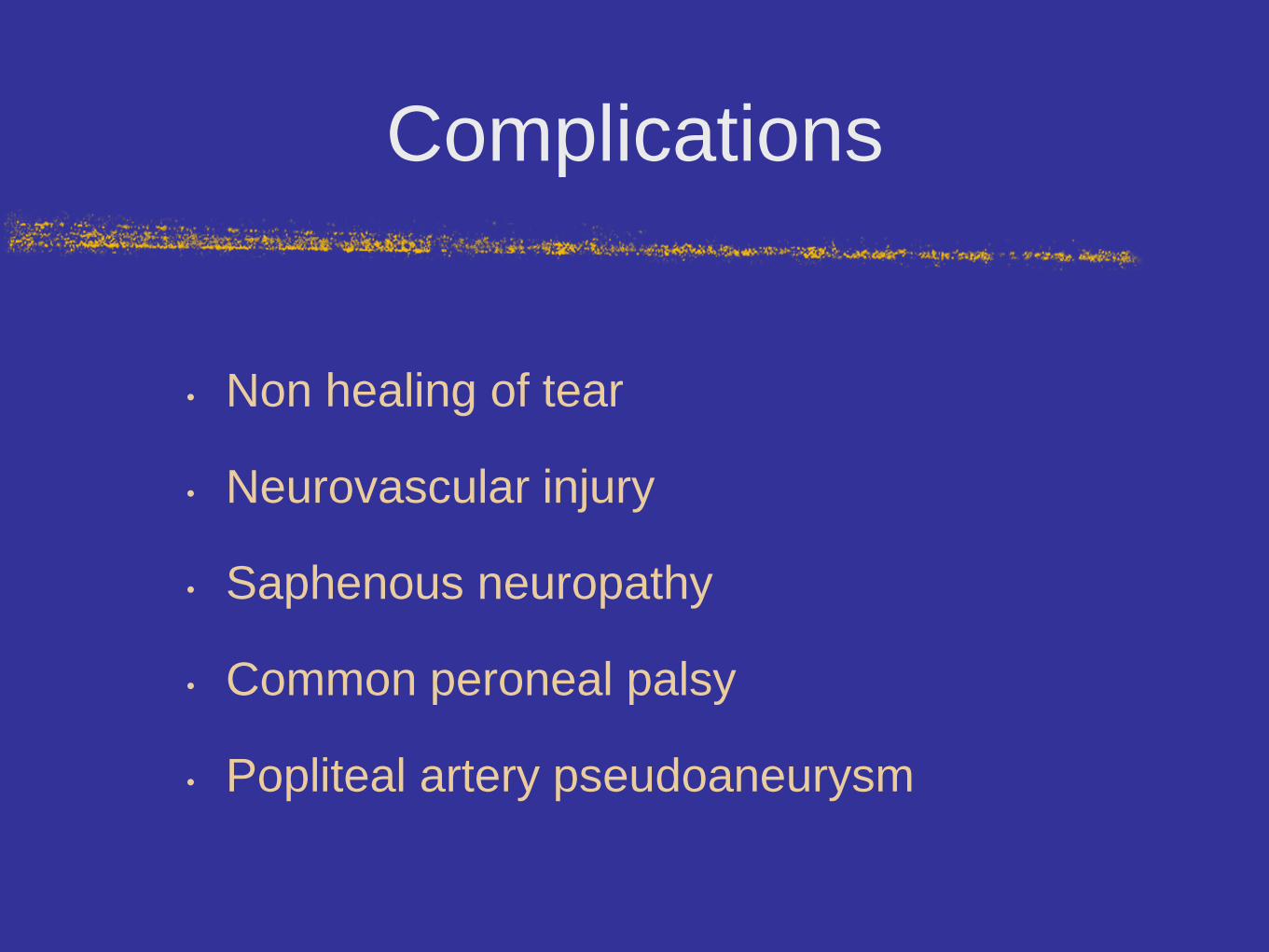
Complications
• Non healing of tear
• Neurovascular injury
• Saphenous neuropathy
• Common peroneal palsy
• Popliteal artery pseudoaneurysm
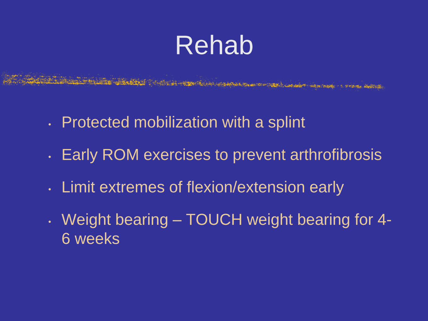
Rehab
• Protected mobilization with a splint
• Early ROM exercises to prevent arthrofibrosis
• Limit extremes of flexion/extension early
• Weight bearing – TOUCH weight bearing for 4-
6 weeks
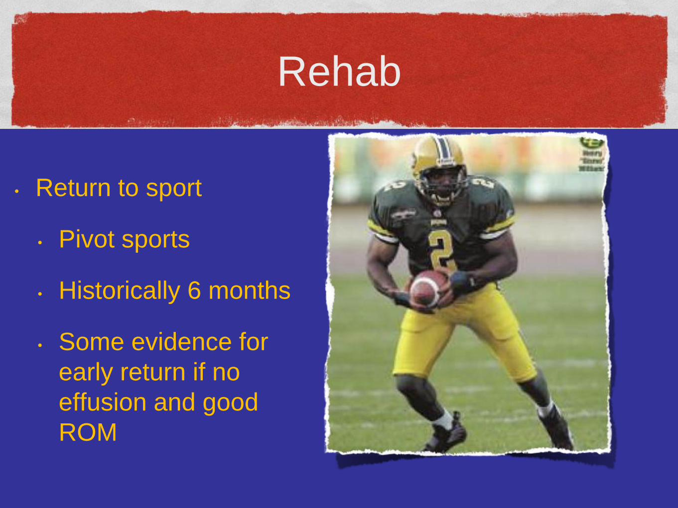
Rehab
• Return to sport
• Pivot sports
• Historically 6 months
• Some evidence for
early return if no
effusion and good
ROM
THANK YOU
THe END