Embed Size (px)
Citation preview
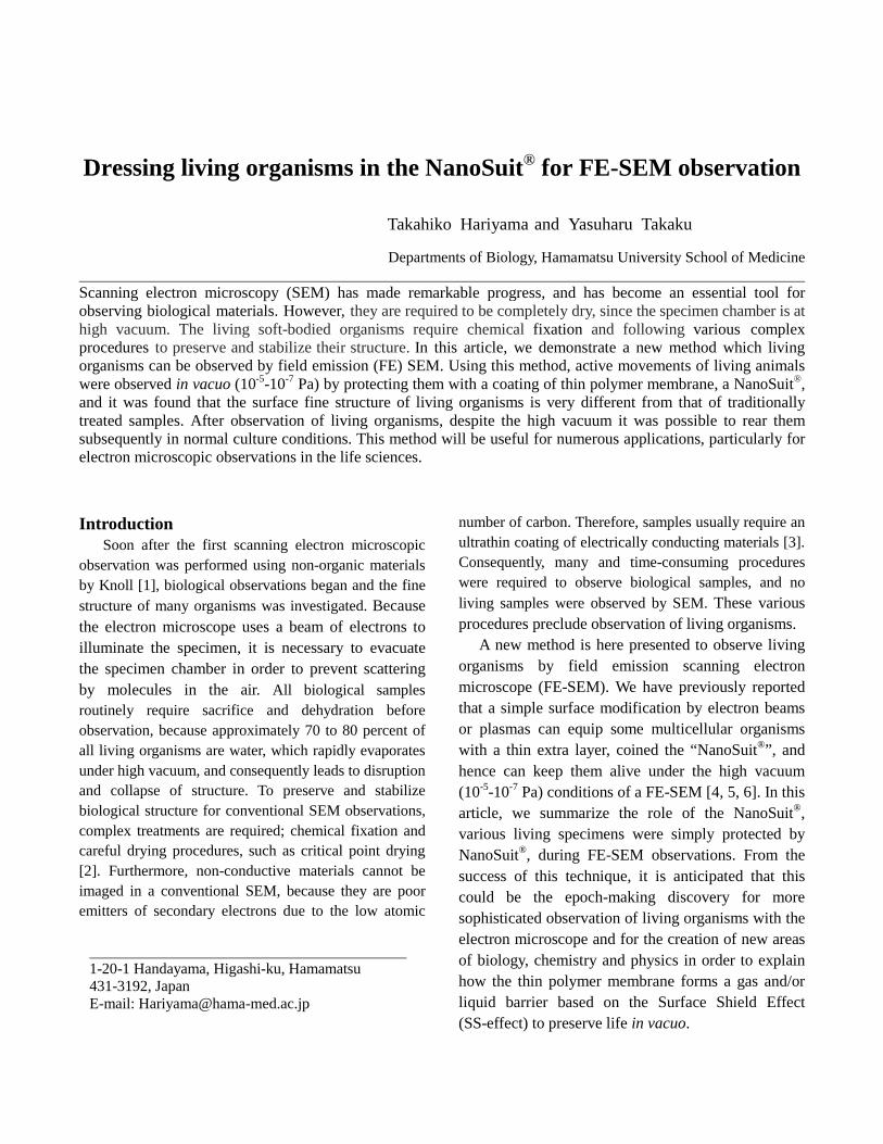
Dressing living organisms in the NanoSuit® for FE-SEM observation
Takahiko Hariyama and Yasuharu Takaku
Departments of Biology, Hamamatsu University School of Medicine Scanning electron microscopy (SEM) has made remarkable progress, and has become an essential tool for observing biological materials. However, they are required to be completely dry, since the specimen chamber is at high vacuum. The living soft-bodied organisms require chemical fixation and following various complex procedures to preserve and stabilize their structure. In this article, we demonstrate a new method which living organisms can be observed by field emission (FE) SEM. Using this method, active movements of living animals were observed in vacuo (10-5-10-7 Pa) by protecting them with a coating of thin polymer membrane, a NanoSuit®, and it was found that the surface fine structure of living organisms is very different from that of traditionally treated samples. After observation of living organisms, despite the high vacuum it was possible to rear them subsequently in normal culture conditions. This method will be useful for numerous applications, particularly for electron microscopic observations in the life sciences.
Introduction Soon after the first scanning electron microscopic observation was performed using non-organic materials by Knoll [1], biological observations began and the fine structure of many organisms was investigated. Because the electron microscope uses a beam of electrons to illuminate the specimen, it is necessary to evacuate the specimen chamber in order to prevent scattering by molecules in the air. All biological samples routinely require sacrifice and dehydration before observation, because approximately 70 to 80 percent of all living organisms are water, which rapidly evaporates under high vacuum, and consequently leads to disruption and collapse of structure. To preserve and stabilize biological structure for conventional SEM observations, complex treatments are required; chemical fixation and careful drying procedures, such as critical point drying [2]. Furthermore, non-conductive materials cannot be imaged in a conventional SEM, because they are poor emitters of secondary electrons due to the low atomic
number of carbon. Therefore, samples usually require an ultrathin coating of electrically conducting materials [3]. Consequently, many and time-consuming procedures were required to observe biological samples, and no living samples were observed by SEM. These various procedures preclude observation of living organisms.
A new method is here presented to observe living organisms by field emission scanning electron microscope (FE-SEM). We have previously reported that a simple surface modification by electron beams or plasmas can equip some multicellular organisms with a thin extra layer, coined the “NanoSuit®”, and hence can keep them alive under the high vacuum (10-5-10-7 Pa) conditions of a FE-SEM [4, 5, 6]. In this article, we summarize the role of the NanoSuit®, various living specimens were simply protected by NanoSuit®, during FE-SEM observations. From the success of this technique, it is anticipated that this could be the epoch-making discovery for more sophisticated observation of living organisms with the electron microscope and for the creation of new areas of biology, chemistry and physics in order to explain how the thin polymer membrane forms a gas and/or liquid barrier based on the Surface Shield Effect (SS-effect) to preserve life in vacuo.
1-20-1 Handayama, Higashi-ku, Hamamatsu 431-3192, Japan E-mail: [email protected]

2
Experimental Experimental living organisms Third-instar larvae (ca. 3 mm in body length) of the fruit fly Drosophila melanogaster (Oregon-R variety) were used. These larvae possess a soft cuticle covered by extracellular substances (ECSs) [7]. To exclude any effects of culture medium, the larvae were washed at 24 ± 1 °C with distilled water several times before the experiment. Fourth-instar larvae of the mosquito Aedes albopictus (ca. 5 mm in body length) and fourth-instar larvae of the mosquito Culex pipiens molestus (ca. 6 mm in body length), collected from puddles and maintained in the same water in which they were found, were also used. These larvae have a soft cuticle not covered by ECS. To exclude any effects of the water, they were transferred to distilled water at 24 ± 1 °C for 2 d before the experiment, with distilled water changes every 12 h. The larvae were rinsed thoroughly in distilled water 1 h before experiments began. The initial weights of single Drosophila and Culex larvae were about 1.2 mg and 2.2 mg, respectively. Five larvae were measured for each trial, and five trials (n = 25) were performed for each species under each condition. The larvae of a chironomid midge, Chironomus yoshimatsui, were collected from the mud of a rice paddy, and then cultured in the muddy water brought from the same paddy. The fourth larval instar (ca. 4 mm in body length) was used. In order to exclude any effects of the water, they were transferred to distilled water at 24 ± 1ºC for two days prior to the experiment, with distilled water changes every 12 h. The larvae were rinsed thoroughly in distilled water one hour before experiments began.
Specimens of the amphipod sandhopper, Talitrus saltator, were collected on a sandy beach in southern Tuscany, transported to the laboratory in plastic boxes containing wet sand, and maintained in an aquarium containing sand moistened with artificial seawater at 20 ± 1ºC. They were fed weekly with dry fish food placed on blotting paper. The experimental animals were rinsed thoroughly in distilled water one hour before experiments began.
Adult specimens of the beetle Lilioceris merdigera were collected and were maintained in the laboratory at 20 ± 1ºC. and fed daily. Individuals chosen for the experiment were rinsed thoroughly in distilled water 1 h prior to experimentation.
Preparation of Tween 20 solutions and sample preparation for FE-SEM to observe living specimens The amphiphilic surfactant compound polyoxyethylene (20) sorbitan monolaurate (Tween 20; Wako Pure Chemical Industries) was used for all the experiments to mimic natural extracellular substances (ECS) [7]. To form the NanoSuit®, the organisms were dipped into 1% (v/v) Tween 20 solution dissolved in distilled water for 1 min, blotted briefly on a dry filter paper to remove excess solution. Then the living specimen was introduced into the SEM to construct a Tween 20 film, without performing any traditional treatments such as chemical fixation or dehydration. Preparation for standard scanning electron microscopy. For standard SEM observation, animals were prefixed with 2% glutaraldehyde in 0.1 M cacodylate buffer (pH 7.4) and postfixed in 1% OsO4 in the same buffer. The specimens were then dehydrated, freeze dried (JFD300, JEOL), and ultra-thin coated with OsO4 (PMC-5000, Meiwa). Microscopy For FE-SEM observations, we used a JEM7100F (JEOL) operated at acceleration voltages of 1.0 kV. The vacuum level of the observation chamber was 10-5 to 10-7 Pa. The detector for secondary electron was a mixture of signal with upper and lower detectors. Other details in conditions were as follows: working distance, 8 mm; aperture size, w 100 mm; scan speed, each beam is 10–15 frames per second. To record the dynamic movements of animals, imaging data from the SEM were directly transferred to a Hi-band digital-formatted video recorder (SONY, BDZ-EW500). Transmission electron microscopy (TEM) observations were carried out using a JEM-1220 (JEOL) Results High vacuum tolerant animal In order to find the nature tolerance of living organisms to high vacuum condition, we introduced numerous living organisms belonging to various taxa directly into the SEM to see how long they survived under high vacuum (10−5 to 10−7 Pa). Almost all of their structural integrity was completely destroyed by

3
rapid evaporation under high vacuum (Fig.1a-c). We, however, found that Drosophila larvae tolerated the high vacuum well. Although they have a soft cuticle covered by extracellular substances (ECSs) [7] they continued to actively move around for 60 min under the SEM (n = 25) (Fig.1d) and subsequently some of them (n=18) developed normally.
Fig. 1. Living organisms were introduced directly into a FE-SEM. Many of them were dead (a-c) except the Drosophila larva (d).
Fig. 2. (a-c) A living Drosophila larva was exposed in high vacuo for 60 min. (e-g) Before SEM observation, a different larva (light micrograph in (f) was introduced into the observation chamber without electron-beam irradiation. It was collapsed thoroughly when observed by SEM subsequently (g). TEM images of vertical sections through the surface of each animal are shown in (d) and (h). The layer between the arrowheads in (d) shows the newly formed outer membrane, not present in (h).
Using Drosophila larva, no apparent structural changes occurred (Fig. 2 a–c). However, when control larvae (Fig. 2f) were placed in the SEM observation chamber for 60 min at an identical vacuum level, but without concurrent electron-beam radiation (Fig.2e), subsequent SEM observations revealed they were all dead and structurally badly distorted (Fig. 2g).
Transmission electron microscopy (TEM) showed that animals subjected to SEM electron bombardment immediately produce an extra thin layer (ca.50 to 100 nm thickness) over their surface (Fig. 2d). No such layer was detected in animals that had been exposed in vacuo for the same time but without electron bombardment (Fig. 2h). Electron-beam and/or plasma irradiation Those results described above led to the hypothesis that electron-beam irradiation [8] enhanced cross-linking within the ECS to form a durable polymer on the surface and that this polymer increased resistance to vacuum conditions. To test this hypothesis, surface polymerization was achieved by a plasma irradiation. The ionized particles of plasma provide the energy necessary to initiate polymer formation, enabling uniform coating of surfaces with solvent-insoluble polymers [9]. Although plasma polymerization so far has been used only in creating new inert industrial materials or modification of their surfaces [10], we applied this technique to the ECS of living animals to construct a protective surface barrier referred to as the NanoSuit®. Plasma-irradiated specimens left in vacuo for 60 min before SEM observation clearly showed features similar to specimen electron-beam irradiation by SEM ab initio: They moved continuously, seemingly unharmed without problems of electrical charging. The weight of each larva was measured just before the experiment and 60 min later following exposure to high vacuum. Nonirradiated control larvae and larvae pretreated by plasma radiation showed mean body weight decreases of 56.3 ± 6.0% and 8.7 ± 3.8%, respectively (n = 25), demonstrating the effectiveness of the surface shield effect by the surface membrane formed by plasma irradiation. Mimicking ECS for other animals To mimic the ECS which could create the NanoSuit®, solutions including amphiphilic molecules were then tested. One of the best results was obtained with a solution of Tween 20, a nontoxic compound commonly used in biological experiments [11]. To test the barrier properties of the nano-suit made by this solution, the surfaces of several different animals previously unable to survive SEM exposure were provided exogenous materials by immersing them in 1% Tween 20 solution before electron or plasma irradiation. All tested animals survived, including the flatworm Dugesia japonica, the ant Pristomyrmex punctatus, and the amphipod . Talitrus saltator. Fig. 2 F–O and Fig. S2 show typical results obtained with larvae of the Asian tiger mosquito,
4
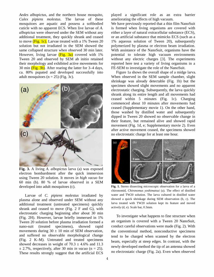
Aedes albopictus, and the northern house mosquito, Culex pipiens molestus. The larvae of these mosquitoes are aquatic and possess a softbodied cuticle with no apparent ECS. When live larvae of A. albopictus were observed under the SEM without any additional treatment, they quickly shrank and ceased to move (Fig. 1c). Larvae treated with a 1% Tween 20 solution but not irradiated in the SEM showed the same collapsed structure when observed 30 min later. However, living larvae (Fig. 3a) covered with 1% Tween 20 and observed by SEM ab initio retained their morphology and exhibited active movements for 30 min (Fig. 3b). After rearing the latter for 3–4 days, ca. 80% pupated and developed successfully into adult mosquitoes (n = 25) (Fig. 3c).
Fig. 3. A living A. albopictus larva (a) was exposed electron bombardment after the quick immersion using Tween 20 solution. It moves in high vacuo for 60 min (b). 80 % of larvae observed in a SEM developed into adult mosquitoes (c). Larvae of C. pipiens molestus irradiated by plasma alone and observed under SEM without any additional treatment (untreated specimens) quickly shrank and ceased to move (Fig. 2 F and G), with electrostatic charging beginning after about 30 min (Fig. 2H). However, larvae briefly immersed in 1% Tween 20 solution before plasma irradiation formed a nano-suit (treated specimens), showed rapid movements during 30 ± 10 min of SEM observation, and suffered no observable morphological change (Fig. 2 K–M). Untreated and treated specimens showed decreases in weight of 70.3 ± 4.6% and 11.3 ± 2.7%, respectively, after 30 min in vacuo (n = 25). These results strongly suggest that the artificial ECS
played a significant role as an extra barrier ameliorating the effects of high vacuum. We have previously reported that a thin film NanoSuit is formed when living organisms are covered with either a layer of natural extracellular substance (ECS), or an artificial substance that mimicks ECS (such as a 1% aqueous solution of Tween 20), subsequently polymerized by plasma or electron beam irradiation. With assistance of the NanoSuit, organisms have the potential to tolerate high vacuum environments without any electric charges [3]. The experiments reported here test a variety of living organisms in a FE-SEM to investigate the role of the NanoSuit.
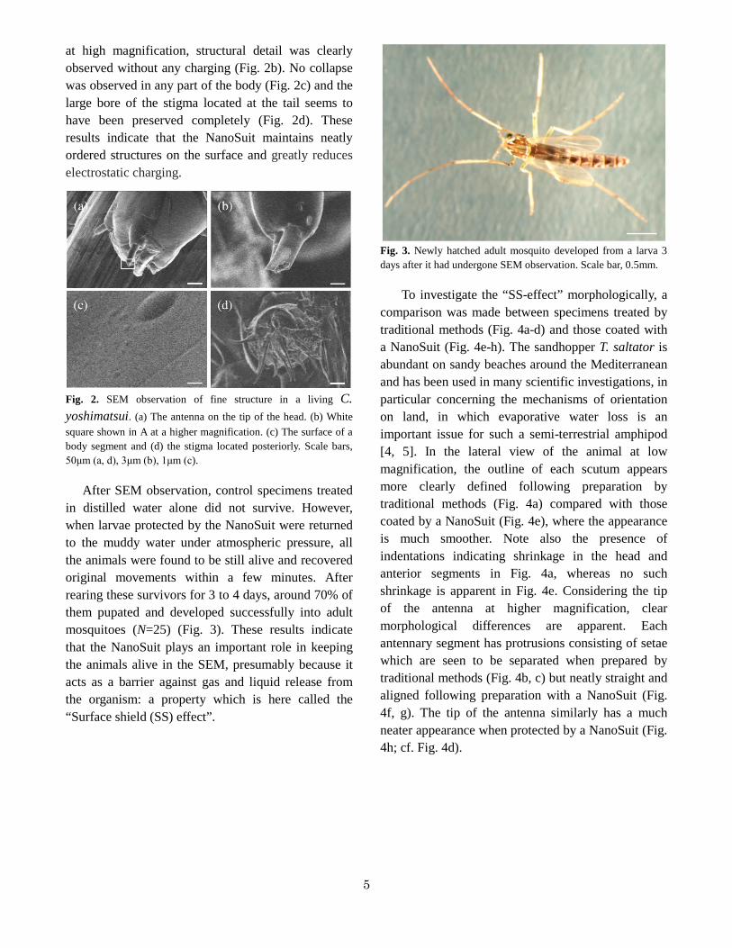
Figure 1a shows the overall shape of a midge larva. When observed in the SEM sample chamber, slight shrinkage was already detectable (Fig. 1b) but the specimen showed slight movements and no apparent electrostatic charging. Subsequently, the larva quickly shrank along its entire length and all movements had ceased within 5 minutes (Fig. 1c). Charging commenced about 10 minutes after movements had ceased (Supplementary movie 1). On the other hand, those washed by distilled water and subsequently dipped in Tween 20 showed no observable change in their feature, but remained alive and showed rapid movement (Fig. 1d, e; Supplementary movie 2). Even after active movement ceased, the specimens showed no electrostatic charge for at least one hour.
Fig. 1. Stereo dissecting microscopic observation for a larva of a chironomid, Chironomus yoshimatsui (a). The effect of distilled water and TW20 solution. The larva cultured in distilled water showed a quick shrinkage during SEM observation (b, c). The larva treated with TW20 solution kept its feature and moved actively (d, e). Scale bar, 0.3mm.
To investigate what happens to fine structure when an organism is covered with a Tween 20 NanoSuit, conduct careful observations were made (Fig. 2). With the conventional method, nonconductive specimens tend to be charged when scanned by the electron beam, especially at steep edges. In contrast, with the newly developed method the tip of an antenna showed no electrostatic charge (Fig. 2a). Even when observed
5
at high magnification, structural detail was clearly observed without any charging (Fig. 2b). No collapse was observed in any part of the body (Fig. 2c) and the large bore of the stigma located at the tail seems to have been preserved completely (Fig. 2d). These results indicate that the NanoSuit maintains neatly ordered structures on the surface and greatly reduces electrostatic charging.
Fig. 2. SEM observation of fine structure in a living C. yoshimatsui. (a) The antenna on the tip of the head. (b) White square shown in A at a higher magnification. (c) The surface of a body segment and (d) the stigma located posteriorly. Scale bars, 50μm (a, d), 3μm (b), 1μm (c).
After SEM observation, control specimens treated
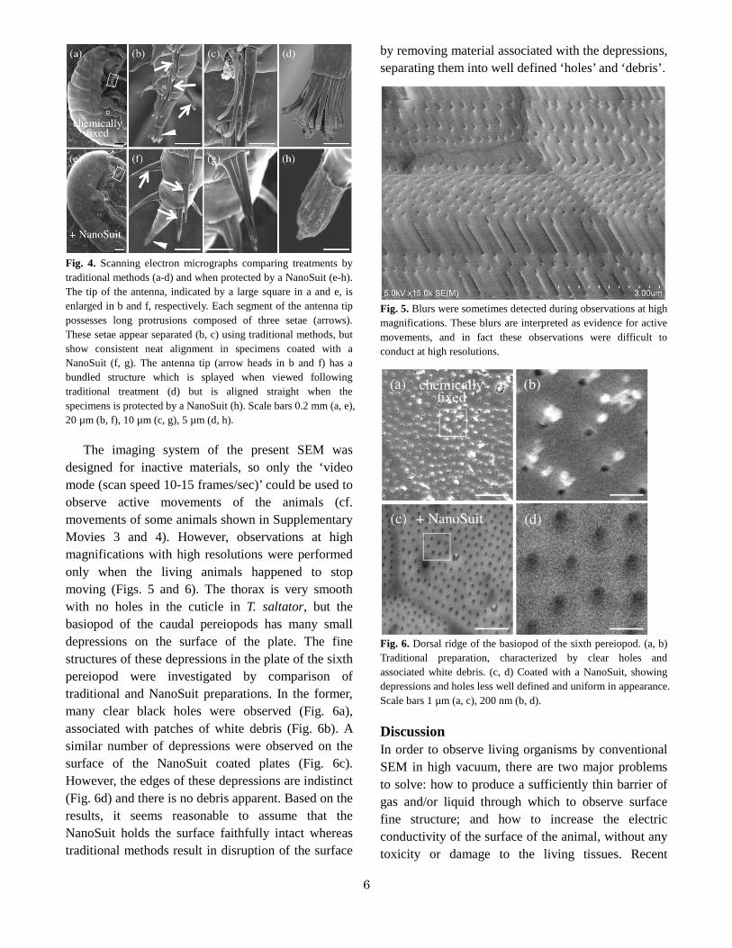
in distilled water alone did not survive. However, when larvae protected by the NanoSuit were returned to the muddy water under atmospheric pressure, all the animals were found to be still alive and recovered original movements within a few minutes. After rearing these survivors for 3 to 4 days, around 70% of them pupated and developed successfully into adult mosquitoes (N=25) (Fig. 3). These results indicate that the NanoSuit plays an important role in keeping the animals alive in the SEM, presumably because it acts as a barrier against gas and liquid release from the organism: a property which is here called the “Surface shield (SS) effect”.
Fig. 3. Newly hatched adult mosquito developed from a larva 3 days after it had undergone SEM observation. Scale bar, 0.5mm.
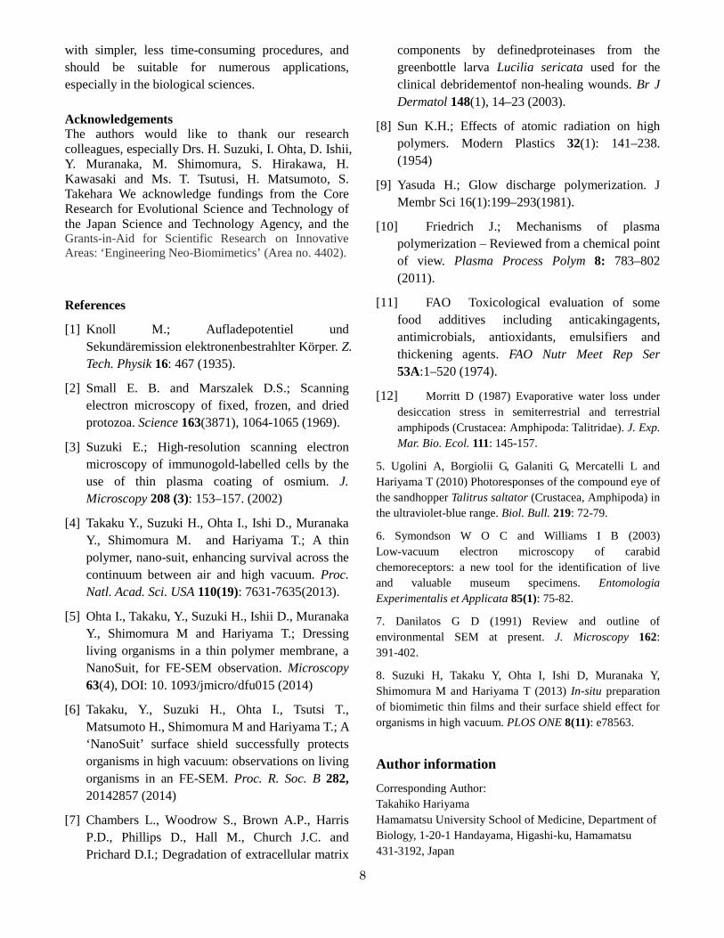
To investigate the “SS-effect” morphologically, a
comparison was made between specimens treated by traditional methods (Fig. 4a-d) and those coated with a NanoSuit (Fig. 4e-h). The sandhopper T. saltator is abundant on sandy beaches around the Mediterranean and has been used in many scientific investigations, in particular concerning the mechanisms of orientation on land, in which evaporative water loss is an important issue for such a semi-terrestrial amphipod [4, 5]. In the lateral view of the animal at low magnification, the outline of each scutum appears more clearly defined following preparation by traditional methods (Fig. 4a) compared with those coated by a NanoSuit (Fig. 4e), where the appearance is much smoother. Note also the presence of indentations indicating shrinkage in the head and anterior segments in Fig. 4a, whereas no such shrinkage is apparent in Fig. 4e. Considering the tip of the antenna at higher magnification, clear morphological differences are apparent. Each antennary segment has protrusions consisting of setae which are seen to be separated when prepared by traditional methods (Fig. 4b, c) but neatly straight and aligned following preparation with a NanoSuit (Fig. 4f, g). The tip of the antenna similarly has a much neater appearance when protected by a NanoSuit (Fig. 4h; cf. Fig. 4d).
6
Fig. 4. Scanning electron micrographs comparing treatments by traditional methods (a-d) and when protected by a NanoSuit (e-h). The tip of the antenna, indicated by a large square in a and e, is enlarged in b and f, respectively. Each segment of the antenna tip possesses long protrusions composed of three setae (arrows). These setae appear separated (b, c) using traditional methods, but show consistent neat alignment in specimens coated with a NanoSuit (f, g). The antenna tip (arrow heads in b and f) has a bundled structure which is splayed when viewed following traditional treatment (d) but is aligned straight when the specimens is protected by a NanoSuit (h). Scale bars 0.2 mm (a, e), 20 µm (b, f), 10 µm (c, g), 5 µm (d, h).
The imaging system of the present SEM was designed for inactive materials, so only the ‘video mode (scan speed 10-15 frames/sec)’ could be used to observe active movements of the animals (cf. movements of some animals shown in Supplementary Movies 3 and 4). However, observations at high magnifications with high resolutions were performed only when the living animals happened to stop moving (Figs. 5 and 6). The thorax is very smooth with no holes in the cuticle in T. saltator, but the basiopod of the caudal pereiopods has many small depressions on the surface of the plate. The fine structures of these depressions in the plate of the sixth pereiopod were investigated by comparison of traditional and NanoSuit preparations. In the former, many clear black holes were observed (Fig. 6a), associated with patches of white debris (Fig. 6b). A similar number of depressions were observed on the surface of the NanoSuit coated plates (Fig. 6c). However, the edges of these depressions are indistinct (Fig. 6d) and there is no debris apparent. Based on the results, it seems reasonable to assume that the NanoSuit holds the surface faithfully intact whereas traditional methods result in disruption of the surface
by removing material associated with the depressions, separating them into well defined ‘holes’ and ‘debris’.
Fig. 5. Blurs were sometimes detected during observations at high magnifications. These blurs are interpreted as evidence for active movements, and in fact these observations were difficult to conduct at high resolutions.
Fig. 6. Dorsal ridge of the basiopod of the sixth pereiopod. (a, b) Traditional preparation, characterized by clear holes and associated white debris. (c, d) Coated with a NanoSuit, showing depressions and holes less well defined and uniform in appearance. Scale bars 1 µm (a, c), 200 nm (b, d).
Discussion In order to observe living organisms by conventional SEM in high vacuum, there are two major problems to solve: how to produce a sufficiently thin barrier of gas and/or liquid through which to observe surface fine structure; and how to increase the electric conductivity of the surface of the animal, without any toxicity or damage to the living tissues. Recent

7
research to improve techniques such as low-vacuum scanning electron microscopy [6] and use of an environmental scanning electron microscope (ESEM) have been developed [7]. However, although both are elegant techniques, low-vacuum SEMs allow semi-wet samples to be imaged without coating but are unable to attain the resolution of conventional SEMs. ESEMs, being wet or contained in low vacuum or gas, also can be used to observe specimens without coating and produce better images than a low-vacuum SEM. However, it is not reliable enough to investigate the movements of living organisms at this level of resolution. We previously found that, with the assistance of a polymerized thin film NanoSuit, organisms possess the potential to tolerate high vacuum environments without any electrical charges [3]. The advance that we have made is to preserve life in high vacuum long enough to observe active movements of living specimens and at good resolution. The reason for improved clarity in fine structure seen with living specimens is due to a surface shield effect (SS-effect) caused by the thin NanoSuit film on the surface of the animal in vacuo.
In the present research, in addition to the apparent barrier effect, no electrostatic charging was observed on any of the animals during their active movements. Animals treated in distilled water alone showed electrostatic charge build-up quickly after they ceased moving (Fig. 1b, c; Supplementary movie 1). In contrast, charging commenced about an hour after movements ceased under NanoSuit protection. Traditionally, biological samples are coated with ultrathin electrical conductors such as gold, palladium, platinum, or osmium, to prevent the accumulation of electrostatic charge at the surface [2]. The living specimens in the present study were observed at good resolution and with no electrostatic charging, which suggests that living organisms would have their own electrical conductors and/or possess certain properties on the surface which inhibit charging. The NanoSuit seems somehow able to preserve life and to prolong the charge-free condition.
Until our discovery, most scientists believed that traditional viewing with the SEM is close to the true appearance of the surface of living animals. However the structures used as examples in Figures 4 and 6 showed many differences from the images of the
living specimens. These morphological differences apparently result from the presence of the NanoSuit on the surface of the living animal providing the SS-effect. One intriguing apparent contradiction here is the appearance of well separated setae in the traditional method (Fig. 4c, g), which seems to show more detail than in the NanoSuit condition where the setae adhere to each other (Fig. 4d, h), implying a more poorly resolved structure. However, the differences suggest that, in the method presented here, the natural surface structure of the living organism is conserved and strongly supports the hypothesis that the NanoSuit is able to preserve the “real life appearance” down to electron microscopic details.
At present, however, the NanoSuit component Tween 20 is not omnipotent. It is obvious that, other than the Tween 20 solution, much more suitable components must be found for each species to live longer in high vacuum. In a different study, this research group has proposed other possible candidates for forming a NanoSuit, and various monomer solutions were fabricated into thin films by plasma polymerization [8].
It is also necessary to investigate whether or not the SS-effect is accomplished by a combination of different substances including unknown compounds which might be produced by the living animal itself. Nevertheless, it is apparent that the NanoSuit SS-effect preserves the morphology of the living animal, decreasing the evaporation rate of gas and/or liquid and facilitate electrical conductivity in the electron beam. When the many intriguing aspects of the SS-effect have been resolved, it is easy to predict that this may be the start of a new era of improvement in the depth of our understanding not only in biology but also in many other fields of science. Concluding remarks The NanoSuit plays an important role in keeping animals alive in the FE-SEM, a new method permitting the use of high vacuum and high resolution of innate fine structurel with living specimens at high magnification imaging (~x500,000). Since the NanoSuit holds the organism surface faithfully intact with high resolution imaging, such a life barrier technique will be a desirable tool for all future work with SEMs to observe real images of living organisms
8
with simpler, less time-consuming procedures, and should be suitable for numerous applications, especially in the biological sciences. Acknowledgements The authors would like to thank our research colleagues, especially Drs. H. Suzuki, I. Ohta, D. Ishii, Y. Muranaka, M. Shimomura, S. Hirakawa, H. Kawasaki and Ms. T. Tsutusi, H. Matsumoto, S. Takehara We acknowledge fundings from the Core Research for Evolutional Science and Technology of the Japan Science and Technology Agency, and the Grants-in-Aid for Scientific Research on Innovative Areas: ‘Engineering Neo-Biomimetics’ (Area no. 4402).
References
[1] Knoll M.; Aufladepotentiel und Sekundäremission elektronenbestrahlter Körper. Z. Tech. Physik 16: 467 (1935).
[2] Small E. B. and Marszalek D.S.; Scanning electron microscopy of fixed, frozen, and dried protozoa. Science 163(3871), 1064-1065 (1969).
[3] Suzuki E.; High-resolution scanning electron microscopy of immunogold-labelled cells by the use of thin plasma coating of osmium. J. Microscopy 208 (3): 153–157. (2002)
[4] Takaku Y., Suzuki H., Ohta I., Ishi D., Muranaka Y., Shimomura M. and Hariyama T.; A thin polymer, nano-suit, enhancing survival across the continuum between air and high vacuum. Proc. Natl. Acad. Sci. USA 110(19): 7631-7635(2013).
[5] Ohta I., Takaku, Y., Suzuki H., Ishii D., Muranaka Y., Shimomura M and Hariyama T.; Dressing living organisms in a thin polymer membrane, a NanoSuit, for FE-SEM observation. Microscopy 63(4), DOI: 10. 1093/jmicro/dfu015 (2014)
[6] Takaku, Y., Suzuki H., Ohta I., Tsutsi T., Matsumoto H., Shimomura M and Hariyama T.; A ‘NanoSuit’ surface shield successfully protects organisms in high vacuum: observations on living organisms in an FE-SEM. Proc. R. Soc. B 282, 20142857 (2014)
[7] Chambers L., Woodrow S., Brown A.P., Harris P.D., Phillips D., Hall M., Church J.C. and Prichard D.I.; Degradation of extracellular matrix
components by definedproteinases from the greenbottle larva Lucilia sericata used for the clinical debridementof non-healing wounds. Br J Dermatol 148(1), 14–23 (2003).
[8] Sun K.H.; Effects of atomic radiation on high polymers. Modern Plastics 32(1): 141–238. (1954)
[9] Yasuda H.; Glow discharge polymerization. J Membr Sci 16(1):199–293(1981).
[10] Friedrich J.; Mechanisms of plasma polymerization – Reviewed from a chemical point of view. Plasma Process Polym 8: 783–802 (2011).
[11] FAO Toxicological evaluation of some food additives including anticakingagents, antimicrobials, antioxidants, emulsifiers and thickening agents. FAO Nutr Meet Rep Ser 53A:1–520 (1974).
[12] Morritt D (1987) Evaporative water loss under desiccation stress in semiterrestrial and terrestrial amphipods (Crustacea: Amphipoda: Talitridae). J. Exp. Mar. Bio. Ecol. 111: 145-157.
5. Ugolini A, Borgiolii G, Galaniti G, Mercatelli L and Hariyama T (2010) Photoresponses of the compound eye of the sandhopper Talitrus saltator (Crustacea, Amphipoda) in the ultraviolet-blue range. Biol. Bull. 219: 72-79.
6. Symondson W O C and Williams I B (2003) Low-vacuum electron microscopy of carabid chemoreceptors: a new tool for the identification of live and valuable museum specimens. Entomologia Experimentalis et Applicata 85(1): 75-82.
7. Danilatos G D (1991) Review and outline of environmental SEM at present. J. Microscopy 162: 391-402.
8. Suzuki H, Takaku Y, Ohta I, Ishi D, Muranaka Y, Shimomura M and Hariyama T (2013) In-situ preparation of biomimetic thin films and their surface shield effect for organisms in high vacuum. PLOS ONE 8(11): e78563.
Author information Corresponding Author: Takahiko Hariyama Hamamatsu University School of Medicine, Department of Biology, 1-20-1 Handayama, Higashi-ku, Hamamatsu 431-3192, Japan