Embed Size (px)
Citation preview
Dr.Aida Fadhel Biawi2013
Mitosis, Genotype and Phenotype
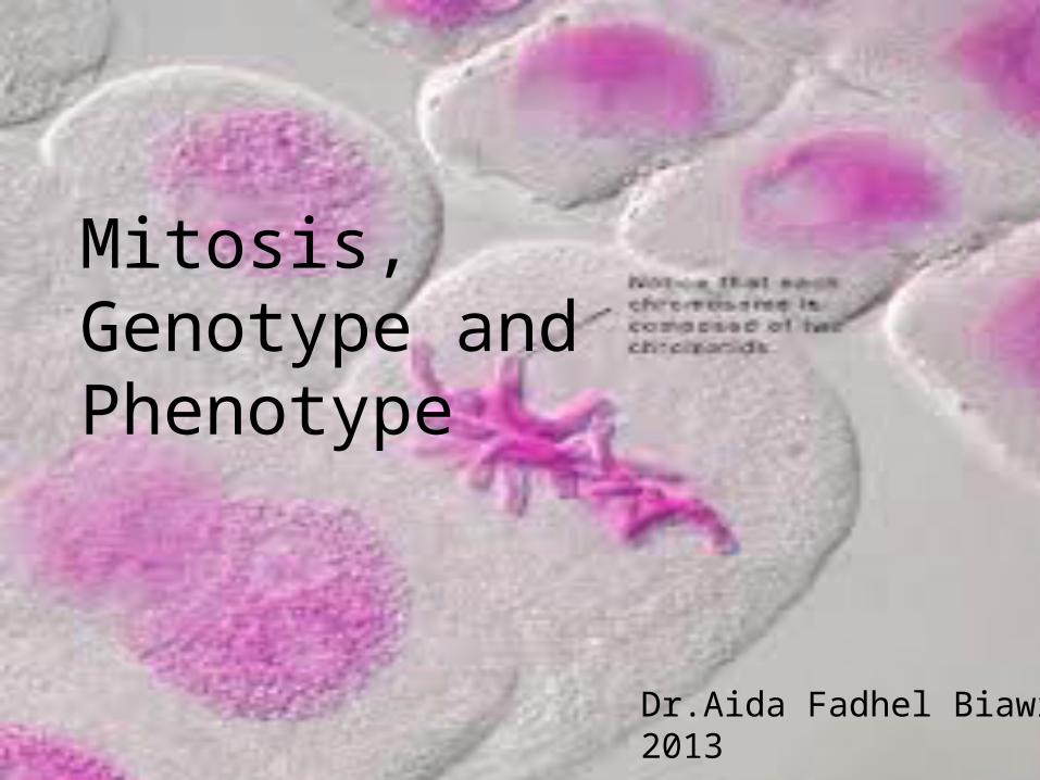
Phases of the Cell Cycle• The cell cycle consists of
– Interphase – normal cell activity– The mitotic phase – cell division ??
INTERPHASE
Growth
G 1(DNA synthesis)
Growth
G2
Cel
l Div
sion
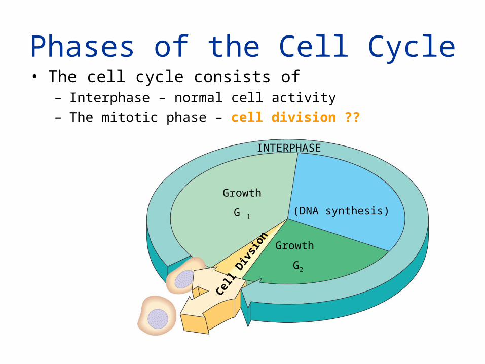
- Reproduction.
- Growth and development.
- Tissue renewal.
What is the benefits of the cell division??
Is all cells still dividing after maturation??
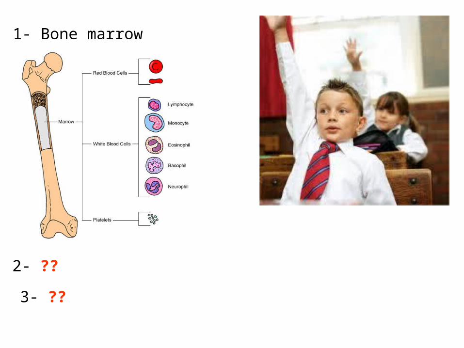
1- Bone marrow
2- ??
3- ??
Cell Division
• Results in genetically identical daughter cells
• Cells duplicate their genetic material– Before they divide, ensuring that each
daughter cell receives an exact copy of the genetic material.
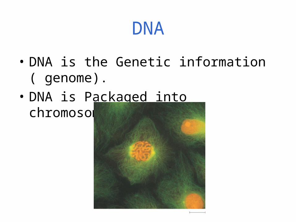
DNA
• DNA is the Genetic information ( genome).
• DNA is Packaged into chromosomes.
DNA And Chromosomes
• An average eukaryotic cell has about 1,000 times more DNA then an average prokaryotic cell.
• The DNA in a eukaryotic cell is organized into several linear chromosomes, whose organization is much more complex than the single, circular DNA molecule in a prokaryotic cell
Chromosomes
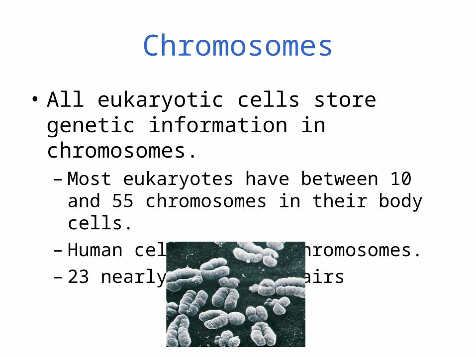
• All eukaryotic cells store genetic information in chromosomes.– Most eukaryotes have between 10 and 55
chromosomes in their body cells.– Human cells have 46 chromosomes.– 23 nearly-identical pairs
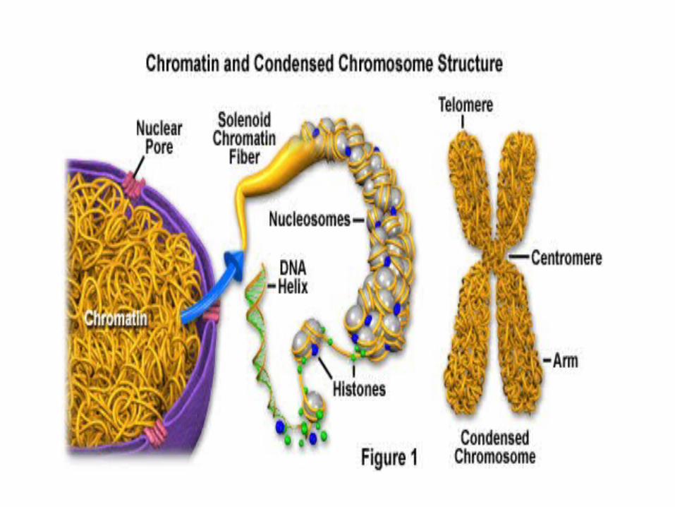
Structure of Chromosomes
• Chromosomes are composed of a complex of DNA and protein called chromatin that condenses during cell division.
• DNA exists as a long, double-stranded fiber extending chromosome’s entire length.
• Each unduplicated chromosome contains one DNA molecule.
• Chromatin is the combination of DNA and proteins that make up the contents of the nucleus of a cell.
The primary functions of chromatin are:
1- to package DNA into a smaller volume to fit in the cell .
2- to strengthen the DNA to allow mitosis and meiosis.
3- to prevent DNA damage
4- to control gene expression and DNA replication .
• DNA coding genes that are actively transcribed ("turned on") are more loosely packaged and are found associated with RNA polymerases (referred to as euchromatin .
• DNA coding inactive genes ("turned off") are found associated with structural proteins and are more tightly packaged )heterochromatin( .
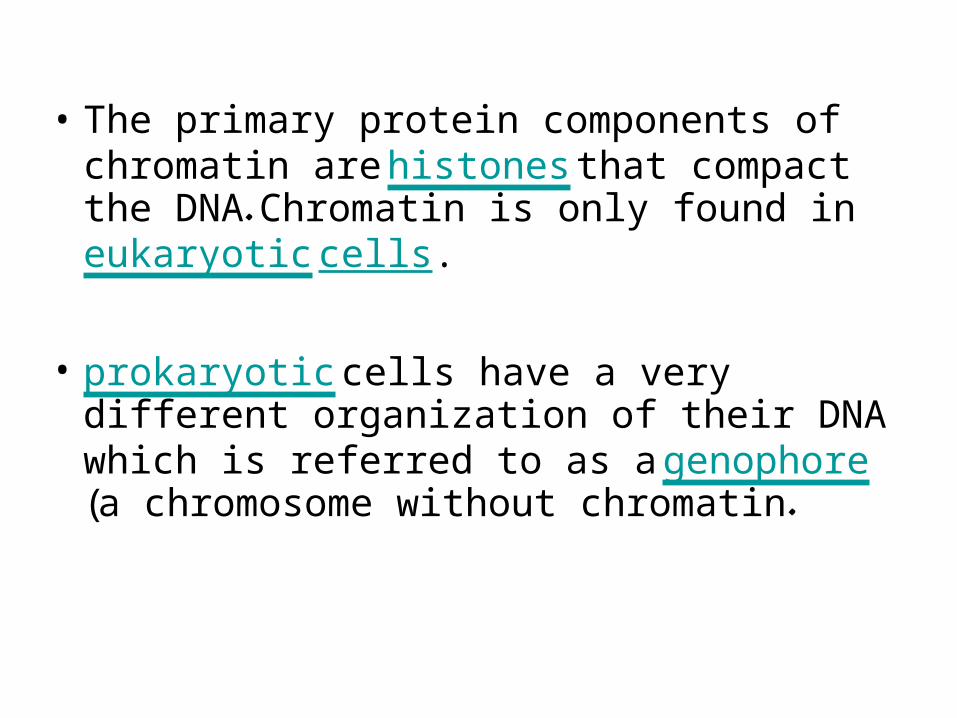
• The primary protein components of chromatin are histones that compact the DNA. Chromatin is only found in eukaryotic cells.
• prokaryotic cells have a very different organization of their DNA which is referred to as a genophore (a chromosome without chromatin.
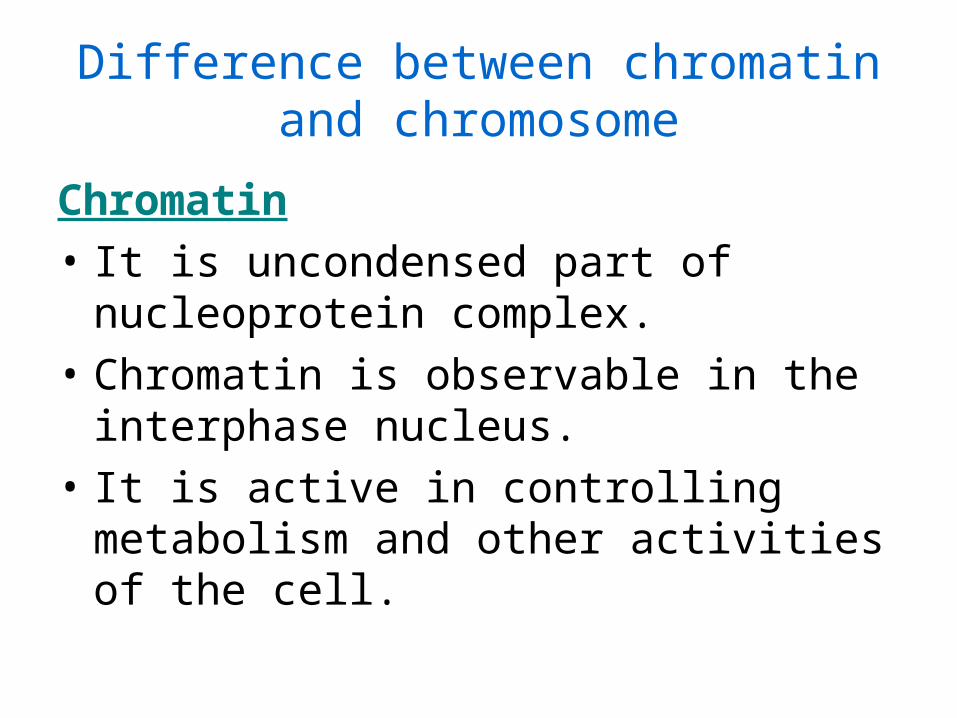
Difference between chromatin and chromosome
Chromatin
• It is uncondensed part of nucleoprotein complex.
• Chromatin is observable in the interphase nucleus.
• It is active in controlling metabolism and other activities of the cell.
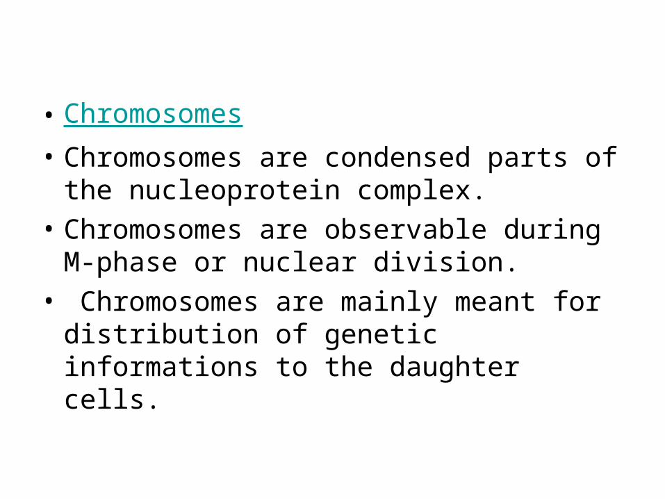
• Chromosomes
• Chromosomes are condensed parts of the nucleoprotein complex.
• Chromosomes are observable during M-phase or nuclear division.
• Chromosomes are mainly meant for distribution of genetic informations to the daughter cells.
Chromosomes
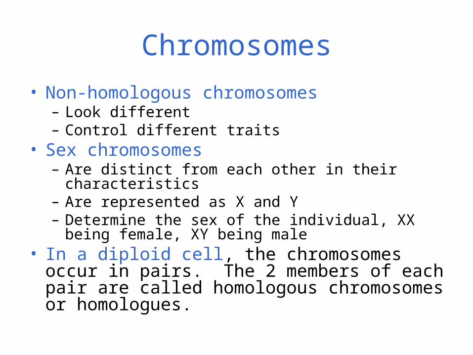
• Non-homologous chromosomes– Look different – Control different traits
• Sex chromosomes– Are distinct from each other in their characteristics– Are represented as X and Y – Determine the sex of the individual, XX being female,
XY being male• In a diploid cell, the chromosomes occur in
pairs. The 2 members of each pair are called homologous chromosomes or homologues.
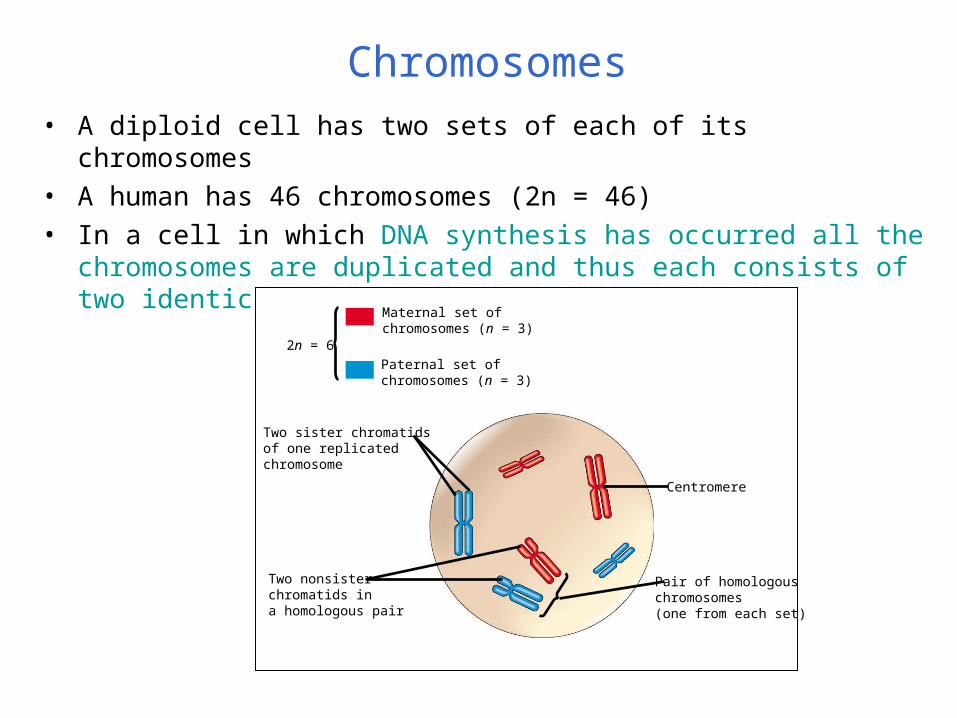
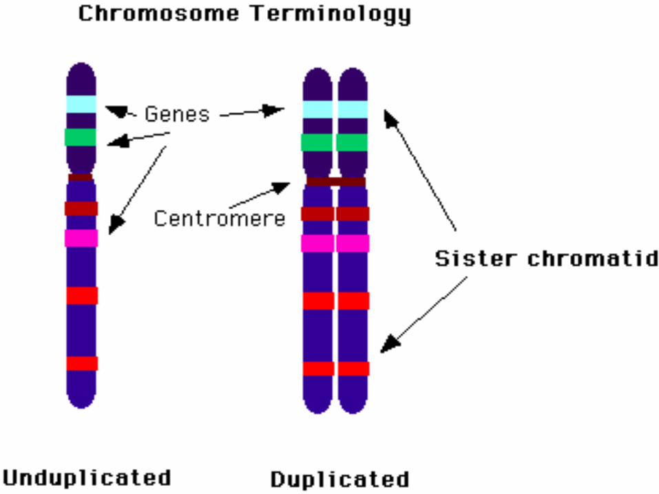
Chromosomes• A diploid cell has two sets of each of its chromosomes• A human has 46 chromosomes (2n = 46)• In a cell in which DNA synthesis has occurred all the chromosomes are
duplicated and thus each consists of two identical sister chromatids
Maternal set ofchromosomes (n = 3)
Paternal set ofchromosomes (n = 3)
2n = 6
Two sister chromatidsof one replicatedchromosome
Two nonsisterchromatids ina homologous pair
Pair of homologouschromosomes(one from each set)
Centromere
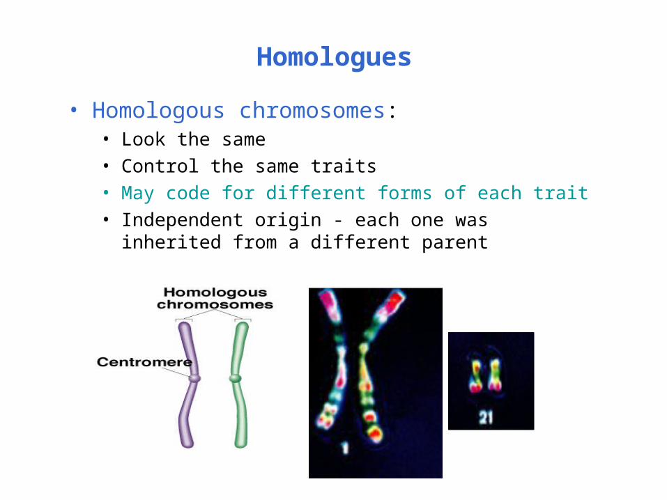
Homologues
• Homologous chromosomes:• Look the same• Control the same traits• May code for different forms of each trait• Independent origin - each one was inherited from a
different parent
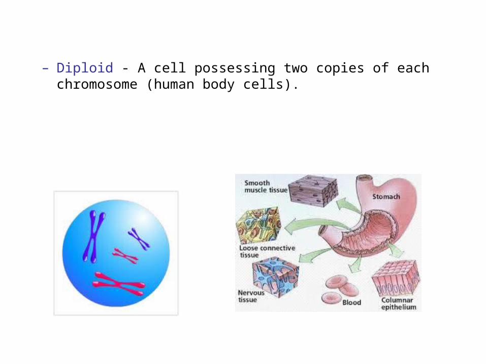
– Diploid - A cell possessing two copies of each chromosome (human body cells).
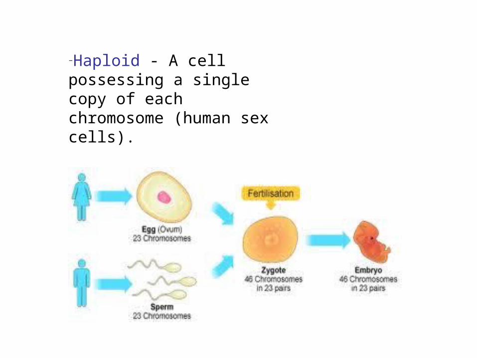
–Haploid - A cell possessing a single copy of each chromosome (human sex cells).
Phases of the Cell Cycle
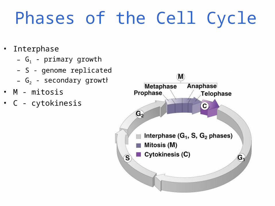
• Interphase– G1 - primary growth
– S - genome replicated
– G2 - secondary growth
• M - mitosis• C - cytokinesis
Interphase
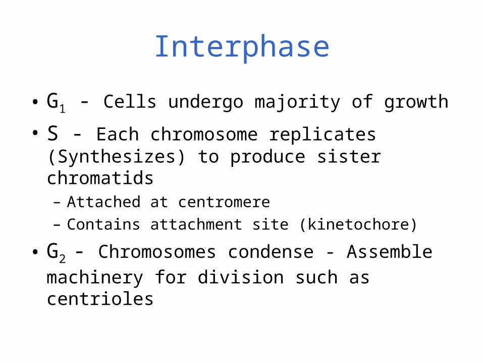
• G1 - Cells undergo majority of growth
• S - Each chromosome replicates (Synthesizes) to produce sister chromatids– Attached at centromere– Contains attachment site (kinetochore)
• G2 - Chromosomes condense - Assemble
machinery for division such as centrioles
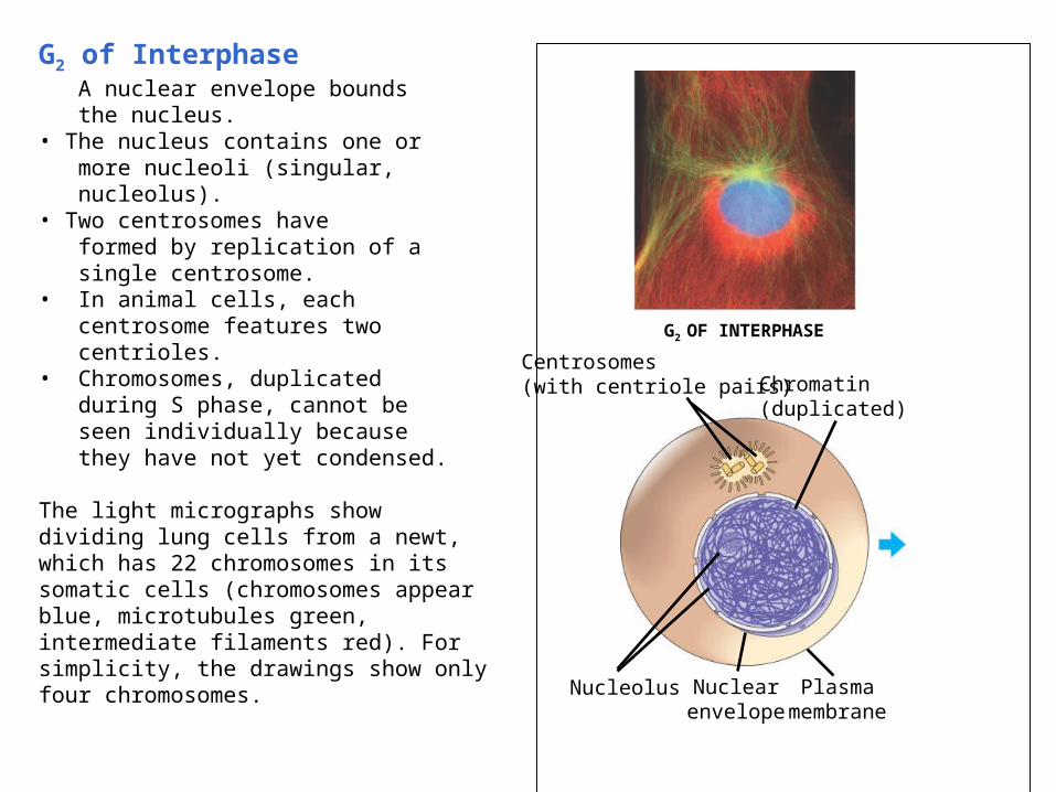
G2 of Interphase• A nuclear envelope bounds the nucleus.• The nucleus contains one or more nucleoli (singular, nucleolus).• Two centrosomes have formed by replication of a single centrosome.• In animal cells, each centrosome features two centrioles.• Chromosomes, duplicated during S phase, cannot be seen individually because they have not yet condensed.
The light micrographs show dividing lung cells from a newt, which has 22 chromosomes in its somatic cells (chromosomes appear blue, microtubules green, intermediate filaments red). For simplicity, the drawings show only four chromosomes.
G2 OF INTERPHASE
Centrosomes(with centriole pairs) Chromatin
(duplicated)
Nucleolus Nuclearenvelope
Plasmamembrane
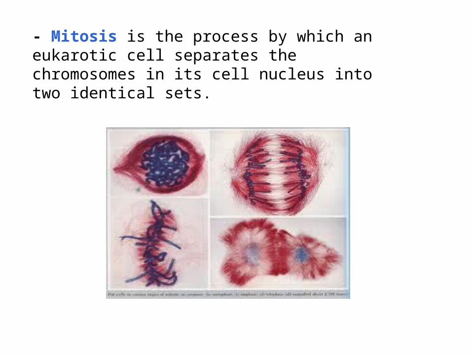
- Mitosis is the process by which an eukarotic cell separates the chromosomes in its cell nucleus into two identical sets.
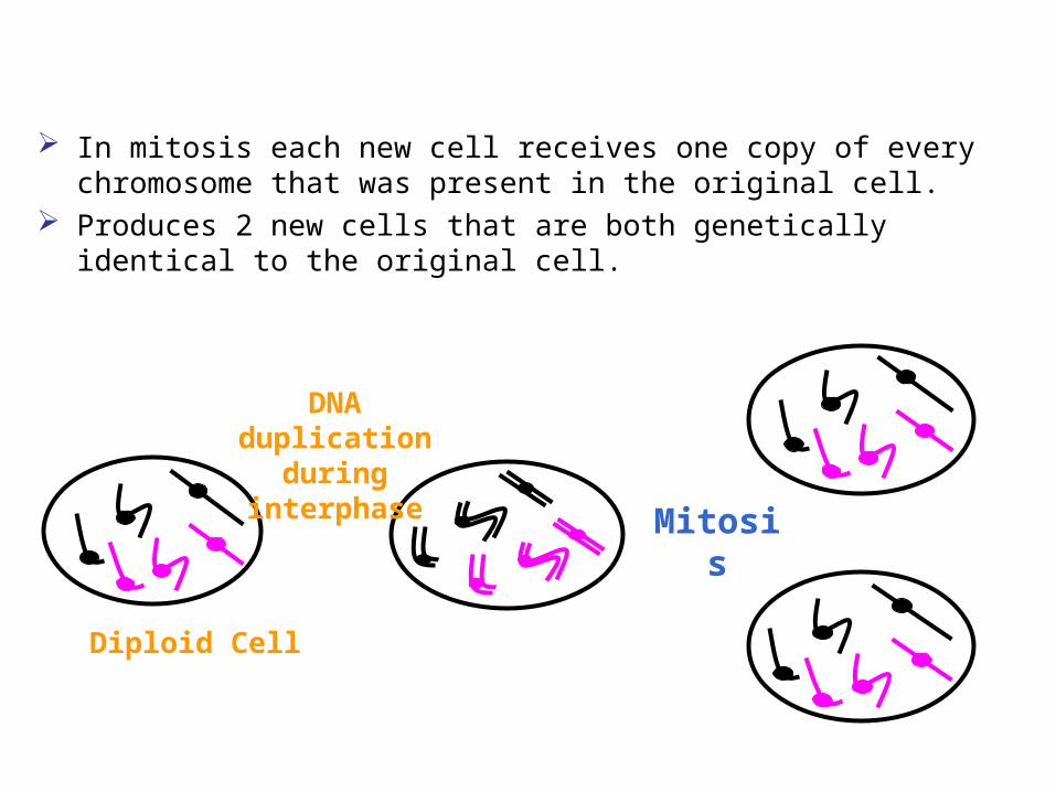
In mitosis each new cell receives one copy of every chromosome that was present in the original cell.
Produces 2 new cells that are both genetically identical to the original cell.
DNA duplication
during interphase Mitosis
Diploid Cell
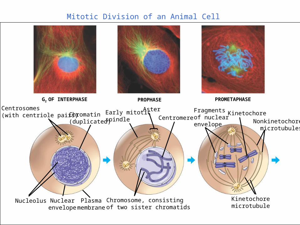
Mitotic Division of an Animal Cell
G2 OF INTERPHASE PROPHASE PROMETAPHASE
Centrosomes(with centriole pairs) Chromatin
(duplicated)
Early mitoticspindle
Aster
CentromereFragmentsof nuclearenvelope
Kinetochore
Nucleolus Nuclearenvelope
Plasmamembrane
Chromosome, consistingof two sister chromatids
Kinetochore microtubule
Nonkinetochoremicrotubules
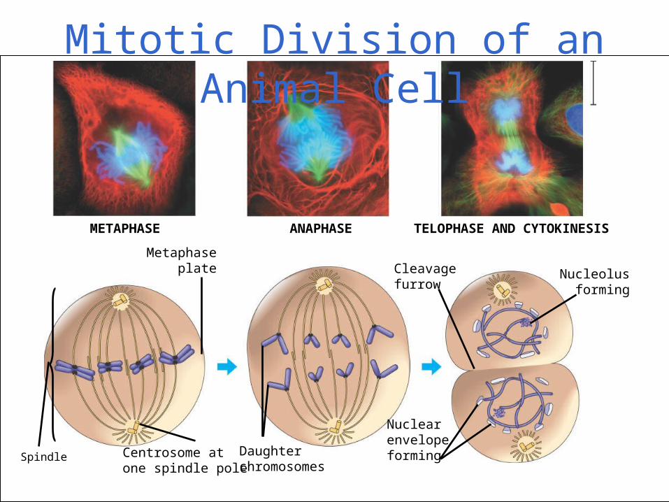
METAPHASE ANAPHASE TELOPHASE AND CYTOKINESIS
Spindle
Metaphaseplate Nucleolus
forming
Cleavagefurrow
Nuclear envelopeformingCentrosome at
one spindle poleDaughter chromosomes
Mitotic Division of an Animal Cell
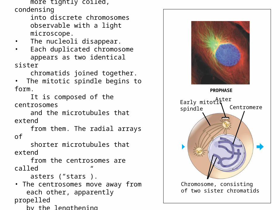
Prophase• The chromatin fibers become more tightly coiled, condensing into discrete chromosomes observable with a light microscope.• The nucleoli disappear.• Each duplicated chromosome appears as two identical sister chromatids joined together.• The mitotic spindle begins to form. It is composed of the centrosomes and the microtubules that extend from them. The radial arrays of shorter microtubules that extend from the centrosomes are called asters (“stars”).• The centrosomes move away from each other, apparently propelled by the lengthening microtubules between them.
PROPHASE
Early mitoticspindle
Aster
Centromere
Chromosome, consistingof two sister chromatids
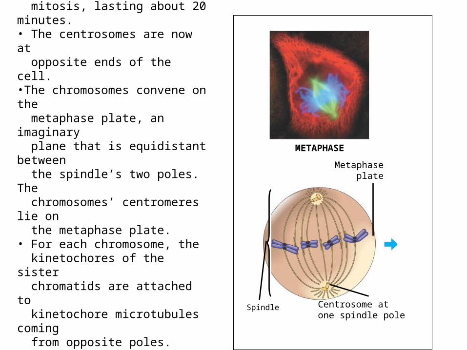
Metaphase• Metaphase is the longest stage of mitosis, lasting about 20 minutes.• The centrosomes are now at opposite ends of the cell. •The chromosomes convene on the metaphase plate, an imaginary plane that is equidistant between the spindle’s two poles. The chromosomes’ centromeres lie on the metaphase plate. • For each chromosome, the kinetochores of the sister chromatids are attached to kinetochore microtubules coming from opposite poles. • The entire apparatus of
microtubules is called the spindle because of its shape.
METAPHASE
Spindle
Metaphaseplate
Centrosome at one spindle pole
The Mitotic Spindle• The spindle includes the centrosomes, the spindle
microtubules, and the asters• The apparatus of microtubules controls
chromosome movement during mitosis• The centrosome replicates, forming two
centrosomes that migrate to opposite ends of the cell
• Assembly of spindle microtubules begins in the centrosome, the microtubule organizing center
• An aster (a radial array of short microtubules) extends from each centrosome
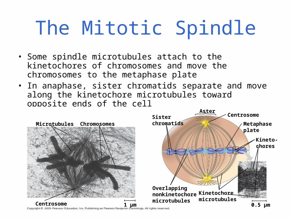
• Some spindle microtubules attach to the kinetochores of chromosomes and move the chromosomes to the metaphase plate
• In anaphase, sister chromatids separate and move along the kinetochore microtubules toward opposite ends of the cell
Microtubules ChromosomesSisterchromatids
AsterCentrosome
Metaphaseplate
Kineto-chores
Kinetochoremicrotubules
0.5 µm
Overlappingnonkinetochoremicrotubules
1 µmCentrosome
The Mitotic Spindle
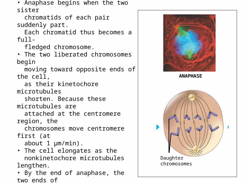
Anaphase• Anaphase is the shortest stage of mitosis, lasting only a few minutes.• Anaphase begins when the two sister chromatids of each pair suddenly part. Each chromatid thus becomes a full- fledged chromosome.• The two liberated chromosomes begin moving toward opposite ends of the cell, as their kinetochore microtubules shorten. Because these microtubules are attached at the centromere region, the chromosomes move centromere first (at about 1 µm/min).• The cell elongates as the nonkinetochore microtubules lengthen.• By the end of anaphase, the two ends of the cell have equivalent—and complete—collections of chromosomes.
ANAPHASE
Daughter chromosomes
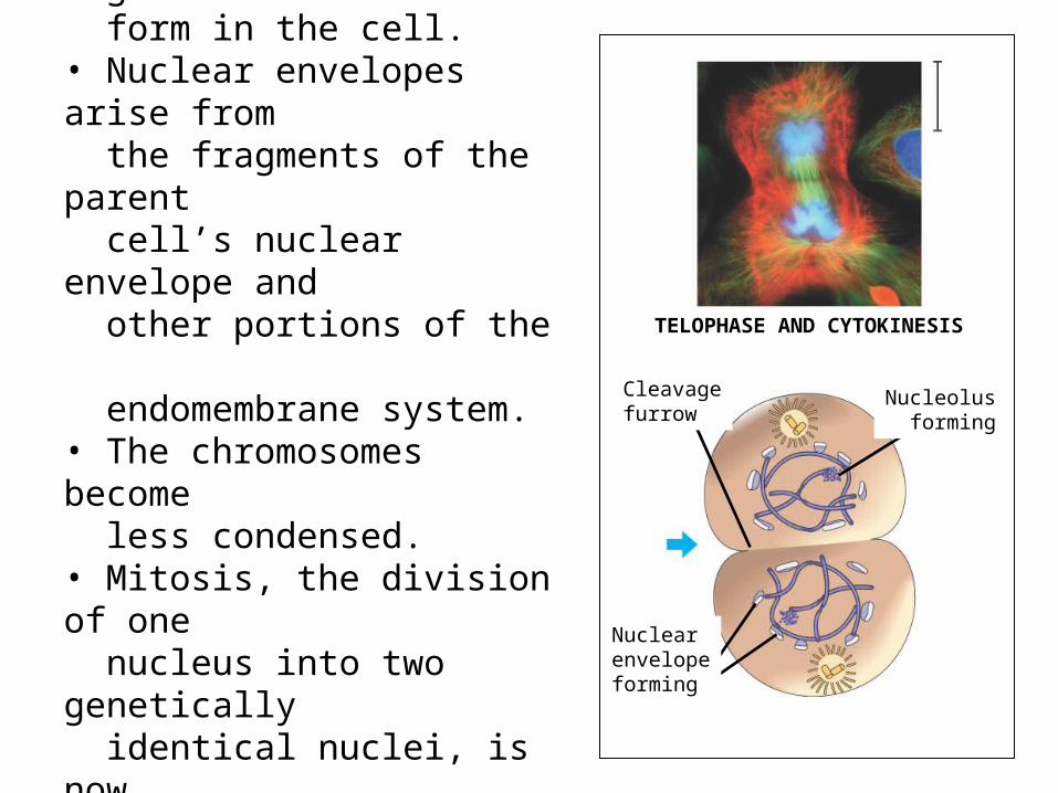
Telophase• Two daughter nuclei begin to form in the cell.• Nuclear envelopes arise from the fragments of the parent cell’s nuclear envelope and other portions of the endomembrane system.• The chromosomes become less condensed.• Mitosis, the division of one nucleus into two genetically identical nuclei, is now complete.
TELOPHASE AND CYTOKINESIS
Nucleolusforming
Cleavagefurrow
Nuclear envelopeforming
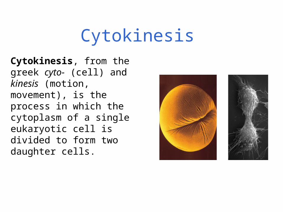
Cytokinesis
Cytokinesis, from the greek cyto- (cell) and kinesis (motion, movement), is the process in which the cytoplasm of a single eukaryotic cell is divided to form two daughter cells.
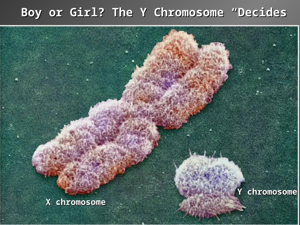
Boy or Girl? The Y Chromosome “Decides”Boy or Girl? The Y Chromosome “Decides”
X chromosomeX chromosomeY chromosomeY chromosome
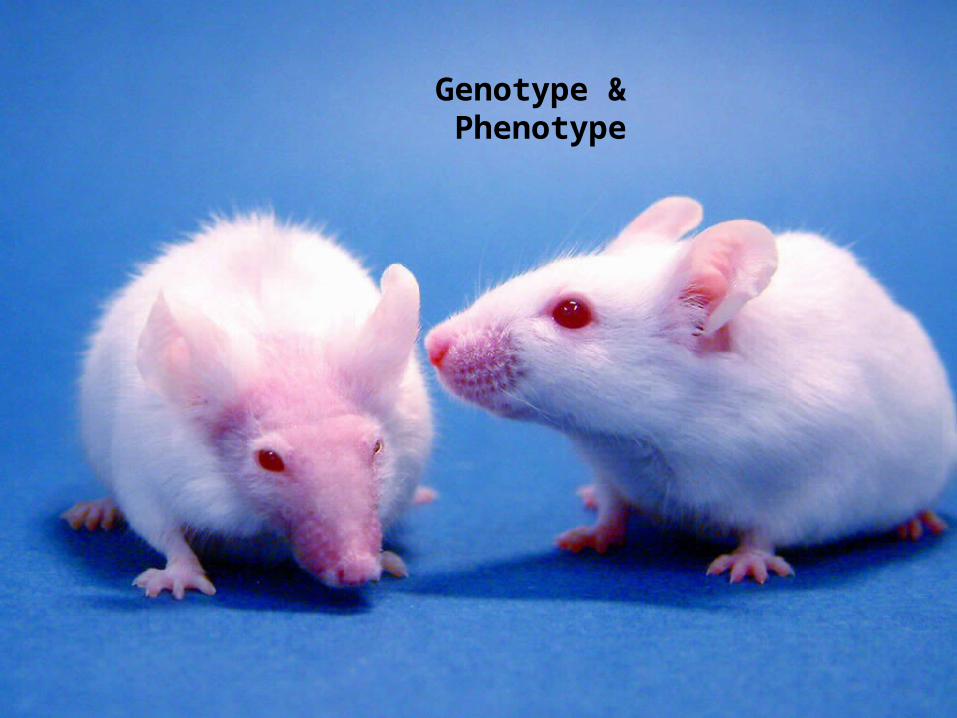
Genotype & Phenotype
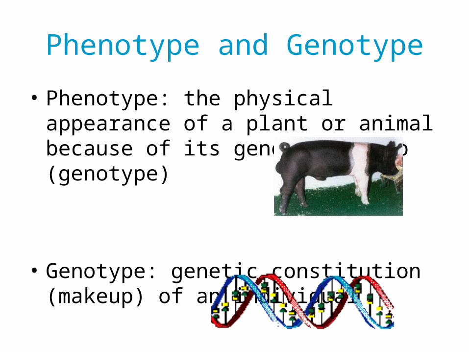
Phenotype and Genotype
• Phenotype: the physical appearance of a plant or animal because of its genetic makeup (genotype)
• Genotype: genetic constitution (makeup) of an individual
www.ansi.okstate.edu/breeds/swine/
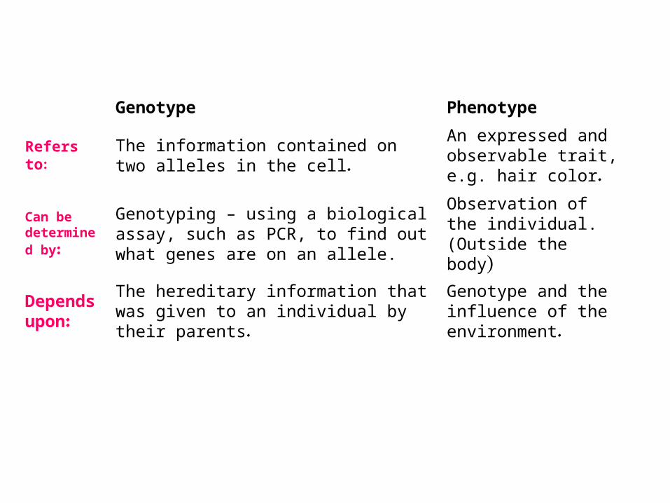
Genotype Phenotype
Refers to:The information contained on two alleles in the cell.
An expressed and observable trait, e.g. hair color.
Can be determined by:
Genotyping – using a biological assay, such as PCR, to find out what genes are on an allele.
Observation of the individual. (Outside the body)
Depends upon:
The hereditary information that was given to an individual by their parents.
Genotype and the influence of the environment.
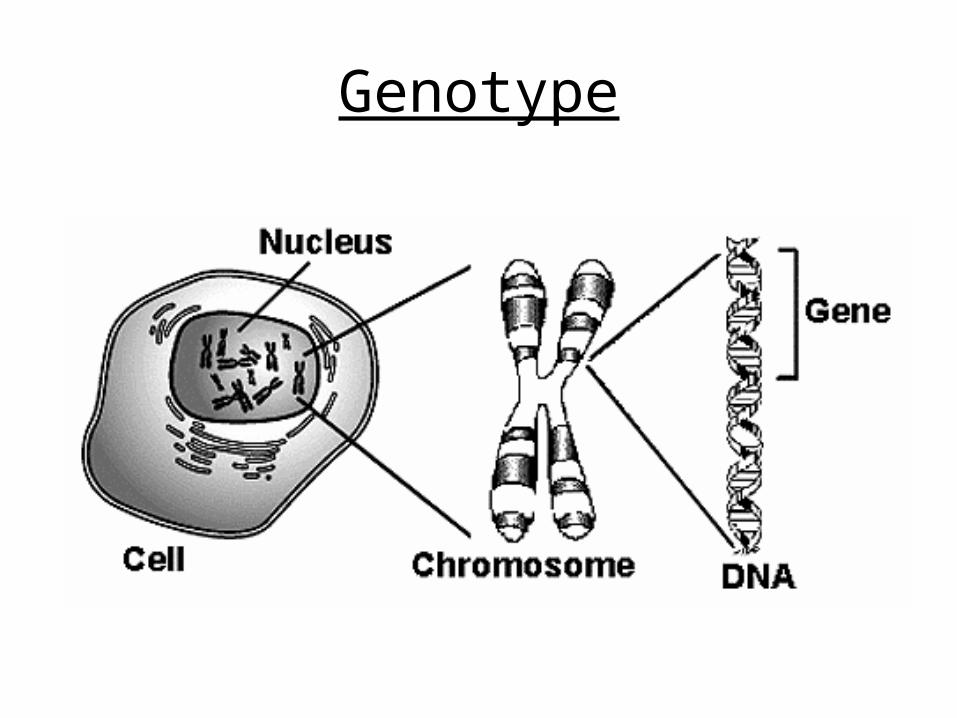
Genotype
Genotype
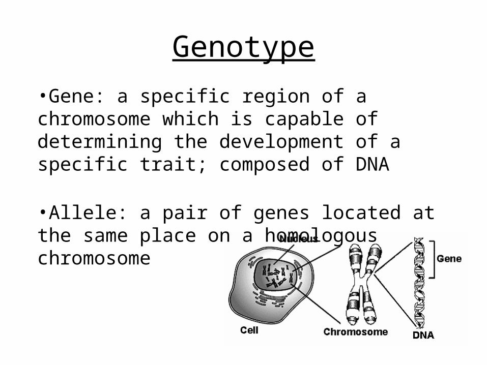
•Gene: a specific region of a chromosome which is capable of determining the development of a specific trait; composed of DNA
•Allele: a pair of genes located at the same place on a homologous chromosome
Allele : is one of a number of alternative forms of the same gene or same genetic locus. Each human has two alleles, one for each chromosome (humans have two of each chromosome).
There are only two possible traits for an allele: dominant or recessive.
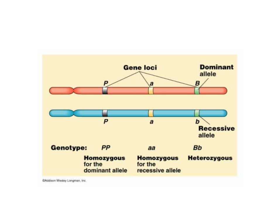
Since everyone has two alleles, an individual with two dominant alleles is termed homozygous dominant, an individual with one dominant and one recessive allele is termed heterozygous, and an individual with two recessive alleles is termed homozygous recessive.
Traits are controlled by genes
• Dominant allele: those that mask the presence of other corresponding allele.
• Recessive allele: those whose physical expression (phenotype) is masked when in the presence of a dominant allele.
Heterozygous and Homozygous
• Heterozygous- when a plant or animal has two genes for different traits .
• Homozygous- when a plant or animal has two genes for the same trait .


![FADHEL M. GHANNOUCHI - Contacts Directorycontacts.ucalgary.ca/info/enel/profiles/168-42875/... · wide-band RF power amplifiers," IET Electronics Letters, 2016 accepted. [7] T. Sharma,](https://img.pdfslide.us/doc/110x75/5f0c18da7e708231d433b9ed/fadhel-m-ghannouchi-contacts-wide-band-rf-power-amplifiers-iet-electronics.jpg)