Embed Size (px)
Citation preview
BIOLOGICAL STAINING
Dr . Samah Kotb Nasr
Eldeen

INTRODUCTION
Biological stains are prepared from dyes which have been
manufactured to rigid specifications; to ensure that they
are suitable for specialized purpose for which they are
made.

Dyes classified into two main groups:
Natural dyes.
Synthetic dyes.

NATURAL DYES1. Haematoxylin – from plant. Is extracted from the
heartwood of the tree Haematoxylum.
2. Carmine – derived from female cochineal bug.
3. Orcein – a vegetable dye extract .

SYNTHETIC DYES
Are derived from hydrocarbon benzene. Simple
benzene compounds have absorption bands in the U.V
range of the spectrum.

Chromophores added to the benzene compound to
move the absorption bands into visible portion of the
spectrum giving the visible color. Benzene
compounds containing chromophores are known as
CHROMAGENS.

Chromagen is a colored substance; but its not a dye.
That because it can easily washed out or removed.
Another radical known as AUXOCHROME is added to
the chromagen to make a dye not easily washed fast ex:
Methylene blue, Eosin and others.
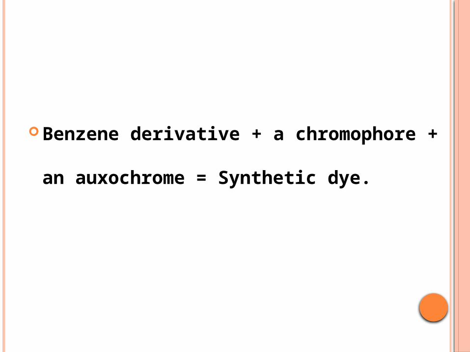
Benzene derivative + a chromophore + an auxochrome
= Synthetic dye.

BASIC, ACID AND NEUTRAL DYES
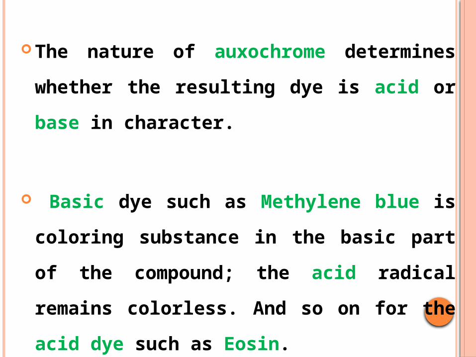
The nature of auxochrome determines whether the
resulting dye is acid or base in character.
Basic dye such as Methylene blue is coloring substance
in the basic part of the compound; the acid radical
remains colorless. And so on for the acid dye such as
Eosin.

Neutral dyes as leishmans’ stain are obtained by
combining aqueous solutions of basic and acid
dyes; And they give more than two colors; the
nuclei take the color of the basic dye while the
cytoplasm stained by the acidic dye and the
granules take the third color.

TERMS USED WHEN STAINING

1. Micro-anatomical stains: used for demonstrating the
general relationship of tissues to each other.
2. Cytological stains: used for demonstration minute
structures in the nucleus and cytoplasm of cells.
3. Indirect staining: stain need a mordant; so the stain can
work.

4. Direct staining: no need to add a mordant to the stain.
5. Simple stain: stain contains only one dye.
6. Compound stain: stain contains more than one stain.
7. Progressive stain: when the different elements in the
tissues are colored in sequence and at the correct time
differential coloration of tissues are achieved . It does not need
differentiation.

8. Regressive stain: when tissues are over-stained and then
destained or differentiated (washed out) by removing excess
stain from the unwanted parts of the tissues.
9. Selective stain: stains more than one element in the same
color, but its easy to identify the element you want to
demonstrate either by morphology or site or by both.

10. Specific stain: when the stain acts only on a specific
constituent or element of the cell or tissue and has no effect
upon other elements.

11. Vital stain:
The living cells may be stained after removal from the
organism and in this case the staining called SUPRAVITAL
stain.
If the stain take place while the elements are still part of the
living organism; the staining called INTRAVITAL stain.
Intravital staining is done by injecting or introducing the stain
into the body.

12. Negative staining: when the organism do not take the
stain, but these unstained organisms are sharply contrasted
against a stained background.
13. Fluorescent staining: staining by flurochrome dyes. there
are two types:
a) Primary or Auto-fluorescence.
B) Secondary or induced fluorescence.

14. Mordant: are metallic substances (Aluminum, Iron &
others) which act as a link between the stain and the tissue to
be stained. Mainly used for indirect staining. These mordants
are used in 3 ways:
1) Before the application of the dye “pre-mordanating”.
2) In conjunction with the stain.
3) After the application of the stain “post-mordanating”.

15. Accentuators: used for indirect stains. They differ from
mordants. They are not essential for chemical union of the dye.
But they increase the intensity and selectivity of the staining.
16. Accelerators: increase the staining power. They used with
the metallic impregnation (not with the dyes).

17. Differentiation: it’s a de-staining or differentiation of an
over stained tissues in a regressive staining techniques.
Mordants and some dyes also act as differentiating agents.
Washing in water or alcohol is a common means of
differentiation.
18. Using controls in histology: when you apply a new
staining solution you have to apply it side by side with your old
working staining solution on sections from the same tissues.