Embed Size (px)
Citation preview

Dr Kate Talks, Consultant Haematologist, Newcastle upon Tyne Hospitals NHS Trust
Nov 2012

What coagulation tests are needed in major haemorrhage
What POCT tests are available
How do you go about setting up a new test
POCT: any analytical test performed for a patient by a healthcare worker outside the laboratory setting

Bleeding and Clotting
Pro-coagulation Anti-coagulation
Endothelial surfacePlatelets
Von Willebrand FactorCoagulation Factors
Natural Anticoagulants
(Drugs)
Inherited or acquired bleeding disorders Inherited clotting tendency
Risk factors
Fibrinolysis

Identification of coagulation abnormalities and results that guide treatmentAbility to monitor individual s response to replacement therapy ?is clot formingPredict bleeding risk ? How strong is the clot? Is the clot stablePredict thrombotic risk hypercoaguability
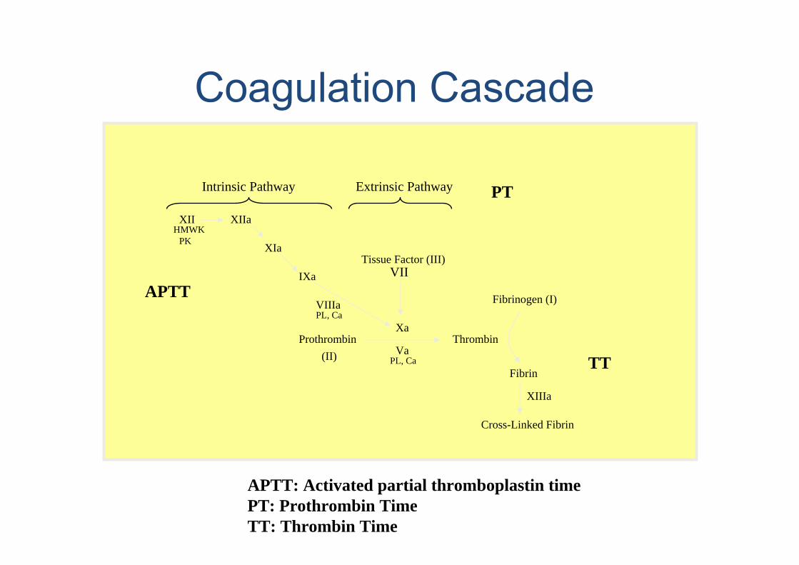
Prothrombin ThrombinXa
Va
Fibrinogen (I)
Fibrin
Cross-Linked Fibrin
XIIIa
Tissue Factor (III)VII
PL, Ca(II)
IXa
XIa
XIIaXIIHMWK
PK
VIIIaPL, Ca
Intrinsic Pathway Extrinsic Pathway
APTT
PT
TT
APTT: Activated partial thromboplastin timePT: Prothrombin TimeTT: Thrombin Time

Existing tests only look at a small part of the processThey use plasma rather than whole bloodThey are tested in a static processThey are not tested at patient s temperatureThey are not generally done at the bedside and analysis takes time

Routine : PT, APTT, Fibrinogen, TT, D-dimersFBC, plateletsGlobal assays endogenous thrombin generation
POCT: Coaguchek INRViscoelastic haemostatic assays (VHA): TEG,ROTEM
POCT: FBC / Haemoglobin concentrationPT/ APTT require sample centrifugation platelet poor plasma and sample/ reagent pipetting; ACT

Establish INR in patients on warfarinInterface with laboratory result systemPotential value in patients who require urgent surgery and may need warfarin reversal egfracture NOF Patients bleeding known to be on warfarinBUT also need to check APTT, fibrinogen and FBC

NRHG GUIDE TO WARFARIN REVERSAL
BLEEDING
Life / Limb /SightThreatening Minor
Retroperitoneal (CT or MRI)Intra-ocular (NOT conjunctival)Spontaneous muscle bleed with compartment syndromePericardial Active bleeding from any orifice plus either BP 90 mmHg systolic, oliguria or 2 g fall in haemoglobin
Vitamin K 5 mg IV2 andProthrombin complex concentrate IV(Beriplex)3 30 units/kg
Vitamin K 2mg PO1
Check INRat 24 hours or sooner if clinical
deterioration
Check INR Immediately
Adequate correction Inadequate correction
Repeat INR & APTT in 4-6 hours
Consider other factors contributing to prolonged coagulation tests eg DIC, Congenital coagulation factor deficiency, Liver disease. A further dose of 20 units/kg Beriplex may be indicated- seek haematological advice
1Oral vitamin K - there are marked differences between formulations of vitamin K. The most effective preparation is IV Konakion (Roche) given orally. The vial contains 10 mg/ml dilute appropriate dose in small amount of juice/water after drawing up in 1 ml insulin syringe. Alternatively the Konakion MM paediatric formulation may be used.
2Vitamin K IV may rarely cause anaphylaxis. Give by slow IV bolus
3Prothrombin complex concentrate (PCC) may induce a prothrombotic state. Use with cautionin patients with DIC or decompensated liver disease
4In serious but non-life-threatening bleeding (e.g. GI bleeding or epistaxis without haemodynamic compromise) prompt reversal with IV vitamin K is indicated.
Significant bleeding4
withouthaemodynamic
compromise
Vitamin K 2 mg IV2
Check INR at 4-6 hours or sooner
if clinical deterioration
Oral vitamin K is safe and adequate treatment for the majority of patients.There may be some clinical circumstances when 1-2 mg IV vitamin K
should be considered e.g. gross over-anticoagulation or unsteady patients
Patients with rapid onset neurological signswhile on warfarin do URGENT INR and CT scan (within 1 hour). If INR>1.5, consider urgent reversal with Beriplex (see below), without waiting for CT scan. NB ensure CT scan is reported and acted on immediately
CONTACT HAEMATOLOGISTIntracranial (CT or MRI)
NB All bleeding in a patient on warfarin should be taken seriously. Bleeding may occur when the INR is therapeutic. If the INR is sub-therapeutic e.g. <1.5, bleeding may be due to factors other than warfarin and reversal may not be appropriate. Always check FBC andcoagulation screen to identify other causes.If in doubt discuss with haematologist.

NRHG GUIDE TO WARFARIN REVERSAL
NEED FOR SURGERY
Assess urgency of surgery and
degree of reversal required
EMERGENCY SURGERY (IMMEDIATE):
Vitamin K 5mg IV2 and Prothrombin complex concentrate IV (Beriplex)3 30 units/kg
URGENT (WITHIN 24 HOURS):
If surgery can be delayed for 6-12 hours, give Vitamin K 2mg IV2. If it can delayed for 24 hours, give Vitamin K 1-2mg po1
1Oral vitamin K - there are marked differences between formulations of vitamin K. The most effective preparation is IV Konakion (Roche) given orally. The vial contains 10 mg/ml -dilute appropriate dose in small amount of juice/water after drawing up in 1 ml insulin syringe. Alternatively the Konakion MM paediatric formulation may be used.
2Vitamin K IV may rarely cause anaphylaxis. Give by slow IV bolus3Prothrombin complex concentrate (PCC) may induce a prothrombotic state. Use with cautionin patients with DIC or decompensated liver disease

Thrombin generation curve
Lag time
Time to peak
Peak thrombin
Endogenous thrombin potential

Thromboelastography (TE) and rotational elastometry (ROTEM)
Thromboelastography [TE] was first described by Hartert in 1948. Thromboelastography®(TEG®) and Thromboelastometry (ROTEM®) provide global information on the dynamics of clot development, stabilisation and dissolution that reflect in vivo haemostasis. Although TE has not been subjected to the same evaluation processes as conventional haemostatic tests, its use as a POCT monitor in complex major surgery has been shown to significantly reduce the use of blood component therapy and overall blood loss.

Thromboelastography (TEG)
Measures the viscoelastic properties of whole blood as it is induced to clot in a low shear environment resembling venous blood flow
Clot formation
Clot kinetics
Clot strength/stability
Clot resolution

Thromboelastography- Basic principles
Heated (370C) oscillating cupPin suspended from torsion wire into bloodDevelopment of fibrin strands couplemotion of cup to pin
Increased tension in wire detected by EM transducer and transmitted to create TEG traceDeflection of trace increases as clot strength increases and decreases as
clot strength decreases

A normal TEG trace
R (Reaction) time = Time taken for blood to clot, i.e. clotting factorsK (Kinetic) time/ angle = Measure of clot kinetics, i.e. speed of fibrin build-up
MA (Maximum amplitude) = Measure of clot strength, i.e. interaction between activated platelets and fibrin
LY (Lysis) 30 = Measure of clot stability, i.e. fibrinolysis

ROTEM


Variable TEG® ROTEM®
Measurement period - Reaction Time [RT]
Time from start to when the waveform reaches 2mm above baseline
R Clotting Time [CT]
The time from 2mm above baseline to 20mm above baseline
K Clot Formation Time [CFT]
Alpha angle [°] [slope between R and K] [angle of tangent at 2mm amplitude]
Maximum angle - CRF
Maximum strength Maximal Amplitude [MA] Maximal Clot Firmness [MCF]
Time to Maximum strength - MCF-t
Amplitude at a specific time A30, A60 A5, A10...
Clot elasticity G MCE
Maximum lysis - CLF
Clot Lysis[CL] at a specific time [minutes]
CL30, CL60 LY30, LY45, LY60
Time to lysis 2mm from MA CLT [10% difference from MCF]

Fibrinolysis The degree of fibrinolysis can be established from either a native sample with no activator, from the Tissue Factor activator or the combined Tissue Factor/kaolin activated TEG. Hyperfibrinolysis is increasingly recognised as a cause of perioperative microvascular bleeding and is readily detected by analysing the clot lysis index on the TEG or ROTEM .The ability to detect and determine the severity of fibrinolysis avoids empirical or inappropriate anti-fibrinolytic therapy.Mathematical derivations of changes in elastic modulus derived from the amplitude have been used to quantify the extent of fibrinolysis in clinical and laboratory settings, as well as to guide antifibrinolytic therapy
Hypercoagulability The TEG may be helpful in screening for hypercoagulable states. TEG analysis of patients with a history of thromboembolic complications showed shorter R values and accelerated clot propagation compared to healthy reference subjects
Fibrinogen and platelet function
The MA is primarily a reflection of clot strength and is affected by changes in fibrinogen, platelet count and function. The MA is established either from a native sample with no activator or from the combined Tissue Factor/kaolin activated TEG. There is a strong linear correlation between the log platelet count and MA. Abciximab is a potent platelet GpIIb/IIIa inhibitor and an abciximab-modified TEG can help to discriminate between hypofibrinogenemia and platelet dysfunction as a cause of decreased MA.

Modification Interpretation
Tissue Factor The use of an activator when undertaking thromboelastography is generally recommended to standardise the initiation of the clotting process. Tissue Factor activation of the TEG enables the maximal amplitude (MA) to be established within 10 minutes but will result in significant shortening of the reaction time (R value) and as a result much of this latter information is lost.
R Time and Heparinase The R value in a native TEG is sensitive to trace amounts of heparin and endogenously released heparan sulphate. The use of a heparinase-coated reaction cuvette for the TEG will demonstrate any heparin present in the sample or in the patient and enables assessment of haemostasis in patients who are fully anticoagulated with heparin e.g. on CPB.
Tissue factor/kaolin activated TEG and the ACT
By incorporating both tissue factor and kaolin into the TEG cuvette, the TEG approximates to the Activated Clotting Time [ACT.]

Rotational Thromboelastometry (ROTEM®)The ROTEM analyser provides a trace similar to that of the TEG with related parameters including clotting time (CT) and maximum clot firmness (MCF). Additional tests include:TestInterpretation
INTEM Contains phospholipid and ellagic acid as activators and provides information similar to that of the APTT (Intrinsic system)EXTEM Contains Tissue Factor as an activator and provides information similar to that of the PT (extrinsic system)HEPTEM Contains lyophilised heparinase for neutralising heparinAPTEM Contains aprotinin for inhibiting fibrinolysisFIBTEM Utilises cytochalasin D, a platelet inhibitor which blocks the platelet contribution to clot formation, allowing qualitative analysis of the functional fibrinogen component.ECATEM Contains Ecarin and so is similar to the Ecarin Clotting Time. This makes it very sensitive to presence of direct thrombin inhibitors.

Use of TEG in cardiac surgery with bypass- Monitoring protocol
When ? Cup type Why?
On induction
Plain Baseline haemostatic profile
At re-warming/ end of CPB
PlainHeparinase
Early identification of coagulopathy
10 mins post protamine
PlainHeparinase
1. Check heparin reversal2. Identify cause of bleeding
Post-op PlainHeparinase
If bleeding

Use of TEG in cardiac surgery with bypass- Treatment protocol
TEG values Clinical cause Suggested treatment
R 11-14 mins
clotting factors X 2 FFP
R > 14 mins clotting factors X 4 FFP
MA 42-47 mm
platelet function 1 platelet pool
MA < 42 mins
platelet function
2 platelet pools
LY30 >7.5% Fibrinolysis Antifibrinolytic
R < 3 mins, MA > 75 mm
Prothrombotic state Anticoagulant
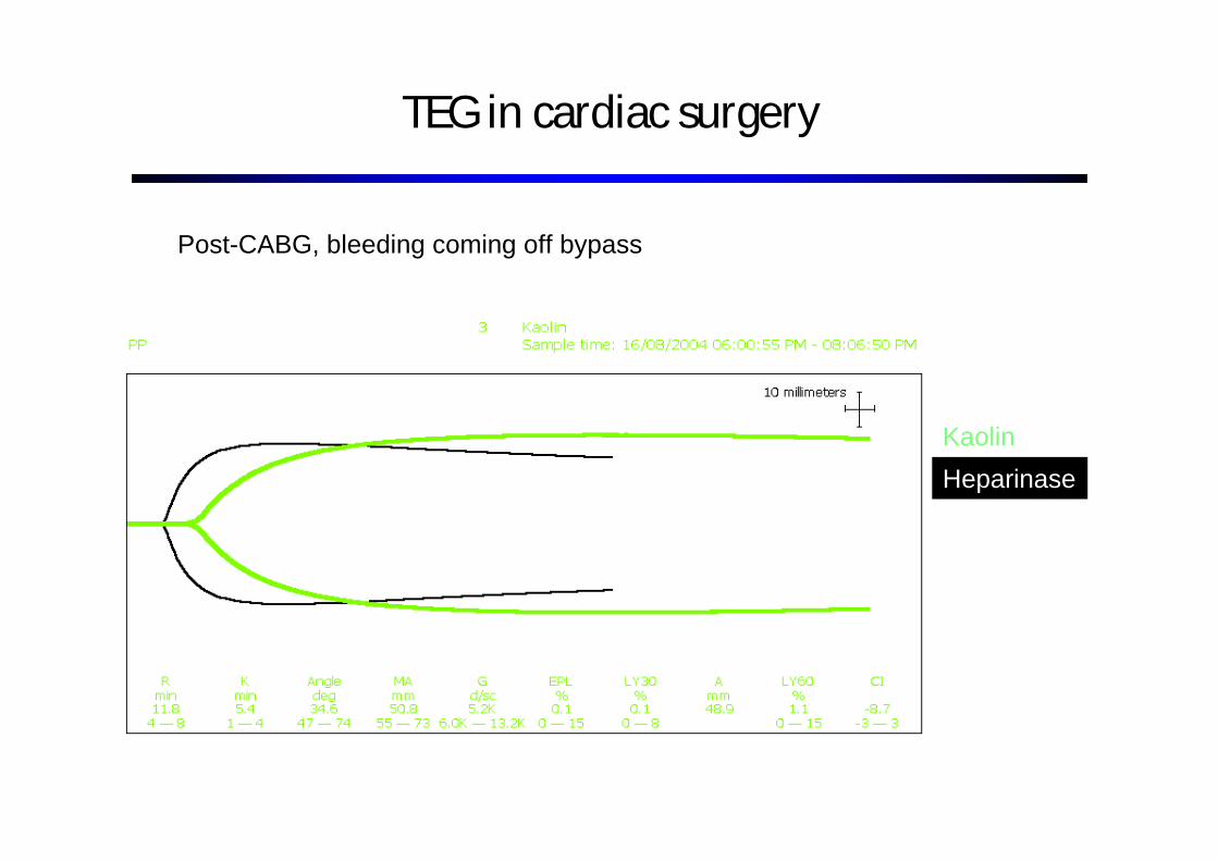
TEG in cardiac surgery
Kaolin
Heparinase
Post-CABG, bleeding coming off bypass

TEG in cardiac surgery- residual heparin
Kaolin
Heparinase
Give more protamine

TEG in cardiac surgery
Normal trace
Patient (heparinase)
During cardiac surgery, patient bleeding +++

TEG in cardiac surgery- Long R time, clotting factor deficiency
Normal trace
Patient (heparinase)
Administer FFP

TEG in cardiac surgery
Normal
Patient (heparinase)
During cardiac surgery, patient bleeding +++

TEG in cardiac surgery- Low MA, platelet dysfunction
Normal
Patient (heparinase)
Administer platelet concentrate

TEG in cardiac surgery
Normal
Patient
During cardiac surgery, patient continuing to bleed despite protamine, FFP and platelet transfusion

TEG in cardiac surgery- Fibrinolysis
Normal
Patient
Administer antifibrinolytic agent

TEG in cardiac surgery
Post-operative bleeding ++ from one mediastinal drain, not on anticoagulant therapy

TEG in cardiac surgery- Surgical bleeding

TEG- Cardiac surgery
Thromboelastography-guided transfusion algorithm reduces transfusions in complex cardiac surgery
Shore-Lesserson et al. Anesth Analg 1999;88:312-9
Reduced hemostatic factor transfusion using heparinase modified TEG during cardiopulmonary bypassVon Kier et al. Br J Anaesthesia 2001;86:575-8
Both used TEG algorithm-directed therapy vs. clinician-guided therapyBoth reported reduced blood product usage in TEG group and one reported lower volumes of blood in mediastinal drains
Early recognition and prompt correction of haemostatic abnormalities during surgery
More rapid recognition of surgical bleedingIdentification of the need for further protamine infusion

(a) Normal ROTEM® tracing.
Roullet S et al. Br. J. Anaesth. 2010;bja.aeq022© The Author [2010]. Published by Oxford University Press on behalf of the British Journal of
Anaesthesia. All rights reserved. For Permissions, please email:[email protected] (a) Normal ROTEM® tracing. (b) ROTEM® tracing depicting severe coagulation abnormalities with hyperfibrinolysis corrected by the addition of aprotinin (APTEM).

ROTEM traces from a trauma patient treated according to the AUVA Trauma Hospital algorithm: a. upon admission to the ER (EXTEM coagulation time and clot formation time are prolonged; maximum clot firmness is reduced; no clot formation in the FIBTEM test). b. 40 minutes after treatment with 2 g tranexamic acid, 10 g fibrinogen concentrate, 1800 U prothrombin complex concentrate and 1250 U factor XIII (normal coagulation).Schöchl et al. Scandinavian Journal of Trauma, Resuscitation and Emergency Medicine 2012 20:15 doi:10.1186/1757-7241-20-15 Download authors' original image

TEG- Trauma
Guiding transfusion of blood productsPotential to reduce blood product use by earlier detection of coagulopathyCan look at fibrinogen activity and hyperfibrinolysisUse of point-of-care RapidTEG(20 minsvs. 30 minsfor Kaolin-activated TEG)Ability to use as basis for goal directed treatment decisions and to monitor response to treatmentAn adjunct to other testsLarger studies needed

In vitro tests done in low shear conditionsAbsence of vascular endotheliumUnmodified VHA cannot be used to assess platelet function with anti-platelet drugsLMWH and warfarin effectDifferent reagents and activators give different results important when establishing algorithms

Wide range of normality important to monitor change within an individual relative to baseline
Moderate complexity of tests operators require training and SOP, IQA and EQA
How to go about setting up POCT
Guidelines on POCTBritish Journal of Haematology, 2008 142, 904 915
Contact your hospital / trust POCT lead for help

The purpose, nature and potential benefits of POCT at a particular site should be defined before initiating the servicePOCT devices should deliver results comparable to the local lab; need IQA an EQACost benefit analysisNeed clear clinical procedural protocols and clear clinical guidelinesOversight of POCT committee for procurement, user training, writing SOP, monitoring and reviewing serviceAudit on patient care, quality and training

There is not a one fit for all POCT solution for all departmentsAny new POCT needs evaluation, planning and then ongoing monitoring to ensure reliability of results; laboratory POCT committee and engagement of clinical usersModels of VHA use in cardiac and liver surgery and military experience support further assessment in trauma, but further evaluation needed

TF
VIIVIIa-TF
X Xa
ProthrombinII
ThrombinIIa
Endothelial surface
VIIIa
IXa
IX
Va V
Fibrinogen Fibrin Monomer
Fibrin PolymerXIII
XIIXIIaPK
HK
CaPL
CaPL
CaXI
XIa
VIII
INITIATION
AMPLIFICATIONand
PROPAGATION
THROMBINBURST
COAGULATIONNETWORK

TEG Pattern Recognition
Normal
Coagulopathy/anticoagulantsLong time
Reduced platelet functionLow MA
Primary fibrinolysisHigh LY30
HypercoagulableShort R time, High MA
DICStage 1 hypercoagulable state with secondary fibrinolysisDICStage 2 hypocoagulable state