Embed Size (px)
Citation preview

• DEVELOPMENT OF SKELETAL & MUSCULAR
SYSTEMS
Dr Jamila EL Medany

OBJECTIVES
At the end of the lecture, students should be able to:List the different parts of mesoderm and the
different divisions of somites.Differentiate bones according to their
embryological origin and mode of ossification.Describe the ossification of long bones.Describe the main steps for development of limbs.Differentiate muscles according to their
embryological origin.
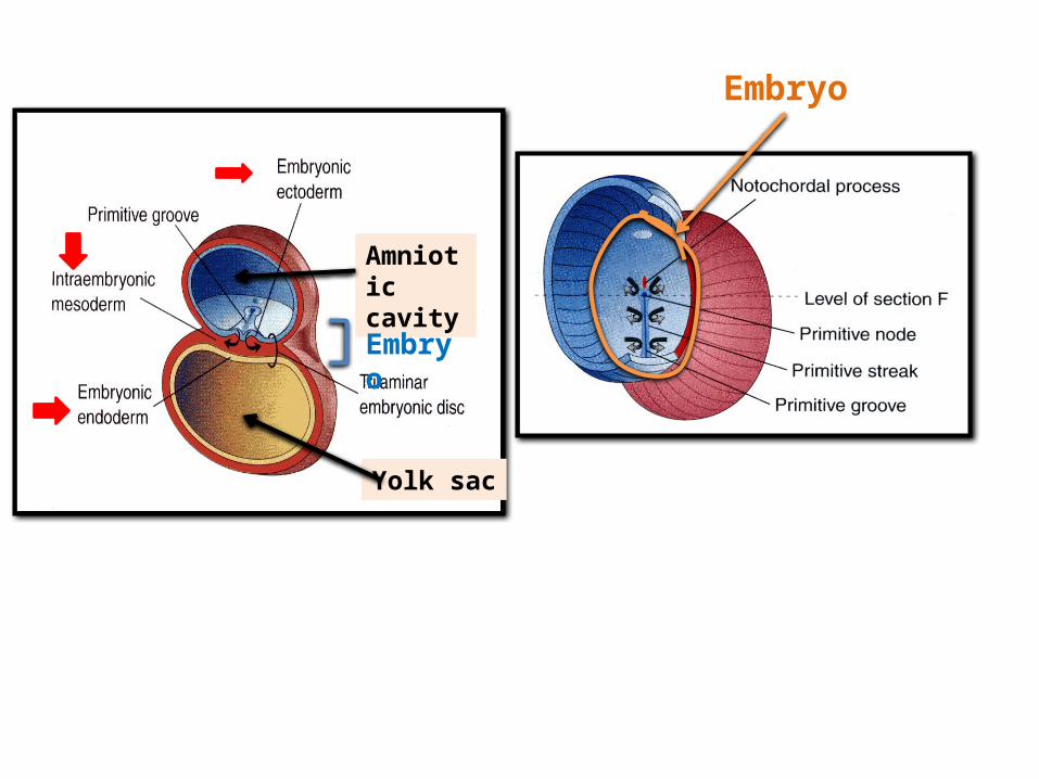
Amniotic cavity
Yolk sac
Embryo
Embryo
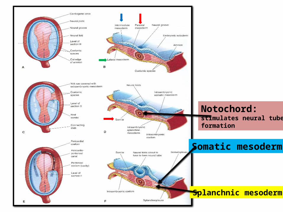
Notochord: stimulates neural tube formation
Somatic mesoderm
Splanchnic mesoderm

INTRAEMBRYONIC MESODERMProliferates between Ectoderm & Endoderm EXCEPT in
the central axis of embryo where NOTOCHORD is found.Differentiates into 3 parts:1. Paraxial mesoderm: on each side of notochord.2. Intermediate mesoderm3. Lateral mesodermParaxial mesoderm divides into units (Somites).Lateral mesoderm divided by intraembryonic coelom
into:1. Somatic mesoderm (between ectoderm & coelom).2. Splanchnic mesoderm (between endoderm & coelom).
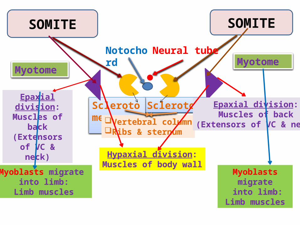
SOMITE SOMITE
Notochord
SclerotomeSclerotome
Neural tube
Myotome
Vertebral columnRibs & sternum
Epaxial division:Muscles of back
(Extensors of VC & neck)
Hypaxial division:Muscles of body wall
Myotome
Myoblasts migrate into limb:
Limb muscles
Myoblasts migrate into limb:
Limb muscles
Epaxial division:Muscles of back
(Extensors of VC & neck)

DEVELOPMENT OF LIMBS - 1
28 DAYS
32 DAYS
The limb Bud appears as an elevation on the ventrolateral body wall resulting from proliferation of mesenchyme of the somatic layer of lateral mesoderm.Each limb bud is surrounded by an area of ectoderm.Upper limb buds Appear at day 26 opposite the lower cervical segments.Lower limb buds Appear at day 28 opposite the lumbar & sacral segments.
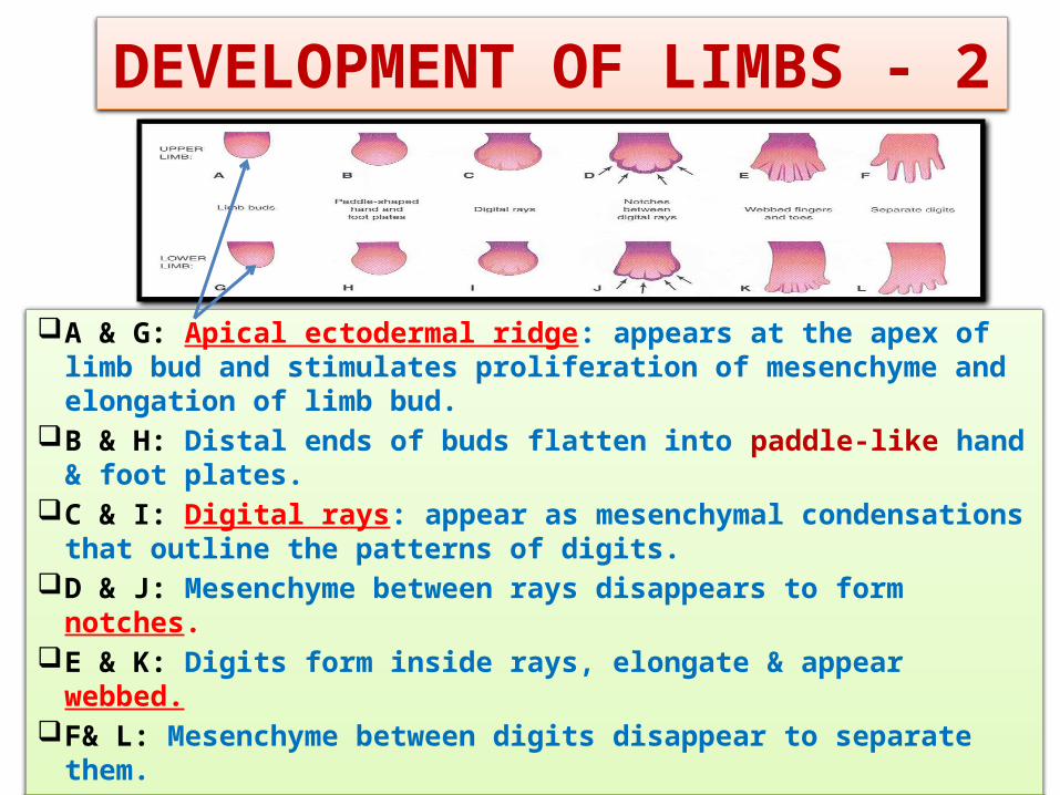
DEVELOPMENT OF LIMBS - 2
A & G: Apical ectodermal ridge: appears at the apex of limb bud and stimulates proliferation of mesenchyme and elongation of limb bud.
B & H: Distal ends of buds flatten into paddle-like hand & foot plates.
C & I: Digital rays: appear as mesenchymal condensations that outline the patterns of digits.
D & J: Mesenchyme between rays disappears to form notches. E & K: Digits form inside rays, elongate & appear webbed. F& L: Mesenchyme between digits disappear to separate them.
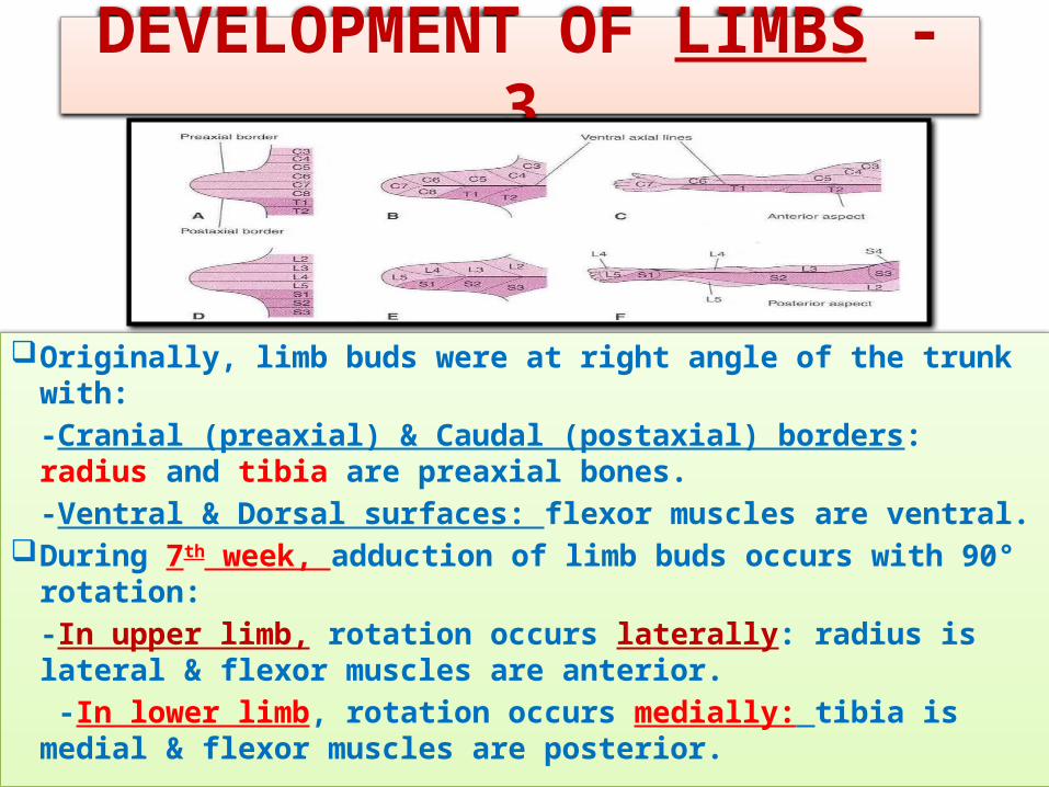
DEVELOPMENT OF LIMBS - 3
Originally, limb buds were at right angle of the trunk with:-Cranial (preaxial) & Caudal (postaxial) borders: radius and tibia are preaxial bones.-Ventral & Dorsal surfaces: flexor muscles are ventral.
During 7th week, adduction of limb buds occurs with 90° rotation:-In upper limb, rotation occurs laterally: radius is lateral & flexor muscles are anterior. -In lower limb, rotation occurs medially: tibia is medial & flexor muscles are posterior.

Mesenchyme from lateral mesoderm
Induces growth of mesenchyme & its transformation into cartilage
Cartilage ossifies by:Endochondral ossification
Myoblasts migrate from myotomes to form:Muscles of limbs

Bone in cartilaginous state
Appearance of primary ossific centers: ossification of diaphysis
Appearance of secondary ossific centers: ossification of epiphysis
Ossification of epiphseal plate: Complete union of epiphysis & diaphysis
Diaphysis
Epiphysis
Epiphyseal plate of cartilage
BIRTH PUBERTY
Diaphysis
Bone increases in length by proliferation of epiphyseal plate
Growth of bone stops
OSSIFICATION OF LONG BONES
Bone age is a good index of general maturation. Bone age is determined by:1. Appearance of ossific centers in diaphysis & epiphysis (specific for each bone & sex)2. Disappearance of epiphyseal plate (specific for each bone & sex)
Epiphysis

DEVELOPMENT OF CRANIUM (SKULL)
The skull develops from mesoderm around the developing brain.
The skull consists of:
1. Neurocranium: protective case for brain
2. Viscerocranium: skeleton of face Bones of skull ossify either by:
*Endochondral ossification or
*Intramembranous ossification
12
FP
ST Z MaxMand
F
F
F
P
P
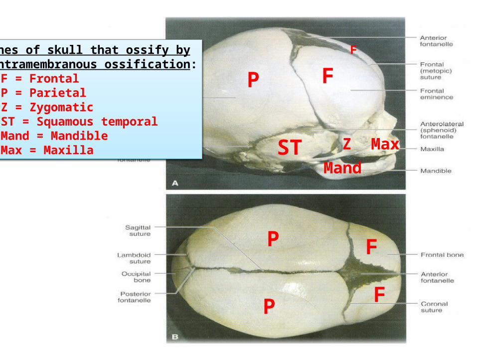
Bones of skull that ossify by intramembranous ossification:1. F = Frontal2. P = Parietal3. Z = Zygomatic4. ST = Squamous temporal5. Mand = Mandible6. Max = Maxilla

SUMMARY OF DEVELOPMENT OF BONE
All bones develop from MESODERM.AXIAL SKELETON:
*Vertebrae, Ribs & Sternum: from Sclerotomes of Somites (Paraxial Mesoderm)
*Skull: from Mesoderm surrounding the BrainAPPENDICULAR SKELETON: from Somatic part
of Lateral Mmesoderm
All bones ossify by Endochondral Ossification EXCEPT:
1. Some bones of Skull
2. Clavicle

JOINTS
They develop from mesoderm between bones:Fibrous joints: mesoderm differentiates into
dense fibrous connective tissue.Cartilaginous joints: mesoderm differentiates
into cartilage.Synovial joints: a synovial cavity is formed
inside mesoderm; mesoderm differentiates into synovial membrane, capsule & ligaments.

SUMMARY OF DEVELOPMENT OF
MUSCLESAll muscles develop from MESODERM EXCEPT:1. Muscles of iris (eyeball)2. Myoepithelial cells of ECTODERM
mammary & sweat glands All skeletal muscles develop from myotomes
of paraxial mesoderm EXCEPT: some Head & Neck muscles from mesoderm
of Pharyngeal Arches

SUMMARY OF DEVELOPMENT OF MUSCLES
Cardiac & Smooth muscles develop from lateral mesoderm:
1. Cardiac muscles from: splanchnic part of lateral mesoderm
2. Smooth muscles:*In the wall of viscera from: splanchnic part of lateral mesoderm* In the wall of blood & lymphatic vessels from: somatic part of lateral mesoderm
QUESTION 1
Which one of the following group of muscles are derivatives from epaxial division of myotomes?
1. Muscles of back2. Muscles of limbs3. Muscles of viscera4. Cardiac muscles
QUESTION 2
Which one of the following bones ossifies by intramembranous ossification?
1. Vertebra2. Humerus3. Ribs4. Mandible
QUESTION 3
Regarding the ossification of long bones, which one of the following statement is correct?
1. Primary ossific centre appears after birth.2. Secondary ossific centre leads into
ossification of diaphysis.3. Long bones ossify by intramembranous
ossification.4. When epiphysis unites with diaphysis, growth
of bone stops.

THANK YOU