Embed Size (px)
Citation preview

© A. L. Neill 5© A. L. Neill 4
The A to Z of Blood
BLOOD GROUPS 164
BLOOD PRESSURE see Blood Flow
BLOOD SPECIMENS
collection capillary puncture ............................................................................................168 venipuncture ....................................................................................................... 170 preparation smears see also Blood Cells ..........................................................................174
BLOOD TESTS
cell counts / stains .................................................................................................. 178 measurements clotting / coagulation see Coagulation RBCs - anaemias ..............................................................................................184 WBCs - leukaemias .........................................................................................186
BONE MARROW
blood supply ................................................................................................................188 cellularity ....................................................................................................................190 organization / structure ......................................................................................194 specimens / biopsies see also Blood Specimens .......................................200
COAGULATION / ANTICOAGULATION
EDTA ................................................................................................................................206 factors - Clotting Factors F1-F13 ....................................................................208 other regulators ................................................................................................229 overview ........................................................................................................................242 fibrin ...............................................................................................................................244 platelets .......................................................................................................................246 tests ...............................................................................................................................248
COPPER see also Iron
copper metabolism .................................................................................................250
ERYTHROPOIESIS see Blood Formation
ERYTHROPOIETIN see Blood Formation
FERRITIN see Iron
HAEM
breakdown ...................................................................................................................252 O2 saturation levels ................................................................................................253 states ..............................................................................................................................254 synthesis .......................................................................................................................258
HAEMODIALYSIS 264
HAEMOGLOBIN
break down - formation of bile see Haem, Blood Cells- RBCs development of ........................................................................................................266 synthesis .....................................................................................................................268
HAEMOLYSIS see RBC destruction
HAEMOSTASIS see also Coagulation 270
HAEMOSIDERIN see Iron
HAEMOZOIN 272
HEPARIN see Coagulation
IRON see also Haem
divalent metal transporter ................................................................................276 ferritin ...........................................................................................................................278 haemosiderin ............................................................................................................280 hepcidin & ferroportin hephaestin see transport homeostatic protein controls of Fe - overview .......................................282 transferrin & transferrin receptors see also transport ...........................284 transport ......................................................................................................................286
MEGAKARYOCYTES see also Platelets 292
MYOGLOBIN see Haem
PLASMA see Blood Proteins
SPLEEN 292
VITAMIN K see also Blood Proteins & Coagulation 298
copyright
Dr A. L. Neill

© A. L. Neill 33© A. L. Neill 32
The A to Z of Blood
Erythrocyte AKA Red Corpuscle AKA red blood cell (RBC): Gk erythros = red + kytos = hollow vessel, changed to -cyte = cell see Red Blood Cell for all alternative names
Erythrocytic Sedimentation Rate (ESR): the rate at which the RBCs move to the bottom of a testube, is indicative of the viscosity of the B - & the number of additional PP present which indicates an upgrading of the ImR / IfR & so the presence of disease
Erythrogenic: produces erythrocytes.
Erythroleukemia: an abnormal condition characterized by proliferation of erythroblastic & myeloblastic cells.
Erythropaenia: a decrease in the number of RBCs in the B.
Erythropoiesis: the production of erythrocytes (RBCs) see MT for more details.
Erythropoietin (EPO) AKA Haematopoietin AKA Haemopoietin: a H produced primarily by the kidneys, stimulates erythropoiesis ( RBCs) see MT for more details
Etiology AS Aetiology
Extracellular (EC): occurring outside the cell
Extravascular: occurring outside the CVS
Extrinsic pathway AKA Tissue Factor Pathway: The main role of this pathway is to facilitate a rapid release of F2a - i.e. THROMBIN. It does this via F7a, the most concentrated CF in the B, & its reaction to F3a - TISSUE FACTOR which is present at the site of any BV damage.
F7a/F3a form a complex which activates F9 & F10, which then activates F2 - beginning a rapid cascade of activating CFs & forming a BClot at the site of injury see MT for more details
Factor (F) AKA Clotting factor (CF): a protein in the B that is needed to form a BClot see MT for more details
Ferritin: a large hollow protein which stores Fe & acts as an indicator of Fe stores in the body. It is composed of 24 identical protein subunits that form a hollow shell, enclosing the Fe to protect Ts from its toxic properties. It is released from the liver & recycled to the RBMa for erythropoiesis see MT for more details
Fibril: a microscopic filament often composed of fibrin.
Fibrin (F1a) see Fibrinogen
Fibrinogen (F1): the inactive form of fibrin. It has α & β globular nodules connected via coiled proteins(C) which join on either side of a central connector(CC) & see also Clot & MT for more details
Fibrinolysin AKA Plasmin
Folate AKA folic acid AKA Vita B9 AKA Vita Bc AKA Vita M: an essential Vita closely associated with Vita B12 & necessary for the formation of RBCs. Absence or inability to process these substances (Folate & Vita B12) results in a hyperchromic, macrocytic anaemia, & may give rise to birth defects.
Fragility Test (Osmotic): a test devised to measure the resistance of the RBCs to break down (haemolyze) when subjected to varying concentrations of hypotonic salt solutions.
Fulminating: sudden & severe.
Think Lance Armstrong
Fe
P
α
β
C CCC
copyright
Dr A. L. Neill

© A. L. Neill 35© A. L. Neill 34
The A to Z of Blood
Ghost cells AKA Lysed cells AKA Selenocytes: CM of lysed RBCs in part or complete, where the contents have been removed or leaked away
Gilbert's syndrome (GS): a genetic liver disorder in 3-12% of the population. Elevated levels of BR-U are produced in the BS (hyperbilirubinaemia) due to of glucuronyltransferase, which conjugates BR (BR-C), (making it water soluble). SS mild jaundice, particularly when stressed. see MT for more details
Giemsa stain (GEEM-suh): is a stain used in cytogenetics & to stain: BSms, BMa smears & to demonstrate the histopathological Dx of malaria & other parasites. Similar but more stable than Wright's stain, it demonstrates different IC structures also human & bacterial cells see MT for more details
Globular protein: a protein which can disperse in H2O to form a colloid e.g. Hb see also Haemoglobin
Glycohaemoglobin (A1cHb): a form of Hb used to measure the average B[glucose] levels for the past 3 months – the average life of RBCs see also A1c test.
Granulation: the accumulation of proteins, lipids, carbohydrates, pigments, & secretory granules w/n the cytoplasm
Granulocyte: a type of WBC that contains specific cytoplasmic granules which are peroxidase positive. Granulocytes include neutrophils, eosinophils, & basophils
Golgi Apparatus (GA): a meshwork of membrane plates w/n the cytoplasm; used to facilitate protein transport.
Granulocytopaenia AKA Granulopaenia: a in the number of granulocytes in the B.
Granulocytosis: the presence of numbers of granulocytes in the B.
Granulopoiesis: the production of granulocytes.
Haem AS Haeme AKA Heme (HEEM): Gk haima = blood is a cofactor for many enzymes, made up of porphyrin ring with a central Fe2+, held in place by the 4 pyrrolic groups, joined by methine rings. Its main use is, as the O2 carrier of the RBC but it is also used in a number of other reactions in the body, relying upon the ability of Fe2+ to further oxidise to Fe3+ . Haem(1) is at the centre of most metalloproteins, i.e. haemoproteins, the most common being Haemoglobin (Hb) of RBCs. It is also present in the following enzymes: catalase, cytochrome P450(2), endothelial NO synthetase, haem peroxidases, & myoglobin
Haematein: a dark purple water-insoluble crystalline substance used as an indicator & biological stain.
1
2
copyright
Dr A. L. Neill

© A. L. Neill 127© A. L. Neill 126
The A to Z of Blood
Platelets AKA Thrombocyte (Plt)
Structure
The platelet is a small discoid piece (1-4µ diameter) of a megakaryocyte. It is not a true cell. When activated it is called a thrombocyte but these terms are used interchangeably. It lives for 7-10 days. A megakaryocyte forms 2-5000/cell.
It plays a major role in the formation of a clot in both pathways but mainly the extrinsic pathway. There are 3 zones in the platelet -
the structural zone = containing 3 types of fibrils (1,2 & 3)
the peripheral zone or hyalomere = relatively clear of organelles (6-7) with light blue cytoplasm
and the centrally located
the organelle zone or chromomere = a granular central region with specific granules related to activating the Plt.
The CM has a glycocalyx coating, increasing its stickiness, & the extensive canaliculi & tubules - allow for rapid transport of Ic substances to the surface - a well developed fibrillar system allows for extensive morphological changes, upon activation.
1 microtubules 2 actin 3 myosin (II)4 glycocalyx - coating outside the CM 5 CM 6 open canalicular system - indenting of the CM to
invaginate shallowly into the cytoplasm - few other organelles
7 dense tubular system - deeper CM invagination - to the chromomere zone
8 δ granules 9 mitochondria 10 α granules 11 glycogen granules 12 λ granules (lysosomes)
1
2
3
6
4
7 6 8 10 9 5 4
128
10
11
copyright
Dr A. L. Neill

© A. L. Neill 175© A. L. Neill 174
The A to Z of Blood
Blood Specimens, Blood Smears (BSms)
Preparation
BSms obtained from any free-flowing B are similar, but capillary specimens from cyanotic, calloused or areas of local stasis, will have higher WBC counts. Hence venous samples are preferred.
A small drop of B no larger than 3-4mm in diameter is placed near one end of a clean glass slide.
Using a second slide at about 30º move it up to the B drop & spread the drop to the edges of both slides before slowly smoothly & lightly pushing the spreader slide towards the opposite end, leaving a thin B film - the Blood Smear (BSm), with a feathered end. The BSm should be labelled (with a pencil to stop the label being lost during the staining process) & completely air-dried before fixing & staining.
Note fixing should occur w/n 1 hour of collection as the WBCs will start to deteriorate after this time. B from a patient on anticoagulant Tx may have varied WBCs morphology, although B samples anticoagulated with EDTA do not show any differences.
Stains - Giemsa, Wright's stains
Giemsa & Wright's stains are differential polychromic stains; simple histologic dye mixtures which facilitate the differentiation of BC types. Classically they are mixtures of eosin (red) - an acidic dye which stains the background & cytoplasm various red shades & methylene blue dyes (blue) - basic dyes, which stain the nucleic & other acids blue.
They are used to stain BSms & BMa aspirates. WBCs are mostly identified by their preference for these dyes, & named after this preference e.g. granules in WBCs with an equal preference for acid & basic dyes are called neutrophils; eosinophils are WBCs with intensely red (basic) granules in their cells, & basophils are the opposite.
Wrights stain, appears similar to the T stains H&E. It is less stable & deteriorates in an alkaline solution. Giemsa, has an added dye Azure B to compensate for this defect, which also accentuates the azurophilic staining (blue) properties, present in lymphocytes. It is also used to identify B parasites, particularly malarial parasites.
2
13
4
1 base slide 2 Blood drop 3 spreader slide 4 BSm
Type of BC or component
Balanced / Good stain
too Acidic too Acidic
RBCs pink - orange
bright red blue - green
nuclei purple - blue
pale blue deep dark blue
eosinophilic granules
granules red
brilliant red deep grey - blue
neutrophilic granules
violet - pink pale pink dark blue
lymphocyte cytoplasm
blue pale blue dark blue
copyright
Dr A. L. Neill

© A. L. Neill 201© A. L. Neill 200
The A to Z of Blood
Bone Marrow (BMa) Specimen Site & Procedure
A sites of RBMa in adult B position to take iliac sample C procedure to obtain aspirate sample D procedure & equipment for Bone Bx E Bone Bx sample
BMa is commonly taken from either the Sternum(1) or Ileum(2) of the hip. In adults the main RBMa is in the axial skeleton. The patient is placed on their side, & the needle for an aspiration approaches inferiorly to the iliac ridge. The needle pierces the outer cortical bone & then sucks up a BMa sample, containing the cells of the BMa but not necessarily the stromal support & endosteal niches. With a Bx the needle(4) is wider & cutting blades(6) are inserted to form a core which is then retrieved after removal of the cutting blades. This sample maintains the structure of the BMa.
1 Sternum - site of RBMa - samples taken from here tended to scar
2 Iliac crest - preferred site for RBMa sampling 3 Sylet 4 Bx needle 5 Obturator 6 Cutting blade
1
2
A
B
C
2
D
3 4 5 6
E
copyright
Dr A. L. Neill

© A. L. Neill 251© A. L. Neill 250
The A to Z of Blood
Copper - Cu
Metabolism
Cu & Fe metabolism are interdependent, but unlike Fe there is an adequate way of eliminating excess Cu from the body. Like Fe it is toxic to the body & only small amounts are tolerated in the BS as a free ion. Most of ingested Cu passes through the body, 95% of the rest is combined with Ceruloplasmin with small amounts found in macroglobulins. Cu deficiency leads to Fe deficient anaemia.
1 average intake of Cu is approx 2mg / day
2 25% is transported to the liver
3 in the liver it is combined with Ceruloplasmin
4 excess Cu is excreted with bile from the GB
5 95% of Cu in the B is bound to ceruloplasmin
6 < 5% of Cu circulates as free Cu in the serum
7 < 5% of Cu is excreted via the urine
8 50% combines with metallothioneins in the duodenum, which is excreted in the faeces
9 25% is excreted in the faeces directly
1
5
23
46
7
9
8
8
8
8
copyright
Dr A. L. Neill

© A. L. Neill 295© A. L. Neill 294
The A to Z of Blood
Spleen
Surface Anatomy & Splenomegaly
The spleen is like a large bag of B, in a thin capsule normally protected by the rib cage. In parasitic Ins (e.g. malaria) it may swell up to 5X and then becomes vulnerable when there is an abdominal trauma to Iy which may result in death, from internal Hg.
1 spleen - gastric surface 2 spleen - renal surface 3 gastrolienal lig 4 splenic a&v5 L lung 6 spleen / m = splenomegaly - ie enlarged outlines 7 diaphragm 8 small intestine 9 rib 9 10 rib 1011 rib 11 12 large intestine13 stomach 14 liver
5
6
7
6m
8
14
13
12
9
10
11
52
3
4
copyright
Dr A. L. Neill
© A. L. Neill 299© A. L. Neill 298
The A to Z of Blood
Vitamin K (Vita K)
Role in protein synthesis
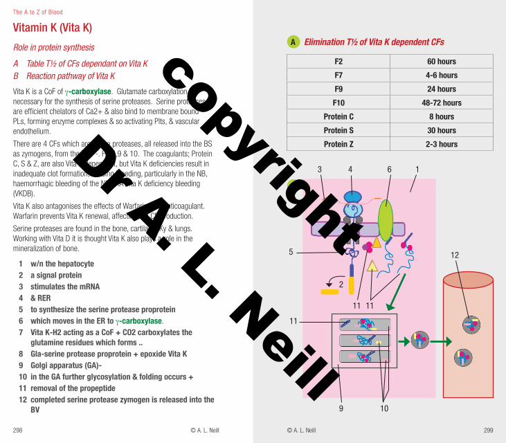
A Table T½ of CFs dependant on Vita K B Reaction pathway of Vita K
Vita K is a CoF of γ-carboxylase. Glutamate carboxylation is necessary for the synthesis of serine proteases. Serine proteases are efficient chelators of Ca2+ & also bind to membrane bound PLs, forming enzyme complexes & so activating Plts, & vascular endothelium.
There are 4 CFs which are serine proteases, all released into the BS as zymogens, from the liver -. F2,7,9 & 10. The coagulants; Protein C, S & Z, are also Vita K dependant, but Vita K deficiencies result in inadequate clot formation, causing bleeding, particularly in the NB, haemorrhagic bleeding of the NB AKA Vita K deficiency bleeding (VKDB).
Vita K also antagonises the effects of Warfarin - an anticoagulant. Warfarin prevents Vita K renewal, affecting the CF production.
Serine proteases are found in the bone, cartilage, Ky & lungs. Working with Vita D it is thought Vita K also plays a role in the mineralization of bone.
1 w/n the hepatocyte 2 a signal protein 3 stimulates the mRNA 4 & RER 5 to synthesize the serine protease proprotein 6 which moves in the ER to γ-carboxylase. 7 Vita K-H2 acting as a CoF + CO2 carboxylates the
glutamine residues which forms .. 8 Gla-serine protease proprotein + epoxide Vita K 9 Golgi apparatus (GA)- 10 in the GA further glycosylation & folding occurs +11 removal of the propeptide 12 completed serine protease zymogen is released into the
BV
12
A
B
Elimination T½ of Vita K dependent CFs
F2 60 hours
F7 4-6 hours
F9 24 hours
F10 48-72 hours
Protein C 8 hours
Protein S 30 hours
Protein Z 2-3 hours
3 4 6 1
5
11 11
9 10
11
2
copyright
Dr A. L. Neill