Embed Size (px)
Citation preview
J Clin Pathol: Mol Pathol 1995;48:M142-M147
Non-invasive detection of bladder cancer byidentification of abnormal CD44 proteins inexfoliated cancer cells in urine
M Sugiyama, A Woodman, T Sugino, S Crowley, K Ho, J Smith, Y Matsumura,D Tarin
AbstractAims-To investigate the expression ofCD44 proteins in exfoliated urothelial cellsand in tumour tissues from bladder cancerpatients. A further objective was to eval-uate the diagnostic potential ofthe changesobserved in the expression of these pro-teins as a marker for non-invasive de-tection of bladder cancer.Methods-Naturally voided urine speci-mens were collected from 47 patients withbladder cancer or severe urothelial dys-plasia (n = 3) and from a control group of43 people with no evidence of neoplasticdisease. Exfoliated urothelial cells floatingin the urine were pelleted by centrifugationand lysed, and their constituent proteinsextracted. The pattern of expression ofCD44 proteins in each sample was ex-amined by western blot analysis using amonoclonal antibody, Hermes 3, which re-cognises an epitope on the polypeptidebackbone of the CD44 protein. Immuno-histochemical studies were performed onneoplastic (n = 10) and normal (n = 4) blad-der tissue specimens which were snapfrozen in liquid nitrogen before ex-amination with antibodies to CD44 geneproducts (CD44s and CD44v6).Results-Western blot analysis revealedseveral high molecular weight CD44 iso-forms >160 kDa in urine cell lysates from75% of patients with histologically con-firmed bladder cancer and in two of thethree patients with severe dysplasia. Suchpatterns were not detected in the urine cellpellets from any persons in the controlgroup. Immunohistochemical studies ofthe tissue distribution of CD44s andCD44v6 showed that the differentiationand maturation ofthe epithelial cells in thenormal bladder mucosa is accompanied bya decrease in CD44 protein expression.However, carcinoma cells overexpressstandard and variant CD44 isoforms andcontinue to do so as they proceed throughthe thickened epithelial layer to the lu-minal surface and after they are shed intothe urine.Conclusions-The abnormal expression ofCD44 proteins in exfoliated cancer cellsmay be a useful marker for the non-invasive diagnosis of bladder cancer.(J Clin Pathol: Mol Pathol 1995;48:M142-M147)
Keywords: Bladder cancer, exfoliated cancer cells,CD44 protein.
CD44, a widely distributed cell surface glyco-protein regarded as an extracellular adhesionmolecule, is generated as various isoforms byalternative splicing of the products of at least10 variant exons (CD44v) into a 37 kDapolypeptide backbone encoded by nine other,constitutively expressed, exons (CD44s) .
The dominant isoform expressed in mostnormal cell types is the 90 kDa "standardform" produced by heavy glycosylation of thebasic backbone, but differing quantities ofthe individual variant isoforms, which aremuch larger, can also be detected in severalnormal tissues and organs.45 The types ofvariants produced differ according to the siteof origin of the tissue.We and other groups have reported marked
changes in CD44 gene splicing patterns invarious types of human cancer.6-" These con-sist of a general increase in CD44 mRNAtranscription with a progressive and dis-proportionate increase in variety and quantityof CD44v isoforms as malignancy advances.Immunohistochemical analysis has indicatedthat there is also a good correlation betweenthe altered quantity and distribution of CD44variant forms (CD44v), particularly CD44v6,and tumour progression.'012 These obser-vations suggest the possibility that evaluationofCD44 gene activity could be useful in tumourdiagnosis and evaluation of prognosis.
Early detection of neoplasia is an importantobjective because many cancers can be curedby surgical resection if diagnosed at an earlystage. Bladder cancer is a common form ofmalignancy in which early diagnosis of primarytumours is difficult because of their in-accesibility. As the lesions have a strong tend-ency to recur, monitoring asymptomaticpatients for recurrence is also problematic. Cur-rently, urography and cystoscopy are the mainmethods for investigating symptomatic disease.However, because they are uncomfortable andlabour intensive procedures, they are not op-timal for routine monitoring of asymptomaticindividuals who may be at risk of primary orrecurrent tumours, or for general screening.Urinary cytology is simple and non-invasivebut has insufficient sensitivity for routine gen-eral use, although it has a place for monitoringhigh risk populations in certain occupationalgroups. It is also expensive and labour intensiveand a realistic alternative would be welcome.Consequently, there is an urgent need for asensitive, reliable, and non-invasive method ofdetection of this disease.
Nuffield Departmentof Pathology andBacteriology(University of Oxford),Oxford RadcliffeHospital, Headington,Oxford OX3 9DUM SugiyamaA WoodmanT SuginoS CrowleyY MatsumuraD Tarin
Department ofUrology, OxfordChurchill Hospital,Headington,Oxford OX3 7LJK HoJ Smith
Correspondence to:Dr Tarin.Accepted for publication20 March 1995
M142
group.bmj.com on October 24, 2017 - Published by http://mp.bmj.com/Downloaded from
Diagnosis of bladder cancer from CD44 proteins in exfoliated cells
In a study which is relevant to this clinicalrequirement, we recently reported abnormaltranscription of the CD44 gene in exfoliatedcancer cells in urine specimens." However, asmRNA is unstable and the technology for itsanalysis is specialised, routine tests based uponthis analyte are not yet widely available. Anassay based upon detection of CD44 proteinmay circumvent these problems. This led usto investigate, by western blotting, whetherthere were any specific abnormalities in CD44protein profile of desquamated urothelial cellsfrom bladder cancer patients which could be auseful marker for early diagnosis. We now pres-ent the results of a study of urine specimensfrom 44 bladder cancer patients, three patientswith severe urothelial dysplasia, and 43 peoplewithout malignancy. In this report the datafrom western blotting procedures are comparedwith the results obtained with immuno-histochemical techniques on the quantity anddistribution of CD44s and CD44v6 isoformsin normal and cancerous bladder tissue bi-opsies. We chose to study the status of ex-pression of exon v6 (exon 11) in these tumoursamples because previous studies, cited above,had indicated that increased expression of thistranscript/epitope correlates with tumour pro-gression.The results indicate that it should be possible
to design an accurate test for bladder cancerbased upon abnormal CD44 gene expression.
MethodsPREPARATION OF URINE CELL LYSATESNaturallyvoided urine specimens ofabout 50 mlvolume were collected from 47 patients withbladder cancer or severe urothelial dysplasia (37male; 10 female) and 43 persons with no evid-ence ofmalignant disease (28 male; 15 female).The urine was collected into vessels containing0-02mM amidinophenylmethane sulphonylfluoride (APMSF) (Sigma) and 1 mM EDTA
(Sigma), as protease inhibitors, and kept on iceduring transport to the laboratory.The cell pellets collected by centrifugation
at 3000 rpm for 15 min were lysed with 1:1(wt/vol) of lysis buffer (20mM tris [pH 8 0],150mM NaCl, 20mM CHAPS, 2mM EDTA,0 04mM APMSF, 20 ,ug/ml aprotinin) on icefor 15 minutes. This lysate was clarified bycentrifugation (12 000 rpm, 15 min). The con-centration of protein was determined (Bio-Radprotein assay kit) and adjusted to 5,g/lane bydilution with phosphate buffered saline con-taining 0-02mM APMSF and 1 mM EDTA.Where necessary, samples were concentratedusing a Microcon 30 membrane (Amicon).
Urine cell smears were prepared by washingthe samples (3000 rpm) with Hank's bufferedsalt solution (HBSS), and spotting the cellsonto poly l-lysine coated slides before fixing airdried preparations in 100% methanol at 4°C.All smears were stored at - 20°C until re-quired.
WESTERN BLOT ANALYSISSamples were boiled with gel loading buffer andsubjected to 6% SDS-PAGE under reducingconditions. The separated proteins were elec-troblotted (100 mA, one hour) onto im-mobilon-P membranes (Millipore) usingtransfer buffer (48mM tris, 39 mM glycine,0-1% SDS, 20% methanol, pH 9 2). Non-specific reactions were blocked with Trisbuffered saline (TBS) containing 5% skimmedmilk before the membrane was incubated withmonoclonal antibody Hermes 3 (anti-CD44s;epitope encoded by exon 5) at 4°C overnight,and then with peroxidase conjugated anti-mouse IgG (Sigma; 1/1000 dilution) for onehour at room temperature. All antibodies werediluted in 5% skimmed milk in TBS and aftereach incubation the membrane was washedwith TBS containing 0-1% Tween 20. Signalswere detected by enhanced chemiluminescencewith an ECL detection kit (Amersham).
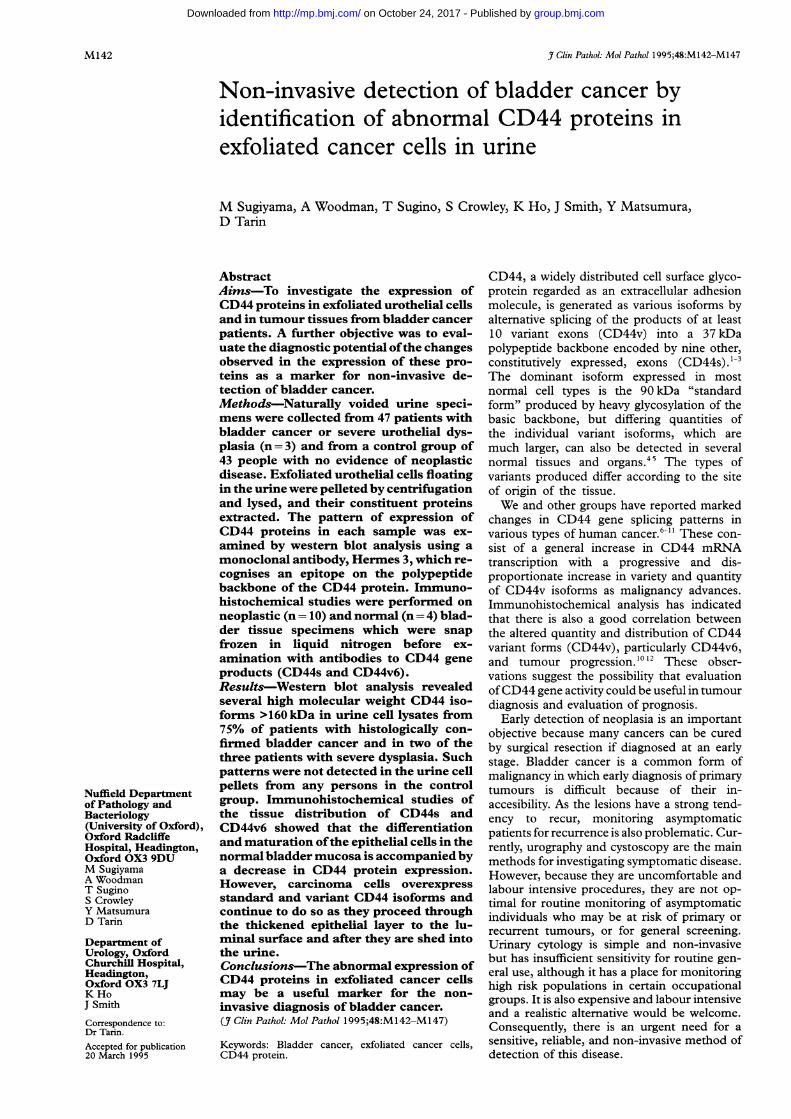
1 2 3 4 5 6 7 8 9 10 11
t._ m_
0 0
Figure 1 Western blot analysis of cell lysates with Hermes 3 (CD44s). "dominant CD44 proteins. Lanes 1, 2, and 3 show results with cell lysatescolon cancer cell line (0 5 pg/lane), RT112 bladder cancer cell line (0-5 pnormal human leucocytes (1 pg/lane), respectively. Major protein bands o- 220 kDa (plus - 85 kDa), and - 90 kDa are seen in HT29, RT112,
respectively. The 90 kDa band corresponds to CD44s, the 150 kDa bandform of CD44, and the 220 kDa band to the largest variant form. The ufrom normal subjects (lanes 4-7) contain one to three band(s) ofMW -
- 120 kDa, whereas several bands of - 70 kDa to - 220 kDa are seen icancer patients (lanes 8-11). Total protein loaded in each lane = S pg.
IMMUNOHISTOCHEMISTRYCystoscopic biopsies and/or surgical resectionspecimens of bladder carcinoma to be used
- 220 kDa for immunohistochemistry (n = 10) were snap4- 170 kDa frozen in liquid nitrogen. Samples of non-neo-
150 kDa plastic urothelial mucosa (n = 4) were obtained- 150 kDa from ureters attached to nephrectomy speci-
*-- 120 kDa mens, from a cadaveric bladder specimen, and
-4- 90 kDafrom a bladder biopsy for a non-malignantcondition. Frozen sections (5 gim) were fixedin cold methanol and preincubated with 20%
.4 70 kDa normal rabbit serum in TBS at 37°C for 45minutes before incubation with the primarymonoclonal antibodies Hermes 3 (CD44s) or2F10 (CD44v6) (R and D Systems) at 4°C
4rrows show overnight and at 37°C for 30 minutes nexts from HT29 morning. Endogenous peroxidase activity wasigilane), and blocked with 0 3% H202 in methanol for 10)f -.lS0kDa,and leucocytes, minutes before addition of biotinylated anti-to the epithelial mouse IgG (Dako; 1/400 dilution; room tem-rine specimens perature, two hours), followed by horseradish7
specimens from peroxidase conjugated ABComplex (Dako;room temperature, one hour). Immunostaining
194 kDa-
116 kDa
85 kDa
M143
group.bmj.com on October 24, 2017 - Published by http://mp.bmj.com/Downloaded from

Sugiyama, Woodman, Sugino, Crowley, Ho, Smith, et al
Results of western blot analysis in urine samples from bladder cancer patients and normalvolunteers. As discussed in the text, samples which showed several bands of over 160 kDawere judged to be positive and those with band(s) < 120 kDa negative in western blottingusing monoclonal antibody Hermes-3 to CD44s
Sex and Histological stage Results inPatient No age (years) and grade of tumours? western blot
1 M29 pT4G3LO +2 M52 pTaGI +3 M72 pTaGl4 F70 pTlGILO5 F63 pTlGlL0 +6 F85 Severe dysplasia +7 M60 pT1G3LO +8 F79 pT2G39 M54 pTlG2L0 +
10 M81 pTlG2L0 +11 M76 pTlG2 +12 M78 pT2G2LO +13 F73 pTlG2LO14 M57 pTlGIL0 +15 F79 pTlGlLO +16 F51 pTlGlL017 M73 pT2G3LO +18 M74 pTlGlLO +19 M82 pTaGILO +20 M68 pTaG1L021 M81 Cancer, NS22 M76 pT1G3L0 +23 M84 pT1G1LO24 M80 pT2G3LO +25 M68 pT1G2LO +26 M72 pTlG2L0 +27 M70 No tumour28 M46 pT1G2LO +29 M73 pT2G2LO +30 M76 pT1G2LO31 M75 pTIGlLO +32 F73 pT1G2LO +33 M69 Severe dysplasia +34 M83 pTlGlLO +35 M56 pT2GlLO36 F82 Severe dysplasia37 M78 pTlGlL0 +38 M72 NS39 M70 pT2G3LO +40 M56 pTlG2L0 +41 M71 pT1G2LO +42 F54 pTIGILO +43 M64 pTlGlLO +44 F74 No tumour45 M54 pTaGlLO +46 M48 pTlG2LO +47 M88 pTlG2L0 +48 M83 pTaG2LO +49 M78 pTlG2LO +
Cancer patient total 44 Numbers positive - 33 (75%)Severe dysplasia 3Normal volunteer total 41 (M18-62, F19-52) Numbers positive - 0 (0%)Non-neoplastic at 2 (M70, F74)cystoscopy
aTumour stage and grade were evaluated according to classification of International Union againstCancer (IUCC): pTa =papillary non-invasive cancer; pT1 = tumour not extending beyond thelamina propria; pT2 = tumour invading superficial smooth muscle; pT3 = tumour invading deepmuscle; pT4 =tumour invading surrounding pelvic tissue; Gi =high degree of differentiation;G2 =medium degree of differentiation; G3 = low degree of differentiation; LO = no lymphaticinvasion; NS= not pathologically staged; +, positive case; -, negative case.
was visualised using diaminobenzidine/H202(Sigma) as substrate and sections were coun-
terstained with haematoxylin. Between all anti-body incubations, sections were washed by3 x 5 minute incubations with TBS on a shak-ing platform. Urine cell smears made fromcell pellets of normal individuals (n = 5) andtumour bearers (n = 15) were also treated withthe above protocol.
ResultsANALYSIS OF URINE CELL SEDIMENTS USINGWESTERN BLOTTING
Western blot analysis of exfoliated urine celllysates from patients with bladder cancer (n =44) using monoclonal antibody Hermes 3(CD44s) showed several high intensity bandswith molecular weights ranging from - 70 kDato 220 kDa (fig 1). However, in lysates from
the control group (n = 43), bands were onlydetected in the 70 kDa to 90 kDa range witha few further minor components up to 120 kDa.Under the same assay conditions two humantumour cell lines (namely HT29, which is de-rived from a colonic carcinoma, and RT1 12,derived from a bladder carcinoma) also dis-played a range of high molecular weight iso-forms ofCD44, well above the size of those seenin normal urine cells or in blood leucocytes.On the basis of all of the above information,
the presence of bands above 160 kDa was, forthe further analysis of the data, defined as apositive criterion of malignancy. The tableshows that samples from 33 of 44 patients(sensitivity = 75%) with histologically provenbladder cancer showed this positive pattern,whereas samples from all 41 asymptomatic vo-lunteers and from two cystocopically negativepatients with non-neoplastic disease showedthe negative pattern, consisting of one to threebands smaller than 120 kDa. There were nofalse positives in this control group (speci-ficity= 100%).Among the test group of patients with proven
malignancy or severe dysplasia, there were sixwith very early (pTaGl) tumours and four ofthese showed the positive high molecularweight pattern defined as positive. Of the threepatients with severe urothelial dysplasia, twoshowed the positive western blotting pattern.
LOCALISATION OF CD44s AND CD44v6 INNORMAL AND MALIGNANT BLADDER TISSUES
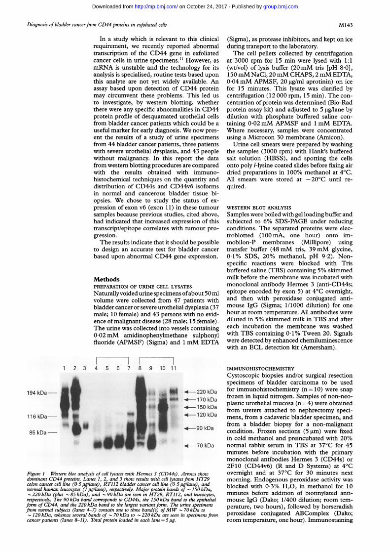
Immunohistochemical studies with mono-clonal antibodies to the standard form ofCD44(Hermes 3) and to CD44v6 (2F10) showeddifferences in the location of the correspondingCD44 epitopes between normal and neoplasticbladder mucosa. Hermes 3 showed that innormal bladder epithelium (n = 4) CD44s pro-tein can be detected only in the basal layers, andis also present in stromal cells, lymphocytes,vascular endothelial cells, and smooth musclecells (fig 2A). CD44v6 is likewise confined tothe basal layers of normal epithelium but isnot detectable in stromal cells (fig 2B). Inneoplastic bladder specimens (n = 10) strongmembranous immunostaining for CD44s andfor CD44v6 was seen both in the basal cellsof the malignant epithelium and in its moresuperficial cells (fig 2C and 2D) in most speci-mens. In some areas there was a degree ofregional heterogeneity in staining, but the trendtowards staining of epithelial cells above thebasal layer was still maintained.
PRESENCE OF CD44s AND CD44v PROTEINS INEXFOLIATED BLADDER CELLS FROM URINE
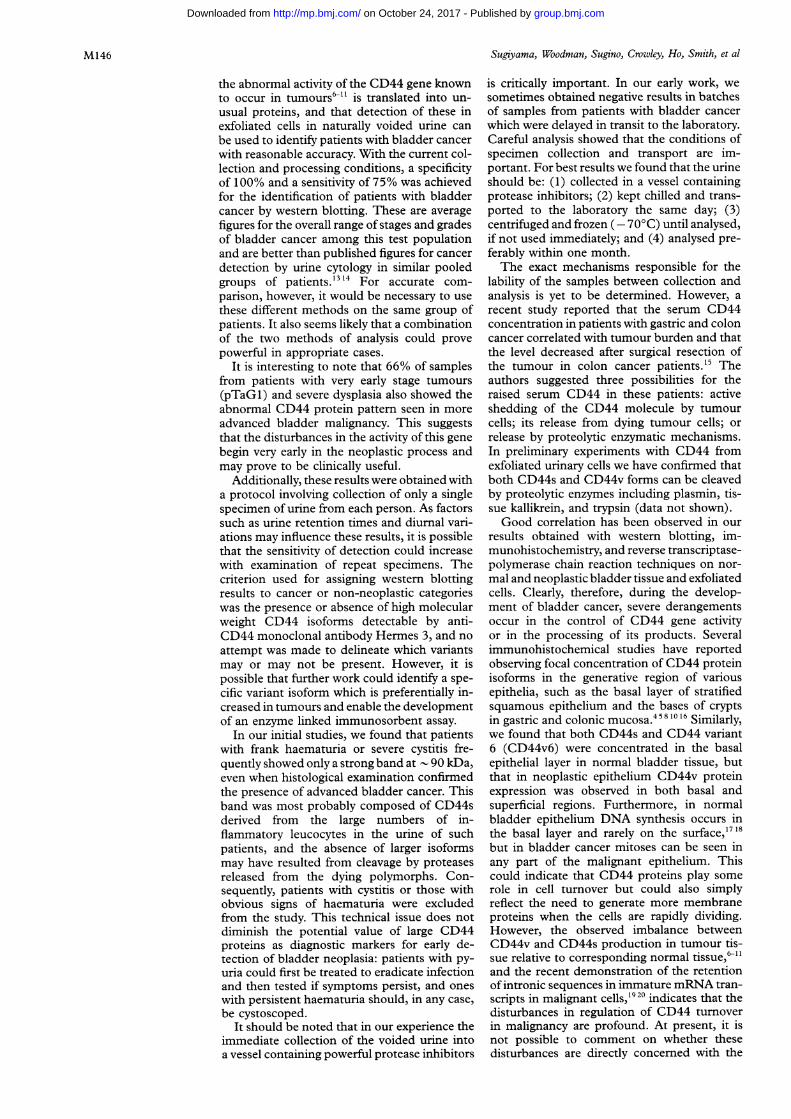
Immunocytochemistry of urine cell smearsshowed that some cancer cells in urine fromcancer patients stained positively for CD44v6(fig 3A). This confirmed that some of theCD44v protein is retained even following ex-foliation, but that the tumour cells show het-erogeneous expression. Inflammatory cellswere negative. As expected, normal and neo-plastic epithelium and inflammatory cells in
M144
group.bmj.com on October 24, 2017 - Published by http://mp.bmj.com/Downloaded from

Diagnosis of bladder cancer front CD44 proteins in exfoliated cells
9~~~~r'' -.e$.tC>~~~~~~~~~~aS7tw £~~~~~~1
'~~~~'Stt20~~ ' v_ __
i s~>
'4 r C.'\
Figure 2 Immunohistochemical localisation of CD44 proteins in normal and neoplastic bladder tissues. The cell membranes of basal epithelial cells ofnormal bladder are moderately stained with monoclonal antibody (mAb) Hermes 3 to CD44s (A) and with mAb 2F10 to CD44v6 (B), whereassuperficial epithelial cells are negative. Underlying stromal cells only react with Hermes 3 and do not contain detectable quantities of v6. In bladdercarcinoma, both superficial and basal epithelial cells stain strongly with Hermes 3 (C) and with 2F10 to CD44v6 (D). (Magnification x 560.)Arrows define the position of the epithelial-stromal boundary and asterisks are positioned over the luminal surface of the epithelium.
B. 0_W if
.-.**4'S
S..
: .^
:e: &
ft
_0
*i",..^
Wr"
Figure 3 Immunohistochemical studies on CD44 protein expression in cells pelleted from the urine of bladder cancer patients. (A) Some exfoliatedcarcinoma cells in urine from a bladder cancer patient retain immunostaining for CD44v6 (2F1 0) but others are negative. Inflanmmatory cells do notshow immunoreactivity for this epitope. (B) Exfoliated cancer cells (large arrows) as well as inflammatory cells (small arrows), including polymorphs,stain for CD44s with Hermes 3. (Magnification x 554.)
the urine all stained for CD44s with Hermes3 (fig 3B). Normal urine smears showed no
staining with monoclonal antibody 2F10 forCD44v6 (data not shown). All of these data are
in accordance with the immunohistochemicalresults on tissue sections and with the westernblotting findings.
DiscussionWe have previously reported that CD44 mRNAis detectable in exfoliated urine cells" and we
have now shown that CD44 protein is alsodetectable in these cells, using western blottingand immunohistochemistry. The present workhas further demonstrated that at least some of
A
M145
gr
r} ^
l..., k.
t
group.bmj.com on October 24, 2017 - Published by http://mp.bmj.com/Downloaded from
Sugzyama, Woodman, Sugino, Crowley, Ho, Smith, et al
the abnormal activity of the CD44 gene knownto occur in tumours"'1 is translated into un-usual proteins, and that detection of these inexfoliated cells in naturally voided urine canbe used to identify patients with bladder cancerwith reasonable accuracy. With the current col-lection and processing conditions, a specificityof 100% and a sensitivity of 75% was achievedfor the identification of patients with bladdercancer by western blotting. These are averagefigures for the overall range of stages and gradesof bladder cancer among this test populationand are better than published figures for cancerdetection by urine cytology in similar pooledgroups of patients.13 14 For accurate com-parison, however, it would be necessary to usethese different methods on the same group ofpatients. It also seems likely that a combinationof the two methods of analysis could provepowerful in appropriate cases.
It is interesting to note that 66% of samplesfrom patients with very early stage tumours(pTaGl) and severe dysplasia also showed theabnormal CD44 protein pattern seen in moreadvanced bladder malignancy. This suggeststhat the disturbances in the activity of this genebegin very early in the neoplastic process andmay prove to be clinically useful.
Additionally, these results were obtained witha protocol involving collection of only a singlespecimen of urine from each person. As factorssuch as urine retention times and diurnal vari-ations may influence these results, it is possiblethat the sensitivity of detection could increasewith examination of repeat specimens. Thecriterion used for assigning western blottingresults to cancer or non-neoplastic categorieswas the presence or absence of high molecularweight CD44 isoforms detectable by anti-CD44 monoclonal antibody Hermes 3, and noattempt was made to delineate which variantsmay or may not be present. However, it ispossible that further work could identify a spe-cific variant isoform which is preferentially in-creased in tumours and enable the developmentof an enzyme linked immunosorbent assay.
In our initial studies, we found that patientswith frank haematuria or severe cystitis fre-quently showed only a strong band at - 90 kDa,even when histological examination confirmedthe presence of advanced bladder cancer. Thisband was most probably composed of CD44sderived from the large numbers of in-flammatory leucocytes in the urine of suchpatients, and the absence of larger isoformsmay have resulted from cleavage by proteasesreleased from the dying polymorphs. Con-sequently, patients with cystitis or those withobvious signs of haematuria were excludedfrom the study. This technical issue does notdiminish the potential value of large CD44proteins as diagnostic markers for early de-tection of bladder neoplasia: patients with py-uria could first be treated to eradicate infectionand then tested if symptoms persist, and oneswith persistent haematuria should, in any case,be cystoscoped.
It should be noted that in our experience theimmediate collection of the voided urine intoa vessel containing powerful protease inhibitors
is critically important. In our early work, wesometimes obtained negative results in batchesof samples from patients with bladder cancerwhich were delayed in transit to the laboratory.Careful analysis showed that the conditions ofspecimen collection and transport are im-portant. For best results we found that the urineshould be: (1) collected in a vessel containingprotease inhibitors; (2) kept chilled and trans-ported to the laboratory the same day; (3)centrifuged and frozen (- 70°C) until analysed,if not used immediately; and (4) analysed pre-ferably within one month.The exact mechanisms responsible for the
lability of the samples between collection andanalysis is yet to be determined. However, arecent study reported that the serum CD44concentration in patients with gastric and coloncancer correlated with tumour burden and thatthe level decreased after surgical resection ofthe tumour in colon cancer patients.15 Theauthors suggested three possibilities for theraised serum CD44 in these patients: activeshedding of the CD44 molecule by tumourcells; its release from dying tumour cells; orrelease by proteolytic enzymatic mechanisms.In preliminary experiments with CD44 fromexfoliated urinary cells we have confirmed thatboth CD44s and CD44v forms can be cleavedby proteolytic enzymes including plasmin, tis-sue kallikrein, and trypsin (data not shown).Good correlation has been observed in our
results obtained with western blotting, im-munohistochemistry, and reverse transcriptase-polymerase chain reaction techniques on nor-mal and neoplastic bladder tissue and exfoliatedcells. Clearly, therefore, during the develop-ment of bladder cancer, severe derangementsoccur in the control of CD44 gene activityor in the processing of its products. Severalimmunohistochemical studies have reportedobserving focal concentration of CD44 proteinisoforms in the generative region of variousepithelia, such as the basal layer of stratifiedsquamous epithelium and the bases of cryptsin gastric and colonic mucosa.4581016 Similarly,we found that both CD44s and CD44 variant6 (CD44v6) were concentrated in the basalepithelial layer in normal bladder tissue, butthat in neoplastic epithelium CD44v proteinexpression was observed in both basal andsuperficial regions. Furthermore, in normalbladder epithelium DNA synthesis occurs inthe basal layer and rarely on the surface,17 18but in bladder cancer mitoses can be seen inany part of the malignant epithelium. Thiscould indicate that CD44 proteins play somerole in cell turnover but could also simplyreflect the need to generate more membraneproteins when the cells are rapidly dividing.However, the observed imbalance betweenCD44v and CD44s production in tumour tis-sue relative to corresponding normal tissue, 6-11and the recent demonstration of the retentionof intronic sequences in immature mRNA tran-scripts in malignant cells,'920 indicates that thedisturbances in regulation of CD44 turnoverin malignancy are profound. At present, it isnot possible to comment on whether thesedisturbances are directly concerned with the
M146
group.bmj.com on October 24, 2017 - Published by http://mp.bmj.com/Downloaded from

Diagnosis of bladder cancer from CD44 proteins in exfoliated cells
development and progression of the neoplasmor are a side effect. However, the increasingseverity of the changes observed in advancedand metastatic malignancy may indicate thatthe disturbances have a causal role.
We thank J O'D McGee, H Bodenmuller, M Kauffman, andK Yoshida for helpful discussions, J Cook and R Robinson forcollection of urine samples, and L Summerville for invaluablehelp in preparing the manuscript. We also wish to acknowledgewith gratitude the generous gift of mAb Hermes 3 by Dr E CButcher. This work was supported by a research contract be-tween Boehringer Mannheim GmbH and Oxford University.
1 Screaton GR, Bell MV, Jackson DG, Cornelis FB, GerthU, Bell JI. Genomic structure of DNA encoding thelymphocyte homing receptor CD44 reveals at least 12alternatively spliced exons. Proc Natl Acad Sci USA 1992;89:12160-4.
2 Tolg C, Hofmann M, Herrlich P, Ponta H. Splicing choicefrom ten variant exons establishes CD44 variability. NucleicAcids Res 1993;21:1225-9.
3 Screaton GR, Bell MV, Bell JI, Jackson DG. The iden-tification of a new alternative exon with highly restrictedtissue expression in transcripts encoding the mousePgp-1 (CD44) homing receptor. Biol Chem 1993;268:12235-8.
4 Fox SB, Fawcett J, Jackson DG, Collins I, Gatter KC,Harris AL, et al. Normal human tissues, in addition tosome tumors, express multiple different CD44 isoforms.Cancer Res 1994;54:4539-46.
5 Mackay CR, Terpe H-J, Stauder R, Marston WL, Stark H,Gunthert U. Expression and modulation of CD44 variantisoforms in humans. Cell Biol 1994;124:71-82.
6 Matsumura Y, Tarin D. Significance ofCD44 gene productsfor cancer diagnosis and disease evaluation. Lancet 1992;340:1053-8.
7 Tanabe KK, Ellis LM, Saya H. Expression of CD44R1adhesion molecule in colon carcinomas and metastases.Lancet 1993;341:725-6.
8 Heider K-H, Dammrich J, Skroch-Angel P, Muller-Her-melink H-K, Vollmers HP, Herrlich P, et al. Differentialexpression of CD44 splice variants in intestinal- and
diffuse-type human gastric carcinomas and normal gastricmucosa. Cancer Res 1993;53:4197-203.
9 Dall P, Heider K-H, Hekele A, von Minchwitz G, KaufmannM, Ponta H, et al. Surface protein expression and mes-senger RNA-splicing analysis of CD44 in uterine cervicalcancer and normal cervical epithelium. Cancer Res 1994;54:3337-41.
10 Wielenga VJM, Heider K-H, Offerhaus GJA, Adolf GR,van den Berg FM, Ponta H, et al. Expression of CD44variant proteins in human colorectal cancer is related totumor progression. Cancer Res 1993;53:4754-6.
11 Matsumura Y, Hanbury D, Smith J, Tarin D. Non-invasivedetection ofmalignancy by identification ofunusual CD44gene activity in exfoliated cancer cells. BMJ7 1994;308:619-24.
12 Mulder J-WR, Kruyt PM, Sewnath M, Oosting J, SeldenrijkCA, Weidema WF, et al. Colorectal cancer prognosis andexpression of exon-v6-containing CD44 proteins. Lancet1994;344: 1470-2.
13 Matzkin H, Moinuddin SM, Soloway MS. Value of urinecytology versus bladder washing in bladder cancer. Urology1992;39:201-3.
14 Koss LG, Deitch D, Tamamathan R, Sherman AB. Diag-nostic value of cytology of voided urine. Acta Cytol 1985;29:810-6.
15 Guo Y-J, Liu G, Wang X, Jin D, Wu M, Ma J, et al. Potentialuse ofsoluble CD44 in serum as indicator oftumor burden'and metastasis in patients with gastric or colon cancer.Cancer Res 1994;54:422-6.
16 Abbasi AM, Chester KA, Talbot IC, Macpherson AS, BoxerG, Forbes A, et al. CD44 is associated with proliferationin normal and neoplastic human colorectal epithelial cells.Eur J Cancer 1993;29A(14):1995-2002.
17 Tsujihashi H, Matsuda H, Uejima S, Akiyama T, Kurita T.Cell proliferation of human bladder tumors. J Urol 1989;142:1113-6.
18 Limas C, Bigler A, Bair R, Bernhart P, Reddy P. Proliferativeactivity of urothelial neoplasms: comparison of BrdU in-corporation, Ki67 expression and nucleolar organiser re-gions. J Clin Pathol 1993;46:159-65.
19 Matsumura Y, Matsumura S, Smith J, Tarin D. Cancerspecific novel abnormalities in splicing of CD44 geneproducts in bladder tumours. In: Proceedings ofthe EighthInternational Conference of the International Society ofDifferentiation (ISD), 1994:199 (abstract P140).
20 Matsumura Y, Sugiyama M, Smith JC, Tarin D. Unusualretention of introns in CD44 gene transcripts in bladdercancer provides new diagnostic and clinical oncologicalopportunities. J Pathol (in press).
M147
group.bmj.com on October 24, 2017 - Published by http://mp.bmj.com/Downloaded from

exfoliated cancer cells in urineidentification of abnormal CD44 proteins in Non-invasive detection of bladder cancer by
Matsumura and D TarinM Sugiyama, A Woodman, T Sugino, S Crowley, K Ho, J Smith, Y
doi: 10.1136/mp.48.3.M1421995 48: M142-M147 Clin Mol Pathol
http://mp.bmj.com/content/48/3/M142Updated information and services can be found at:
These include:
serviceEmail alerting
box at the top right corner of the online article. Receive free email alerts when new articles cite this article. Sign up in the
Notes
http://group.bmj.com/group/rights-licensing/permissionsTo request permissions go to:
http://journals.bmj.com/cgi/reprintformTo order reprints go to:
http://group.bmj.com/subscribe/To subscribe to BMJ go to:
group.bmj.com on October 24, 2017 - Published by http://mp.bmj.com/Downloaded from