Embed Size (px)
Citation preview
Evaluation of the accuracy in detectingcervical lesions by nurses versus doctorsusing a stationary colposcope andGynocular in a low-resource setting
Ashrafun Nessa,1 Joya Shree Roy,2 Most Afroza Chowdhury,1 Quayuma Khanam,1
Romena Afroz,1 Charlotte Wistrand,3 Marcus Thuresson,4 Malin Thorsell,3
Isaac Shemer,5 Elisabeth Andrea Wikström Shemer3
To cite: Nessa A, Roy JS,Chowdhury MA, et al.Evaluation of the accuracy indetecting cervical lesions bynurses versus doctors usinga stationary colposcope andGynocular in a low-resourcesetting. BMJ Open 2014;4:e005313. doi:10.1136/bmjopen-2014-005313
▸ Prepublication history forthis paper is available online.To view these files pleasevisit the journal online(http://dx.doi.org/10.1136/bmjopen-2014-005313).
Received 21 March 2014Revised 9 October 2014Accepted 16 October 2014
For numbered affiliations seeend of article.
Correspondence toDr Elisabeth WikströmShemer;[email protected]
ABSTRACTObjectives: Evaluation of the performance of VIA(visual inspection with acetic acid) trained nurses tolearn colposcopy and the Swede score method todetect cervical lesions by using stationary colposcopeor a portable, hand-held colposcope; the Gynocular, ascompared to doctors.Design: A crossover randomised clinical trial.Setting: The Colposcopy Clinic of BangabandhuSheikh Mujib Medical University (BSMMU), Dhaka,Bangladesh.Participants: 932 women attending the clinic aseither screening naïve for VIA screening (404) orwomen referred as VIA positive (528) from other VIAscreening centres in the Dhaka region.Intervention: VIA trained nurses were trained on-sitein colposcopy and in the Swede score systematiccolposcopy method. The Swede score grade cervicalacetowhiteness, margins plus surface. vessel pattern,lesion size and iodine staining. The women wererandomised to start the examination by either astationary colposcope or the Gynocular. Swede scoreswere first obtained by a nurse and the same patientwas equally evaluated by a doctor.Primary and secondary outcome measures:Agreement between nurses and doctors in Swedescores was evaluated using the weighted κ statistic forthe Gynocular and standard colposcope. The ability topredict CIN 2+ (CIN 2, CIN 3 and invasive cervicalcancer) using Swede scores was evaluated usingreceiver-operating characteristic curves.Results: The Swede scores obtained by nurses anddoctors using the Gynocular and stationary colposcopeshowed high agreement with a κ statistic of 0.858 and0.859, respectively, and no difference in detectingcervical lesions in biopsy. Biopsy detected CIN 2+ in39 (4.2%) women.Conclusions: Our study showed that VIA nurses canperform colposcopy. There was no significantdifferences compared to doctors in detecting cervicallesions by stationary colposcope or the Gynocularusing the Swede score system. Swede scores obtained
by nurses using the Gynocular could offer an accuratecervical diagnostic approach in low resource settings.Trial registration number: ISRCTN53264564.
INTRODUCTIONCervical cancer is a common cause of deathin women from low-resource settings. Recentdata indicate that each year 528 000 womenare diagnosed with cervical cancer worldwideand 266 000 die from the disease.1 A
Strengths and limitations of this study
▪ The main strength of our study is its randomisedcrossover design including both screening naïvewomen and women referred as visual inspectionwith acetic acid (VIA positive), thus giving theexaminers a wide range of normal to patho-logical colposcopic impressions, and a reductionof the risk of selection bias. The crossover ran-domised design was used to reduce the risk ofintraobserver variability. Another strength is thatall the biopsies were analysed in a single-sitelaboratory.
▪ The main limitation of our study is that not all thewomen examined had a biopsy, which may havebiased our results. The crossover design may alsohave influenced the scoring of the secondinstrument.
▪ Other study designs were considered but wouldhave been difficult to implement in a low-resource setting where many poor women mightnever return to the colposcopy clinic. Also,blinding of the instrument that was used was notpossible due to the nature of the instruments.However, by using a crossover study design,block randomisation and the large sample size,we consider the risk of bias reduced.
Nessa A, et al. BMJ Open 2014;4:e005313. doi:10.1136/bmjopen-2014-005313 1
Open Access Research
on 29 May 2018 by guest. P
rotected by copyright.http://bm
jopen.bmj.com
/B
MJ O
pen: first published as 10.1136/bmjopen-2014-005313 on 3 N
ovember 2014. D
ownloaded from

majority (87%) of women diagnosed with cervicalcancer live in less developed regions of the world.1
Mortality varies highly, ranging from less than 2/100 000in developed regions to more than 20/100 000 in areassuch as Melanesia and Middle and Eastern Africa.1
In Bangladesh, 11 956 new cases of cervical cancer arediscovered yearly and each year 6582 women die fromthe disease.1 Moreover, as the incidence rises sharplyamong women over 35 years of age, cervical cancer hasa vast impact on communities at a time when a woman’spresence is vital to social and economical stability.2 3
According to the International Agency for Research onCancer (IARC’s) projections for the coming decades,deaths from cervical cancer will continue to rise.1
In high-resource settings, cervical cytology screeningprogrammes have successfully reduced the rates of cer-vical cancer.4 In many low-resource settings, few womenhave access to cytology screening programmes due tothe absence of national screening programmes, lack ofequipment and skilled technicians.5 As a result, otherlow-cost methods have been implemented for screening,where opportunistic visual inspection with acetic acid(VIA) by trained nurses and doctors is the mostcommon approach.6–8 Often, VIA positive women arereferred for colposcopy in order to reduce the risk ofovertreatment.6–9
The role of colposcopy is to visually assess the size andextent of cervical lesions with strong illumination andmagnification, and to select the most abnormal lesions forbiopsy.10 In low-resource settings, colposcopy is often usedto assess the severity of the lesion in VIA positive women aspart of a ‘see and treat’ policy, as it may be more practicalto treat the women immediately if a severe lesion is diag-nosed, as she might not be able to return to the clinic.11
Colpscopy may also be used for primary screening inlow-resource settings, especially when using the Swedescore systematic colposcopy system, where the total scoregives an indication of the severity of the visual impressionof the cervix.11–15 However, the limited access of doctorsand the limitations of stationary colposcopes (heavy,requires an electrical grid and technical support) arereasons why screening colposcopy has not reached wide-spread use in rural areas with poor infrastructure.11 12 14 15
In high-resource settings, nurse colposcopists havebeen trained to increase accessibility to colposcopy andachieve similar diagnostic accuracy as doctors,16–18 and asimilar approach in low-resource settings, with diagnosisand screening by the portable battery-driven colposcope,the Gynocular, could offer an accurate, cost-effectiveand pragmatic approach to combat cervical cancer.13–15
By using the Swede score systematic colposcopy method,the findings of the colposcopic findings of the cervixcan be organised and scored as a structured report ofthe colposcopic examination.12 13 The Swede scorediffers from other cervical scoring systems by adding thelesion size as a variable.12 13
The main aim of this study was to evaluate if nurse-ledSwede score colposcopy in a low-resource setting had a
similar performance to that of a doctor Swede score col-poscopy in detecting cervical lesions. The second aim ofthe study was to evaluate if a nurse or doctor Gynocularcolposcopy had similar performances as compared to astationary colposcopy in detecting cervical lesions. Thethird aim of the study was to evaluate if nurse-led Swedescore colposcopy could be a future option for detectionof cervical lesions in low-resource settings.
METHODSDesign, participants and proceduresThis study was a randomised crossover clinical trial forevaluating the accuracy of Swede scores of VIA nursestrained in the Swede score colposcopy method in detect-ing cervical lesions using biopsy as a criterion standard.All the participating women were randomised to beexamined using both the stationary colposcope and theGynocular in a crossover design. The nurses and thedoctors examined the same women, but were blindedfrom each other’s Swede scores.In Bangladesh, 2.3% of the women have been
screened with VIA so far, and among those who havebeen screened, approximately 4.8% were VIA positive.6
The opportunistic VIA screening programme inBangladesh was initiated in the year 2004 by the govern-ment of Bangladesh. Trained paramedics, senior staffnurses and doctors offer VIA to married women 30 yearsand above to detect the precancer and early cervicalcancer among women visiting VIA centres of differentdistricts in Bangladesh.18–20 VIA-positive women arereferred to BSMMU and government medical collegehospitals for colposcopic evaluation and manage-ment.18–20 The cervical cancer screening programme isan ongoing programme and colposcopy becomes animportant part of this prevention programme. A womanis considered to be VIA positive when sharp, distinct,well-defined, dense acetowhite areas on the cervix arenoticed during examination with or without raisedmargins, close to the squamocolumnar junction in thetransformation zone.6 8
The inclusion criteria were: (1) women positive for VIAat opportunistic screening by trained family welfare visi-tors, senior staff nurses and doctors in the Dhaka region,Bangladesh referred for colposcopy, or women comingfor VIA cervical screening at the colposcopy clinic ofBangabandhu Sheikh Mujib Medical University(BSMMU) during 1 June to 31 September 2012. Otherinclusion criteria were: (2) ability to understand writtenand oral information, (3) women signing an informedconsent form to participate in the study after receivingoral and written information from a social worker.Exclusion criteria were: (1) ongoing vaginal bleeding,(2) any previous gynaecological examinations less than1 week (3) pregnancy. Women who chose not to take partin the study had a standard colposcopy examination.In total, 932 women were included in the study, of
them 404 attending the clinic as screening naïve and528 women referred as VIA positive. Two VIA nurses
2 Nessa A, et al. BMJ Open 2014;4:e005313. doi:10.1136/bmjopen-2014-005313
Open Access
on 29 May 2018 by guest. P
rotected by copyright.http://bm
jopen.bmj.com
/B
MJ O
pen: first published as 10.1136/bmjopen-2014-005313 on 3 N
ovember 2014. D
ownloaded from

were trained on-site in colposcopy and the Swede scoresystematic colposcopy method for 2 weeks before startingthe study. The colposcopy specialists were accreditedphysicians or gynaecologists who were trained in colpos-copy, cold coagulation and the loop electrical excisionprocedure at the colposcopy clinic of BSMMU.7 Allwomen in the study were examined by one of the twonurses and one of the six colposcopy specialists andboth by the stationary colposcope and the Gynocular.During the colposcopy examination, one of the two
standard colposcopes (Leisegang 1DF, Leisegang,Feinmechanik-Optik GmbH, Berling, Germany or KarlKaps Som 52, Karl Kaps GmbH & Co.KG, Asslar/Wetzlar,Germany) and the Gynocular (Gynius AB, Stockholm,Sweden) were used. Women were randomly allocated inblocks of 50 to start the examination by one of thenurses, followed by an examination by one of the doctorswith either the stationary colposcope or the Gynocular.Then the same examiners examined the woman with thesecond instrument in order to assess the performance ofagreement between the Swede scores of the nurse and ofthe doctor and of the two instruments. The crossoverdesign was chosen in order to lessen possible observervariability.21 A total of 524 women started the examin-ation with the stationary colposcope and 408 women withthe Gynocular. During the Swede score examination,each of the five colposcopic variables (acetowhiteness,margins plus surface, vessel pattern, lesion size andiodine staining) was given a score of 0, 1 or 2 points.12–15
A self-holding non-lubricated speculum was placed inthe vagina and the cervix was visualised. The nurse startedthe examination with an inspection of the cervical vesselsof mosaic pattern, punctuation, atypical vessels or absenceof vessels as randomised with the colposcope or theGynocular using the red-free (green filter) mode andswitched instruments. Then the doctor evaluated thevessel patterns with both instruments as randomised. Thisprocedure differs slightly from the original Swede score,12
where the vessels are inspected after application of aceticacid to the cervix. However, after application of acetic acid,the whitening effect on the cervix slowly fades. Therefore,we chose to first inspect the vessel pattern, and then applythe acetic acid to ensure that both nurses and doctorswould have enough time to accurately estimate the aceto-whitening effect before fading. Thus, the cervix was wipedwith 5% acetic acid for 1 min, followed by evaluation bythe nurse of three Swede score variables (acetowhiteness,margins plus surface and lesion size) and scored by thenurse by both instruments and then again by the doctor.Next, the cervix was swabbed with 5% Lugol iodine solu-tion, and the nurse scored the Swede score’s fifth variable(iodine staining) with both instruments as randomisedand then again by the doctor. The results from the exami-nations were reported by the nurses and the doctors to thestudy nurse in a separate location. The other examinerscould not overhear the reports and they could not see theresults of the previous examiner. The nurses and thedoctors were also not allowed to communicate the
patient’s examination results. The examination was com-pleted with one or more biopsies taken from areas of sus-pected cervical lesions. Punch biopsies of the cervix wereperformed in all women in whom the doctor had scored aSwede score ≥4,12 13 15 as doctors using the Swede scorehave been validated and biopsy recommended whenSwede scores are 6 and above,12 13 and this study aimed tovalidate the nurse colposcopist’s Swede scoring in a low-resource setting (which has not been validated before).The cervical biopsies were analysed at the histopathologylaboratory of BSMMU. The histopathology diagnoses weregraded according to the CIN classification system and con-sidered as the gold standard.22
Women with CIN1 lesions were given the choice ofdirect treatment or a follow-up examination after6 months. Women with CIN2+ were offered a loop elec-trical excision procedure. Women with invasive cervicalcancers were referred to the Gynecological OncologyUnit of the Department of Obstetrics and Gynaecologyat BSMMU for management. This study was approved bythe local ethics committees in Bangladesh and inSweden: the Institutional Review Board of BSMMU; DnrBSMMU/2012/3176 and the Stockholm RegionalEthical Review Board; Dnr 2012/545-31/1. The studywas registered as ISRCTN53264564 at http://www.controlled-trials.com after enrolment of the participantsstarted, due to the publishing restraints of the parallelpatenting process of the Gynocular.The study protocol can be accessed at the Department
of Obstetrics and Gynecology, Danderyd Hospital,Stockholm, Sweden.The Gynocular (Gynius AB, Stockholm, Sweden) is a
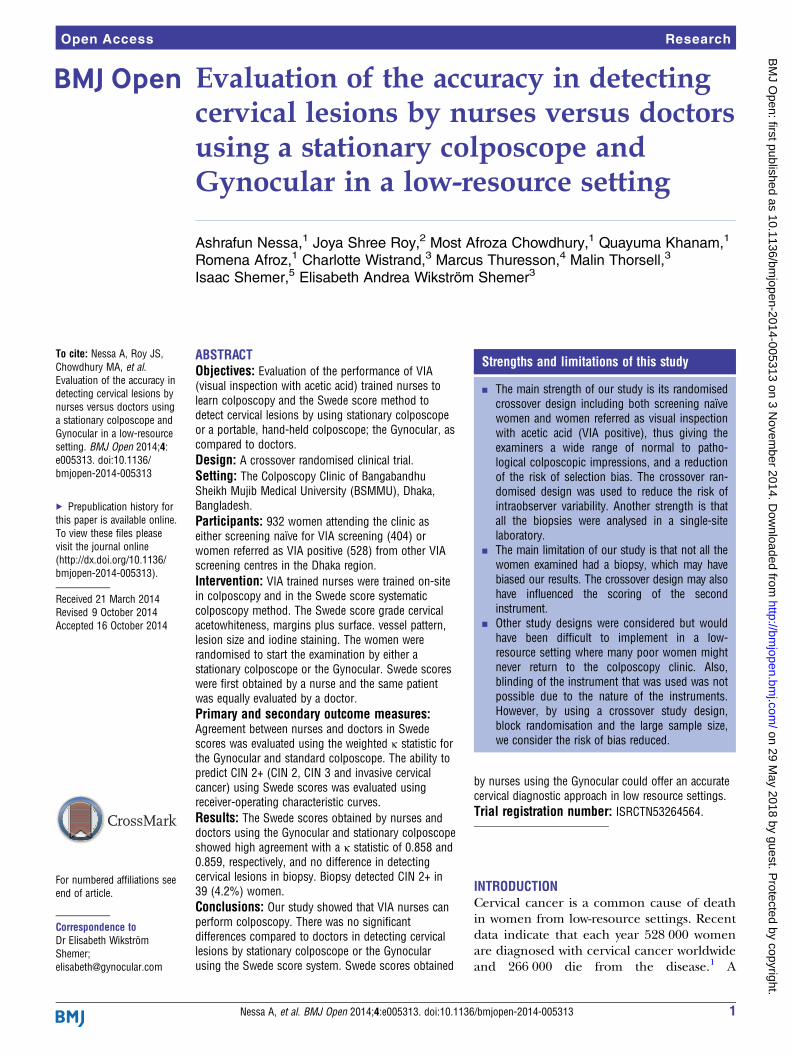
high resolution monocular colposcope with similar specifi-cations to stationary colposcopes.14 15 The Gynocular is asmall, hand-held, battery-driven, measuring 50×33×166mm with 300 mm focal distance, and three magnifications:5×, 8× and 12×.14 15 It has a tripod mounting clip thatscrews into any standard tripod, allowing the medical pro-fessional to perform colposcopy in a hands-free mode forease of biopsy (figure 1). The Gynocular has high-intensitylight-emitting diodes for warm white illumination, a greenfilter light, and is powered by a rechargeable lithium-ionbattery. It is a product approved by the Swedish NationalDrug Authority as a non-invasive medical diagnostic class Itool, CE marked and Food and Drug Administrationapproved.
Statistical analysisAll statistical analyses have been performed using RV.2.14.23 The baseline patient characteristics of thewomen were summarised using means and SD for con-tinuous variables and absolute and relative frequenciesfor categorical variables. To test the level of agreementbetween the colposcope and the Gynocular, the percent-age agreement and the weighted κ statistic was calcu-lated.24 Cervical lesions were classified by the Swedescores system using the Gynocular and the stationary col-poscope.12–15 Detection rates of CIN 1, CIN 2, CIN 3,
Nessa A, et al. BMJ Open 2014;4:e005313. doi:10.1136/bmjopen-2014-005313 3
Open Access
on 29 May 2018 by guest. P
rotected by copyright.http://bm
jopen.bmj.com
/B
MJ O
pen: first published as 10.1136/bmjopen-2014-005313 on 3 N
ovember 2014. D
ownloaded from
ICC (invasive cervical cancer), AIS (adenocarcinoma insitu), benign cervicitis and cervical tuberculosis in cer-vical punch biopsies were calculated. A positive biopsyresult was defined as CIN2+ (CIN 2, CIN 3, CIN 3+) andwe calculated the Swede score’s sensitivity, specificity,positive predictive value (PPV) and negative predictivevalue (NPV) using biopsy as a gold standard for all cut-offlevels of Swede scores between 4 and 10 for doctors and 0and 10 for nurses. The results are presented in tables andas receiver operating characteristic (ROC) curves as wellas the area under the curve (AUC). The comparison ofAUC of the ROC curves was performed using the roc.testfunction in the pROC package.
Sample sizeThe sample size was estimated based on the expectednumber of positive biopsy results (defined as CIN2+)and not in terms of statistical power. In Bangladesh,there are no published data on CIN2+ in a previouslyunscreened population in Bangladesh verified bycytology, colposcopy and biopsy, only of CIN2+ in VIApositive women. However, data from India show a rate of2.7% CIN2+ in unscreened women.25 Thus, we assumeda similar rate in Bangladesh with an expected rate ofCIN2+ of 2.5% in naïve women and 7.5% in VIA-positivewomen as a sample size of 500 naïve and 500VIA-positive women would generate 50 positive biopsyresults, which were considered to give sufficient preci-sion to the nurses colposcopists’ accuracy and Swedescoring compared to doctors.12 13 Thus, the aim was toinclude a total of approximately 1000 women.
In a retrospective power analysis based on the resultsfrom the present study, we estimated that approximately1500 biopsies would have been needed (as compared tothe 228 biopsies in women with a Swede score above 4 inthis study) for 80% power to detect a difference of 0.05 inthe AUC of the ROC curves at a 5% significance level.
RESULTSA total of 932 women were included in the study, of which404 (43%) were screening naïve. The women’s baselinecharacteristics are presented in table 1. A total of 256women had a Swede score of at least 4 by a doctor, and ofthem 228 had a biopsy and 28 refused biopsy (excludedfrom the ROC analyses). Fifty-nine biopsies were takenoutside the research protocol (excluded from ROC ana-lyses). Twenty-seven VIA positive women had CIN2+ and 5screening naïve women had CIN2+. Punch biopsy wasbenign in 7 (1.8%), chronic cervicitis in 23 (5.8%), CIN1in 19 (4.8%) and CIN2 in 4 (1.0%). No women had CIN3and 4 (1.0%) had ICC (CIN3+). In 1 (0.2%), the woman’sbiopsy showed tuberculosis. The Swede score was <4 in 342(85.5%) women, and in those women no biopsy was taken.Among the referred VIA-positive women, punch
biopsy was benign in 13 (2.5%), chronic cervicitis in 82(15.7%), CIN1 in 90 (17.3%) and CIN2 in 21 (4%).Four women (0.8%) had CIN3 and 6 (1.2%) had ICC(CIN3+). Two (0.4%) women had tuberculosis in thebiopsy. In 303 (57.4%) women, the Swede score was <4and no biopsy was taken.When cross tabulating Swede scores by the Gynocular
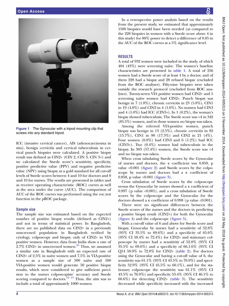
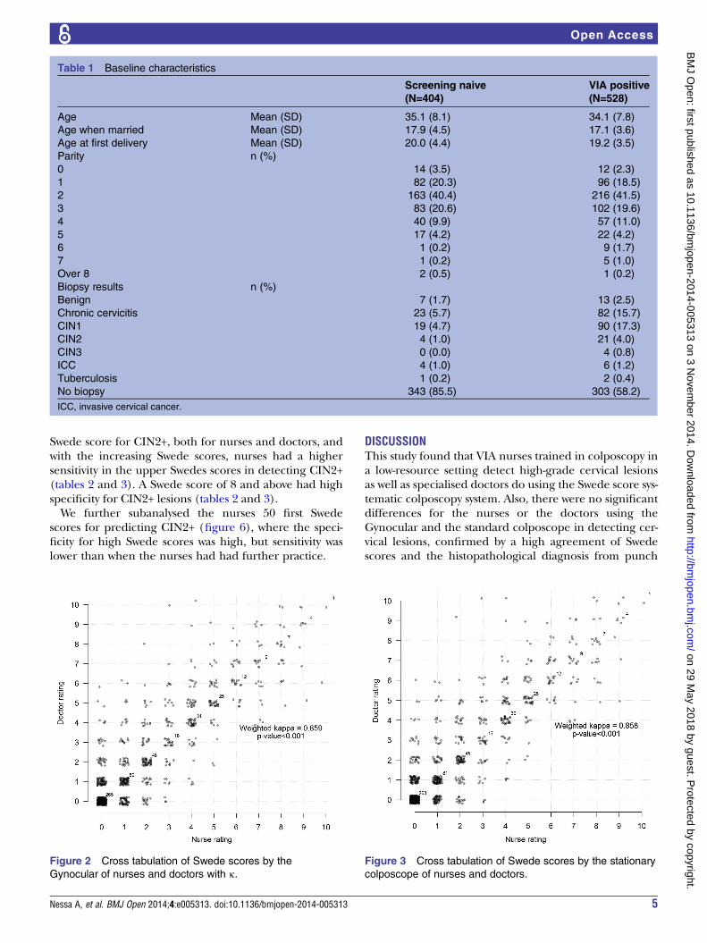
of nurses and doctors, the κ coefficient was 0.859, pvalue <0.001 (figure 2) and Swede scores by the colpo-scope by nurses and doctors had a κ coefficient of0.858, p value <0.001 (figure 3).Cross tabulation of Swede scores by the colposcope
versus the Gynocular by nurses showed a κ coefficient of0.997 (p value <0.001), and a cross tabulation of Swedescores by the colposcope and the Gynocular for thedoctors showed a κ coefficient of 0.998 (p value <0.001).There were no significant differences between the
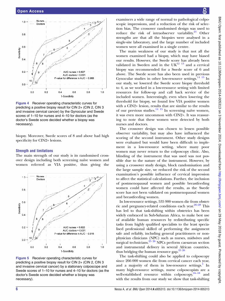
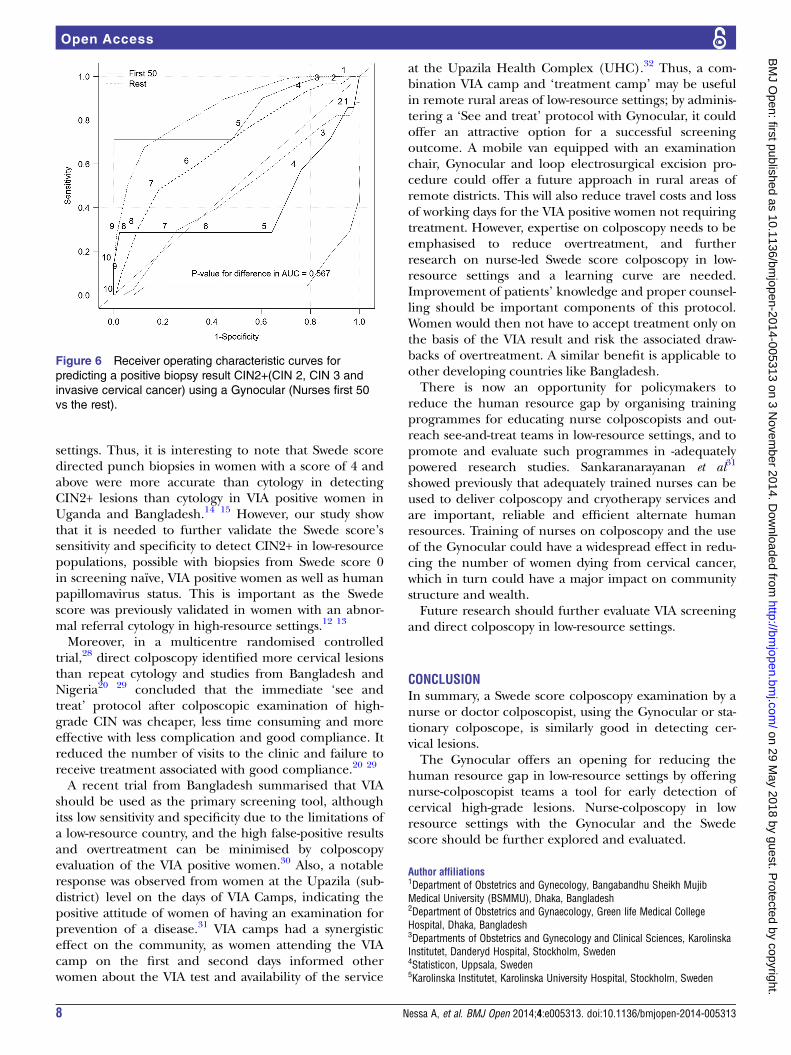
Swede scores of the nurses and the doctors in predictinga positive biopsy result (CIN2+) for both the Gynocular(figure 4) and the colposcope (figure 5).With a cut-off value of 6 and above for Swede score and
biopsy, Gynocular by nurses had a sensitivity of 52.8%(95% CI 35.5% to 69.6%) and a specificity of 65.6%(95% CI 58.4% to 72.4%) for CIN2+ and stationary col-poscope by nurses had a sensitivity of 52.8% (95% CI35.5% to 69.6%) and a specificity of 66.1.6% (95% CIQ14 58.9% to 72.8% for CIN2+ (table 2). For doctorsusing the Gynocular and having a cut-off value of 6, thesensitivity was 61.1% (95% CI 43.5% to 76.9%) and speci-ficity 52.9% (95% CI 45.5% to 60.1%), and for the sta-tionary colposcope the sensitivity was 61.1% (95% CI43.5% to 76.9%) and specificity 53.4% (95% CI 46.1% to60.6%) for detecting CIN2+ (table 3). The sensitivitydecreased while specificity increased with the increased
Figure 1 The Gynocular with a tripod mounting clip that
screws into any standard tripod.
4 Nessa A, et al. BMJ Open 2014;4:e005313. doi:10.1136/bmjopen-2014-005313
Open Access
on 29 May 2018 by guest. P
rotected by copyright.http://bm
jopen.bmj.com
/B
MJ O
pen: first published as 10.1136/bmjopen-2014-005313 on 3 N
ovember 2014. D
ownloaded from
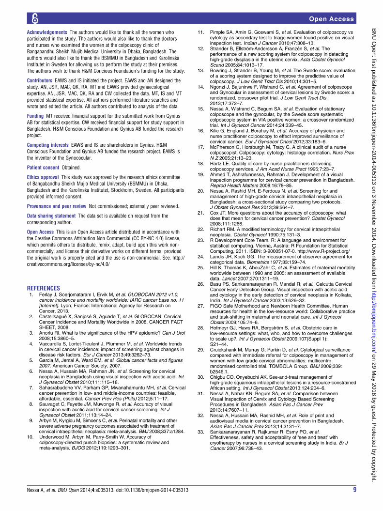
Swede score for CIN2+, both for nurses and doctors, andwith the increasing Swede scores, nurses had a highersensitivity in the upper Swedes scores in detecting CIN2+(tables 2 and 3). A Swede score of 8 and above had highspecificity for CIN2+ lesions (tables 2 and 3).We further subanalysed the nurses 50 first Swede
scores for predicting CIN2+ (figure 6), where the speci-ficity for high Swede scores was high, but sensitivity waslower than when the nurses had had further practice.
DISCUSSIONThis study found that VIA nurses trained in colposcopy ina low-resource setting detect high-grade cervical lesionsas well as specialised doctors do using the Swede score sys-tematic colposcopy system. Also, there were no significantdifferences for the nurses or the doctors using theGynocular and the standard colposcope in detecting cer-vical lesions, confirmed by a high agreement of Swedescores and the histopathological diagnosis from punch
Figure 3 Cross tabulation of Swede scores by the stationary
colposcope of nurses and doctors.
Figure 2 Cross tabulation of Swede scores by the
Gynocular of nurses and doctors with κ.
Table 1 Baseline characteristics
Screening naive VIA positive(N=404) (N=528)
Age Mean (SD) 35.1 (8.1) 34.1 (7.8)
Age when married Mean (SD) 17.9 (4.5) 17.1 (3.6)
Age at first delivery Mean (SD) 20.0 (4.4) 19.2 (3.5)
Parity n (%)
0 14 (3.5) 12 (2.3)
1 82 (20.3) 96 (18.5)
2 163 (40.4) 216 (41.5)
3 83 (20.6) 102 (19.6)
4 40 (9.9) 57 (11.0)
5 17 (4.2) 22 (4.2)
6 1 (0.2) 9 (1.7)
7 1 (0.2) 5 (1.0)
Over 8 2 (0.5) 1 (0.2)
Biopsy results n (%)
Benign 7 (1.7) 13 (2.5)
Chronic cervicitis 23 (5.7) 82 (15.7)
CIN1 19 (4.7) 90 (17.3)
CIN2 4 (1.0) 21 (4.0)
CIN3 0 (0.0) 4 (0.8)
ICC 4 (1.0) 6 (1.2)
Tuberculosis 1 (0.2) 2 (0.4)
No biopsy 343 (85.5) 303 (58.2)
ICC, invasive cervical cancer.
Nessa A, et al. BMJ Open 2014;4:e005313. doi:10.1136/bmjopen-2014-005313 5
Open Access
on 29 May 2018 by guest. P
rotected by copyright.http://bm
jopen.bmj.com
/B
MJ O
pen: first published as 10.1136/bmjopen-2014-005313 on 3 N
ovember 2014. D
ownloaded from
biopsy. Moreover, Swede scores of 8 and above had highspecificity for CIN2+ lesions.
Strength and limitationsThe main strength of our study is its randomised cross-over design including both screening naïve women andwomen referred as VIA positive, thus giving the
examiners a wide range of normal to pathological colpo-scopic impressions, and a reduction of the risk of selec-tion bias. The crossover randomised design was used toreduce the risk of intraobserver variability.21 Otherstrengths are that all the biopsies were analysed in asingle-site laboratory, and the large number of includedwomen were all examined in a single centre.The main weakness of our study is that not all the
women examined had a biopsy, which may have biasedour results. However, the Swede score has already beenvalidated in Sweden and in the UK12 13 and a cervicalbiopsy was recommended for a Swede score of 6 andabove. The Swede score has also been used in previousGynocular studies in other low-resource settings.14 15 Inour study, we lowered the Swede score biopsy thresholdto 4, as we worked in a low-resource setting with limitedresources for follow-up and call back service of theincluded women. Interestingly, even when lowering thethreshold for biopsy, we found few VIA positive womenwith a CIN2+ lesion, results that are similar to the resultsof our previous studies.14 15 In screening naïve women,it was even more uncommon with CIN2+. It was reassur-ing to note that these women were detected by bothnurses and doctors.The crossover design was chosen to lessen possible
observer variability, but may also have influenced thescoring of the second instrument. Other study designswere evaluated but would have been difficult to imple-ment in a low-resource setting, where many poorwomen may never return to the colposcopy clinic. Also,blinding of the instrument that was used was not pos-sible due to the nature of the instrument. However, byusing a crossover study design, block randomisation andthe large sample size, we reduced the risk of the secondexamination’s possible influence of cervical impressionto affect the statistical calculations. Further, the inclusionof postmenopausal women and possible breastfeedingwomen could have affected the results, as the Swedescore has not been validated on postmenopausal womenand breastfeeding women.In low-resource settings, 535 900 women die from obstet-
ric and pregnancy-related conditions each year.26–28 Thishas led to that task-shifting within obstetrics has beenwidely embraced in Sub-Saharan Africa, to make best useof available human resources by redistributing specifictasks from highly qualified specialists to the least specia-lised professional skilled of performing the assignmentsafe and reliably, including general practitioners or non-physician clinicians (NPC) such as nurses, midwives andsurgical technicians.25 26 NPCs perform caesarean sectionand instrumental delivery in several African countries,thus bridging the human resource gap.25 26
The task-shifting could also be applied to colposcopysince 266 000 women die from cervical cancer each year,and a majority of them in low-resource settings.1 Inmany high-resource settings, nurse colposcopists are awell-established resource within colposcopy,16–18 andwith the results from our study we show that task-shifting
Figure 4 Receiver operating characteristic curves for
predicting a positive biopsy result for CIN 2+ (CIN 2, CIN 3
and invasive cervical cancer) by the Gynocular and Swede
scores of 1–10 for nurses and 4–10 for doctors (as the
doctor’s Swede score decided whether a biopsy was
necessary).
Figure 5 Receiver operating characteristic curves for
predicting a positive biopsy result for CIN 2+ (CIN 2, CIN 3
and invasive cervical cancer) by a stationary colposcope and
Swede scores of 1–10 for nurses and 4–10 for doctors (as the
doctor’s Swede score decided whether a biopsy was
necessary).
6 Nessa A, et al. BMJ Open 2014;4:e005313. doi:10.1136/bmjopen-2014-005313
Open Access
on 29 May 2018 by guest. P
rotected by copyright.http://bm
jopen.bmj.com
/B
MJ O
pen: first published as 10.1136/bmjopen-2014-005313 on 3 N
ovember 2014. D
ownloaded from

within colposcopy and with the Gynocular is also a feas-ible and safe opportunity to lessen the human resourcegap within colposcopy in low-resource settings. Thispragmatic but also highly accurate approach may havewidespread implications to lower the epidemic high inci-dence of cervical cancer.Bowring et al13 showed that trainee unaccredited colpos-
copists were as accurate as accredited colposcopists indetecting cervical lesions using the Swede score, findingsanalogous to our findings of VIA nurse colposcopists com-pared to accredited doctor colposcopists. Our study also
showed that a VIA nurse colposcopist Swede score of 8 orabove had parallel high specificities of CIN2+ as the Swedescore of the doctors. These results are comparable toCIN2+ specificities in Swede score trials by doctors fromboth high-resource11 12 and low-resource settings.13 14
The Swede score colposcopy system works well withvarious healthcare professionals and economicalsettings.It has been suggested13–15 that the Swede score may
be used as a primary cervical screening as well as a seeand treat method of cervical lesions in low-resource
Table 2 Sensitivity and specificity for different cut-off levels for CIN 2+ (CIN 2, CIN 3 and invasive cervical cancer; nurses,
n=228)
Sensitivity (95% CI) Specificity (95% CI) PPV (95% CI) NPV (95% CI)
Gynocular
10 vs <10 2.8% (0.1% to 14.5%) 99.5% (97.1% to 100.0%) 84.3% (78.9% to 88.8%) 50.0% (1.3% to 98.7%)
≥9 vs <9 13.9% (4.7% to 29.5%) 96.8% (93.2% to 98.8%) 85.5% (80.1% to 89.9%) 45.5% (16.7% to 76.6%)
≥8 vs <8 30.6% (16.3% to 48.1%) 90.5% (85.4% to 94.3%) 87.2% (81.7% to 91.6%) 37.9% (20.7% to 57.7%)
≥7 vs <7 44.4% (27.9% to 61.9%) 80.4% (74.0% to 85.8%) 88.4% (82.6% to 92.8%) 30.2% (18.3% to 44.3%)
≥6 vs <6 52.8% (35.5% to 69.6%) 65.6% (58.4% to 72.4%) 87.9% (81.4% to 92.8%) 22.6% (14.2% to 33.0%)
≥5 vs <5 66.7% (49.0% to 81.4%) 43.9% (36.7% to 51.3%) 87.4% (79.0% to 93.3%) 18.5% (12.2% to 26.2%)
≥4 vs <4 86.1% (70.5% to 95.3%) 22.2% (16.5% to 28.8%) 89.4% (76.9% to 96.5%) 17.4% (12.2% to 23.8%)
≥3 vs <3 91.7% (77.5% to 98.2%) 13.8% (9.2% to 19.5%) 89.7% (72.6% to 97.8%) 16.8% (11.9% to 22.8%)
≥2 vs <2 94.4% (81.3% to 99.3%) 6.9% (3.7% to 11.5%) 86.7% (59.5% to 98.3%) 16.2% (11.5% to 21.9%)
≥1 vs 0 97.2% (85.5% to 99.9%) 3.2% (1.2% to 6.8%) 85.7% (42.1% to 99.6%) 16.1% (11.4% to 21.6%)
Colposcope
10 vs <10 0.0% (0.0% to 9.7%) 99.5% (97.1% to 100.0%) 83.9% (78.5% to 88.5%) 0.0% (0.0% to 97.5%)
≥9 vs <9 11.1% (3.1% to 26.1%) 96.8% (93.2% to 98.8%) 85.1% (79.6% to 89.6%) 40.0% (12.2% to 73.8%)
≥8 vs <8 30.6% (16.3% to 48.1%) 90.5% (85.4% to 94.3%) 87.2% (81.7% to 91.6%) 37.9% (20.7% to 57.7%)
≥7 vs <7 44.4% (27.9% to 61.9%) 81.0% (74.6% to 86.3%) 88.4% (82.7% to 92.8%) 30.8% (18.7% to 45.1%)
≥6 vs <6 52.8% (35.5% to 69.6%) 66.1% (58.9% to 72.8%) 88.0% (81.5% to 92.9%) 22.9% (14.4% to 33.4%)
≥5 vs <5 66.7% (49.0% to 81.4%) 45.0% (37.7% to 52.4%) 87.6% (79.4% to 93.4%) 18.8% (12.4% to 26.6%)
≥4 vs <4 83.3% (67.2% to 93.6%) 22.2% (16.5% to 28.8%) 87.5% (74.8% to 95.3%) 16.9% (11.7% to 23.3%)
≥3 vs <3 91.7% (77.5% to 98.2%) 14.3% (9.6% to 20.1%) 90.0% (73.5% to 97.9%) 16.9% (11.9% to 22.9%)
≥2 vs <2 94.4% (81.3% to 99.3%) 6.9% (3.7% to 11.5%) 86.7% (59.5% to 98.3%) 16.2% (11.5% to 21.9%)
≥1 vs 0 97.2% (85.5% to 99.9%) 3.2% (1.2% to 6.8%) 85.7% (42.1% to 99.6%) 16.1% (11.4% to 21.6%)
PPV, positive predictive value; NPV, negative predictive value.
Table 3 Sensitivity and specificity for different cut-off levels for CIN 2+ (CIN 2, CIN 3 and invasive cervical cancer (doctors,
n=228))
Sensitivity (95% CI) Specificity (95% CI) PPV (95% CI) NPV (95% CI)
Gynocular
10 vs <10 5.6% (0.7% to 18.7%) 97.4% (94.0% to 99.1%) 84.5% (79.1% to 89.1%) 28.6% (3.7% to 71.0%)
≥9 vs <9 22.2% (10.1% to 39.2%) 93.7% (89.3% to 96.7%) 86.5% (81.0% to 90.8%) 40.0% (19.1% to 63.9%)
≥8 vs <8 36.1% (20.8% to 53.8%) 88.0% (82.5% to 92.2%) 88.0% (82.5% to 92.2%) 36.1% (20.8% to 53.8%)
≥7 vs <7 52.8% (35.5% to 69.6%) 74.9% (68.1% to 80.9%) 89.4% (83.5% to 93.7%) 28.4% (18.0% to 40.7%)
≥6 vs <6 61.1% (43.5% to 76.9%) 52.9% (45.5% to 60.1%) 87.8% (80.4% to 93.2%) 19.6% (12.7% to 28.2%)
≥5 vs 4 83.3% (67.2% to 93.6%) 22.0% (16.3% to 28.5%) 87.5% (74.8% to 95.3%) 16.8% (11.6% to 23.1%)
Colposcope
10 vs <10 5.6% (0.7% to 18.7%) 97.4% (94.0% to 99.1%) 84.5% (79.1% to 89.1%) 28.6% (3.7% to 71.0%)
≥9 vs <9 19.4% (8.2% to 36.0%) 93.2% (88.6% to 96.3%) 86.0% (80.5% to 90.4%) 35.0% (15.4% to 59.2%)
≥8 vs <8 36.1% (20.8% to 53.8%) 88.0% (82.5% to 92.2%) 88.0% (82.5% to 92.2%) 36.1% (20.8% to 53.8%)
≥7 vs <7 52.8% (35.5% to 69.6%) 75.4% (68.7% to 81.3%) 89.4% (83.6% to 93.7%) 28.8% (18.3% to 41.3%)
≥6 vs <6 61.1% (43.5% to 76.9%) 53.4% (46.1% to 60.6%) 87.9% (80.6% to 93.2%) 19.8% (12.9% to 28.5%)
≥5 vs 4 83.3% (67.2% to 93.6%) 22.5% (16.8% to 29.1%) 87.8% (75.2% to 95.4%) 16.9% (11.7% to 23.2%)
PPV, positive predictive value; NPV, negative predictive value.
Nessa A, et al. BMJ Open 2014;4:e005313. doi:10.1136/bmjopen-2014-005313 7
Open Access
on 29 May 2018 by guest. P
rotected by copyright.http://bm
jopen.bmj.com
/B
MJ O
pen: first published as 10.1136/bmjopen-2014-005313 on 3 N
ovember 2014. D
ownloaded from
settings. Thus, it is interesting to note that Swede scoredirected punch biopsies in women with a score of 4 andabove were more accurate than cytology in detectingCIN2+ lesions than cytology in VIA positive women inUganda and Bangladesh.14 15 However, our study showthat it is needed to further validate the Swede score’ssensitivity and specificity to detect CIN2+ in low-resourcepopulations, possible with biopsies from Swede score 0in screening naïve, VIA positive women as well as humanpapillomavirus status. This is important as the Swedescore was previously validated in women with an abnor-mal referral cytology in high-resource settings.12 13
Moreover, in a multicentre randomised controlledtrial,28 direct colposcopy identified more cervical lesionsthan repeat cytology and studies from Bangladesh andNigeria20 29 concluded that the immediate ‘see andtreat’ protocol after colposcopic examination of high-grade CIN was cheaper, less time consuming and moreeffective with less complication and good compliance. Itreduced the number of visits to the clinic and failure toreceive treatment associated with good compliance.20 29
A recent trial from Bangladesh summarised that VIAshould be used as the primary screening tool, althoughitss low sensitivity and specificity due to the limitations ofa low-resource country, and the high false-positive resultsand overtreatment can be minimised by colposcopyevaluation of the VIA positive women.30 Also, a notableresponse was observed from women at the Upazila (sub-district) level on the days of VIA Camps, indicating thepositive attitude of women of having an examination forprevention of a disease.31 VIA camps had a synergisticeffect on the community, as women attending the VIAcamp on the first and second days informed otherwomen about the VIA test and availability of the service
at the Upazila Health Complex (UHC).32 Thus, a com-bination VIA camp and ‘treatment camp’ may be usefulin remote rural areas of low-resource settings; by adminis-tering a ‘See and treat’ protocol with Gynocular, it couldoffer an attractive option for a successful screeningoutcome. A mobile van equipped with an examinationchair, Gynocular and loop electrosurgical excision pro-cedure could offer a future approach in rural areas ofremote districts. This will also reduce travel costs and lossof working days for the VIA positive women not requiringtreatment. However, expertise on colposcopy needs to beemphasised to reduce overtreatment, and furtherresearch on nurse-led Swede score colposcopy in low-resource settings and a learning curve are needed.Improvement of patients’ knowledge and proper counsel-ling should be important components of this protocol.Women would then not have to accept treatment only onthe basis of the VIA result and risk the associated draw-backs of overtreatment. A similar benefit is applicable toother developing countries like Bangladesh.There is now an opportunity for policymakers to
reduce the human resource gap by organising trainingprogrammes for educating nurse colposcopists and out-reach see-and-treat teams in low-resource settings, and topromote and evaluate such programmes in -adequatelypowered research studies. Sankaranarayanan et al31
showed previously that adequately trained nurses can beused to deliver colposcopy and cryotherapy services andare important, reliable and efficient alternate humanresources. Training of nurses on colposcopy and the useof the Gynocular could have a widespread effect in redu-cing the number of women dying from cervical cancer,which in turn could have a major impact on communitystructure and wealth.Future research should further evaluate VIA screening
and direct colposcopy in low-resource settings.
CONCLUSIONIn summary, a Swede score colposcopy examination by anurse or doctor colposcopist, using the Gynocular or sta-tionary colposcope, is similarly good in detecting cer-vical lesions.The Gynocular offers an opening for reducing the
human resource gap in low-resource settings by offeringnurse-colposcopist teams a tool for early detection ofcervical high-grade lesions. Nurse-colposcopy in lowresource settings with the Gynocular and the Swedescore should be further explored and evaluated.
Author affiliations1Department of Obstetrics and Gynecology, Bangabandhu Sheikh MujibMedical University (BSMMU), Dhaka, Bangladesh2Department of Obstetrics and Gynaecology, Green life Medical CollegeHospital, Dhaka, Bangladesh3Departments of Obstetrics and Gynecology and Clinical Sciences, KarolinskaInstitutet, Danderyd Hospital, Stockholm, Sweden4Statisticon, Uppsala, Sweden5Karolinska Institutet, Karolinska University Hospital, Stockholm, Sweden
Figure 6 Receiver operating characteristic curves for
predicting a positive biopsy result CIN2+(CIN 2, CIN 3 and
invasive cervical cancer) using a Gynocular (Nurses first 50
vs the rest).
8 Nessa A, et al. BMJ Open 2014;4:e005313. doi:10.1136/bmjopen-2014-005313
Open Access
on 29 May 2018 by guest. P
rotected by copyright.http://bm
jopen.bmj.com
/B
MJ O
pen: first published as 10.1136/bmjopen-2014-005313 on 3 N
ovember 2014. D
ownloaded from
Acknowledgements The authors would like to thank all the women whoparticipated in the study. The authors would also like to thank the doctorsand nurses who examined the women at the colposcopy clinic ofBangabandhu Sheikh Mujib Medical University in Dhaka, Bangladesh. Theauthors would also like to thank the BSMMU in Bangladesh and KarolinskaInstitutet in Sweden for allowing us to perform the study at their premises.The authors wish to thank H&M Concious Foundation's funding for the study.
Contributors EAWS and IS initiated the project. EAWS and AN designed thestudy. AN, JSR, MAC, QK, RA, MT and EAWS provided gynaecologicalexpertise. AN, JSR, MAC, QK, RA and CW collected the data. MT, IS and MTprovided statistical expertise. All authors performed literature searches andwrote and edited the article. All authors contributed to analysis of the data.
Funding MT received financial support for the submitted work from GyniusAB for statistical expertise. CW received financial support for study support inBangladesh. H&M Conscious Foundation and Gynius AB funded the researchproject.
Competing interests EAWS and IS are shareholders in Gynius. H&MConscious Foundation and Gynius AB funded the research project. EAWS isthe inventor of the Gynococular.
Patient consent Obtained.
Ethics approval This study was approved by the research ethics committeeof Bangabandhu Sheikh Mujib Medical University (BSMMU) in Dhaka,Bangladesh and the Karolinska Institutet, Stockholm, Sweden. All participantsprovided informed consent.
Provenance and peer review Not commissioned; externally peer reviewed.
Data sharing statement The data set is available on request from thecorresponding author.
Open Access This is an Open Access article distributed in accordance withthe Creative Commons Attribution Non Commercial (CC BY-NC 4.0) license,which permits others to distribute, remix, adapt, build upon this work non-commercially, and license their derivative works on different terms, providedthe original work is properly cited and the use is non-commercial. See: http://creativecommons.org/licenses/by-nc/4.0/
REFERENCES1. Ferlay J, Soerjomataram I, Ervik M, et al. GLOBOCAN 2012 v1.0,
cancer incidence and mortality worldwide: IARC cancer base no. 11[Internet]. Lyon, France: International Agency for Research onCancer, 2013.
2. Castellsagué X, Sanjosé S, Aguado T, et al. GLOBOCAN: CervicalCancer Incidence and Mortality Worldwide in 2008. CANCER FACTSHEET, 2008.
3. Anorlu RI. What is the significance of the HPV epidemic? Can J Urol2008;15:3860–5.
4. Vaccarella S, Lortet-Tieulent J, Plummer M, et al. Worldwide trendsin cervical cancer incidence: impact of screening against changes indisease risk factors. Eur J Cancer 2013;49:3262–73.
5. Garcia M, Jemal A, Ward EM, et al. Global cancer facts and figures2007. American Cancer Society, 2007.
6. Nessa A, Hussain MA, Rahman JN, et al. Screening for cervicalneoplasia in Bangladesh using visual inspection with acetic acid. IntJ Gynaecol Obstet 2010;111:115–18.
7. Sahasrabuddhe VV, Parham GP, Mwanahamuntu MH, et al. Cervicalcancer prevention in low- and middle-income countries: feasible,affordable, essential. Cancer Prev Res (Phila) 2012;5:11–17.
8. Sauvaget C, Fayette JM, Muwonge R, et al. Accuracy of visualinspection with acetic acid for cervical cancer screening. Int JGynaecol Obstet 2011;113:14–24.
9. Arbyn M, Kyrgiou M, Simoens C, et al. Perinatal mortality and othersevere adverse pregnancy outcomes associated with treatment ofcervical intraepithelial neoplasia: meta-analysis. BMJ 2008;337:a1284.
10. Underwood M, Arbyn M, Parry-Smith W, Accuracy ofcolposcopy-directed punch biopsies: a systematic review andmeta-analysis. BJOG 2012;119:1293–301.
11. Pimple SA, Amin G, Goswami S, et al. Evaluation of colposcopy vscytology as secondary test to triage women found positive on visualinspection test. Indian J Cancer 2010;47:308–13.
12. Strander B, Ellström-Andersson A, Franzén S, et al. Theperformance of a new scoring system for colposcopy in detectinghigh-grade dysplasia in the uterine cervix. Acta Obstet GynecolScand 2005;84:1013–17.
13. Bowring J, Strander B, Young M, et al. The Swede score: evaluationof a scoring system designed to improve the predictive value ofcolposcopy. J Low Genit Tract Dis 2010;14:301–5.
14. Ngonzi J, Bajunirwe F, Wistrand C, et al. Agreement of colposcopeand Gynocular in assessment of cervical lesions by Swede score: arandomized, crossover pilot trial. J Low Genit Tract Dis2013;17:372–7.
15. Nessa A, Wistrand C, Begum SA, et al. Evaluation of stationarycolposcope and the gynocular, by the Swede score systematiccolposcopic system in VIA positive women: a crossover randomizedtrial. Int J Gynecol Cancer 2014;24:339–45.
16. Kilic G, England J, Borahay M, et al. Accuracy of physician andnurse practitioner colposcopy to effect improved surveillance ofcervical cancer. Eur J Gynaecol Oncol 2012;33:183–6.
17. McPherson G, Horsburgh M, Tracy C. A clinical audit of a nursecolposcopist. Colposcopy: cytology: histology correlation. Nurs PraxN Z 2005;21:13–23.
18. Hartz LE. Quality of care by nurse practitioners deliveringcolposcopy services. J Am Acad Nurse Pract 1995;7:23–7.
19. Ahmed T, Ashrafunnessa, Rahman J. Development of a visualinspection programme for cervical cancer prevention in Bangladesh.Reprod Health Matters 2008;16:78–85.
20. Nessa A, Rashid MH, E-Ferdous N, et al. Screening for andmanagement of high-grade cervical intraepithelial neoplasia inBangladesh: a cross-sectional study comparing two protocols.J Obstet Gynaecol Res 2013;39:564–7.
21. Cox JT. More questions about the accuracy of colposcopy: whatdoes that mean for cervical cancer prevention? Obstet Gynecol2008;111:1266.
22. Richart RM. A modified terminology for cervical intraepithelialneoplasia. Obstet Gynecol 1990;75:131–3.
23. R Development Core Team. R: A language and environment forstatistical computing. Vienna, Austria: R Foundation for StatisticalComputing, 2011. ISBN: 3-900051-07-0. http://www.R-project.org/
24. Landis JR, Koch GG. The measurement of observer agreement forcategorical data. Biometrics 1977;33:159–74.
25. Hill K, Thomas K, AbouZahr C, et al. Estimates of maternal mortalityworldwide between 1990 and 2005: an assessment of availabledata. Lancet 2007;370:1311–19.
26. Basu PS, Sankaranarayanan R, Mandal R, et al.; Calcutta CervicalCancer Early Detection Group. Visual inspection with acetic acidand cytology in the early detection of cervical neoplasia in Kolkata,India. Int J Gynecol Cancer 2003;13:626–32.
27. FIGO Safe Motherhood and Newborn Health Committee. Humanresources for health in the low-resource world: Collaborative practiceand task-shifting in maternal and neonatal care. Int J GynecolObstet 2009;105:74–6.
28. Hofmeyr GJ, Haws RA, Bergström S, et al. Obstetric care inlow-resource settings: what, who, and how to overcome challengesto scale up?. Int J Gynaecol Obstet 2009;107(Suppl 1):S21–44.
29. Cruickshank M, Murray G, Parkin D, et al. Cytological surveillancecompared with immediate referral for colposcopy in management ofwomen with low grade cervical abnormalities: multicentrerandomised controlled trial. TOMBOLA Group. BMJ 2009;339:b2546.1.
30. Chigbu CO, Onyebuchi AK. See-and-treat management ofhigh-grade squamous intraepithelial lesions in a resource-constrainedAfrican setting. Int J Gynaecol Obstet 2013;124:204–6.
31. Nessa A, Nahar KN, Begum SA, et al. Comparison betweenVisual Inspection of Cervix and Cytology Based ScreeningProcedures in Bangladesh. Asian Pac J Cancer Prev2013;14:7607–11.
32. Nessa A, Hussain MA, Rashid MH, et al. Role of print andaudiovisual media in cervical cancer prevention in Bangladesh.Asian Pac J Cancer Prev 2013;14:3131–7.
33. Sankaranarayanan R, Rajkumar R, Esmy PO, et al.Effectiveness, safety and acceptability of ‘see and treat’ withcryotherapy by nurses in a cervical screening study in India. Br JCancer 2007;96:738–43.
Nessa A, et al. BMJ Open 2014;4:e005313. doi:10.1136/bmjopen-2014-005313 9
Open Access
on 29 May 2018 by guest. P
rotected by copyright.http://bm
jopen.bmj.com
/B
MJ O
pen: first published as 10.1136/bmjopen-2014-005313 on 3 N
ovember 2014. D
ownloaded from