Embed Size (px)
Citation preview

Contribution of Citrulline Ureidase to Francisella tularensis Strain Schu S4 1
Pathogenesis 2
3
4
5
6
7
8
9
10
11
12
13
14
15
Manish Mahawar1, Girish S. Kirimanjeswara
1, Dennis W Metzger
1
and Chandra Shekhar Bakshi1*
1 Center for Immunology and Microbial Disease, Albany Medical College, Albany, NY
12208
*Corresponding Author: Chandra Shekhar Bakshi
Center for Immunology and Microbial Disease
Albany Medical College, MC-151
47 New Scotland Avenue,
Albany, New York 12208-3479
E-mail: [email protected], Tel. (518) 262-6263, Fax (518) 262-6161 16
17
18
Short title: Characterization of citrulline ureidase of F. tularensis strain Schu S4.
1
Copyright © 2009, American Society for Microbiology and/or the Listed Authors/Institutions. All Rights Reserved.J. Bacteriol. doi:10.1128/JB.00212-09 JB Accepts, published online ahead of print on 5 June 2009
on Decem
ber 30, 2019 by guesthttp://jb.asm
.org/D
ownloaded from

ABSTRACT 19
20
21
22
23
24
25
26
27
28
29
30
31
32
33
34
35
36
37
38
39
The citrulline ureidase (CTU) activity has been shown to be associated with highly
virulent F. tularensis strains including Schu S4, while absent in avirulent or less virulent
strains. A definitive role of the ctu gene in virulence and pathogenesis of F. tularensis Schu
S4 has not been assessed thus, an understanding of the significance of this phenotype is long
overdue. CTU is a carbon-nitrogen hydrolase encoded by citrulline ureidase (ctu) gene
(FTT0435) on F. tularensis Schu S4 genome. In the present study, we evaluated the
contribution of the ctu gene in the virulence of category A agent F. tularensis Schu S4 by
generating a non-polar deletion mutant, 〉ctu. The deletion of the ctu gene resulted in loss of
CTU activity which was restored by transcomplementing the ctu gene. The 〉ctu mutant did
not exhibit any growth defect under acellular growth conditions; however, it was impaired
for intramacrophage growth in resting as well as interferon-gamma (IFN-i) stimulated
macrophages. The 〉ctu mutant was further tested for its virulence attributes in a mouse
model of respiratory tularemia. Mice infected intranasally with the 〉ctu mutant showed
significantly reduced bacterial burden in the lungs, liver and spleen as compared to WT Schu
S4 infected mice. The reduced bacterial burden in mice infected with the 〉ctu mutant was
also associated with significantly lower histopathological scores in the lungs. Mice infected
with the 〉ctu mutant succumbed to infection, but they survived longer and showed
significantly extended median time to death as compared to WT Schu S4 infected mice. To
conclude, this study demonstrates that ctu contributes to intracellular survival, in-vivo growth
and pathogenesis. However, ctu is not an absolute requirement for the virulence of F.
tularensis Schu S4 in mice.
40
41
2
on Decem
ber 30, 2019 by guesthttp://jb.asm
.org/D
ownloaded from

INTRODUCTION 42
43
44
45
46
47
48
49
50
51
52
53
54
55
56
57
58
59
60
Francisella tularensis, the etiological agent of tularemia, is a category A bioterrorism
agent. High infectivity, ease of intentional aerosol dissemination and lack of a licensed
vaccine has made Francisella a potential biowarfare agent (5,12,34). The two major
subspecies of Francisella have been divided on the basis of virulence, epidemiological
distribution and biochemical reactions (51). F. tularensis subspecies tularensis (Type A
strain) is highly virulent and the major cause of tularemia in North America whereas F.
tularensis subspecies holarctica (Type B), prevalent in Europe and Asia, is less virulent.
Biochemically, type A strains produce acid from glycerol and exhibit citrulline ureidase
(CTU) activity, while type B strains do not exhibit these activities (21). In contrast to these
biochemical differences, very limited variation is seen at the genetic level (25,41) suggesting
that differences in virulence between type A and B strains may arise from differential gene
expression by nearly homologous genomes. The highly virulent Schu S4 strain represents
type A F. tularensis subspecies tularensis and was originally isolated from a clinical case of
tularemia in Ohio in 1941. To date, only a few virulence associated genes have been
characterized in this strain (22,36,37,48) and its virulence determinants still remain poorly
understood.
CTU, a member of the carbon-nitrogen hydrolase family protein encoded on the F.
tularensis genome (FTT0435), degrades citrulline into ornithine, carbon dioxide and
ammonia (10). Citrulline is generated during the catabolism of arginine by bacterial arginine 61
deiminase (ADI) (40,47). Ornithine generated by citrulline degradation is either exchanged 62
63 for arginine by an arginine-ornithine transporter, or utilized for the generation of polyamines
64 and energy in the form of ATP (40). Citrulline is also produced by macrophages during
3
on Decem
ber 30, 2019 by guesthttp://jb.asm
.org/D
ownloaded from

65
66
67
68
69
70
71
72
73
74
75
76
77
78
79
80
81
82
83
84
85
86
conversion of L-arginine and oxygen to nitric oxide (NO) by inducible nitric oxide synthase
(iNOS). Citrulline thus formed can be recycled to L-arginine through an arginine-citrulline
cycle which not only regulates intracellular availability of L-arginine, but in turn maintains a
sustained production of NO by macrophages (19). However unlike citrulline, macrophages
have little or no capacity to convert ornithine; the breakdown product of citrulline into L-
arginine (4). Recent reports have demonstrated that reactive nitrogen species (RNS) derived
from NO are critical for clearance of F. tularensis (27,29). In addition, ammonia generated
by degradation of citrulline has been proposed to play a role in alkalization of endosomal pH
leading to phagosomal maturation arrest (25). Thus, interruption of the arginine-citrulline
cycle through the degradation of citrulline into ornithine, CO2 and ammonia by CTU may
assume an important role in the virulence of F. tularensis.
Until recently, CTU activity has been used to differentiate strains of F. tularensis
with high virulence from strains with low virulence or avirulent strains (45). Previous
studies have shown that the majority of virulent F. tularensis type A strains exhibit high CTU
activity whereas strains lacking this enzyme activity are either less virulent or avirulent
(10,11). However, a direct relationship between CTU activity and virulence of F. tularensis
could not be established. As majority of these previous studies were based on comparison of
a CTU activity in naturally occurring wild type (WT) virulent Type A strains with less or
avirulent Type B variants of F. tularensis. In the current study a genetic approach was used
to directly assess the role of CTU activity in pathogenesis and virulence of the F. tularensis
Schu S4 strain.
4
on Decem
ber 30, 2019 by guesthttp://jb.asm
.org/D
ownloaded from
MATERIALS AND METHODS 87
88
89
90
91
92
93
94
95
96
97
98
99
100
101
102
103
104
105
106
107
108
109
110
Bacterial strains. F. tularensis Schu S4, originally isolated from a human case of tularemia,
was obtained from the U.S. Army Medical Research Institute for Infectious Diseases
(USAMRIID) (Frederick, MD). F. tularensis LVS (ATCC 29684; American Type Culture
Collection, Rockville, MD) was kindly provided by Dr. Karen Elkins (U.S. Food and Drug
Administration, Bethesda, MD). The bacteria were cultured on modified Mueller-Hinton
(MH) chocolate agar plates (2,13) or in MH broth (Difco Laboratories, Lawrence, KA)
supplemented with ferric pyrophosphate and Iso-Vitalex (BD Biosciences, San Jose, CA).
Active mid-log phase bacteria were harvested and stored in liquid nitrogen; one ml aliquots
were thawed periodically for use.
Generation of 〉ctu mutant and transcomplementation. F. tularensis Schu S4 was used
for the generation of an in-frame gene deletion mutant of the ctu gene (〉ctu). All genetic
manipulations of the Schu S4 strain conformed to Center for Disease Control guidelines, and
were performed in a BSL3/ABSL3 facility at the Albany Medical College. The sequences
and location of the primers, bacterial strains used, and plasmid constructs generated in this
study are shown in Table 1. An allelic replacement method was adapted for the generation of
the 〉ctu mutant of F. tularensis Schu S4 (15). A suicide plasmid vector pDMK, kindly
provided by Dr Anders Sjostedt (University of Umeå, Sweden) was used for mutagenesis
(27). A previously described splicing by overlap extension (SOE) PCR method was used to
generate a deletion within the coding region of the ctu gene in such a way that only the
flanking regions of the gene remained (26). The PCR amplified fragment containing up and
downstream regions minus the coding region of the ctu gene was cleaved with SalI/SpeI
5
on Decem
ber 30, 2019 by guesthttp://jb.asm
.org/D
ownloaded from

111
112
113
114
115
116
117
118
119
120
121
122
123
124
125
126
127
128
129
130
131
132
133
134
restriction enzymes and ligated into a similarly digested pDMK vector. The resultant
pDMK:〉ctu was transformed into chemically competent E. coli S17-1 cells to yield E.
coli:pDMK:〉ctu and the colonies were selected on Luria-Bertani (LB) plates containing
kanamycin (20 µg/ml). Early log phase cultures of E. coli:pDMK:〉ctu (~107 CFU/ml) and
F. tularensis Schu S4 (~109 CFU/ml) grown in Chamberlain’s chemically defined medium
(7) were prepared for conjugation according to the method described earlier (2,13). The
transconjugants were selected on modified chocolate agar plates containing kanamycin (10
µg/ml) and polymyxin B (100 µg/ml) (2). The resultant colonies were screened for loss of
both resistance to kanamycin and sensitivity to sucrose. The mutant colonies exhibiting such
a phenotype were selected and deletion of ctu was confirmed by PCR using flanking primers
and DNA sequencing. The 〉ctu mutant was further characterized for its virulence attributes
by macrophage invasion assays and mouse survival studies.
For transcomplementation of the 〉ctu mutant, a pKK214:gfp vector expressing green
fluorescent protein kindly provided by Dr T. Kawula, (University of North Carolina, Chapel
Hill) was used. The ctu gene was cloned downstream of the F. tularensis GroEL promoter
by replacing gfp in the pKK214:gfp vector. Briefly, the ctu gene was amplified using Schu
S4 genomic DNA as a template employing primers CTUpkkF and CTUpkkR (Table 1).
Simultaneously, kanamycin cassette was amplified from plasmid pkk214:gfp using pkk kan
CTU-F and pkk kan-R PstI primers (Table 1). Both the ctu gene and kanamycin cassette
PCR products were fused together by overlap extension. The final PCR product was
digested with PstI and ligated into similarly digested pKK214. This method allowed us to
replace gfp with ctu gene while keeping kanamycin gene in-frame. This construct was
termed as pctu and checked for the orientation of the ctu gene by PCR. The pctu containing
the cloned ctu gene in the correct orientation was electroporated into the 〉ctu mutant as
6
on Decem
ber 30, 2019 by guesthttp://jb.asm
.org/D
ownloaded from

described earlier (3) to generate the transcomplemented strain 〉ctu+pctu. The expression of
CTU in 〉ctu+pctu was confirmed by reverse transcriptase PCR (RT-PCR) and a citrulline
ureidase activity assay.
135
136
137
138
139
140
141
142
143
144
145
146
147
148
149
150
151
152
153
154
155
156
157
For RT-PCR, RNA was isolated from overnight bacterial cultures using TRIZOL®
reagent (Invitrogen, Carlsbad, CA) and one og of RNA was reverse transcribed using ctu
gene specific primers (CTUpkkR) by SuperScriptTM
II RT kit (Invitrogen, Carlsbad, CA).
The cDNA was amplified using CTUpkkF and CTUpkkR primers. The amplified products
were electrophoresed on a 1.5% agarose gel and visualized on a UV transilluminator after
staining with ethidium bromide.
CTU activity assay. For assessment of CTU activity in WT F. tularensis Schu S4, 〉ctu and
〉ctu+pctu strains, a thin layer chromatography (TLC) based approach as described earlier
was used (20,24). The bacterial cultures were resuspended in 25 たl of 0.1M PBS (pH 6.5) to
yield a concentration of 1·1010
CFU/ml and lysed by ultrasonication. The lysates were
filtered using a 0.22o filter. Forty microliters of the filtrate was incubated with an equal
volume of 0.7% (w/v) citrulline (Sigma, St. Louis, MO) at 30ºC for 20 hrs. Three microliters
of the reaction mix were spotted on silica gel TLC plates (Partisil® DIAMOND K6F,
Schleicher & Schuell, Keene, NH). The spots were dried and the TLC was carried out using
n-butanol:acetic acid:water solvents mixed at a ratio of 40:10:17 in a glass chamber until the
liquid front reached the top of the plates. The plates were removed from the glass chamber,
dried and sprayed with 0.5% ninhydrin dissolved in n-butanol. The plates were dried again
in a fume hood and developed at 60ºC for 30 min to visualize the colored spots. F. tularensis
LVS, which does not exhibit CTU activity (38,39), and bacterial lysates not treated with
7
on Decem
ber 30, 2019 by guesthttp://jb.asm
.org/D
ownloaded from

158
159
160
161
162
163
164
165
166
167
168
169
170
171
172
173
174
175
176
177
178
179
180
181
citrulline were used as negative controls. Citrulline (0.7% w/v) and ornithine (100mM) were
spotted as positive controls.
Growth curves. WT Schu S4, the 〉ctu and the 〉ctu+pctu strains were cultured in MH-
broth for 48 hrs at 37qC in a shaking incubator. Aliquots were withdrawn at four hr intervals
and absorbance was recorded at 600 nm (OD600). To enumerate CFU, the aliquots were
serially diluted in sterile PBS and plated on MH-chocolate agar plates. The plates were
incubated for 48 hrs and the colonies were counted and expressed as Log10 CFU/ml.
Macrophage invasion assay. To address the effect of ctu gene deletion on intramacrophage
survival, a macrophage cell culture invasion assay was performed as described earlier
(23,31,32). Bone marrow derived macrophages (BMDMs) isolated from WT and inos-/-
C57BL/6 mice, and the MH-S, a murine alveolar macrophage cell line (33) were used in
these assays. MH-S or BMDMs were either left untreated or treated with recombinant IFN-i
(Sigma, St. Louis, MO) (100 ng/ml) for 16 hrs prior to infection, and thereafter. The
macrophages were infected with WT Schu S4, 〉ctu or 〉ctu+pctu at an MOI of 100. The
infection was synchronized by centrifuging the plates at 1000·g for 5 min at 4qC. Two hrs
after infection, the growth medium was replaced with medium containing gentamicin (100
µg/ml) to kill all adherent and extracellular bacteria. One hr later, the medium containing
gentamicin was replaced with growth medium without any antibiotics and the cells were
incubated at 37qC in the presence of 5% CO2. The cells were lysed with 0.1% sodium
deoxycholate 24 and 48 hrs later, diluted ten-fold in sterile PBS and spread on chocolate agar
plates (BD Biosciences, San Jose, CA) to quantitate the number of bacteria that replicated
intracellularly. The 〉ctu+pctu strain was plated on chocolate agar plates containing
8
on Decem
ber 30, 2019 by guesthttp://jb.asm
.org/D
ownloaded from

kanamycin (10µg/ml) to ensure that recovered bacteria still carried the pctu plasmid. The
results were expressed as Log10 CFU/ml.
182
183
184
Measurement of NO. The concentration of nitrite (NO2
–), the oxidized metabolite of NO, 185
186 was assessed by the Griess reaction. The culture supernatants of BMDMs unstimulated or
stimulated with 100ng/ml of IFN-i and infected with 〉ctu mutant or the WT Schu S4 were 187
collected at 24 and 48 hrs and analyzed for NO2– levels. One hundred microliters of the 188
189 culture supernatants were mixed with an equal volume of Griess reagent (Promega, Madison,
190 WI) and incubated at room temperature for 10 min in the dark. The OD readings were
191 recorded at 545 nm. A standard curve generated with varying concentrations (2.5 to 10µM)
of sodium nitrite (NaNO2) was used for determining NO2– concentration in culture 192
supernatants. The data were expressed as oM concentrations of NO2–. 193
194
195
196
197
198
199
200
201
202
203
204
205
Mice experiments. All experiments were conducted using six to eight week-old BALB/c
mice (Taconic, Germantown, NY) of both sexes. The mice were maintained in a specific
pathogen free environment in the Animal Resource Facility at Albany Medical College. All
Schu S4 challenge experiments were performed in a CDC-approved Animal Biosafety Level
3 (ABSL-3) facility at Albany Medical College and conformed to the Institutional Animal
Care and Use Committee guidelines.
Kinetics of bacterial clearance. The effect of the ctu mutation on bacterial survival under
in-vivo conditions was determined by performing a kinetic experiment in mice. Six to eight
week old BALB/c mice were infected intranasally with 25 CFU of WT Schu S4, 〉ctu or
〉ctu+pctu strains. Mice were sacrificed on day one, three or five post-infection (PI) and
9
on Decem
ber 30, 2019 by guesthttp://jb.asm
.org/D
ownloaded from

206
207
208
209
210
211
212
213
214
215
216
217
218
219
220
221
222
223
224
225
226
227
228
229
bacterial numbers were quantified in the lung, liver and spleen of the infected mice. Briefly,
the organs were subjected to mechanical homogenization using a Mini-Bead Beater-8™
(BioSpec Products Inc. Bartlesville, OK). The tissue homogenates were spun at 1000·g for
10 sec in a microcentrifuge to pellet tissue debris. The supernatants were diluted 10-fold in
sterile PBS and 10 ol of each dilution was spotted on MH-chocolate agar plates in duplicate
and incubated at 37qC for 48-72 hr in the presence of 5% CO2. The colonies on the plates
were counted and expressed as CFU per organ as reported earlier (1,49).
Histopathology. The lungs from WT Schu S4, 〉ctu or 〉ctu+pctu infected BALB/c mice
were excised and fixed in 10% neutral buffered formalin for histological evaluation. The
lungs were collected on days one, three and five PI. The lungs were inflated via instillation
of PBS into the trachea prior to fixation and processed using standard histological
procedures. The paraffin embedded sections were stained with hematoxylin-eosin (H & E)
and examined by light microscopy. The H & E stained sections were analyzed in a blind
fashion using a histopathological (HSP) scoring system described earlier (1).
Survival experiments. To analyze the role of ctu in virulence, time to death experiments
were performed. BALB/c mice were deeply anesthetized via intraperitoneal injection of a
cocktail of Ketamine (Fort Dodge Animal Health, Fort Dodge, IA) and Xylazine (Phoenix
Scientific, St. Joseph, MO). Mice were challenged intranasally with 25 CFU of WT Schu S4,
〉ctu or 〉ctu+pctu strains in a volume of 20 µl PBS (10 µl/nare). The mice were monitored
for a period of 21 days for morbidity and mortality. The survival results were plotted as
Kaplan-Meier curves and the statistical significance was determined by Log-Rank test.
10
on Decem
ber 30, 2019 by guesthttp://jb.asm
.org/D
ownloaded from

Statistical analysis. All results were expressed as mean ‒ SEM and comparisons between
the groups were made using one-way ANOVA followed by Bonferroni’s correction,
nonparametric Mann-Whitney test, or Student’s t-test. The survival data were analyzed using
Log-rank test and P values were determined. Differences between the experimental groups
were considered significant at a P< 0.05 level.
230
231
232
233
234
11
on Decem
ber 30, 2019 by guesthttp://jb.asm
.org/D
ownloaded from
RESULTS 235
236
237
238
239
240
241
242
243
244
245
246
247
248
249
250
251
252
253
254
255
256
257
258
The ctu gene is interrupted in avirulent or less virulent strains of F. tularensis. The
CTU protein in Schu S4 is 286 amino acids long and has a molecular weight of 32.29 kDa.
Computer based comparative analysis showed that the amino acid sequences of CTU from
Schu S4 and virulent type A strains, FSC198 and WY96-3418, were 100% identical. The
CTU sequence of type A strains also exhibited 97% sequence homology with the attenuated
F. tularensis sub-species holarctica type B live vaccine strain (LVS), FTA and OSU18.
However, unlike the single open reading frame (ORF) of type A strains, the CTU ORF in
Type B strains, including LVS, was found to be interrupted by stop codons at amino acids 54
and 141, resulting in a truncated protein product. In addition, the CTU sequence in LVS and
other type B strains revealed amino acid changes/substitutions at positions 113, 136, 183,
204, 222 and 248. Thus, sequence analysis revealed that while the virulent type A strains of
F. tularensis are all strongly CTU positive owing to the presence of an uninterrupted ORF,
the avirulent or less virulent type B strains are CTU negative due to mutations in the ctu
gene. The findings indicate that ctu gene sequence analysis may form a strong basis for the
rapid differentiation of type A and B strains of F. tularensis.
Verification of ctu gene deletion from F. tularensis Schu S4. The genomic organization of
the ctu gene is shown in the Fig. 1A. Deletion of the ctu gene in the 〉ctu mutant was
confirmed by PCR, DNA sequencing and RT-PCR. A colony PCR using primer pairs
located up and downstream of the ctu gene resulted in a smaller fragment (~1.27 kb) in the
〉ctu mutant as compared to the WT Schu S4 (~2.13 kb) confirming the gene deletion (Fig.
1B). DNA sequencing of the regions flanking the deleted ctu gene revealed that deletion of
12
on Decem
ber 30, 2019 by guesthttp://jb.asm
.org/D
ownloaded from

the ctu gene was in frame and ORFs up and downstream of ctu gene were unaltered (data not
shown). RT-PCR was performed using ctu gene specific primers. This analysis also
confirmed deletion of the ctu gene as no transcripts were amplified in the 〉ctu mutant;
however, ctu specific transcripts were observed in WT Schu S4 and the transcomplemented
strain (Fig. 1C).
259
260
261
262
263
264
265
266
267
268
269
270
271
272
273
274
275
276
277
278
279
280
Deletion of ctu gene results in the loss of CTU activity. We further characterized the 〉ctu
mutant for CTU activity by TLC. The lysates from WT Schu S4 degraded citrulline into
ornithine; whereas the 〉ctu mutant, similar to F. tularensis LVS, lost its citrulline degrading
capability. CTU activity was restored by complementing the ctu gene in-trans in the
〉ctu+pctu strain (Fig. 2). The results demonstrate that the ctu gene in Schu S4 is required
for degradation of citrulline into ornithine and that this function is specific to the ctu gene.
The results also confirm findings from sequence analysis that LVS does not have a functional
ctu gene.
Loss CTU does not affect acellular growth. The role of the ctu gene under acellular
growth conditions was assessed by comparing growth curves of the 〉ctu mutant with WT
Schu S4 and the 〉ctu+pctu transcomplemented strain. The 〉ctu mutant did not exhibit any
growth defect and its growth rate was similar to the WT Schu S4 and the transcomplemented
strain (Fig. 3). This result suggests that CTU activity is not required for growth under
normal, acellular growth conditions.
13
on Decem
ber 30, 2019 by guesthttp://jb.asm
.org/D
ownloaded from

Loss of ctu attenuates intramacrophage survival. Based on the association of CTU
activity with highly virulent type A strains of F. tularensis, we hypothesized that deletion of
the ctu gene would lead to attenuation of intramacrophage growth. We performed
macrophage cell culture invasion assay in BMDMs and the MH-S cell line using WT Schu
S4, the 〉ctu mutant, and the 〉ctu+pctu transcomplemented strain for the quantitation of
intramacrophage survival and replication. Despite equal numbers of bacteria recovered at
three hrs PI, significantly lower numbers (5-7 fold) of 〉ctu mutants relative to WT Schu S4
and the transcomplemented strain were recovered in BMDMs and the MH-S cells at 24 and
48 hrs PI (Fig. 4, left panels). Significantly reduced numbers of the 〉ctu mutant compared to
the WT and transcomplemented strain were also recovered from IFN-i treated BMDMs and
MH-S cells (Fig. 4, right panels).
281
282
283
284
285
286
287
288
289
290
Transcomplementation restored growth of the 〉ctu mutant 291
within BMDMs and MH-S cells to levels intermediate between the 〉ctu mutant and the WT 292
F. tularensis Schu S4 strain. These results demonstrate that CTU contributes to the
intramacrophage survival of F. tularensis Schu S4. However, in the absence of a complete
clearance of the 〉ctu mutant by infected macrophages, our results indicate that ctu is not the
sole factor responsible for the intracellular lifestyle of F. tularensis Schu S4 and other factors
in conjunction with ctu contributes to its intramacrophage survival and replication.
293
294
295
296
297
298
299
300
301
302
303
Enhanced killing of the 〉ctu mutant may be attributed to NO in infected macrophages.
We hypothesized that suppression of NO production by CTU via interruption of the arginine-
citrulline cycle would enhance intramacrophage survival of F. tularensis Schu S4. Since
RNS is instrumental in intramacrophage killing of F. tularensis (27-29), to examine if the
enhanced killing of the 〉ctu mutant relative to WT Schu S4 strain was due to differences in
14
on Decem
ber 30, 2019 by guesthttp://jb.asm
.org/D
ownloaded from
the levels of NO produced by infected macrophages. Levels of nitrite/nitrates, the stable
oxidative product of NO, were measured by the Griess reaction and used as an indicator of
NO production in culture supernatants of infected macrophages with or without IFN-i
treatment. Elevated nitrite levels were observed in culture supernatants of the 〉ctu infected
BMDMs as compared to WT Schu S4 infected cells (Fig. 5A). Conversely,
304
305
306
307
in BMDMs 308
deficient for iNOS, intramacrophage survival of the 〉ctu mutant was restored similar to WT 309
310
311
312
313
314
315
316
317
318
Schu S4 (Fig. 5B). These results demonstrate that NO contributes to the enhanced
intramacrophage killing of the 〉ctu mutant.
〉ctu mutant-infected mice exhibit significantly reduced bacterial burden and
histopathology. It was next investigated whether the enhanced killing of the 〉ctu mutant in
macrophages could be replicated under in-vivo conditions. BALB/c mice were infected
intranasally with 25 CFU of WT Schu S4, 〉ctu or 〉ctu+pctu transcomplemented bacteria.
Mice were sacrificed at the indicated times and bacterial burdens were quantitated in the
lung, liver and spleen. At days three and five PI, at which 100% of Schu S4 infected mice
succumb to infection, significantly lower bacterial loads were observed in the lung, liver and
spleen of the 〉ctu infected mice compared to Schu S4 infected counterparts (Fig. 6A). The 319
transcomplemented strain, 〉ctu+pctu exhibited partial restoration of the parental Schu S4 320
321 phenotype and was recovered in nearly ten-fold higher numbers in the lungs at day three PI,
and in the liver and spleen at day five PI than the 〉ctu mutant strain. These results
demonstrate that ctu, in addition to its role in intramacrophage survival, is required for in-
vivo replication of F. tularensis Schu S4.
322
323
324
Histological lesions in the lungs of mice infected with the 〉ctu mutant were 325
quantitated using a previously described HPS system (1,31), and the scores were compared 326
15
on Decem
ber 30, 2019 by guesthttp://jb.asm
.org/D
ownloaded from

with WT Schu S4 and 〉ctu+pctu infected mice. Consistent with reduced bacterial burdens, 327
significantly lower histopathological scores were observed in lungs at days three and five PI 328
(Fig. 6B), and at day three PI in the liver and spleen of mice infected with the 〉ctu mutant 329
compared to Schu S4 infected mice (data not shown). Transcomplementation increased the 330
severity of the lung lesions in 〉ctu+pctu as compared to 〉ctu mutant infected mice. 331
However, the severity of the lesions in the 〉ctu+pctu infected mice never reached the extent 332
to that observed for WT Schu S4 infected mice. Collectively, these results demonstrate that 333
ctu participates in the pathogenesis of F. tularensis Schu S4 strain. 334
335
The 〉ctu mutant of Schu S4 is partially attenuated for virulence in mice. An attenuation 336
in intramacrophage survival and reduced bacterial burden in mice infected with the 〉ctu 337
mutant prompted us to further investigated the effect of ctu gene deletion on virulence in 338
mice using an intranasal challenge protocol (1,2,31,32). BALB/c mice are extremely 339
susceptible to Schu S4 infection and a dose as low as one CFU administered intranasally can 340
cause death of the infected mice [(22) and our unpublished data]. Groups of 15 mice each 341
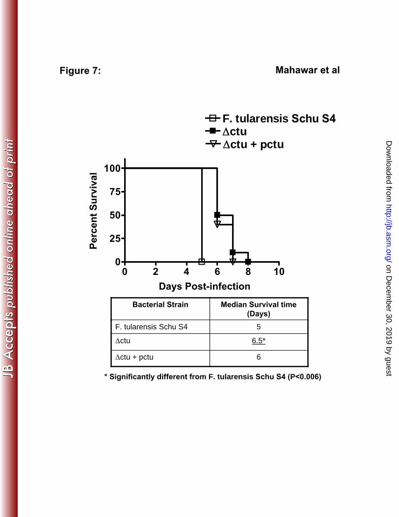
were inoculated intranasally with 25 CFU of WT Schu S4, the 〉ctu or the 342
transcomplemented strain. The mice were monitored twice daily for morbidity and mortality 343
for a period of 21 days. All mice inoculated with 25 CFU of Schu S4 succumbed to infection 344
by day five PI. Although, 100% of the mice infected with 25 CFU of 〉ctu also succumbed 345
to infection, a significantly extended median time to death as compared to Schu S4 infected 346
mice was observed (Fig. 7). Mice infected with the 〉ctu+pctu transcomplemented strain had 347
an intermediate virulence phenotype. The results suggest that 〉ctu undergoes slow 348
replication in infected mice compared to WT Schu S4 strain; however, given the extremely 349
16
on Decem
ber 30, 2019 by guesthttp://jb.asm
.org/D
ownloaded from

high virulence and very low LD100 of Schu S4, subsequent increases in bacterial numbers are 350
sufficient to cause death in the infected mice. These results also indicate that ctu gene 351
deletion causes only a partial attenuation of virulence in mice. 352
17
on Decem
ber 30, 2019 by guesthttp://jb.asm
.org/D
ownloaded from

DISCUSSION 353
354
355
356
357
358
359
360
361
CTU activity has essentially been used as a marker to differentiate highly virulent
strains of F. tularensis from less virulent or avirulent strains (10,11,38,39,42). Despite this
exclusive association with a highly virulent phenotype, the actual contribution of CTU to
virulence and pathogenesis of F. tularensis is not known. We attempted to address this
important issue by generating a non-polar deletion mutant of the ctu gene in highly virulent
Schu S4 strain of F. tularensis and further characterized this mutant for its virulence
attributes in macrophages and mice. DNA sequence analysis of the 〉ctu mutant revealed
that deletion of the ctu gene was in-frame and did not alter the transcription of upstream
genes (data not shown) as ctu is the last gene of the operon. Our in vitro analysis confirmed 362
that the 〉ctu mutant was not growth defective under acellular conditions. However, the 〉ctu
mutant was attenuated for intramacrophage survival and showed reduced virulence in
363
364
365 intranasally infected mice.
Francisella utilizes L-arginine as a carbon and/or nitrogen source (7). The ctu gene 366
of F. tularensis Schu S4 is encoded on an operon that resembles the ADI system required for 367
arginine utilization in several bacterial pathogens (6,9,18). The ctu (FTT0435), arginine 368
deiminase (FTT0434) and arginine decarboxylase [(speA) (FTT0432)] genes similar to those 369
found on other bacterial ADI operon may serve to carryout arginine catabolism in 370
Francisella whereas, spermidine synthase [(speE) (FTT0431)] and S-adenosylmethionine 371
decarboxylase [(speH) (FTT0430)] are required for polyamine biosynthesis (Fig. 1A). A 372
recent report has shown that transcription of all these genes, including the ctu gene is
significantly upregulated following infection of macrophages by F. tularensis Schu S4 (50).
373
374
The genomic organization of ctu with genes involved in arginine utilization, and their 375
18
on Decem
ber 30, 2019 by guesthttp://jb.asm
.org/D
ownloaded from

376 transcriptional upregulation following macrophage infection (50) raises the possibility that
deletion of ctu diminishes the ability of 〉ctu mutant to grow in a nutrient-limiting 377
378 macrophage environment. However, arginine decarboxylase (FTT0432), an enzyme that
379 degrades arginine into agmatine provides an additional arginine metabolism mechanism in
Francisella that may compensate for the loss of CTU. Our laboratory is now in the process 380
381 of creating deletion mutants of additional genes involved in arginine utilization in the
382 virulent Schu S4 strain. These mutant strains will allow us to explore further whether
arginine is a major substrate that is required for intramacrophage survival of Francisella. 383
384
385
The NO produced by IFN-け activated murine macrophages reduces infectivity of F.
tularensis LVS and Schu S4 (17,27,30,35). Similarly, iNOS is required to resolve LVS
infection in mouse models (29). Our results have shown elevated NO levels in culture 386
supernatants from the 〉ctu mutant infected macrophages (Fig. 5A). Additionally, the 〉ctu 387
mutant survived similar to the WT Schu S4 in inos-/-
macrophages (Fig. 5B) suggesting an
NO dependent mechanism for killing of the 〉ctu mutant.
388
In activated macrophages,
increased NO levels are associated with increased citrulline generated as a result of
breakdown of arginine by iNOS. The citrulline is recycled to generate arginine via an
arginine-citrulline cycle in the macrophages (19). The CTU of F. tularensis SchuS4
degrades citrulline to ornithine and ammonia and thus, may inhibit arginine resynthesis in the
infected macrophages. However,
389
390
391
392
393
this process might require secretion of CTU by F. 394
tularensis Schu S4. The PsortB software analysis of CTU did not predict its exact 395
subcellular localization and provided identical scores for all possible locations, including the 396
397 secreted form (data not shown). In the absence of concrete evidence on the secretory nature
of CTU, we speculate that Francisella depletes the arginine pool in macrophages by an 398
active uptake, and metabolism of arginine via CTU and Arginine decarboxylase thereby 399
19
on Decem
ber 30, 2019 by guesthttp://jb.asm
.org/D
ownloaded from

400 reducing the substrate for iNOS and subsequent NO production. Inhibiting this aspect of the
innate immune response
could help Francisella resist killing by macrophages. 401
Chlamydophila and Helicobacter pylori also use a similar strategy and deplete
arginine for 402
403 reducing the iNOS activity and NO abundance (14,46).
It has been shown that Francisella when grown in an acidic medium causes
alkalization of the pH due to generation of ammonia (7). The ammonia produced via
404
405
406 deamination of amino acids also serves to stabilize bacterial cytoplasmic pH on exposure to
407 an acidic environment, such as in the phagosomal vacuoles (40). The ammonia generated by
408 CTU has been proposed to play a role in neutralization of endosomal pH that leads to
phagosomal maturation arrest (25). It has also been reported that inhibition of acidification
and phagosomal maturation enhances intramacrophage survival of Francisella (8,43),
Helicobacter pylori (44) and Mycobacterium tuberculosis (16). On the other hand,
neutralization of phagosomal pH by ammonium chloride (NH4Cl) treatment of macrophages
restores intramacrophage survival of H. pylori urease mutants, which are deficient for
ammonia production (44).
409
410
411
412
413
Thus, an inability to modulate the phagosomal environment or to 414
maintain the bacterial pH homeostasis in the absence of ctu may also offer an explanation for 415
attenuated intramacrophage survival of the 〉ctu mutant. However, NH4Cl treatment of
macrophages resulted in a modest two to three fold increase in the survival of ingested 〉ctu
mutant at 24 hrs PI (data not shown). This small improvement in survival of the 〉ctu mutant
following NH4Cl treatment of infected macrophages suggests that CTU alone may not cause
a significant change in the phagosomal environment. Other genes such as asparaginase,
glutaminase and arginine deiminase may still produce ammonia in the absence of CTU.
416
417
418
419
420
The 421
422 presence of these multi-factorial and redundant mechanisms potentially argues in favor of our
20
on Decem
ber 30, 2019 by guesthttp://jb.asm
.org/D
ownloaded from

observation that the 〉ctu mutant was not cleared completely by the infected macrophages. It 423
is possible that in the absence of ctu these redundant mechanisms compensate for its loss. 424
The transcomplementation studies provided the evidence that the 〉ctu mutation itself 425
is responsible for the reduced-virulence phenotype. Transcomplementation of 〉ctu restored 426
virulence to the levels intermediate between the WT and the 〉ctu mutant phenotype in cell 427
428 culture based assays and mouse model of respiratory tularemia. This partial rather than full
429 restoration of the mutant to the WT phenotype could be attributed to plasmid loss in the
absence of kanamycin selection in cellular and mouse infection models. The loss of pctu in 430
431 the absence of antibiotic selection pressure may have compromised the growth of the
432 complemented mutant, resulting in an intermediate phenotype. Similar observations have
been reported earlier for transcomplemented Schu S4 mutant strain of F. tularensis (37). 433
434
435
436
437
438
439
440
441
442
Mice infected with the 〉ctu mutant showed significantly extended median time to
death as compared to the WT Schu S4 infected mice, but all mice eventually succumbed to
infection. The results are not unexpected, as several other mutants of Schu S4 that are
defective for intracellular survival have also been shown to retain virulence in mice
(27,36,37). A possible explanation could be the existence of unidentified redundant
virulence mechanisms in Schu S4 that mask the effect of a single gene deletion. In addition,
due to the extremely high virulence of Schu S4, even small increases in bacterial numbers are
sufficient to cause death in infected mice.
To conclude, this study provides definitive evidence that CTU activity contributes to
intramacrophage survival and tularemia pathogenesis, but is not the primary virulence factor
of F. tularensis SchuS4
443
. However, the association of the ctu gene with virulence may 444
21
on Decem
ber 30, 2019 by guesthttp://jb.asm
.org/D
ownloaded from

445
446
constitute a strong and rapid method for differentiation of highly virulent type A strains from
less virulent or avirulent type B strains of F. tularensis.
22
on Decem
ber 30, 2019 by guesthttp://jb.asm
.org/D
ownloaded from

ACKNOWLEDGEMENTS 447
448
449
450
Excellent technical support was provided by Michelle Wyland-O’Brien and Sherie
O’Connell. We also thank Dr Anders Sjöstedt, Umea University Umea, Sweden, for
providing the pDMK vector. This work was supported by NIH grant P01 AI056320.
23
on Decem
ber 30, 2019 by guesthttp://jb.asm
.org/D
ownloaded from

451
452
453
454
455
456
457
458
459
460
461
462
463
464
465
466
467
468
469
470
471
472
473
474
475
476
REFERENCES
Reference List
1. Bakshi, C. S., M. Malik, M. Mahawar, G. S. Kirimanjeswara, K. R. Hazlett, L. E.
Palmer, M. B. Furie, R. Singh, J. A. Melendez, T. J. Sellati, and D. W. Metzger.
2008. An improved vaccine for prevention of respiratory tularemia caused by
Francisella tularensis SchuS4 strain. Vaccine 26:5276-5288.
2. Bakshi, C. S., M. Malik, K. Regan, J. A. Melendez, D. W. Metzger, V. M. Pavlov,
and T. J. Sellati. 2006. Superoxide dismutase B gene (sodB)-deficient mutants of
Francisella tularensis demonstrate hypersensitivity to oxidative stress and attenuated
virulence. J. Bacteriol. 188:6443-6448.
3. Baron, G. S., S. V. Myltseva, and F. E. Nano. 1995. Electroporation of Francisella
tularensis. Methods Mol. Biol. 47:149-154.
4. Benninghoff, B., V. Lehmann, H. P. Eck, and W. Droge. 1991. Production of
citrulline and ornithine by interferon-gamma treated macrophages. Int. Immunol.
3:413-417.
5. Bossi, P. and F. Bricaire. 2003. [Tularemia, a potential bioterrorism weapon]. Presse
Med. 32:1126-1130.
6. Casiano-Colon, A. and R. E. Marquis. 1988. Role of the arginine deiminase system
in protecting oral bacteria and an enzymatic basis for acid tolerance. Appl. Environ.
Microbiol. 54:1318-1324.
7. Chamberlain, R. E. 1965. Evaluation of live tularemia vaccine prepared in a
chemically defined medium. Appl. Microbiol. 13:232-235.
8. Clemens, D. L., B. Y. Lee, and M. A. Horwitz. 2004. Virulent and avirulent strains of
Francisella tularensis prevent acidification and maturation of their phagosomes and
escape into the cytoplasm in human macrophages. Infect. Immun. 72:3204-3217.
24
on Decem
ber 30, 2019 by guesthttp://jb.asm
.org/D
ownloaded from

9. Cunin, R., N. Glansdorff, A. Pierard, and V. Stalon. 1986. Biosynthesis and
metabolism of arginine in bacteria. Microbiol. Mol. Biol. Rev. 50:314-352.
477
478
479
480
481
482
483
484
485
486
487
488
489
490
491
492
493
494
495
496
497
498
499
500
501
502
503
10. Fleming, D. E. and L. Foshay. 1955. Studies on the physiology of virulence of
Pasteurella tularensis. I. Citrulline ureidase and deamidase activity. J. Bacteriol.
70:345-349.
11. Fleming, D. E. and L. Foshay. 1956. Studies on the physiology of virulence of
Pasteurella tularensis. II. Serine deaminase and transaminase activity. J. Bacteriol.
71:324-327.
12. Gallagher-Smith, M., J. Kim, R. Al-Bawardy, and D. Josko. 2004. Francisella
tularensis: possible agent in bioterrorism. Clin. Lab Sci. 17:35-39.
13. Gil, H., G. J. Platz, C. A. Forestal, M. Monfett, C. S. Bakshi, T. J. Sellati, M. B.
Furie, J. L. Benach, and D. G. Thanassi. 2006. Deletion of TolC orthologs in
Francisella tularensis identifies roles in multidrug resistance and virulence. Proc. Natl.
Acad. Sci. U. S. A 103:12897-12902.
14. Gobert, A. P., D. J. McGee, M. Akhtar, G. L. Mendz, J. C. Newton, Y. Cheng, H.
L. T. Mobley, and K. T. Wilson. 2001. Helicobacter pylori arginase inhibits nitric
oxide production by eukaryotic cells: A strategy for bacterial survival. Proceedings of
the National Academy of Sciences of the United States of America 98:13844-13849.
15. Golovliov, I., A. Sjostedt, A. Mokrievich, and V. Pavlov. 2003. A method for allelic
replacement in Francisella tularensis. FEMS Microbiol. Lett. 222:273-280.
16. Gordon, A. H., P. D. Hart, and M. R. Young. 1980. Ammonia inhibitsphagosome-
lysosome fusion in macrophages. Nature 286:79-80.
17. Green, S. J., C. A. Nacy, R. D. Schreiber, D. L. Granger, R. M. Crawford, M. S.
Meltzer, and A. H. Fortier. 1993. Neutralization of gamma interferon and tumor
necrosis factor alpha blocks in vivo synthesis of nitrogen oxides from L-arginine and
protection against Francisella tularensis infection in Mycobacterium bovis BCG-treated
mice. Infect. Immun. 61:689-698.
25
on Decem
ber 30, 2019 by guesthttp://jb.asm
.org/D
ownloaded from

18. Gruening, P., M. Fulde, P. Valentin-Weigand, and R. Goethe. 2006. Structure,
Regulation, and Putative Function of the Arginine Deiminase System of Streptococcus
suis. J. Bacteriol. 188:361-369.
504
505
506
507
508
509
510
511
512
513
514
515
516
517
518
519
520
521
522
523
524
525
526
527
528
19. Guoyao, W. U. and J. T. Brosnon. 1992. Macrophages can convert citrulline into
arginine. Biochemistry 281:45-48.
20. Hill, D. L. and J. Van Eys. 1960. The relationship between arginine, citrulline and
ornithine in Tetrahymena pyriformis. J. Protozool. 12:259-265.
21. Jellison, W. L. 1980. Tularemia in North America, p. 161-193. In Steel J.H. (ed.), CRC
Handbook Series in Zoonosis, Vol.2. CRC Press, Boca raton, Fla.
22. Kadzhaev, K., C. Zingmark, I. Golovliov, M. Bolanowski, H. Shen, W. Conlan,
and A. Sj+¦stedt. 2009. Identification of Genes Contributing to the Virulence of
Francisella tularensis SCHU S4 in a Mouse Intradermal Infection Model. PLoS ONE
4:e5463.
23. Kirimanjeswara, G. S., J. M. Golden, C. S. Bakshi, and D. W. Metzger. 2007.
Prophylactic and therapeutic use of antibodies for protection against respiratory
infection with Francisella tularensis. J. Immunol. 179:532-539.
24. Kumar, V. B., A. E. Bernardo, M. M. Alshaher, M. Buddhiraju, R.
Purushothaman, and J. E. Morley. 1999. Rapid Assay for Nitric Oxide Synthase
Using Thin-Layer Chromatography. Analytical Biochemistry 269:17-20.
25. Larsson, P., P. C. Oyston, P. Chain, M. C. Chu, M. Duffield, H. H. Fuxelius, E.
Garcia, G. Halltorp, D. Johansson, K. E. Isherwood, P. D. Karp, E. Larsson, Y.
Liu, S. Michell, J. Prior, R. Prior, S. Malfatti, A. Sjostedt, K. Svensson, N.
Thompson, L. Vergez, J. K. Wagg, B. W. Wren, L. E. Lindler, S. G. Andersson,
M. Forsman, and R. W. Titball. 2005. The complete genome sequence of Francisella
tularensis, the causative agent of tularemia. Nat. Genet. 37:153-159.
26
on Decem
ber 30, 2019 by guesthttp://jb.asm
.org/D
ownloaded from

26. Lauriano, C. M., J. R. Barker, F. E. Nano, B. P. Arulanandam, and K. E. Klose.
2003. Allelic exchange in Francisella tularensis using PCR products. FEMS Microbiol.
Lett. 229:195-202.
529
530
531
532
533
534
535
536
537
538
539
540
541
542
543
544
545
546
547
548
549
550
551
552
553
554
555
27. Lindgren, H., H. Shen, C. Zingmark, I. Golovliov, W. Conlan, and A. Sjostedt.
2007. Resistance of Francisella tularensis strains against reactive nitrogen and oxygen
species with special reference to the role of KatG. Infect. Immun. 75:1303-1309.
28. Lindgren, H., L. Stenman, A. Tarnvik, and A. Sjostedt. 2005. The contribution of
reactive nitrogen and oxygen species to the killing of Francisella tularensis LVS by
murine macrophages. Microbes. Infect. 7:467-475.
29. Lindgren, H., S. Stenmark, W. Chen, A. Tarnvik, and A. Sjostedt. 2004. Distinct
roles of reactive nitrogen and oxygen species to control infection with the facultative
intracellular bacterium Francisella tularensis. Infect. Immun. 72:7172-7182.
30. Loegering, D. J., J. R. Drake, J. A. Banas, T. L. McNealy, D. G. Mc Arthur, L. M.
Webster, and M. R. Lennartz. 2006. Francisella tularensis LVS grown in
macrophages has reduced ability to stimulate the secretion of inflammatory cytokines
by macrophages in vitro. Microb. Pathog. 41:218-225.
31. Malik, M., C. S. Bakshi, K. McCabe, S. V. Catlett, A. Shah, R. Singh, P. L.
Jackson, A. Gaggar, D. W. Metzger, J. A. Melendez, J. E. Blalock, and T. J.
Sellati. 2007. Matrix metalloproteinase 9 activity enhances host susceptibility to
pulmonary infection with type A and B strains of Francisella tularensis. J. Immunol.
178:1013-1020.
32. Malik, M., C. S. Bakshi, B. Sahay, A. Shah, S. A. Lotz, and T. J. Sellati. 2006. Toll-
like receptor 2 is required for control of pulmonary infection with Francisella tularensis.
Infect. Immun. 74:3657-3662.
33. Mbawuike, I. N. and H. B. Herscowitz. 1989. MH-S, a murine alveolar macrophage
cell line: morphological, cytochemical, and functional characteristics. J Leukoc Biol
46:119-127.
27
on Decem
ber 30, 2019 by guesthttp://jb.asm
.org/D
ownloaded from

34. Oyston, P. C., A. Sjostedt, and R. W. Titball. 2004. Tularaemia: bioterrorism defence
renews interest in Francisella tularensis. Nat. Rev. Microbiol. 2:967-978.
556
557
558
559
560
561
562
563
564
565
566
567
568
569
570
571
572
573
574
575
576
577
578
579
580
581
582
35. Polsinelli, T., M. S. Meltzer, and A. H. Fortier. 1994. Nitric oxide-independent
killing of Francisella tularensis by IFN-gamma-stimulated murine alveolar
macrophages. J. Immunol. 153:1238-1245.
36. Qin, A. and B. J. Mann. 2006. Identification of transposon insertion mutants of
Francisella tularensis tularensis strain Schu S4 deficient in intracellular replication in
the hepatic cell line HepG2. BMC. Microbiol. 6:69.
37. Qin, A., D. W. Scott, and B. J. Mann. 2008. Francisella tularensis subsp. tularensis
Schu S4 disulfide bond formation protein B, but not an RND-type efflux pump, is
required for virulence. Infect. Immun. 76:3086-3092.
38. Rodionova, I. V. 1968. [Citrulline ureidase activity in geographical races of Francisella
tularensis]. Dokl. Akad. Nauk SSSR 179:457-460.
39. Rodionova, I. V. 1970. [Differentiation of geographic races of Francisella tularensis on
the basis of citrulline ureidase activity]. Lab Delo 1:42-43.
40. Ryan, S., M. Begley, C. G. Gahan, and C. Hill. 2009. Molecular characterization of
the arginine deiminase system in Listeria monocytogenes: regulation and role in acid
tolerance. Environ. Microbiol. 11:432-445.
41. Samrakandi, M. M., C. Zhang, M. Zhang, J. Nietfeldt, J. Kim, P. C. Iwen, M. E.
Olson, P. D. Fey, G. E. Duhamel, S. H. Hinrichs, J. D. Cirillo, and A. K. Benson.
2004. Genome diversity among regional populations of Francisella tularensis
subspecies tularensis and Francisella tularensis subspecies holarctica isolated from the
US. FEMS Microbiol. Lett. 237:9-17.
42. Sandstrom, G., A. Sjostedt, M. Forsman, N. V. Pavlovich, and B. N. Mishankin.
1992. Characterization and classification of strains of Francisella tularensis isolated in
the central Asian focus of the Soviet Union and in Japan. J. Clin. Microbiol. 30:172-
175.
28
on Decem
ber 30, 2019 by guesthttp://jb.asm
.org/D
ownloaded from

43. Santic, M., M. Molmeret, K. E. Klose, and K. Y. Abu. 2006. Francisella tularensis
travels a novel, twisted road within macrophages. Trends Microbiol. 14:37-44.
583
584
585
586
587
588
589
590
591
592
593
594
595
596
597
598
599
600
601
602
603
604
605
606
607
608
609
44. Schwartz, J. T. and L. A. Allen. 2006. Role of urease in megasome formation and
Helicobacter pylori survival in macrophages. J Leukoc Biol 79:1214-1225.
45. Sjostedt, A. 2004. Gram negative aerobic cocci. Family XVII. Francisellae, p. 200-
210. In D. J. Brenner (ed.), Bergey's Manual of Systematic Bacteriology. Springer,
New York.
46. Smith, C. B. and D. E. Graham. 2008. Outer and Inner Membrane Proteins Compose
an Arginine-Agmatine Exchange System in Chlamydophila pneumoniae. J. Bacteriol.
190:7431-7440.
47. Sonck, K. A., G. Kint, G. Schoofs, W. C. Vander, J. Vanderleyden, and S. C. De
Keersmaecker. 2009. The proteome of Salmonella Typhimurium grown under in vivo-
mimicking conditions. Proteomics. 9:565-579.
48. Twine, S., M. Bystrom, W. Chen, M. Forsman, I. Golovliov, A. Johansson, J.
Kelly, H. Lindgren, K. Svensson, C. Zingmark, W. Conlan, and A. Sjostedt. 2005.
A mutant of Francisella tularensis strain SCHU S4 lacking the ability to express a 58-
kilodalton protein is attenuated for virulence and is an effective live vaccine. Infect.
Immun. 73:8345-8352.
49. Wayne, C. J., H. Shen, R. Kuolee, X. Zhao, and W. Chen. 2005. Aerosol-, but not
intradermal-immunization with the live vaccine strain of Francisella tularensis protects
mice against subsequent aerosol challenge with a highly virulent type A strain of the
pathogen by an alphabeta T cell- and interferon gamma- dependent mechanism.
Vaccine 23:2477-2485.
50. Wehrly, T. D., A. Chong, K. Virtaneva, D. E. Sturdevant, R. Child, J. A. Edwards,
D. Brouwer, V. Nair, E. R. Fischer, L. Wicke, A. J. Curda, J. J. Kupko Iii, C.
Martens, D. D. Crane, C. M. Bosio, S. F. Porcella, and J. Celli. 2009. Intracellular
biology and virulence determinants of Francisella tularensis revealed by transcriptional
29
on Decem
ber 30, 2019 by guesthttp://jb.asm
.org/D
ownloaded from

610
611
612
613
profiling inside macrophages. Cell Microbiol. doi:10.1111/j.1462-5822.2009.01316.x
(Epub ahead of print).
51. Wong, J. D. and S. Shapiro. 1999. Francisella, p. 647-651. In P. R. Murray (ed.),
Manual of Clinical Microbiology. ASM Press, Washington, DC.
30
on Decem
ber 30, 2019 by guesthttp://jb.asm
.org/D
ownloaded from

FIGURE LEGENDS 614
615
616
617
618
619
620
621
622
623
624
625
626
627
628
629
630
631
632
633
634
635
636
FIG. 1. Verification of ctu gene deletion from F. tularensis Schu S4. (A) Genomic
organization of the ctu gene (FTT0435) of Schu S4. Small arrows indicate primer locations.
(B) Confirmation of the ctu gene deletion by PCR. Flanking primers A+D were used for
confirmation of ctu gene deletion. Lane 1, WT F. tularensis Schu S4; lane 2, a merodiploid
stage indicating integration of pDMK:〉ctu in Schu S4 genome; lanes 3 and 4, the Äctu
mutants. An amplification product of ~1.27 kbp (lanes 3 and 4) compared to 2.13 Kbp in
WT Schu S4 (lane 1) confirmed the ctu gene deletion. (C) RT-PCR analysis; lane 1 WT
Schu S4; lane 2 the 〉ctu mutant; lane 3 〉ctu+pctu transcomplemented strain.
FIG. 2: CTU activity assay: The CTU activity assay was performed as described in the
Materials and Methods section. Citrulline and ornithine were run as positive controls.
The arrow indicates the direction of run.
FIG. 3: In vitro growth analysis of the 〉ctu mutant of F. tularensis SchuS4. Growth
curve for the 〉ctu mutant was generated and compared with WT SchuS4 and the 〉ctu+pctu
transcomplemented bacteria. The O.D.600 (upper panel) and the corresponding CFU (lower
panel) were recorded at the indicated times.
FIG. 4. The 〉ctu mutant of Schu S4 is deficient for intramacrophage survival. A
macrophage cell culture invasion assay was performed in BMDMs and the MH-S cells
untreated (left panel) or treated with IFN-i (right panel). The intracellular replication was
quantitated at the indicated times and expressed as CFU/ml. The values represent mean ‒ SE
31
on Decem
ber 30, 2019 by guesthttp://jb.asm
.org/D
ownloaded from

of quadruplicate samples and are cumulative of three experiments conducted. P values were
determined using one-way ANOVA (*P<0.05; **P<0.01 and ***P<0.001).
637
638
639
FIG. 5. Enhanced intramacrophage killing of the 〉ctu mutant is NO dependent. (A)
Culture supernatants from the invasion assay were analyzed for nitrite levels using a
commercial Griess reagent. The detection limit of the Griess reaction was 2.5µM. The
results are expressed as mean ‒ SE from four replicate samples tested in triplicate and are
representative of two independent experiments conducted.
640
641
642
643
(B). BMDMs derived from 644
C57BL/6 WT and inos-/-
mice were stimulated with IFN-i (100ng/ml) for 16 hrs prior to and 645
after infection with F. tularensis Schu S4, 〉ctu or 〉ctu+pctu strains at an MOI of 100. The 646
647 infected BMDMs were lysed 24 hrs PI and bacterial numbers quantitated. The results are
648 expressed as mean ‒ S.D. The statistical analysis was performed using unpaired Student’s t
test. (**P<0.01 and ***P<0.001). 649
650
651
652
653
654
655
656
657
658
659
660
FIG. 6. Quantitation of bacterial burden and histopathological lesions. Mice (n = 4-6
per group), infected with 25 CFU of WT Schu S4, 〉ctu and the transcomplemented
〉ctu+pctu strain were sacrificed at the indicated times. The bacterial burden in the lungs,
liver and spleen (A) and the histopathological lesions in lungs (B) were quantitated. The
results are expressed as Mean ‒ S.E. and are cumulative of two independent experiments
conducted. P values were determined using one-way ANOVA (*P<0.05; **P<0.01 and
***P<0.001).
FIG. 7. Mouse survival studies: Six to eight week old BALB/c mice (n=15 mice per group)
were infected intranasally with 25 CFU of the WT SchuS4, 〉ctu mutant and the 〉ctu+pctu
32
on Decem
ber 30, 2019 by guesthttp://jb.asm
.org/D
ownloaded from

661
662
663
664
665
transcomplemented strains. The mice were monitored for morbidity and mortality for a
period of 21 days. The results are expressed as Kaplan-Meier survival curves. The median
survival time is shown in the table. The P values were determined using Log-Rank test
(*P<0.006).
33
on Decem
ber 30, 2019 by guesthttp://jb.asm
.org/D
ownloaded from
34
666
667
TABLE 1: Primer sequences, bacterial strains, and plasmids used and generated in this
study.
Primer Primer sequence
A. Up ctuF
5’CACGCGTCGACACATTCAAATGGTCAAGGAGT
TTT 3’
B. Up ctuR
5’ ATCAAATCTCCTTTATAGCCGGT 3’
C. Dn ctuF
5’CGGCTATAAAGGAGATTTGATATTGATAAAAAT
GTTTTTAGTTTAGAG 3’
D. Dn ctuR
5’GGGACTAGTTCATCTAAACGCTAATCATGCTG
3’
CTUpKKF
5’AAAACTGCAGATGGCGAATATAAAAGTTGCAG
3’
CTU pKKR
5’ TTAATACTTTCTAACAATTTCTTCA 3’
pKK kan CTU-F 5’TGAAGAAATTGTTAGAAAGTATTAAGTCGACTA
TTAAAAAAATTCATCAAG 3’
pKK kan CTU-R pst1
5’ GGAGTAACTGCAGTATGTCACAT 3’
668
Bacterial Strains and plasmids
Strain* Source/Reference
F. tularensis Schu S4 USAMRIID
pDMK (27)
pDMK:〉ctu This Study
S17-1:pDMK:〉ctu This Study
F. tularensis Schu S4:〉ctu (〉ctu) This study
F. tularensis Schu S4:〉ctu+pctu This study
*E. coli S17-1 strain available in CIMD was used for conjugation experiments. 669
on Decem
ber 30, 2019 by guesthttp://jb.asm
.org/D
ownloaded from

Figure 1:
Citrulline
Ureidase
A
B
speE speA
FTT0434FTT0433
Up CT F
(A)
Dn CT F
(C)Up CT R
(B)
Dn CT R
(D)
FTT0436
FTT0435
Mahawar et al
1 2 3 4 1 2 3
C
Arginine
DecarboxylasePeptidyl
Arginine
Deiminase
1270 bp
2130 bp
speH
FTT0432FTT0431
FTT0430
on Decem
ber 30, 2019 by guesthttp://jb.asm
.org/D
ownloaded from

F. tularensis LVS
F. tularensis Schu S
4
〉ctu〉ctu
+ pctu
F. tularensis
LVS
F. tularensis
Schu S4
〉ctu〉ctu
+ pctu
Citrulli
ne
Orn
ithin
e
Citrulline - - - - + + + + Positive
controls
Figure 2: Mahawar et al
on Decem
ber 30, 2019 by guesthttp://jb.asm
.org/D
ownloaded from

0 10 20 30 40 50 607
8
9
10
11
0 10 20 30 40 50 600.0
0.5
1.0
1.5
2.0
F. tularensis Schu S4
ctuctu +pctu
O.D
. 60
0 n
mL
og
10
CF
U/m
l
Hours
Figure 3: Mahawar et al
on Decem
ber 30, 2019 by guesthttp://jb.asm
.org/D
ownloaded from

24 48 10 5
10 6
10 7
10 8
10 9
F. tularensis Schu S4ctuctu + pctu
24 48 105
106
107
108
109
24 48 105
106
107
108
24 48 105
106
107
108
Hours Post-infection
CF
U/m
lFigure 4:
* *
* *
*** **
* *
*
*
*
*
BMDM - IFN BMDM + IFN
MH-S- IFN MH-S + IFN
Mahawar et al
*
*
*
on Decem
ber 30, 2019 by guesthttp://jb.asm
.org/D
ownloaded from
Figure 5:
4 24 482.5
5.0
7.5
10.0
F. tularensis Schu S4F. tularensis Schu S4 + IFN-iFctuFctu + IFN-i
Hours Post-infection
Nit
rite
/Nit
rate
(M
)
Mahawar et al
104
105
106
107
C57BL/6 WT
C57BL/6 inos-/-
CF
U/m
l
F. tular
ensis
Schu S4 〉c
tu
〉ctu
+pctu
** *** **
A
B
on Decem
ber 30, 2019 by guesthttp://jb.asm
.org/D
ownloaded from

Figure 6:
1 3 5103
105
107
109
10 11
CF
U
Liver
ND ND
****
***
1 3 5103
105
107
109
10 11
Days Post-infection
CF
U
Spleen
ND ND
****
***
1 3 5103
105
107
109
10 11
F. tularensis Schu S4
ctuctu + pctu
CF
U
Lung **
***
**
*
Mahawar et al
A
on Decem
ber 30, 2019 by guesthttp://jb.asm
.org/D
ownloaded from

1 3 50
5
10
15
20
F. tularensis Schu S4
ctuctu + pctu
Lung
Days Post-infection
His
top
ath
olo
gy S
co
re
Mahawar et al
B
Figure 6:
***
*
on Decem
ber 30, 2019 by guesthttp://jb.asm
.org/D
ownloaded from
0 2 4 6 8 100
25
50
75
100
F. tularensis Schu S4
ctu + pctuctu
Days Post-infection
Perc
en
t S
urv
ival
Figure 7: Mahawar et al
Bacterial Strain Median Survival time
(Days)
F. tularensis Schu S4 5
〉ctu 6.5*
〉ctu + pctu 6
* Significantly different from F. tularensis Schu S4 (P<0.006)
on Decem
ber 30, 2019 by guesthttp://jb.asm
.org/D
ownloaded from