Embed Size (px)
Citation preview

Orientation of Pseudomonas aeruginosa ExsA monomers bound to promoter 1
DNA and base-specific contacts with the PexoT promoter 2
3
Jessica M. King, Evan D. Brutinel1*, Anne E. Marsden, 4
Florian D. Schubot2, and Timothy L. Yahr3 5
Department of Microbiology, University of Iowa 6
7
Running title: ExsA-DNA contacts 8
9
Keywords: Pseudomonas aeruginosa, type III secretion, ExsA, 10
transcriptional activator, ExsD, AraC 11
12
13
*JMK and EDB contributed equally to this work 14
15 1Present address 16 University of Minnesota 17 Biotechnology Institute 18 1479 Gortner Ave, Rm 356 19 St. Paul, MN 55108 20 21 2Virginia Polytechnic Institute and State University 22 Department of Biological Sciences 23 125 Life Sciences I Building 24 Washington Street 25 Blacksburg, VA 24061-0910 26 27 3Corresponding author 28 University of Iowa 29 Department of Microbiology 30 540B Eckstein Medical Research Building 31 Iowa City, IA 52242-1101 32 [email protected] 33 Tel: 319-335-9688 34 Fax: 319-335-8228 35 36
Copyright © 2012, American Society for Microbiology. All Rights Reserved.J. Bacteriol. doi:10.1128/JB.00107-12 JB Accepts, published online ahead of print on 9 March 2012
on July 8, 2017 by guesthttp://jb.asm
.org/D
ownloaded from
King et al. ExsA-DNA Contacts
2
ABSTRACT 37
ExsA is a transcriptional activator of the Pseudomonas aeruginosa type III 38
secretion system (T3SS) and a member of the AraC/XylS protein family. Each of the 39
ten ExsA-dependent promoter regions that define the T3SS regulon has two adjacent 40
binding sites for monomeric ExsA. Whereas the promoter-proximal sites (binding site 1) 41
contain highly conserved GnC and TGnnA sequences that are separated by ~10 bp, the 42
promoter-distal sites (binding site 2) share no obvious sequence similarity to each other 43
or to the binding site 1 consensus. In the present study we use Fe-BABE footprinting to 44
demonstrate that the two ExsA monomers bind to the PexsC, PexsD, PexoT, and PpcrG 45
promoters in a head-to-tail orientation. The footprinting data further indicate that the 46
conserved GnC and TGnnA sequences constitute binding site 1. When bound to site 1, 47
the first helix-turn-helix (HTH) motif of ExsA interacts with the conserved GnC sequence 48
and the second HTH interacts at or near the TGnnA sequences. Genetic data using the 49
PexoT promoter indicate that residues L198 and T199 in the first HTH motif of ExsA 50
contact the guanine in the GnC sequence and that residue K202, also in the first HTH 51
motif, contacts the cytosine. Likewise, evidence is presented that residues Q248, Y250, 52
T252, and R257 located in the second HTH motif contribute to recognition of the TGnnA 53
sequence. These combined data define interactions of ExsA with site 1 on the PexoT 54
promoter and provide insight into the nature of the interactions involved in recognition of 55
binding site 2. 56
57
58
on July 8, 2017 by guesthttp://jb.asm
.org/D
ownloaded from

King et al. ExsA-DNA Contacts
3
INTRODUCTION 59
The Gram-negative opportunistic pathogen P. aeruginosa is a significant cause 60
of morbidity and mortality in immunocompromised patients (32, 33). A primary virulence 61
factor of P. aeruginosa is a type III secretion system (T3SS). The T3SS consists of a 62
large macromolecular complex that is assembled in the bacterial cell envelope and 63
functions by translocating effector proteins into eukaryotic host cells (1, 16, 21, 23, 37). 64
The translocated effectors, which include ExoS, ExoT, ExoU, and ExoY, generally 65
promote virulence through inhibition of phagocytosis, modulation of host inflammatory 66
responses, and host cell killing (2, 16, 36). Mutants lacking a functional T3SS are 67
severely attenuated for virulence in animal infection models (20, 21, 23, 37) and a role 68
for the T3SS has been implicated in human disease severity and progression (35). 69
Several studies have examined therapeutic interventions targeting the T3SS including 70
inhibition of translocase activity (37), effector activity (25), and T3SS gene transcription 71
(15). The latter approach involved isolation of small molecule inhibitors of ExsA, the 72
primary transcriptional regulator of T3SS gene expression. 73
ExsA is a member of the AraC/XylS family of transcription factors that activates 74
ten promoters controlling expression of the T3SS effectors and their chaperones, the 75
secretion and translocation machinery, and regulators of T3SS gene expression (10, 76
44). ExsA-dependent transcription is coupled to T3SS secretory activity by a partner-77
switching mechanism involving ExsD, ExsC, and ExsE (10, 44). ExsD functions as an 78
anti-activator by binding to ExsA and inhibiting ExsA-dependent transcription (7, 27, 39). 79
ExsC is a type III secretion chaperone that can form complexes with either ExsD or 80
ExsE, a secreted substrate of the T3SS (9, 45). Under non-inducing conditions for 81
on July 8, 2017 by guesthttp://jb.asm
.org/D
ownloaded from
King et al. ExsA-DNA Contacts
4
T3SS gene expression (e.g., high Ca2+ growth medium) the T3SS machinery is 82
secretion-incompetent, resulting in intracellular accumulation of ExsE and preferential 83
formation of ExsE•ExsC and ExsD•ExsA complexes. Inducing signals, which include 84
Ca2+-limiting growth conditions and contact of P. aeruginosa with host cells, trigger 85
T3SS-dependent secretion and/or translocation of ExsE into host cells (34, 40, 41). The 86
resulting decrease in ExsE concentration in the bacterial cytoplasm triggers ExsC 87
partner switching whereby formation of the ExsC•ExsD complex is favored and free 88
ExsA is available to activate T3SS gene expression (9, 45). 89
The ExsA consensus-binding sequence (AaAAAnwmMygrCynnnmYTGayAk) 90
is similarly positioned in each of the ten ExsA-dependent promoter regions and contains 91
three conserved elements, an adenine-rich region, a GnC sequence, and a TGnnA 92
sequence, the centers of each being separated by ~10 bp (6). ExsA-dependent 93
promoter regions contain two adjacent binding sites for monomeric ExsA which are 94
centered at the -41 (site 1) and -65 (site 2) positions relative to the start of transcription 95
(6). Genetic analyses of the PexoT promoter suggested that the GnC and TGnnA 96
sequences constitute binding site 1. Binding of ExsA to the promoter located upstream 97
of exoT (PexoT) occurs via a monomer assembly pathway whereby monomeric ExsA first 98
binds to site 1 and then recruits a second ExsA monomer to the lower affinity site 2 (6). 99
Efficient filling of site 2 is dependent upon the adenine rich region (5 nucleotides) 100
located at the boundary between binding sites 1 and 2, and on self-association between 101
ExsA monomers which is mediated by the amino-terminal domain of ExsA (6, 8, 22). 102
While the determinants for binding site 1 are fairly well-defined, binding sites 1 and 2 103
on July 8, 2017 by guesthttp://jb.asm
.org/D
ownloaded from

King et al. ExsA-DNA Contacts
5
share no recognizable sequence similarity and the elements within binding site 2 that 104
are required for ExsA binding are currently unknown. 105
Most members of the AraC/XylS family including ExsA consist of two distinct 106
domains connected by a flexible linker. The amino-terminal domain is poorly conserved 107
among family members but is generally involved in oligomerization and/or ligand binding 108
(13, 26). The amino-terminus of ExsA mediates self-interaction of the two monomers 109
when bound to promoter DNA and is also the target for binding by the ExsD anti-110
activator (8). The carboxy-terminal domain of AraC/XylS family members consists of 111
two HTH DNA-binding motifs. The high resolution crystal structure of the AraC/XylS 112
family member MarA bound to target DNA provides a structural model for DNA binding 113
by AraC/XylS proteins (31). Each MarA HTH motif has a recognition helix (helices 3 114
and 6 in MarA) that inserts into adjacent major grooves on the same face of the DNA, 115
and makes base-specific contacts with target DNA (31). The high level of conservation 116
in the DNA binding domains of AraC/XylS family members suggests a common 117
mechanism for recognition and binding to target DNA. 118
In the present study we performed Fe-BABE footprinting experiments to define 119
the promoter regions recognized by ExsA at binding sites 1 and 2, and found that two 120
ExsA monomers bind to the PexoT, PexsC, PexsD, and PpcrG promoters in a head-to-tail 121
orientation. Using genetic screens we determined that the two HTH motifs of an ExsA 122
monomer interact with the conserved GnC and TGnnA sequences in binding site 1 and 123
identified amino acid residues located within the recognition helices of the HTH motifs 124
that participate in recognition of the GnC and TGnnA sequences. Based on these 125
on July 8, 2017 by guesthttp://jb.asm
.org/D
ownloaded from

King et al. ExsA-DNA Contacts
6
findings we propose that specific recognition of binding site 1 at each of the ExsA-126
dependent promoters involves a common mechanism. 127
128
METHODS AND MATERIALS 129
Bacterial strains, culture conditions, and sample preparation. Escherichia 130
coli DH5α was used for all cloning and was maintained on Luria-Bertani (LB) agar 131
plates with gentamicin [15 µg/ml] or ampicillin [100 µg/ml] as necessary. P. aeruginosa 132
strains were cultured on Vogel Bonner minimal medium (VBMM) agar plates with 100 133
µg/ml gentamicin as required (42). To measure β-galactosidase activity P. aeruginosa 134
strains were cultured to an absorbance (A600) of 1.0 in trypticase soy broth (TSB) 135
supplemented with 100 mM monosodium glutamate, 1% glycerol, and 2 mM EGTA. In 136
some experiments the P. aeruginosa strains were cultured in the presence of arabinose 137
(as indicated in the Figure Legends) to elevate expression of ExsA and the ExsA 138
alanine substitution mutants from the arabinose inducible expression vectors. β-139
galactosidase activity was determined using ONPG as previously described (9). The 140
reported values for the β-galactosidase assays denote the average of at least three 141
independent experiments and error bars represent the standard error of the mean 142
(SEM). Statistical analyses (two-tailed unpaired t-tests) were performed using Prism 5 143
(GraphPad Software Inc, La Jolla, CA). 144
Whole cell lysate samples were prepared by pelleting 1.25 mls of cell culture 145
(A600 = 1.0), suspending the pellet in 0.25 ml of SDS-PAGE sample buffer, and 146
sonicating for 10 seconds. Samples were analyzed by SDS-PAGE followed by 147
on July 8, 2017 by guesthttp://jb.asm
.org/D
ownloaded from

King et al. ExsA-DNA Contacts
7
immunoblotting using antibodies directed against ExsA and developed using enhanced 148
chemiluminescent fluorescence detection reagents (Thermo Scientific, Rockford, IL). 149
Mutagenesis. Random mutagenesis of exsA was performed using the Diversify 150
PCR Random Mutagenesis Kit (Clontech, Mountain View, CA) and oligonucleotide 151
primers flanking exsA (primers 29590679 and 42148424, Table S2). The resulting PCR 152
products were digested with XbaI and SacI, and ligated into the corresponding 153
restriction sites in the pJN105 arabinose-inducible expression vector (28). E. coli DH5α 154
was transformed with the resulting ligation mixture and a plasmid library was generated 155
by pooling colonies from LB agar plates containing gentamycin. The resulting plasmid 156
library was introduced into the PA1O3exsA::Ω mini-CTX-PexoT(C-45A)-lacZ reporter strain by 157
electroporation and screening was performed on VBMM agar plates containing 20 µg/ml 158
5-bromo-4-chloro-3-indolyl-β-D-galactoside (X-gal). 159
Libraries of exsA alleles carrying random mutations targeted within recognition 160
helix 1 or 2 were generated using a two-step PCR as previously described with the 161
following modifications (6). In the first step megaprimers were generated by PCR using 162
a common reverse primer (primer 42148424) and a specific forward primer randomized 163
at the nucleotide positions corresponding to codons L198, T199, T200, K202, E203, 164
L204, Q248, S249, Y250, T252, Q253, or S254 (as indicated in Table S2). The 165
megaprimers were gel purified (Qiagen Inc), mixed at an equimolar ratio for RH1 (L198, 166
T199, T200, K202, E203, and L204) or RH2 (Q248, S249, Y250, T252, Q253, and 167
S254), and used in subsequent PCR reactions with a common forward primer (primer 168
29590679). The resulting PCR products were cloned into the XbaI-SacI restriction sites 169
of pJN105 and introduced into E. coli DH5α by standard transformation. A plasmid 170
on July 8, 2017 by guesthttp://jb.asm
.org/D
ownloaded from
King et al. ExsA-DNA Contacts
8
library derived from ~7,000 colonies was isolated as above, introduced into the 171
indicated reporter strains, and screened on VBMM plates with 20 µg/ml X-Gal. 172
Tables S1-S2 show the oligonucleotide primers used for site-directed 173
mutagenesis of exsA and the PexoT promoter. A previously described two-step PCR 174
method was used to generate mutants (6). The exsA mutants were cloned as XbaI-175
SacI restriction fragments in the arabinose-inducible expression vector pJN105 (28). 176
Promoter mutants were cloned as HindIII-EcoRI fragments in mini-CTX-lacZ (19) and 177
integrated onto the PA1O3 chromosome as previously described (3, 19), or used as 178
PCR templates to generate DNA probes for electrophoretic mobility shift assays 179
(EMSA) or footprinting experiments. 180
FeBABE conjugation and footprinting. To generate the cysteine-less 181
derivative of the exsAHis allele (pET16b exsAHis∆cys) each of the seven cysteine 182
residues in exsA was changed to alanine using the QuikChange Multi Site-Directed 183
Mutagenesis Kit (Stratagene) according to the manufacturer’s instructions and the 184
oligonucleotide primers indicated in Table S2. Novel cysteine residues were then 185
introduced into pET16b exsAHis∆cys at glutamate 193 (E193C), methionine 196 186
(M196C), glycine 244 (G244C), or serine 246 (S246C) using the QuikChange XL Site-187
Directed Mutagenesis Kit (Agilent Technologies) with the primers indicated in Table S2. 188
ExsAHis∆cys M196C and ExsAHis∆cys S246C were expressed in E. coli and 189
purified by Ni2+-NTA chromatography as previously described for ExsA (6). Prior to 190
FeBABE conjugation, purified ExsAHis∆cys M196C and ExsAHis∆cys S246C were 191
exchanged into conjugation buffer (20 mM MOPS [pH 8.0], 200 mM NaCl, 1 mM EDTA, 192
and 5% glycerol) using a PD-10 column (GE Healthcare). A 10-fold molar excess of 193
on July 8, 2017 by guesthttp://jb.asm
.org/D
ownloaded from
King et al. ExsA-DNA Contacts
9
FeBABE reagent (Pierce) was then added and reactions were incubated in the dark for 194
2 hours at 25⁰C. Conjugated ExsA was then exchanged into conjugation buffer, diluted 195
with an equal volume of glycerol, and stored at -20⁰C. 196
DNA probes were generated by PCR using the primers and methodology for 197
single end-labeling with 32P as previously described (6). Binding reactions (20 µl) 198
containing 1 nM promoter probe in DNA binding buffer (10 mM Tris [pH 8.0], 50 mM 199
KCl, 2 mM MgCl2, 0.1 mM EDTA, and 5% glycerol), 2.5 ng/µl poly(2’-deoxyinosinic-2’-200
deoxycytidylic acid) (Sigma), 100 ng/µl bovine serum albumin (New England Biolabs), 201
and 100 nM ExsA were incubated for 15 minutes at 25⁰C. Cleavage reactions were 202
initiated by the addition of 2.5 µl ascorbate solution (40 mM sodium ascorbate, 10 mM 203
EDTA) for one minute followed by the addition of 2.5 µl H2O2 solution (40 mM H2O2, 10 204
mM EDTA) for 2 minutes. Cleavage was terminated by adding 5 µl 100 mM thiourea 205
and the DNA was precipitated with sodium acetate and ethanol. The precipitate was 206
suspended in formamide loading buffer (95% formamide, 10 mM NaOH, 0.05% xylene 207
cyanol, 0.05% bromophenol blue) and samples were subjected to electrophoresis on a 208
1X TBE sequencing gel as previously described (6). Imaging was performed using an 209
FLA-7000 phosphoimager (Fujifilm) and Multigauge v3.0 software (Fujifilm). 210
Electrophoretic mobility shift assays (EMSA). The indicated alanine 211
substitution mutants were PCR amplified from their respective pEB124 expression 212
vectors (Table S1) using primers 42308574-80815243 (Table S2). The resulting PCR 213
products were cloned into the NdeI and BamHI restriction sites in the expression vector 214
pET16b. The proteins were expressed in E. coli Tuner (DE3) as previously described 215
(6) and whole cell extracts were prepared by harvesting 1.7 x 10¹⁰ cells, suspending in 216
on July 8, 2017 by guesthttp://jb.asm
.org/D
ownloaded from

King et al. ExsA-DNA Contacts
10
250 µl ExsA buffer (20 mM Tris [pH 8.0], 500 mM NaCl, 1 mM dithiothreitol, and 0.5% 217
Tween 20) containing protease inhibitor cocktail tablets (Complete Mini, EDTA-free, 218
Roche Diagnostics, Indianapolis, IN) and lysing the cells by sonication. The cell lysate 219
was cleared by centrifugation (16,000 x g, 30 min, 4˚C) and the supernatant was 220
immediately aliquoted and flash frozen in liquid nitrogen. The cell extracts were thawed, 221
diluted in DNA binding buffer, and subjected to EMSA experiments as previously 222
described (6) and anti-ExsA immunoblots to determine the level of specific DNA-binding 223
activity for each alanine substitution mutant relative to wt ExsA. Imaging was performed 224
using an FLA-7000 phosphoimager (Fujifilm) and Multigage v3.0 software (Fujifilm) for 225
data analyses. 226
Missing nucleoside footprinting. The missing nucleoside footprinting 227
experiments were performed according to a protocol adapted from Hayes and Tullius 228
(17). PCR primers (50 pmol) were end-labeled using 20 U polynucleotide kinase (New 229
England Biolabs) and 120 μCi [γ-32P]-ATP (Perkin Elmer). Reactions were applied to 230
NucAway columns (Ambion) to remove unincorporated [γ-32P]-ATP. End-labeled PexsD, 231
PexoT, PexsC, and PpcrG promoter probes were generated in a PCR reaction using labeled 232
(above) and unlabeled primer (20 pmol), and the PCR products were gel purified 233
(Qiagen). Nucleosides were randomly excised by treating the probes (16 µl in 1X DNA 234
binding buffer) with 1 µL each of the following solutions for 1 min; 160 mM sodium 235
ascorbate, Fe(II)-EDTA (3.2 mM (NH4)2Fe(II)(SO4)2x6H2O, 6.4 mM EDTA, and 4.8% 236
H2O2. The reactions were quenched by adding 2 µl 100 mM thiourea. Probes were 237
ethanol precipitated, dissolved in 10 mM Tris-Cl [pH 8.0], 0.1 mM-EDTA, and used in an 238
EMSA reaction as previously described (6). The unbound and ExsA-bound probes 239
on July 8, 2017 by guesthttp://jb.asm
.org/D
ownloaded from

King et al. ExsA-DNA Contacts
11
were excised as a gel slice. Probes were eluted from the gel slices by incubating for 16 240
hr at 37˚C in 0.5 M ammonium acetate, 1 mM EDTA [pH 7.5], 0.1% SDS, and 10 mM 241
MgCl2. Eluted probes were ethanol precipitated, washed, dried, suspended in 242
formamide loading buffer, and analyzed on a 6% sequencing gel. 243
244
RESULTS 245
Two ExsA monomers bind to T3SS promoter regions in a head-to-tail 246
orientation. AraC/XylS family members are characterized by the presence of two HTH 247
DNA binding motifs (Fig. 1A). Each binding motif contains a recognition helix that 248
contacts the target DNA. Amino acid residues that directly contact the target DNA have 249
been determined for a number of AraC/XylS family members and fall within discrete 250
regions of the recognition helices (Fig. 1A) (4, 5, 11, 14, 24, 31). To define the 251
interaction of ExsA with target DNA we generated structural models based on the 252
crystal structures of MarA and Rob bound to target DNA (Fig. 1B-C) (24, 31). Both 253
models predicted that the HTH recognition helices of ExsA (RH1 and RH2) interact with 254
promoter sequences located ~10 bases apart. This observation is consistent with the 255
previous identification of two highly conserved sequences (GnC and TGnnA) which are 256
separated by ~10 bp and present in binding site 1 of all ExsA-dependent promoter 257
regions (6). 258
To determine where RH1 and RH2 interact with the promoter region, we 259
employed FeBABE footprinting. FeBABE is a protein-labeling reagent that can be 260
conjugated to cysteine residues. When conjugated to a DNA binding protein the 261
immobilized FeBABE generates hydroxyl radicals that cleave target DNA in the 262
on July 8, 2017 by guesthttp://jb.asm
.org/D
ownloaded from
King et al. ExsA-DNA Contacts
12
immediate vicinity (within 7-16 angstroms) of the interaction site (12, 30). To adapt this 263
system to ExsA we first generated a cysteine-less exsA allele (exsAHis-∆cys) by 264
changing the seven native cysteine residues (C15, C50, C102, C121, C139, C261, and 265
C271) to alanine. As shown in Fig. 2A, ExsAHis-∆cys activates expression of a PexsD-lacZ 266
transcriptional reporter to levels comparable to wt ExsA. Based on the modeled 267
structure of the ExsA DNA binding domain we then identified amino acid residues 268
positioned within the “turn” regions adjacent to RH1 (E193 and M196) or RH2 (G244 269
and S246) (Figs. 1A-C) and individually changed each to a cysteine. When expressed 270
from a plasmid all four cysteine substitution mutants complemented an exsA mutant for 271
expression of the PexsD-lacZ reporter, albeit with varying degrees of efficiency (Fig. 2A). 272
ExsAHis∆cys M196C and ExsAHis∆cys S246C were chosen for subsequent experiments 273
as they possessed the highest levels of activity and were stably expressed in P. 274
aeruginosa (Fig. 2A). 275
The ExsAHis∆cys M196C and ExsAHis∆cys S246C proteins were expressed in E. 276
coli, partially purified by Ni+2 affinity chromatography (Fig. 2B), and conjugated with 277
FeBABE at the unique cysteine residue (referred to as the RH1 and RH2 conjugants, 278
respectively). The RH1- or RH2-FeBABE conjugants were then incubated with 32P end-279
labeled probes (~200 bp) derived from the ExsA-dependent PexsC, PpcrG, PexsD, and PexoT 280
promoters and subjected to FeBABE-mediated cleavage. Cleavage by the RH1-281
FeBABE conjugant was similar for all four promoters tested with cleavage sites located 282
near the -75 and -50 regions on both the top (Fig. 2C-D, filled circles) and bottom (Fig. 283
2D) strands of the DNA. The one anomaly was an absence of cleavage on the bottom 284
strand at the -50 region of the PexsC promoter. The cleavage patterns generated by the 285
on July 8, 2017 by guesthttp://jb.asm
.org/D
ownloaded from

King et al. ExsA-DNA Contacts
13
RH2-FeBABE conjugant were more variable. At binding site 1 the RH2-generated 286
cleavage patterns were similar for each promoter with cleavage of the top strand 287
centered at -40 and cleavage of the bottom strand centered at -33 and -43 (Fig. 2C-D, 288
(open circles). Cleavage by the RH2-conjugant at binding site 2, however, was only 289
observed for the PexsC and PpcrG promoters where it was centered at -59 and -61 on the 290
top and bottom strands, respectively. The combined RH1 and RH2 cleavage patterns 291
are consistent with our previous data showing the presence of two binding sites for 292
monomeric ExsA on the PexsC, PexsD, and PexoT promoters (6). In addition, the cleavage 293
patterns in binding site 1 indicate that RH1 interacts near the GnC sequence and that 294
RH2 interacts near the TGnnA sequence (Fig. 2D). It should be noted that the FeBABE 295
cleavage patterns were not expected to perfectly coincide with the GnC and TGnnA 296
sequences because the FeBABE is conjugated to the “turn” regions located adjacent to 297
RH1 or RH2 (Fig. 1B-C). Finally, the cleavage patterns at the PexsC and PpcrG promoters 298
indicate that the two ExsA monomers bind in a head-to-tail configuration (i.e., in the 299
same orientation) because the RH1-conjugant cleaves upstream of the RH2-conjugant 300
at both binding sites 1 and 2. 301
Recognition helices 1 and 2 of ExsA make specific contacts with the GnC 302
and TGnnA regions of the PexoT promoter. To determine which amino acids within 303
RH1 and RH2 contact the conserved GnC and TGnnA sequences in binding site 1 of 304
the PexoT promoter we screened a library of PCR-mutagenized exsA alleles for gain-of-305
function mutants that could activate PexoT-lacZ transcriptional reporters carrying mutations 306
within the GnC or TGnnA sequences (Fig. 3A). In the first experiment we utilized a 307
PexoT(C-45A)-lacZ reporter which carries a GnA substitution for the conserved GnC 308
on July 8, 2017 by guesthttp://jb.asm
.org/D
ownloaded from

King et al. ExsA-DNA Contacts
14
sequence. As previously reported, activation of the PexoT(C-45A)-lacZ reporter by wt ExsA is 309
reduced ~14-fold when compared to the wt PexoT-lacZ reporter (6) (Fig. 3B). The 310
mutagenized ExsA expression library was introduced into the PA1O3 exsA::Ω PexoT(C-311
45A)-lacZ reporter strain and ~20,000 colonies were screened for increased β-312
galactosidase activity on VBMM agar plates containing X-Gal. This approach 313
repeatedly identified a single substitution in RH1 (lysine 202 changed to an arginine 314
[K202R]) that resulted in a 4-fold increase in expression from the PexoT(C-45A)-lacZ reporter 315
and a 3.8-fold reduction in PexoT-lacZ expression (Fig. 3B). Immunoblotting of whole cell 316
lysates with ExsA antiserum confirmed that the steady state expression level of ExsA 317
K202R was similar to that of wt ExsA (inset in Fig. 3B). The finding is consistent with 318
the results of the Fe-BABE footprinting data showing that RH1 interacts near the GnC 319
sequence and suggests that K202 in RH1 contacts the -45 cytosine in the PexoT 320
promoter. Note that each of the contacts identified throughout this study will be defined 321
relative to the sequence of the coding (top) strand of the DNA. Although the genetic 322
data cannot differentiate whether the contact is occurring with the coding or noncoding 323
strand, an experiment to address this ambiguity is presented later in Fig. 9. 324
The above approach involved random mutagenesis of the entire ExsA coding 325
sequence. Since only a single gain-of-function mutant was identified, we used directed 326
mutagenesis to increase coverage of RH1 by generating a library of exsA alleles 327
randomized at each of six positions (residues 198-200 and 202-204) within RH1 (Fig. 328
1A). Residues 201 and 205 were excluded from the library as they form part of the 329
conserved hydrophobic core of the HTH motif and residues 206-208 were excluded 330
because the structural models predicted that each of the side chains was buried within 331
on July 8, 2017 by guesthttp://jb.asm
.org/D
ownloaded from
King et al. ExsA-DNA Contacts
15
the structure of ExsA (Fig. 1A). The RH1-targeted library was screened as above and 332
again only the K202R substitution mutant demonstrated a gain-of-function using the 333
PexoT(C-45A)-lacZ reporter. 334
To identify amino acids that interact with the guanine of the GnC sequence we 335
repeated the screen using the RH1-targeted library and a reporter carrying an AnC 336
substitution (PexoT(G-47A)-lacZ) (Fig. 3A) (6). Activation of the PexoT(G-47A)-lacZ reporter by wt 337
ExsA is reduced ~12-fold compared to the wt PexoT-lacZ reporter (6) (Fig. 3C). One 338
substitution in RH1 (T199S) resulted in a significant increase (2.9-fold) in β-339
galactosidase expression from the PexoT(G-47A)-lacZ reporter and a corresponding 1.7-fold 340
decrease in expression of the wt reporter. These combined results suggest that 341
residues T199 and K202 in RH1 interact with the conserved GnC sequence. 342
The Fe-BABE footprinting data and the finding that RH1 contacts the GnC 343
sequence suggested that RH2 might contact the TGnnA sequence centered 10 base 344
pairs downstream. To identify amino acids that interact with the adenine of the TGnnA 345
sequence in the PexoT promoter we generated a targeted library of ExsA alleles 346
randomized at the six positions (residues 252-254 and 256-258) in RH2 predicted to 347
make base specific contacts with the promoter (Fig. 1A). Amino acids that form the 348
conserved hydrophobic core of the HTH motif (residues 251 and 255) or predicted to be 349
buried in the structure of the HTH motif (residues 248-250) were excluded from the 350
library (Fig. 1A). The initial screens were complicated by the fact that Q253 351
substitutions to valine, leucine, or isoleucine resulted in a super-activation phenotype 352
such that expression from both the wt PexoT-lacZ and PexoT(A-34C)-lacZ reporters was ~10-fold 353
higher than with wt ExsA (data not shown). For this reason we regenerated the RH2-354
on July 8, 2017 by guesthttp://jb.asm
.org/D
ownloaded from
King et al. ExsA-DNA Contacts
16
targeted library but excluded Q253. Using this new library we identified a T252S gain-355
of-function mutant within RH2 that resulted in a 3-fold increase in expression of the 356
PexoT(A-34C)-lacZ reporter (Fig. 3D) and reduced expression (130-fold) of the wt PexoT-lacZ 357
reporter. In contrast, activation of the PexoT(A-34C)-lacZ reporter by wt ExsA is reduced 358
~70-fold when compared to the wt PexoT-lacZ reporter (6) (Fig. 3D). These data suggest 359
that RH2 contacts the TGnnA sequence in binding site 1 of the PexoT promoter. 360
Loss-of-contact analyses identify base-specific contacts mediated by T199 361
and K202. To further examine the roles of residues T199, K202, and T252 we 362
employed a genetic loss-of-contact approach, previously used to define base-specific 363
contacts for a number of AraC/XylS proteins for which structural information is 364
unavailable (4, 11, 18). The approach is based on the rationale that a DNA binding 365
protein with an alanine substitution at a residue involved in a critical base-specific 366
contact will be insensitive to the identity of that base. For instance, our data would 367
predict an ExsA T199A mutant to be significantly impaired for activation of the wt PexoT-368
lacZ reporter due to loss of the contact between residue T199 in RH1 and the guanine at 369
position -47. The residual activation of the PexoT-lacZ reporter by the T199A mutant, 370
therefore, should be insensitive to the identity of the base at position -47. Conversely, 371
residual activation by T199A would remain sensitive to promoter mutations that disrupt 372
unrelated base-specific contacts. 373
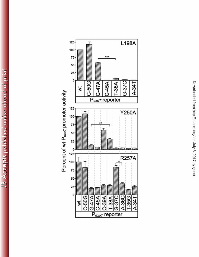
For these analyses we used a previously generated panel of mutant PexoT-lacZ 374
reporters with nucleotide substitutions at the conserved GnC (G-47A and C-45A) and 375
TGnnA (T-38A, G-37C and A-34T) positions in binding site 1 (summarized in Fig. 4A) 376
(6). Each of these substitutions results in a significant reduction in ExsA-dependent 377
on July 8, 2017 by guesthttp://jb.asm
.org/D
ownloaded from
King et al. ExsA-DNA Contacts
17
activity (Fig. 4B). The PexoT(C-50G)-lacZ reporter has a substitution at the non-critical -50 378
position and served as a negative control. We also introduced nucleotide substitutions 379
at positions -39, -36, and -35, which represent conserved but more degenerative 380
nucleotides in the ExsA-binding consensus site (6). Whereas the substitutions at 381
positions -35 and -36 resulted in a significant defect in activation by wt ExsA, the -39 382
mutation had little effect on reporter activity. Based on the latter finding, therefore, we 383
expected both the PexoT(C-50G)-lacZ and PexoT(G-39A)-lacZ reporters to serve as negative 384
controls. 385
To screen for loss-of-contact phenotypes we introduced wt ExsA or the T199A, 386
K202A, and T252A expression plasmids into an exsA::Ω mutant carrying the wt PexoT-lacZ 387
or PexsC-lacZ reporters. The resulting strains were cultured under inducing conditions for 388
T3SS gene expression (low Ca2+) in the absence or presence of arabinose and assayed 389
for β-galactosidase activity. Each of the alanine substitution mutants demonstrated a 390
significant reduction in activation of the wt PexoT-lacZ and PexsC-lacZ reporters when cultured 391
in the absence of arabinose (Table S3). Growth in the presence of arabinose, however, 392
resulted in a significant increase in reporter activity by the T199A and K202A mutants 393
(Fig. 4C, Table S3). The subsequent loss-of-contact analyses, therefore, were 394
performed in the presence of 0.1% arabinose to maximize the sensitivity of the assay. 395
In contrast to T199A and K202A, the activity of the T252A mutant was not significantly 396
recovered by growth in the presence of arabinose. Examination of the steady-state 397
expression levels of wt ExsA, T199A, K202A, and T252A offers an explanation for this 398
since the T252A mutant was poorly expressed (Fig. 4C). This finding was unexpected 399
on July 8, 2017 by guesthttp://jb.asm
.org/D
ownloaded from

King et al. ExsA-DNA Contacts
18
because the T252S substitution identified in the gain-of-function screen was stably 400
expressed (Fig. 3D). 401
Although activation of the wt PexoT-lacZ reporter by the T199A and K202A mutants 402
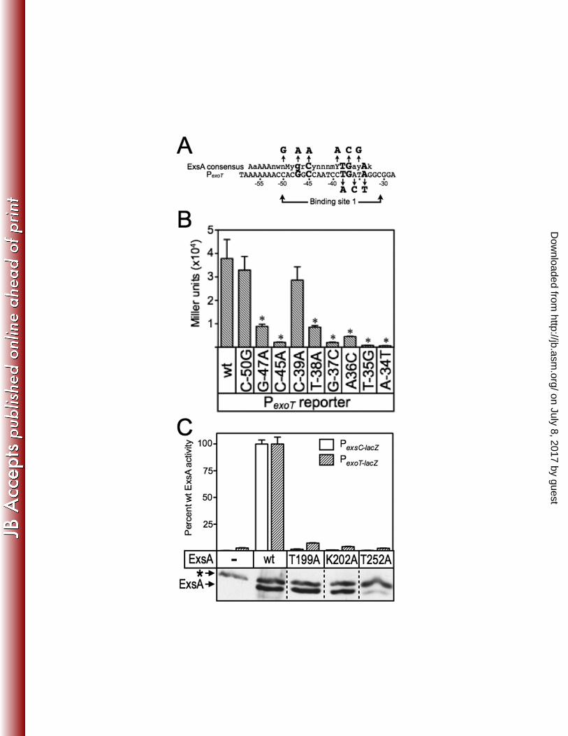
was significantly impaired, both mutants retained residual activity above the vector 403
alone (Table S3). This residual activity allowed us to compare T199A- and K202R-404
dependent activation of the wt PexoT-lacZ reporter (normalized to 100% activity in Fig. 5) to 405
each of the mutant PexoT-lacZ reporters. Reporters with bp substitutions in the conserved 406
-45, -38, -37, and -34 positions led to a further decrease in activation by the T199A 407
mutant. This decrease in activity is consistent with loss of a second base-specific 408
contact that further impairs DNA binding (Fig. 5). In contrast, PexoT-lacZ reporters with 409
substitutions at the non-critical -50 position or at the conserved -47 guanine had activity 410
comparable to the wt PexoT-lacZ reporter (Fig. 5). An important prediction is that wt ExsA 411
should be more sensitive to the C-47A substitution relative to the T199A mutant. 412
Comparison of Figs. 4 and 5 shows that the T199A mutant activates both the wt PexoT-413
lacZ and PexoT(C-47A)-lacZ reporters to similar levels. In contrast, activation PexoT(C-47A)-lacZ 414
reporter by wt ExsA is reduced 4-fold relative to the wt PexoT-lacZ. The findings indicate 415
that the T199A mutant is insensitive to the identity of the base at the -47 position and 416
supports our conclusion that T199A makes a base-specific contact with the -47 417
cytosine. 418
A similar result was seen for K202, predicted to contact cytosine -45 by the gain-419
of-function screen. In the loss-of-contact assay, the K202A mutant was relatively 420
insensitive to a substitution at the -45 position in the PexoT-lacZ reporter (50% activity) 421
(Fig. 5). Importantly, activation by K202A was sensitive to mutations at other critical 422
on July 8, 2017 by guesthttp://jb.asm
.org/D
ownloaded from
King et al. ExsA-DNA Contacts
19
nucleotide positions in the promoter, As expected, wt ExsA is far more sensitive to the 423
C-45A substitution in PexoT (20-fold reduction compared to wt PexoT) relative to the 424
K202R mutant which demonstrated only a 2-fold reduction (Fig. 4B and 5). These 425
combined findings provide strong genetic evidence that ExsA bound to site 1 on the 426
PexoT promoter utilizes residues T199 and K202 to contact the -47 guanine and -45 427
cytosine, respectively. 428
The loss-of-contact approach was also used to examine residue T252, predicted 429
by the gain-of-function screen to contact the adenine at position -34. In contrast to our 430
findings for T199A and K202A, none of the mutant PexoT-lacZ reporters with substitutions 431
in the GnC and TGnnA sequences demonstrated a loss-of-contact phenotype for the 432
T252A mutant (Fig. 5). We considered the possibility that residue T252 contacts 433
another position in the ExsA-consensus binding sequence by using PexoT-lacZ reporters 434
with substitutions at the less highly conserved -36 and -35 positions and again failed to 435
detect a loss-of-contact phenotype. Curiously, the negative controls demonstrated 436
opposite properties. Whereas activation of the PexoT(C-50G)-lacZ reporter by T252A was 3-437
fold higher than the wt PexoT-lacZ reporter, the activity of the PexoT(G-39A)-lacZ reporter was 438
significantly reduced. While an explanation for this is not evident, it should be noted 439
that the T252A mutant is severely impaired for activation of the PexoT-lacZ reporter (Table 440
S3). We suspect that the defective nature of the T252A mutant results in 441
hypersensitivity to promoter mutations that only modestly alter binding affinity. In 442
summary, the loss-of-contact findings are not supportive of a role for T252 in base-443
specific recognition of the TGnnA sequence but the T252S gain-of-function phenotype 444
argues otherwise. The interpretation we favor is that T252 participates in base-specific 445
on July 8, 2017 by guesthttp://jb.asm
.org/D
ownloaded from
King et al. ExsA-DNA Contacts
20
recognition of the TGnnA sequence but that the low residual activity of the T252A 446
mutant precludes detection of loss-of-contact phenotypes. 447
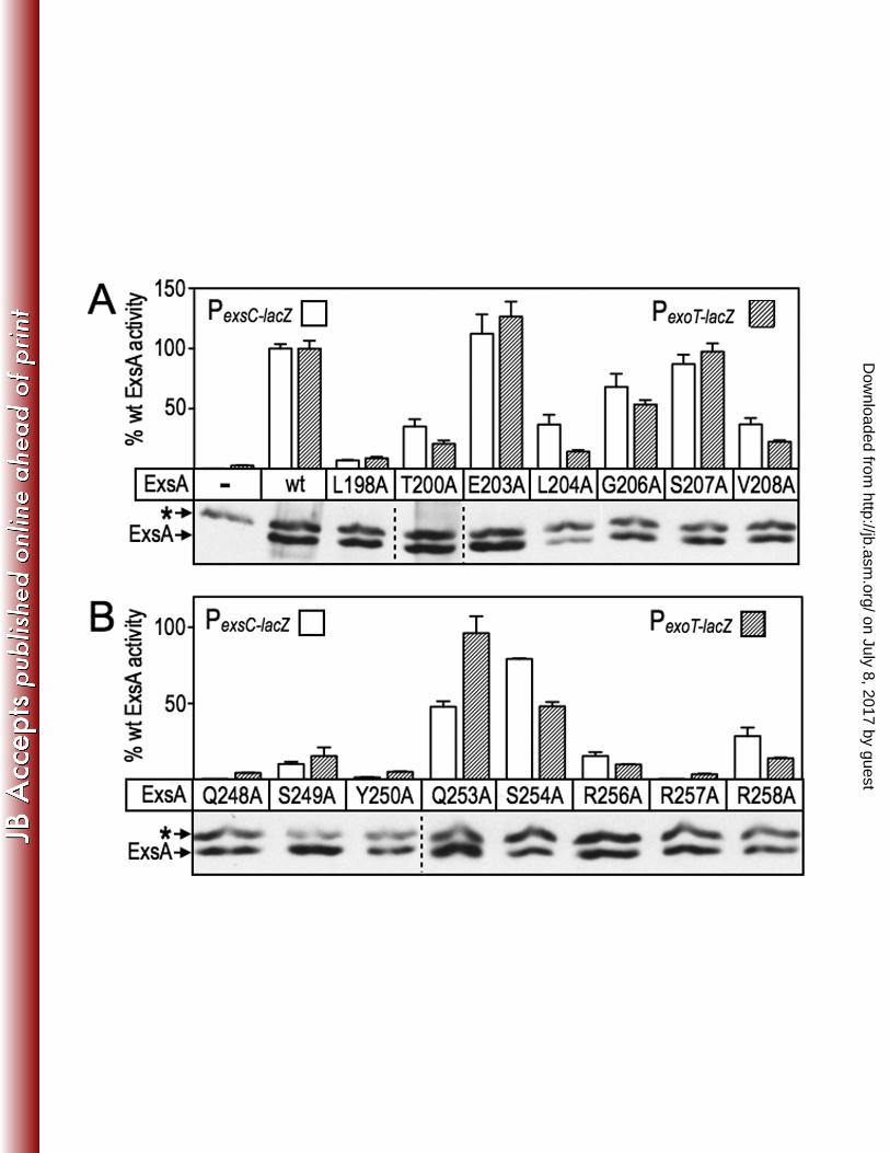
Alanine scanning mutagenesis of RH1 and RH2. While the loss-of-contact 448
approach can be used to confirm base-specific interactions, it can also be used to 449
discover unknown interactions. To determine whether additional residues contribute to 450
ExsA activity we performed alanine-scanning mutagenesis of the remaining residues 451
that constitute RH1 and RH2. Seven alanine substitutions were introduced into RH1 452
and eight substitutions in RH2 using site-directed mutagenesis (Fig. 6A-B). As in the 453
gain-of-function screen described above, residues F201 and F205 in RH1, and F251 454
and Y255 in RH2, were excluded from the analyses because they form the hydrophobic 455
core of the HTH motifs. 456
Most of the alanine-substituted mutant activated both the PexoT-lacZ and PexsC-lacZ 457
transcriptional reporters to similar levels although some modest differences (≤ 2-fold) 458
existed with L204A, Q253A, S254A, and R258A being most notable (Fig. 6A-B, Table 459
S3). The alanine substitution mutants were classified into three primary groups based 460
on activity relative to wt ExsA; (i) mutants at positions 203, 206 and 207 in RH1 (Fig. 461
6A) and at positions 253 and 254 in RH2 (Fig. 6B) retained >50% the activity of wt ExsA 462
and were excluded from further consideration, (ii) mutants at positions T200, L204, 463
V208, S249, and R258 had activities in the range of 10-49%, and (iii) mutants at 464
positions L198, Q248, Y250, R256, and R257 were the most severe with <10% the 465
activity of wt ExsA. A trivial explanation for reduced activation by the mutant proteins is 466
that the alanine substitutions render the proteins unstable. To address this possibility 467
we measured the steady-state expression levels using α-ExsA immunoblots. With the 468
on July 8, 2017 by guesthttp://jb.asm
.org/D
ownloaded from
King et al. ExsA-DNA Contacts
21
exception of L204A, however, each of the mutant proteins was expressed at levels 469
similar to wt ExsA (Fig. 6A-B). 470
The loss of contact approach was applied to each of the alanine substitution 471
mutants that retained <50% activity relative to wt ExsA (Fig. 7). The only alanine 472
substitution mutant in RH1 to demonstrate loss of base-specificity was L198A whereby 473
activation of the PexoT-lacZ reporter was relatively insensitive to a nucleotide substitution 474
at the -47 position (Fig. 7) and wt ExsA was more sensitive to the G-47A substitution 475
relative to the L198A mutant (Fig. 4B). Base-specific interactions for the T200A, L204A, 476
and V208A mutants were not detected (Fig S2A). From this we conclude that the 477
primary specificity determinants in RH1 are residues L198 and T199, which contribute 478
to base-specific recognition of the guanine at position -47, and K202, which interacts 479
with the cytosine at position -45. Two alanine substitution mutants in RH2 480
demonstrated potential interactions with or near the TGnnA sequence. The Y250A 481
mutant was somewhat insensitive to the substitution at position 38 and the R257A 482
mutant was completely insensitive to the substitution at position -37 (Fig. 7). Base 483
specific interactions for the Q248A, S249A, R256A, and R258A mutants were not 484
detected (Fig. S2B). 485
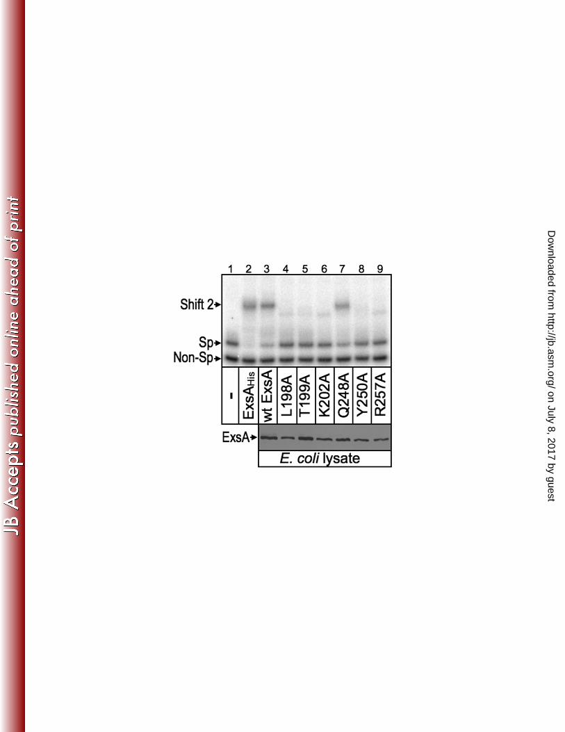
DNA-binding activity of selected alanine scanning mutants. The L198A, 486
T199A, K202A, Y250A, T252A, and R257A mutants each demonstrated a significant 487
reduction in expression of the PexoT-lacZ and PexsC-lacZ reporters, and potential base-488
specific interactions with the PexoT promoter. While these findings were consistent with 489
a primary defect in DNA-binding activity, impaired recruitment of RNA polymerase could 490
also account for the reduced activity of the mutant proteins. To address this question 491
on July 8, 2017 by guesthttp://jb.asm
.org/D
ownloaded from
King et al. ExsA-DNA Contacts
22
whole cell extracts were prepared from E. coli expressing either wt ExsA or the 492
indicated alanine substitution mutants. The Q248A mutant, defective for activation of 493
the PexoT-lacZ and PexsC-lacZ reporters but lacking a loss-of-contact phenotype, was 494
included as a control. The extracts were then normalized for ExsA content (Fig. 8, 495
lower panel) and incubated with a radiolabeled PexsC promoter probe and a non-specific 496
control probe, and analyzed by electrophoretic mobility shift assays. As shown in Fig. 8 497
(upper panel), purified ExsAHis and the E. coli extracts containing either wt ExsA or the 498
Q248A mutant bound specifically to the PexsC promoter probe resulting in the formation 499
of shift product 2 which represents ExsA bound to binding sites 1 and 2. None of the 500
remaining extracts, however, possessed specific DNA binding activity. Subsequent 501
titrations of the extracts indicate that the DNA-binding activity of the L198A, T199A, 502
K202A, Y250A, and R257A mutants is at least 12-fold lower than that of wt ExsA. 503
These findings indicate that reduced activation of the PexoT-lacZ and PexsC-lacZ reporters by 504
these mutants results from a primary defect in DNA binding activity. This finding, 505
however, does not exclude the possibility that some mutants also possess defects in 506
recruitment of RNA polymerase that further contribute to reduced expression of the 507
PexoT-lacZ and PexsC-lacZ reporters in vivo. In fact, impaired recruitment of RNA 508
polymerase most likely accounts for the phenotype of the Q248A mutant, which has 509
DNA-binding activity similar to wt ExsA but a 50-fold defect in activation of the PexoT-lacZ 510
reporter (Table S3). 511
Strand specific contacts by RH1 and RH2. The data presented above define 512
several base-specific contacts with the PexoT promoter region but could not differentiate 513
whether the contacted nucleosides are located on the top or bottom strands of the DNA. 514
on July 8, 2017 by guesthttp://jb.asm
.org/D
ownloaded from

King et al. ExsA-DNA Contacts
23
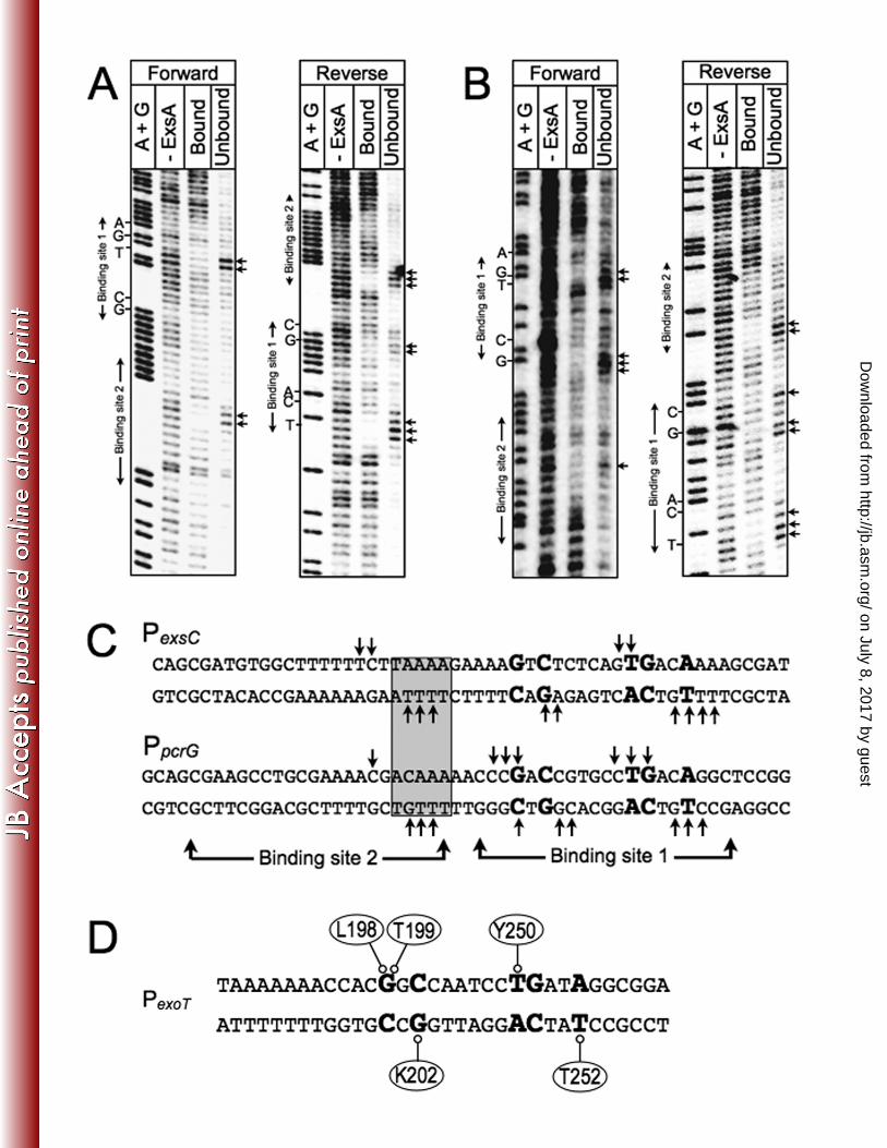
To address this question we used the missing nucleoside footprinting approach (17). 515
For this technique nucleosides are randomly excised from end-labeled promoter probes 516
through brief exposure to hydroxyl radicals. The treated probes are then used in an 517
EMSA reaction, and the ExsA-bound and unbound probes are excised and separated 518
on a sequencing gel to identify nucleosides important for ExsA binding. Comparison of 519
the ExsA bound and unbound samples yielded complimentary data whereby the most 520
prominent bands in the unbound samples were reduced or absent from the ExsA bound 521
samples for both the PexsC and PpcrG promoter probes (Fig. 9). The identity of the bands 522
in the unbound samples indicate that binding of ExsA to site 1 requires the -38 thymine 523
and -39 guanosine on the top strand, and the -34 thymine on the bottom strand. 524
Although the pattern was not as clear with regards to the GnC sequence, the data for 525
the PpcrG promoter suggests that ExsA binding requires the guanosines at positions -47 526
and -45 on the top and bottom strands, respectively. This interpretation is consistent 527
with the molecular modeling data shown in Fig. S3, which predicts that residues L198 528
and T199 in RH1 interact with the -47 guanosine on one strand, and that K202 interacts 529
with the -45 cytosine on the opposite strand. Despite repeated attempts to footprint the 530
PexoT and PexsD promoter probes using the missing nucleoside approach, our efforts 531
were unsuccessful and we suspect this reflects a sensitivity issue related to the low 532
yield of shift product 2. Previous EMSA experiments using PexsC and PpcrG promoter 533
probes found that the yield of shift product 2 (i.e., ExsA bound to both sites 1 and 2) is 534
much higher than when using the PexoT and PexsD promoter probes (6). 535
536
DISCUSSION 537
on July 8, 2017 by guesthttp://jb.asm
.org/D
ownloaded from

King et al. ExsA-DNA Contacts
24
In the present study we tested the hypothesis that the RH1 and RH2 recognition 538
helices of a single ExsA monomer interact with the GnC and TGnnA sequences in 539
binding site 1. Using Fe-BABE footprinting we found that the ExsA monomers are in the 540
same orientation (i.e., head-to-tail) when bound to sites 1 and 2 on the PexoT, PexsC, 541
PexsD, and PpcrG promoter probes (Fig. 2C). This binding configuration is also seen with 542
other AraC family members that function as class II activators including AraC, MelR, 543
ToxT, and XylS, although variations on this theme can occur at different promoters (11, 544
14, 26, 43). A previous study found that cooperative binding by ExsA and efficient filling 545
of site 2 is dependent upon self-association between the ExsA monomers (8). ExsA 546
self-association occurs through interactions mediated by the amino-terminal domain. 547
The crystal structure of the AraC dimerization and ligand binding domain indicates that 548
dimerization occurs in a head-to-head orientation (38). Assuming that ExsA monomers 549
also associate in a head-to-head orientation, the amino-terminal domain of one ExsA 550
monomer would have to be rotated 180° relative to the carboxy-terminal DNA binding 551
domain to allow for both monomers to bind in a head-to-tail orientation. This is 552
consistent with a previous study suggesting that the amino- and carboxy-terminal 553
domains of ExsA are connected by a flexible linker region located between residues 554
149-157 (8). That study found that amino- and carboxyl-terminal domains could be 555
separated at the predicted linker region and still retain function. Whereas the amino-556
terminal domain mediates ExsA self-association and ExsD binding, the carboxy-terminal 557
domain retains DNA-binding activity and the ability to recruit RNA polymerase to the 558
promoter. This linker region likely confers the flexibility required to dimerize in a head-559
to-head orientation and still bind DNA in a head-to-tail orientation. 560
on July 8, 2017 by guesthttp://jb.asm
.org/D
ownloaded from
King et al. ExsA-DNA Contacts
25
In addition to establishing the orientation of the bound ExsA monomers, the Fe-561
BABE footprinting data also defined the general location of the RH1- and RH2-mediated 562
contacts at binding site 1 (Fig. 2D). Cleavage by the RH1 conjugant was located 1-5 bp 563
upstream of the GnC sequence on the top strand of the promoter probes and 3-7 bp 564
upstream of the GnC sequence on the bottom strands. Although the RH1 cleavage 565
patterns could alternatively be described relative to the adenine-rich region (i.e., 566
immediately downstream), three pieces of data indicate that RH1 contacts the GnC 567
sequence as opposed to the adenine-rich region. First, the modeled structure of ExsA 568
bound to DNA indicates that the “turn” region is located amino-terminal to RH1 (Fig. 569
S4A). For this reason we expected and observed that the RH1 conjugant cleaves on 570
the upstream (5’) side of the GnC sequence (Fig. 2). Second, the gain-of-function 571
mutants and genetic loss-of-contact data indicate that RH1 makes base-specific 572
contacts with the GnC sequence (Figs. 3-4). Finally, the midpoints of RH1 and RH2 573
cleavage on the top strand of the promoter are separated by ~11 bp at binding site 1. 574
This spacing is typical of promoter elements recognized by the two recognition helices 575
of a single AraC/XylS subunit (11, 29). 576
Cleavage by the RH2 conjugant at binding site 1 coincided with the TG portion of 577
the TGnnA sequence on the top strand of the promoter probes, and at two locations 578
positioned 2-4 bp upstream and downstream of the TGnnA sequence on the bottom 579
strand (Fig. 2C). The two areas of cleavage on the bottom strand are separated by 580
almost a full helical turn of the DNA or ~34 angstroms. This finding might indicate that 581
the residue conjugated with Fe-BABE (S246) is positioned equidistant from both areas 582
of cleavage since the outer limit that the Fe-BABE generated hydroxyl radicals can 583
on July 8, 2017 by guesthttp://jb.asm
.org/D
ownloaded from
King et al. ExsA-DNA Contacts
26
diffuse before being neutralized by water is ~17 angstroms (12, 30). Another possible 584
explanation for the bipartite cleavage pattern is that the previously observed ExsA-585
dependent bending of the promoters (8) brings both regions close enough to permit 586
cleavage by the RH2 conjugant. 587
The cleavage pattern generated by the RH2 conjugant indicates that RH2 does 588
not closely interact with binding site 2 at the PexsD and PexoT promoters. The MarA and 589
Rob crystal structures bound to target DNA demonstrate two distinct binding modes, 590
which could explain the lack of interactions detected for ExsA at binding site 2 (24, 31). 591
Whereas both recognition helices of MarA are fully inserted into adjacent major grooves 592
on the DNA, only recognition helix 1 of Rob engages the major groove of the binding 593
site while recognition helix 2 lies on the surface of the DNA through contacts with the 594
phosphodiester backbone. Such a binding arrangement might position the RH2-595
conjugant far enough away from the DNA backbone to eliminate observable cleavage. 596
Chemical footprinting and mutational data show that a dimer of AraC uses both modes 597
to bind the araI site (5, 18, 29). Whereas one molecule of the AraC dimer binds the 598
upstream araI1 sites like MarA, the other molecule binds the downstream araI2 site like 599
Rob. In an analogous scenario, our data indicate that ExsA binds to site 1 in all of the 600
promoters like MarA with both helices fully inserted into the DNA. Binding to site 2 at 601
the PexsD and PexoT promoters, however, may more closely resemble the binding 602
properties of Rob. 603
Having determined that ExsA interacts at or near the GnC and TGnnA 604
sequences, we utilized gain-of-function screens and loss-of-contact analyses to identify 605
amino acid residues in RH1 and RH2 involved in making base-specific contact with 606
on July 8, 2017 by guesthttp://jb.asm
.org/D
ownloaded from

King et al. ExsA-DNA Contacts
27
DNA. Combined data from those studies make for a compelling case that residues 607
L198, T199, and K202 in RH1 are the primary, and possibly only, residues involved in 608
base-specific recognition of the GnC sequence (Figs. 3 and 5). The finding for T199A is 609
in agreement with previous studies of AraC, MelR, RhaS, XylS, Rob, and MarA, each 610
reporting that the residue equivalent to T199 participates in base-specific interactions 611
with target DNA (Fig. 1A). Similarly, the equivalent of residue K202 in Rob and MarA 612
also interacts with target DNA. Based on our findings we asked whether the MarA-DNA 613
complex structure could be used to more precisely model the interaction of ExsA with 614
the PexoT promoter at binding site 1. MarA was chosen because binding of an ExsA 615
monomer to binding site 1 of the PexoT promoter bends the DNA to a similar angle as 616
seen in the MarA-DNA complex structure (8, 31). Assuming that the -47 guanine is the 617
nucleotide closest to residue T199, we indeed found that L198 is also positioned 618
favorably to interact with the -47 guanine and that K202 is oriented towards the -45 619
base pair with the side chain pointing to the opposing strand (Fig. S3A). These finding 620
are consistent with the missing nucleoside footprinting data at the PpcrG promoter (Fig. 621
9). 622
In comparison to RH1, interpreting the gain-of-function and loss-of-contact data 623
for RH2 was not as straightforward. The only gain-of-function mutant identified in RH2 624
was T252S, specific for the -34 adenine of the TGnnA sequence. The T252A mutant, 625
however, was poorly expressed and failed to demonstrate a base-specific interaction in 626
the loss-of-contact analyses. Although there is no evidence that the corresponding 627
residue participates in base-specific interactions for other AraC/XylS family members, 628
the equivalent of ExsA residue 253 in XylS and MarA is important for DNA recognition 629
on July 8, 2017 by guesthttp://jb.asm
.org/D
ownloaded from
King et al. ExsA-DNA Contacts
28
(Fig. 1A) (11, 31). An alternative explanation for the T252S gain-of-function phenotype 630
is that T252 normally functions to stabilize a neighboring residue involved in base-631
specific interactions. Under this scenario, however, a loss of contact phenotype would 632
still be expected through secondary effects on the neighboring residue. Nevertheless, a 633
role for protein stability is indicated through the observation that the conservative T252S 634
substitution had no effect on protein stability whereas the T252A substitution resulted in 635
a significant decrease in steady state expression levels. Based upon our combined 636
data we conclude that T252 is involved in a base-specific contact with the -34 adenine 637
but cannot distinguish whether the effect of the T252A substitution is direct or indirect. 638
Attempts to isolate gain-of-function mutants specific for the thymine or guanine of 639
the conserved TGnnA sequence were unsuccessful. One possible reason is that the 640
specific promoter substitutions used in the screens (PexoT(T-38A)-lacZ and PexoT(G-37C)-lacZ) 641
were not exhaustive and may not have been permissive for isolating gain-of-function 642
mutants. Another important point to consider is that ExsA-dependent activation of the 643
PexoT-lacZ reporter is dependent upon occupation of both binding sites 1 and 2 (6). For 644
this reason we expected gain-of-function mutants to be difficult to isolate since the 645
mutant proteins would have to bind both the native sequence at binding site 2 and the 646
mutant sequence at site 1. In fact, when compared to activation of the wt PexoT-lacZ 647
reporter by wt ExsA, neither the T199S, K202R, nor T252S mutants actually 648
demonstrated increased activation of their cognate reporters when compared to the wt 649
PexoT-lacZ reporter (Fig. 3B-D). The simplest interpretation of this finding is that the gain-650
of-function for each mutant protein at binding site 1 is offset by reduced function at site 651
2 and, when combined, results in a net reduction in overall function when compared to 652
on July 8, 2017 by guesthttp://jb.asm
.org/D
ownloaded from
King et al. ExsA-DNA Contacts
29
wt ExsA. An equally plausible explanation, however, is that reduced activation by the 653
T199S, K202R, and/or T252S mutants results solely from suboptimal interactions at 654
binding site 2. 655
The Y250A and R257A mutants each demonstrated a significant defect in DNA 656
binding activity and a potential interaction with the TGnnA sequence in the loss-of-657
contact experiments (Fig. 7-8). The Y250 residue is predicted to interact with position 658
-38 (adenine) in the PexoT promoter. Involvement of Y250 is consistent with previous 659
studies demonstrating that the equivalent of residue Y250 in MelR, XylS, and MarA also 660
participates in base-specific recognition of target DNA (Fig. 1A). Missing nucleoside 661
footprinting data indicated that Y250 interacts with the coding strand (Fig. 9), a finding 662
that is supported by with the modeling data presented in Fig. S3B which also indicates 663
that the Y250 side chain is orientated towards the -38 adenine. The loss-of-contact 664
data for R257 were suggestive of an interaction with the -37 position (Fig. 7). Assuming 665
that Y250 and T252 interact with the -39 and -34 positions, however, it is difficult to 666
explain how residue R257, located at the far end of the recognition helix (Fig. 1A), could 667
interact with the -37 position. The modeling data also indicate that R257 is located too 668
far away from the -37 position to participate in a direct interaction (data not shown). The 669
model does suggest that R257 could interact with residue Q253, which is a prime 670
position to bind to the -37 site. The Q253A mutant, however, had only a modest defect 671
in activation of the wt PexoT-lacZ and PexsC-lacZ reporters. We conclude that the MarA-DNA 672
complex is only a fair model of the interactions between RH2 and the TGnnA sequence. 673
Ultimately, structural studies will be required to fully understand the nature of the 674
contacts occurring at the TGnnA sequence. 675
on July 8, 2017 by guesthttp://jb.asm
.org/D
ownloaded from
King et al. ExsA-DNA Contacts
30
676
ACKNOWLEDGEMENTS 677
This study was supported by the National Institutes of Health (RO1- AI055042 to TLY). 678
The authors would like to thank Gary Gussin, Mark Urbanowski, and David Weiss for 679
critical insight and suggestions. 680
681
REFERENCES 682
1. Apodaca, G., M. Bomsel, R. Lindstedt, J. Engel, D. Frank, K. E. Mostov, and J. 683
Wiener-Kronish. 1995. Characterization of Pseudomonas aeruginosa-induced MDCK 684
cell injury: glycosylation-defective host cells are resistant to bacterial killing. Infect 685
Immun 63:1541-1551. 686
2. Barbieri, J. T., and J. Sun. 2004. Pseudomonas aeruginosa ExoS and ExoT. Rev 687
Physiol Biochem Pharmacol 152:79-92. 688
3. Becher, A., and H. P. Schweizer. 2000. Integration-proficient Pseudomonas aeruginosa 689
vectors for isolation of single-copy chromosomal lacZ and lux gene fusions. 690
BioTechniques 29:948-950, 952. 691
4. Bhende, P. M., and S. M. Egan. 1999. Amino acid-DNA contacts by RhaS: an AraC 692
family transcription activator. J Bacteriol 181:5185-5192. 693
5. Brunelle, A., and R. Schleif. 1989. Determining residue-base interactions between AraC 694
protein and araI DNA. Journal of molecular biology 209:607-622. 695
6. Brutinel, E. D., C. A. Vakulskas, K. M. Brady, and T. L. Yahr. 2008. 696
Characterization of ExsA and of ExsA-dependent promoters required for expression of 697
the Pseudomonas aeruginosa type III secretion system. Mol Microbiol 68:657-671. 698
on July 8, 2017 by guesthttp://jb.asm
.org/D
ownloaded from

King et al. ExsA-DNA Contacts
31
7. Brutinel, E. D., C. A. Vakulskas, and T. L. Yahr. 2010. ExsD inhibits expression of 699
the Pseudomonas aeruginosa type III secretion system by disrupting ExsA self-700
association and DNA binding activity. J Bacteriol 192:1479-1486. 701
8. Brutinel, E. D., C. A. Vakulskas, and T. L. Yahr. 2009. Functional domains of ExsA, 702
the transcriptional activator of the Pseudomonas aeruginosa type III secretion system. J 703
Bacteriol 191:3811-3821. 704
9. Dasgupta, N., G. L. Lykken, M. C. Wolfgang, and T. L. Yahr. 2004. A novel anti-705
anti-activator mechanism regulates expression of the Pseudomonas aeruginosa type III 706
secretion system. Mol Microbiol 53:297-308. 707
10. Diaz, M. R., J. M. King, and T. L. Yahr. 2011. Intrinsic and Extrinsic Regulation of 708
Type III Secretion Gene Expression in Pseudomonas Aeruginosa. Front Microbiol 2:89. 709
11. Dominguez-Cuevas, P., P. Marin, S. Marques, and J. L. Ramos. 2008. XylS-Pm 710
promoter interactions through two helix-turn-helix motifs: identifying XylS residues 711
important for DNA binding and activation. Journal of molecular biology 375:59-69. 712
12. Ebright, Y. W., Y. Chen, P. S. Pendergrast, and R. H. Ebright. 1992. Incorporation of 713
an EDTA-metal complex at a rationally selected site within a protein: application to 714
EDTA-iron DNA affinity cleaving with catabolite gene activator protein (CAP) and Cro. 715
Biochemistry 31:10664-10670. 716
13. Egan, S. M. 2002. Growing repertoire of AraC/XylS activators. J Bacteriol 184:5529-717
5532. 718
14. Grainger, D. C., T. A. Belyaeva, D. J. Lee, E. I. Hyde, and S. J. Busby. 2003. Binding 719
of the Escherichia coli MelR protein to the melAB promoter: orientation of MelR 720
subunits and investigation of MelR-DNA contacts. Mol Microbiol 48:335-348. 721
on July 8, 2017 by guesthttp://jb.asm
.org/D
ownloaded from

King et al. ExsA-DNA Contacts
32
15. Grier, M. C., L. K. Garrity-Ryan, V. J. Bartlett, K. A. Klausner, P. J. Donovan, C. 722
Dudley, M. N. Alekshun, S. K. Tanaka, M. P. Draper, S. B. Levy, and O. K. Kim. 723
2010. N-Hydroxybenzimidazole inhibitors of ExsA MAR transcription factor in 724
Pseudomonas aeruginosa: In vitro anti-virulence activity and metabolic stability. 725
Bioorganic & medicinal chemistry letters 20:3380-3383. 726
16. Hauser, A. R. 2009. The type III secretion system of Pseudomonas aeruginosa: infection 727
by injection. Nat Rev Microbiol 7:654-665. 728
17. Hayes, J. J., and T. D. Tullius. 1989. The missing nucleoside experiment: a new 729
technique to study recognition of DNA by protein. Biochemistry 28:9521-9527. 730
18. Hendrickson, W., and R. Schleif. 1985. A dimer of AraC protein contacts three adjacent 731
major groove regions of the araI DNA site. Proc Natl Acad Sci U S A 82:3129-3133. 732
19. Hoang, T. T., A. J. Kutchma, A. Becher, and H. P. Schweizer. 2000. Integration-733
proficient plasmids for Pseudomonas aeruginosa: site-specific integration and use for 734
engineering of reporter and expression strains. Plasmid 43:59-72. 735
20. Holder, I. A., A. N. Neely, and D. W. Frank. 2001. PcrV immunization enhances 736
survival of burned Pseudomonas aeruginosa-infected mice. Infect Immun 69:5908-5910. 737
21. Holder, I. A., A. N. Neely, and D. W. Frank. 2001. Type III secretion/intoxication 738
system important in virulence of Pseudomonas aeruginosa infections in burns. Burns 739
27:129-130. 740
22. Hovey, A. K., and D. W. Frank. 1995. Analyses of the DNA-binding and transcriptional 741
activation properties of ExsA, the transcriptional activator of the Pseudomonas 742
aeruginosa exoenzyme S regulon. J Bacteriol 177:4427-4436. 743
on July 8, 2017 by guesthttp://jb.asm
.org/D
ownloaded from
King et al. ExsA-DNA Contacts
33
23. Kurahashi, K., O. Kajikawa, T. Sawa, M. Ohara, M. A. Gropper, D. W. Frank, T. 744
R. Martin, and J. P. Wiener-Kronish. 1999. Pathogenesis of septic shock in 745
Pseudomonas aeruginosa pneumonia. J Clin Invest 104:743-750. 746
24. Kwon, H. J., M. H. Bennik, B. Demple, and T. Ellenberger. 2000. Crystal structure of 747
the Escherichia coli Rob transcription factor in complex with DNA. Nat Struct Biol 748
7:424-430. 749
25. Lee, V. T., S. Pukatzki, H. Sato, E. Kikawada, A. A. Kazimirova, J. Huang, X. Li, J. 750
P. Arm, D. W. Frank, and S. Lory. 2007. Pseudolipasin A is a specific inhibitor for 751
phospholipase A2 activity of Pseudomonas aeruginosa cytotoxin ExoU. Infect Immun 752
75:1089-1098. 753
26. Martin, R. G., and J. L. Rosner. 2001. The AraC transcriptional activators. Curr Opin 754
Microbiol 4:132-137. 755
27. McCaw, M. L., G. L. Lykken, P. K. Singh, and T. L. Yahr. 2002. ExsD is a negative 756
regulator of the Pseudomonas aeruginosa type III secretion regulon. Mol Microbiol 757
46:1123-1133. 758
28. Newman, J. R., and C. Fuqua. 1999. Broad-host-range expression vectors that carry the 759
L-arabinose-inducible Escherichia coli araBAD promoter and the araC regulator. Gene 760
227:197-203. 761
29. Niland, P., R. Huhne, and B. Muller-Hill. 1996. How AraC interacts specifically with 762
its target DNAs. Journal of molecular biology 264:667-674. 763
30. Rana, T. M., and C. F. Meares. 1991. Transfer of oxygen from an artificial protease to 764
peptide carbon during proteolysis. Proc Natl Acad Sci U S A 88:10578-10582. 765
on July 8, 2017 by guesthttp://jb.asm
.org/D
ownloaded from

King et al. ExsA-DNA Contacts
34
31. Rhee, S., R. G. Martin, J. L. Rosner, and D. R. Davies. 1998. A novel DNA-binding 766
motif in MarA: the first structure for an AraC family transcriptional activator. Proc Natl 767
Acad Sci U S A 95:10413-10418. 768
32. Richards, M. J., J. R. Edwards, D. H. Culver, and R. P. Gaynes. 2000. Nosocomial 769
infections in combined medical-surgical intensive care units in the United States. Infect 770
Control Hosp Epidemiol 21:510-515. 771
33. Richards, M. J., J. R. Edwards, D. H. Culver, and R. P. Gaynes. 1999. Nosocomial 772
infections in medical intensive care units in the United States. National Nosocomial 773
Infections Surveillance System. Crit Care Med 27:887-892. 774
34. Rietsch, A., I. Vallet-Gely, S. L. Dove, and J. J. Mekalanos. 2005. ExsE, a secreted 775
regulator of type III secretion genes in Pseudomonas aeruginosa. Proc Natl Acad Sci U S 776
A 102:8006-8011. 777
35. Roy-Burman, A., R. H. Savel, S. Racine, B. L. Swanson, N. S. Revadigar, J. 778
Fujimoto, T. Sawa, D. W. Frank, and J. P. Wiener-Kronish. 2001. Type III protein 779
secretion is associated with death in lower respiratory and systemic Pseudomonas 780
aeruginosa infections. The Journal of infectious diseases 183:1767-1774. 781
36. Sato, H., and D. W. Frank. 2004. ExoU is a potent intracellular phospholipase. Mol 782
Microbiol 53:1279-1290. 783
37. Sawa, T., T. L. Yahr, M. Ohara, K. Kurahashi, M. A. Gropper, J. P. Wiener-784
Kronish, and D. W. Frank. 1999. Active and passive immunization with the 785
Pseudomonas V antigen protects against type III intoxication and lung injury. Nat Med 786
5:392-398. 787
on July 8, 2017 by guesthttp://jb.asm
.org/D
ownloaded from
King et al. ExsA-DNA Contacts
35
38. Soisson, S. M., B. MacDougall-Shackleton, R. Schleif, and C. Wolberger. 1997. 788
Structural basis for ligand-regulated oligomerization of AraC. Science 276:421-425. 789
39. Thibault, J., E. Faudry, C. Ebel, I. Attree, and S. Elsen. 2009. Anti-activator ExsD 790
forms a 1:1 complex with ExsA to inhibit transcription of type III secretion operons. J 791
Biol Chem 284:15762-15770. 792
40. Urbanowski, M. L., E. D. Brutinel, and T. L. Yahr. 2007. Translocation of ExsE into 793
Chinese hamster ovary cells is required for transcriptional induction of the Pseudomonas 794
aeruginosa type III secretion system. Infect Immun 75:4432-4439. 795
41. Urbanowski, M. L., G. L. Lykken, and T. L. Yahr. 2005. A secreted regulatory protein 796
couples transcription to the secretory activity of the Pseudomonas aeruginosa type III 797
secretion system. Proc Natl Acad Sci U S A 102:9930-9935. 798
42. Vogel, H. J., and D. M. Bonner. 1956. Acetylornithinase of Escherichia coli: partial 799
purification and some properties. J Biol Chem 218:97-106. 800
43. Withey, J. H., and V. J. DiRita. 2006. The toxbox: specific DNA sequence 801
requirements for activation of Vibrio cholerae virulence genes by ToxT. Mol Microbiol 802
59:1779-1789. 803
44. Yahr, T. L., and M. C. Wolfgang. 2006. Transcriptional regulation of the Pseudomonas 804
aeruginosa type III secretion system. Mol Microbiol 62:631-640. 805
45. Zheng, Z., G. Chen, S. Joshi, E. D. Brutinel, T. L. Yahr, and L. Chen. 2007. 806
Biochemical characterization of a regulatory cascade controlling transcription of the 807
Pseudomonas aeruginosa type III secretion system. J Biol Chem 282:6136-6142. 808
809
810
on July 8, 2017 by guesthttp://jb.asm
.org/D
ownloaded from
King et al. ExsA-DNA Contacts
36
811 FIGURE LEGENDS 812
Figure 1. (A) ClustalW alignment of the DNA-binding domains from selected AraC-813
family members (AraC, MelR, RhaS, XylS, Rob, MarA, and ExsA). Residues that form 814
the hydrophobic core of the DNA-binding domains are boxed in grey and residues 815
experimentally shown to make base specific contacts with the promoter DNA are boxed 816
in black. The predicted α-helices are indicated by rounded rectangles and based upon 817
the MarA crystal structure (31). The predicted recognition helices for the two helix-turn-818
helix motifs are labeled RH1 and RH2, respectively. RH1 and RH2 in ExsA consist of 819
residues L198-Y209 and Q248-F259, respectively. (B and C) Tertiary structure 820
prediction of the ExsA DNA-binding domain bound to target DNA based upon the crystal 821
structures of MarA (B) and Rob (C). The DNA binding domain of ExsA (residues 159-822
278) was threaded onto the crystal structure of MarA (residues 1-127) or Rob (residues 823
1-121) using the PHYRE 0.2 server (www.sbg.bio.ic.ac.uk/~phyre/), and the predicted 824
structures were visualized using the PyMOL Molecular Graphics System 825
(www.pymol.org). Residues located within the “turn” regions of RH1 and RH2 (M196 826
and S246) that were changed to cysteine for the FeBABE footprinting experiments in 827
Fig. 2 are indicated. 828
829
Figure 2. Fe-BABE footprints of the ExsA RH1- and RH2-FeBABE conjugants bound to 830
the PexsC, PpcrG, PexsD, and PexoT promoter probes. (A) PA1O3 exsA::Ω carrying a PexsD-831
lacZ reporter fusion was transformed with either a vector control or plasmids expressing 832
wt ExsA, ExsA lacking all seven of the native cysteine residues (∆cys), or ExsA ∆cys 833
with unique cysteines introduced at positions E193, M196, G244, or S246. 834
on July 8, 2017 by guesthttp://jb.asm
.org/D
ownloaded from
King et al. ExsA-DNA Contacts
37
Transformants were grown under inducing conditions for T3SS gene expression and 835
assayed for β-galactosidase activity. Whole cell lyates (normalized by cell number) 836
were immunoblotted with ExsA antiserum to examine steady state expression levels. 837
(B) Silver-stained SDS-polyacrylamide gel of wt ExsAHis, ExsAHis M196C, and ExsAHis 838
S246C mutants following purification by Ni2+-affinity chromatography (1 µg each). Note 839
that wt ExsA was further purified using a heparin column. The migration points of 840
molecular weight standards are indicated on the left side of the gel. (C) The indicated 841
promoter probes labeled with 32P on the forward strand were incubated in the absence 842
(-) or presence of the RH1- or -RH2-FeBABE conjugants for 15 min at 25˚C as 843
indicated. Binding reactions were subjected to Fe-BABE cleavage, separated by 844
electrophoresis, and visualized by phosphorimaging. Maxam-Gilbert sequencing 845
ladders (A+G) were included for orientation and the positions of the GnC and TGnnA 846
sequences in ExsA binding site 1 are indicated. (D) Summary of the FeBABE 847
footprinting data for the forward (Fig. 2C) and reverse strands (data not shown). The -848
10 hexamers (boxed), conserved GnC and TGnnA sequences (bold with larger 849
typeface), adenine rich region (grey box), and position of ExsA binding sites 1 and 2 are 850
indicated. The regions of PexsC, PexsD, and PexoT previously shown to be protected from 851
DNase I cleavage by ExsA are underlined (6). The protected region of PpcrG is based 852
upon the DNase I footprint presented in Fig. S1. The cleavage events generated by the 853
RH1- and RH2-FeBABE conjugants are indicated with filled and open circles, 854
respectively. The open and filled circles located above and below the nucleotide 855
sequence represent the Fe-BABE footprints for the forward and reverse strands, 856
respectively. 857
on July 8, 2017 by guesthttp://jb.asm
.org/D
ownloaded from
King et al. ExsA-DNA Contacts
38
858
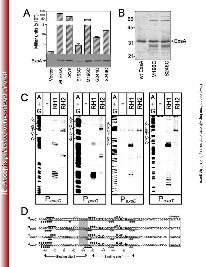
Figure 3. ExsA gain-of-function mutants with altered promoter specificity. (A) Diagram 859
of the nucleotide substitutions in the PexoT(C-45A)-lacZ, PexoT(G-47A)-lacZ, and PexoT(A-34C)-lacZ 860
mutant reporters that were used for the gain-of-function screen. (B-D) PA1O3 exsA::Ω 861
carrying either the wild type PexoT-lacZ reporter or a mutant reporter [(B) PexoT(C-45A)-lacZ, 862
(C) PexoT(G-47A)-lacZ, or (D) PexoT(A-34C)-lacZ ] was transformed with either a vector control (-), 863
an ExsA expression plasmid (wt), or a plasmid expressing ExsA with the indicated 864
amino acid substitutions (K202R, T199S, or T252S). Transformants were cultured 865
under inducing conditions for T3SS gene expression (in panel D the strains were 866
cultured in the presence of 0.05% arabinose to enhance detection of T252S-dependent 867
activity) and assayed for β-galactosidase activity. The reported values for the β-868
galactosidase assays (Miller units) throughout this study represent the average of at 869
least three independent experiments and error bars represent the standard error of the 870
mean (SEM); ** p < 0.01. Anti-ExsA immunoblots demonstrating the steady state 871
expression levels of ExsA and the K202R, T199S, or T252S mutants are inset into each 872
panel. 873
874
Figure 4. (A) Map of the PexoT promoter showing the ExsA-consensus binding 875
sequence, and the conserved GnC and TGnnA sequences (bold with larger typeface) 876
located within binding site 1. The nucleotide substitutions for each of the mutant PexoT 877
reporters used in the genetic loss-of-contact experiments are indicated with arrows. (B) 878
PA1O3 exsA::Ω carrying the mutant PexoT-lacZ reporters and expressing wt ExsA from an 879
arabinose-inducible expression vector was grown in the presence of EGTA and 0.1% 880
on July 8, 2017 by guesthttp://jb.asm
.org/D
ownloaded from
King et al. ExsA-DNA Contacts
39
arabinose, and assayed for β-galactosidase activity. (C) PA1O3 exsA::Ω carrying either 881
a PexsC-lacZ (open bars) or a PexoT-lacZ (hatched bars) transcriptional reporter was 882
transformed with vectors expressing wt ExsA or the indicated ExsA alanine substitution 883
mutants. The resulting strains were cultured under inducing (+EGTA) conditions for 884
T3SS gene expression with 0.1% arabinose and assayed for β-galactosidase activity. 885
The reported values are the percent activity for each alanine substitution mutant relative 886
to wt ExsA; * p < 0.05. Immunoblots demonstrating the steady state expression levels 887
of ExsA and the alanine scanning mutants are inset in each panel. The asterisk 888
indicates a cross-reactive band that served as a loading control. 889
890
Figure 5. Genetic loss-of-contact analyses of the T199A, K202A, and T252A mutants. 891
The T199A, K202A, and T252A expression plasmid were introduced into PA1O3 892
exsA::Ω carrying the indicated mutant PexoT-lacZ reporters. Transformants were cultured 893
in the presence of EGTA and 0.1% arabinose, and assayed for β-galactosidase activity. 894
The reported values are the percent activity at the mutant promoter normalized to the 895
activity of each alanine substitution mutant at the wt PexoT-lacZ reporter; ***, p < 0.001. 896
897
Figure 6. Alanine scanning mutagenesis of RH1 and RH2. PA1O3 exsA::Ω carrying 898
either a PexsC-lacZ (open bars) or PexoT-lacZ (hatched bars) transcriptional reporter was 899
transformed with a vector expressing wt ExsA or the indicated alanine substitution 900
mutants in RH1 (A) and RH2 (B). The resulting strains were grown in the presence of 901
EGTA and 0.1% arabinose, and assayed for β-galactosidase activity. The reported 902
values are the percent activity for each alanine substitution mutant relative to wt ExsA; 903
on July 8, 2017 by guesthttp://jb.asm
.org/D
ownloaded from
King et al. ExsA-DNA Contacts
40
***, p < 0.001 and **, p < 0.01. Anti-ExsA immunoblots demonstrating the steady state 904
expression levels of ExsA and the alanine-substituted mutants are shown below each 905
panel. The asterisk indicates a cross-reactive band that served as a loading control. 906
907
Figure 7. Additional base-specific contacts mediated by residues in RH1 and RH2. 908
PA1O3 exsA::Ω expressing either L198A, Y250A, or R257A were assayed for reporter 909
activity using the panel of mutant PexoT-lacZ reporter described in Fig. 4A. The strains 910
were cultured in the presence of EGTA and 0.1% arabinose, and assayed for β-911
galactosidase activity. The reported values are the percent activity at the mutant 912
promoter compared to the activity at the wt PexoT-lacZ reporter; **, p < 0.01 and *, p < 913
0.05. 914
915
Figure 8. DNA binding activity of selected alanine substitution mutants. For the EMSA 916
experiment (upper panel), an equimolar mixture of non-specific (Non-SP, derived from 917
the coding region of pscF) and specific (SP, derived from the wt PexsC reporter) 918
radiolabeled probes (0.05 nM each) was incubated for 15 min at 25˚C in the absence (-) 919
or presence of 40 nM purified ExsAHis, or 2 µl of whole cell lysate prepared from E. coli 920
expressing wt ExsA or the indicated alanine substitution mutants. Samples were 921
analyzed by native polyacrylamide gel electrophoresis and phosphorimaging. Anti-922
ExsA immunoblots (lower panel) indicate that the E. coli lysates used in the EMSA 923
experiment contain comparable levels of wt ExsA and the indicated alanine substitution 924
mutants. 925
926
on July 8, 2017 by guesthttp://jb.asm
.org/D
ownloaded from
King et al. ExsA-DNA Contacts
41
Figure 9. Missing nucleoside footprints of the PexsC and PpcrG promoter regions. PexsC 927
(A) or PpcrG (B) promoter probes labeled with 32P on the forward or reverse strands were 928
treated with hydroxyl radicals, incubated with ExsAHis, and subjected to non-denaturing 929
electrophoresis in a typical EMSA reaction. The unbound band and the ExsA-bound 930
band corresponding to shift product 2 (representing ExsA bound to both sites 1 and 2) 931
were eluted from the gel, separated by denaturing electrophoresis, and visualized by 932
phosphorimaging. Maxam-Gilbert sequencing ladders (A+G) were included for 933
orientation. The locations of the GnC and TGnnA sequences in binding site 1, and 934
binding site 2, are indicated. (C) Summary of the footprinting data for the forward and 935
reverse strands. The conserved GnC and TGnnA sequences (bold with larger 936
typeface), adenine rich region (grey box), and position of ExsA binding sites 1 and 2 are 937
indicated. Nucleotides indicated with arrows are those present in the unbound samples 938
and required for maximal ExsA binding. (D) Proposed model for the binding of ExsA to 939
site 1 in the PexoT promoter region. The amino acid residues that are proposed to 940
contribute to base-specific recognition of the coding or noncoding strand of the PexoT 941
promoter region are indicated. 942
943
on July 8, 2017 by guesthttp://jb.asm
.org/D
ownloaded from