Embed Size (px)
Citation preview

J Am Acad Audiol 19:257–266 (2008)
*Mayo Clinic Florida
David A. Zapala, Ph.D., Audiology Section, Mayo Clinic Florida, 4500 San Pablo Rd., Jacksonville, FL 32224; Phone: (904) 953-0468; Fax: (904) 953-2489; E-mail: [email protected]
Abstract
Down-beating positional nystagmus is typically associated with central ner-vous system disease. Anterior canal benign paroxysmal positional vertigo (AC-BPPV) can mimic down-beating positional nystagmus of central origin, particularly when it is bilateral. Factors that increase the probability of bi-lateral AC-BPPV include a history of bilateral multicanal BPPV, transient down-beating and torsional nystagmus that follows the plane of the pro-voked canal, and the absence of co-occurring neurologic signs and symp-toms of central nervous system dysfunction. With neurologic clearance for canalith repositioning, exploration for AC-BPPV and canalith repositioning trials may alleviate symptoms even when the nystagmus does not appear to fatigue. In the case presented, the use of a side-lying maneuver with the nose down to provoke AC-BPPV symptoms and the use of a reversed Epley to clear AC-BPPV symptoms are highlighted. This approach is helpful when the diagnosis is unclear and neck hyperextension is to be avoided.
Key Words: Anterior semicircular canal, benign paroxysmal positional ver-tigo, canalithiasis, down-beating nystagmus, horizontal semicircular canal, posterior semicircular canal
Abbreviations: AC-BPPV = anterior canal benign paroxysmal positional vertigo; BPPV = benign paroxysmal positional vertigo; CRP = canalith re-positioning procedure; HC-BPPV = horizontal canal benign paroxysmal po-sitional vertigo; PC-BPPV = posterior canal benign paroxysmal positional vertigo; RALP = right anterior/left posterior
Sumario
El nistagmo posicional de batida hacia abajo está típicamente asociado con enfermedades del sistema nervioso central. El vértigo posicional pa-roxístico benigno del canal anterior (AC-BPPV) puede simular un nistag-mo posicional de batida hacia abajo de origen central, particularmente
David A. Zapala*
Down-Beating Nystagmus in Anterior Canal Benign Paroxysmal Positional VertigoDOI: 10.3766/jaaa.19.3.10
Journal of the American Academy of Audiology / Volume 19, Number 3, 2008
258
cuando es bilateral. Los factores que influyen sobre la probabilidad de un AC-BPPV bilateral incluyen una historia de BPPV multicanal bilateral, de nistagmo transitorio de batida hacia abajo o de nistagmo de torsión, que sigue el plano del canal provocado, y la ausencia de signos y síntomas neurológicos concurrentes por una disfunción del sistema nervioso central. Con una depuración neurológica de reposición canalicular, la exploración buscando AC-BPPV y los intentos de reposicionamiento canalicular pue-den aliviar los síntomas aún cuando el nistagmo no parezca fatigarse. En el caso presentado, se destaca el uso de la maniobra de decúbito lateral con descenso de la nariz para provocar los síntomas del AC-BPPV. Este enfoque es útil cuando el diagnóstico no es claro y se debe evitar la hiper-extensión del cuello.
Palabras Clave: Canal semicircular anterior, vértigo posicional paroxístico benigno, canalolitiasis, nistagmo con batida hacia abajo, canal semicircu-lar horizontal, canal semicircular posterior
Abreviaturas: AC-BPPV = vértigo posicional paroxístico benigno del canal anterior; BPPV = vértigo posicional paroxístico benigno; CRP = procedi-miento de reposicionamiento canalicular; HC-BPPV = vértigo posicional paroxístico benigno del canal horizontal; PC-BPPV = vértigo posicional paroxístico benigno del canal posterior; RALP = anterior-derecho/posterior izquierdo
Down-beating nystagmus is commonly associated with central nervous sys-tem lesions, typically in the lower
posterior fossa or cervical medullary junction (Bronstein et al, 1987; Alpini et al, 2001; Bertholon et al, 2002a). In most vestibular clinics, central down-beating nystagmus is rarely encountered. However, because it may imply central nervous system disease, it is always an important condition to identify.
Benign paroxysmal positional vertigo (BPPV) is a form of vestibulopathy commonly encountered in adults (Katsarkas, 1978; Ekvall Hansson et al, 2005). Canalithiasis (loose debris in the semicircular canals) most commonly occurs in the posterior semicircular canal. It can be detected using the Dix-Hallpike maneuver. Posterior canal BPPV (PC-BPPV) typically provokes a tor-sional and up-beating nystagmus on the Dix-Hallpike maneuver. PC-BPPV can be treated with well-established reposition-ing procedures (Epley, 1992; Baloh and Baringer, 1998; Furman and Cass, 1999; Herdman et al, 2000).
Canalithiasis may occur in other semicir-cular canals as well. The key to detecting these BPPV variations is the ability to cor-relate the gravity vector associated with the provoking head movement or position
and the plane of the induced eye move-ments with the planes of the semicircular canals. For example, horizontal canal BPPV (HC-BPPV) provokes a horizontal nys-tagmus (Crevits, 2005; Han et al, 2006), typically with head movements that align the horizontal canal with the gravity vec-tor. Anterior canal canalithiasis provokes a torsional and down-beating nystagmus (Brantberg and Bergenius, 2002; Crevits, 2004; Jackson et al, 2007). Anterior canal BPPV (AC-BPPV) may be provoked when the “long arm” of the anterior canal (the por-tion of the canal between the cupula and the common curis) is sufficiently lower than the cupula. Canalithic debris drifts downward, away from the cupula, and toward the pull of gravity in this head orientation. The falling debris provokes a vacuum that distends the cupula, which in turn provokes nystagmus in the plane of the anterior canal. These expectations follow directly from Ewald’s first law and the physics of canalithiasis.
Theoretically, other otogenic forms of posi-tional vertigo can provoke down-beating nystagmus. Examples might include ante-rior canal cupulolathiasis (canalithic debris mechanically attached to one or both cupulae of the anterior canal [House and Honrubia, 2003]); Waldenstrom’s disease and macroglobulinemia
259
Down-Beating Nystagmus in Anterior BPPV / Zapala
(Brandt, 1990); conditions that change the rela-tive density of the cupula and endolymph such as postalcohol or heavy water effects (Brandt, 1990); and possibly ganglion damage under the cristae (Gacek, 1985). These end-organ condi-tions may provoke nystagmus that persists as long as the provocative position is held, making it difficult to distinguish from central position-ing nystagmus.
Multicanal and bilateral BPPV represent special diagnostic challenges. In theory, stimulation of multiple canals simultane-ously can provoke any type of naturally occurring eye movement (other than conver-gence). This is the design of the vestibular ocular reflex. In most cases, bilateral BPPV will involve semicircular canals that are not coplanar, and so different types of nys-tagmus are provoked with different head movements. Similarly, when different canals are simultaneously involved (multicanal BPPV), the orientation of the semicircular canals with different head movements will provoke BPPV-induced eye movements with different magnitudes and different time con-stants. For example, a simultaneous right HC-BPPV and PC-BPPV may show a strong torsional nystagmus in response to a right Dix-Hallpike maneuver, which may then evolve into a less intense geotropic horizon-tal nystagmus. The torsional nystagmus occurs first and is stronger because the Dix-Hallpike maneuver positions the posterior semicircular canal vertically, optimizing the pull of gravity on any debris suspended in the canal. The horizontal semicircular canal is less efficiently positioned along the gravity vector in the Dix-Hallpike maneu-ver. As a result, all other factors being equal, the posterior canal will be stimulated more strongly than the horizontal canal, and debris will reach the low point in the posterior canal sooner than in the horizon-tal canal.
In contrast, when a head thrust maneu-ver to the right is performed on the same patient (simultaneous right HC-BPPV and PC-BPPV) positioned so that the horizontal canals are vertical, the right horizontal canal will fire stronger and sooner than the posterior canal, provoking a brisk geo-tropic horizontal nystagmus. A torsional nystagmus, if present at all, will be barely detectable. This follows because the head thrust movement more efficiently positions the loose debris in the horizontal canal into
the gravity vector. In contrast, the gravity vector pulling debris in the posterior canal is quite small.
Other eye movement patterns may also occur in multicanal BPPV, reflecting the sum of the effects in each of the provoked canals. For example, a transient oblique nystagmus may also be seen in simultane-ous PC-BPPV and HC-BPPV when a head movement causes debris in both canals to move simultaneously. This nystagmus simply reflects the vector sum of the inputs from the provoked semicircular canals.
Perplexing findings can present in cases of bilateral AC-BPPV. In these cases, the torsional component commonly seen in uni-lateral AC-BPPV can be difficult to detect. The result is an eye movement that is difficult to distinguish from a central down-beating positional nystagmus. This article presents a complicated case of bilateral, multicanal BPPV presenting at some points with a down-beating position-dependent nystagmus. Some of the modifications I have developed to distinguish this form of BPPV from central positioning nystagmus are described. Specifically, modifications of positioning maneuvers to provoke AC-BPPV are presented, as well as the use of a “reversed Epley” maneuver to clear anterior canal canalithiasis through repositioning.
CASE REPORT
A 78-year-old independently living woman entertained dinner guests one
evening. Although it was not her usual custom, she drank several glasses of wine during the meal. At the end of the evening, she went to sleep in her normal routine. She awoke at about 1:00 a.m. lying in her bathroom, having cut her lip and bruised her left foot. She did not remember getting out of bed or falling. She returned to her bed, and in the morning, concerned that she could not remember falling, she went to the emergency department at a local hospital. Other than her obvious injuries, she was judged healthy and discharged. Her pri-mary care physician subsequently referred her for neurologic assessment of an episode of amnesia. This was accomplished and was unrevealing. She was followed closely by her primary care physician.
Approximately seven months later she experienced a sudden onset of vertigo while performing floor exercises at a local gym.
Journal of the American Academy of Audiology / Volume 19, Number 3, 2008
260
She apparently rolled over, became very dizzy and nauseous, and vomited once but did not lose consciousness. She was taken to the emergency department, where a cardiac evaluation and computed tomography scan of the head were performed and found to be normal. She was told she had a urinary tract infection and was released.
During a follow-up with her primary care physician, a Dix-Hallpike maneuver was performed, the results of which were equiv-ocal. Although no nystagmus was observed, the patient was dizzy when returning to the sitting position. Since her dizziness had shown some improvement at that point, no referral was made for further assessment.
Six months later, she presented to her family physician with an acute complaint of positional dizziness. The episode began as she was lying in bed and rolled over to turn off her alarm clock. She experienced a very strong but brief episode of vertigo with nausea. After this episode she reported residual unsteadiness, and sudden head movements were provocative. Her internist performed a Dix-Hallpike, which now pro-voked nystagmus with either ear down. She was referred to audiology for assessment of suspected BPPV and canalith repositioning procedures (CRPs) as needed.
The patient was initially diagnosed with right PC-BPPV, which was successfully treated with a CRP (Epley maneuver). However, while right ear symptoms abated, she was seen three more times over the next two weeks for left PC-BPPV. On one occasion, she demonstrated a transient horizontal canal conversion while treating the left posterior canal. This apparently resolved on its own.
At this point, the patient carried the diagnosis of bilateral multicanal BPPV, with the presumed causative factor being the head trauma from the nighttime fall in the bathroom. She improved after the initial series of CRPs but returned three weeks after her last CRP with a recurrence of transient positional vertigo symptoms. These were provoked when bending over and rolling over in bed. There were no new otologic symptoms (i.e., no change in hear-ing, tinnitus, otalgia, or aural pressure or fullness) or neurologic symptoms (spe-cifically, no diplopia, dysarthria, dysphagia, other sensory or motor changes, or reported changes in cognition or communication).
A bedside physical examination was per-formed, following the methods described by Walker and Zee (2000). Briefly, her eye movements were grossly intact, and there was no nystagmus noted in room light or under frenzel lenses. The facial animation and sensation to touch were symmetri-cal. Oral motor movements and neck and shoulder strength were all normal appear-ing. Neck range of motion was adequate for BPPV assessment and CRPs. Neck hyperextension without tilting the head did not provoke dizziness, light-headedness, or other symptoms. Cerebellar tests (heel to shin, finger to nose) were satisfactorily accomplished. Head thrust testing in the horizontal plane was normal. Romberg was modestly unsteady without falls.
The Dix-Hallpike maneuver was posi-tive with the left ear down, provoking a torsional counterclockwise nystagmus (described relative to the patient’s per-spective), with latency, crescendo, and gradual diminishment demonstrated.1 This would be consistent with left PC-BPPV—canalithiasis variety. However, with the right ear down, a transient down-beating nystagmus was noted lasting about 20 sec. The clinical impression at that time was
Left PC-BPPV—symptomatic1.
Transient down-beating nystagmus in 2. the setting of recalcitrant, bilateral, multicanal BPPV, with a suspicion of bilateral AC-BPPV. Central positioning nystagmus also a possibility, possibly medication induced (she had been given an antiemetic medication to manage her nausea).
The possibility of a central positioning nystagmus was brought to the attention of the referring physician and our staff neuro-otologist. After this medical review and clearance and with patient consent, it was decided to proceed with canalith repo-sitioning. An Epley CRP addressing the left posterior canal (and hopefully the right ante-rior canal) was accomplished three times. On the third trial, there were no posterior canal symptoms. However, a very subtle, residual, down-beating nystagmus could be detected on Dix-Hallpike maneuvers in either direc-tion. This nystagmus persisted as long as the head was maintained in the Hallpike position (that is, it did not “fatigue”).
261
Down-Beating Nystagmus in Anterior BPPV / Zapala
As was our standard at the time this patient was seen (prior to 2004), postreposi-tioning instructions were given to the patient, including the recommendation to keep the head upright for the next 24 hours and to sleep with the head elevated that evening. Several studies have suggested that these instructions are not helpful in improving repositioning outcomes for PC-BPPV symp-toms (Massoud and Ireland, 1996; Marciano and Marcelli, 2002; Cohen and Kimball, 2004). We currently use these postreposition-ing instructions only in cases of recalcitrant or recurring BPPV symptoms.
Because of the residual down-beating nystagmus, a follow-up audiology appoint-ment was scheduled. Additionally, because this was not a typical case of BPPV and the residual down-beating nystagmus remained, a neurologic follow-up appointment was scheduled to be sure we were not missing an unrecognized neurologic process.
Two weeks later, the patient returned to audiology for follow-up. She was feeling bet-ter but still experienced transient positional vertigo when getting out of bed. Physical examination was unchanged. On direct test-ing, the left Dix-Hallpike maneuver provoked a burst of counterclockwise torsional nystag-mus (again described relative to the patient’s perspective), consistent with left PC-BPPV. After this nystagmus dissipated, a clockwise and down-beating nystagmus developed. This secondary nystagmus was not as strong but lasted for a longer duration.2 The down-beating component was stronger with the eyes directed to the right, and the torsional component was more in evidence with the eyes directed toward the left. This would be in keeping with nystagmus evoked from a right anterior/left posterior (RALP) canal plane. The down-beating component would be consistent with right anterior canal hyper-activity, as in AC-BPPV. The Dix-Hallpike to the right did not provoke any nystagmus. These results were interpreted as indicat-ing co-occurring left PC-BPPV and right AC-BPPV. Of note, this RALP plane effect was not seen in the earlier visit, raising the possibility that the left anterior canal had cleared following repositioning.
The standard Epley addressing the left posterior canal was accomplished twice because this was the more problematic canal. It was hoped that the Epley maneu-ver would affect the suspected anterior canal
symptoms as well because it involves move-ments in the RALP plane. However, this was not the direct target of the treatment. Interestingly, an up-beating nystagmus was noted in position three (side lying, nose down) on the first trial. On the second cycle, no nystagmus or vertigo was noted. The same post-CRP instructions were given, and a follow-up appointment was made.
Two days later, the patient returned for reassessment of what was considered by now a case of bilateral, recalcitrant, multicanal BPPV. She reported no new symptoms in the intervening interval but did note persistent light-headedness. Her bedside evaluation was unchanged. The right Dix-Hallpike was negative. The left Dix-Hallpike provoked a clockwise (described relative to the patient) torsional and down-beating nystagmus, with latency, crescendo, and gradual diminish-ment evidenced. Note that this nystagmus was in the opposite direction from that expected in left PC-BPPV. Head-roll maneu-vers were negative bilaterally.
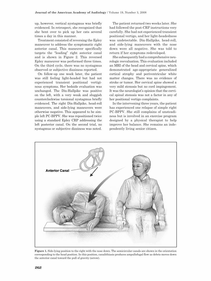
When the patient was placed in the right side-lying position with the nose down (see Figure 1), there was a brisk down-beating nystagmus with a clockwise torsional compo-nent evidenced when the eyes were directed to the left. This nystagmus disappeared over approximately 30 sec. When in the left side-lying position with the nose down, no nystagmus was observed. These results were interpreted as being consistent with right AC-BPPV.
At this point, the continual recurrence of anterior canal symptoms was perplexing. More probing questions were asked about how the patient was moving when she expe-rienced her transient vertiginous symptoms. She was emphatic that she kept her head upright and did not look up or down follow-ing the CRPs. She also related that she slept with her head propped up as recommended. For the most part, she felt better following the CRPs. However, she typically experi-enced a transient positional vertigo within 24 hours of repositioning, first noted when bending over to pick up one of her cats. She was asked to demonstrate how she would pick up the cat. She proceeded to stand erect with her knees essentially locked; she flexed sharply at her hips and touched the floor. In doing this, she immediately experienced transient positional vertigo, but no nystag-mus was observed. When she straightened
Journal of the American Academy of Audiology / Volume 19, Number 3, 2008
262
up, however, vertical nystagmus was briefly evidenced. In retrospect, she recognized that she bent over to pick up her cats several times a day in this manner.
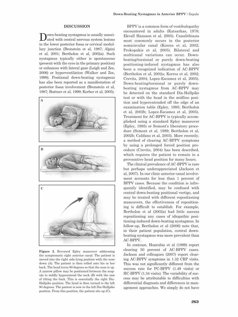
Treatment consisted of reversing the Epley maneuver to address the symptomatic right anterior canal. This maneuver specifically targets the “leading” right anterior canal and is shown in Figure 2. This reversed Epley maneuver was performed three times. On the third cycle, there was no nystagmus observed or subjective dizziness reported.
On follow-up one week later, the patient was still feeling light-headed but had not experienced transient positional vertigi-nous symptoms. Her bedside evaluation was unchanged. The Dix-Hallpike was positive on the left, with a very weak and sluggish counterclockwise torsional nystagmus briefly evidenced. The right Dix-Hallpike, head-roll maneuvers, and side-lying maneuvers were otherwise negative. This appeared to be sim-ple left PC-BPPV. She was repositioned twice using a standard Epley CRP addressing the left posterior canal. On the second trial, no nystagmus or subjective dizziness was noted.
The patient returned two weeks later. She had followed the post-CRP instructions very carefully. She had not experienced transient positional vertigo, and her light-headedness was undetectable. Dix-Hallpike, head-roll, and side-lying maneuvers with the nose down were all negative. She was told to return if her symptoms redeveloped.
She subsequently had a comprehensive neu-rologic reevaluation. This evaluation included an MRI of the head and cervical spine, which demonstrated age-appropriate generalized cortical atrophy and periventricular white matter changes. There was no evidence of stroke or tumor. Her cervical spine showed a very mild stenosis but no cord impingement. It was the neurologist’s opinion that the cervi-cal spinal stenosis was not a factor in any of her positional vertigo complaints.
In the intervening three years, the patient has experienced one relapse of simple right PC-BPPV. She still complains of unsteadi-ness but is involved in an exercise program designed by a physical therapist to help improve her balance. She remains an inde-pendently living senior citizen.
Figure 1. Side-lying position to the right with the nose down. The semicircular canals are shown in the orientation corresponding to the head position. In this position, canalithiasis produces ampullofugal flow as debris moves down the anterior canal toward the pull of gravity (arrow).
263
Down-Beating Nystagmus in Anterior BPPV / Zapala
DISCUSSION
Down-beating nystagmus is usually associ-ated with central nervous system lesions
in the lower posterior fossa or cervical medul-lary junction (Bronstein et al, 1987; Alpini et al, 2001; Bertholon et al, 2002a). Such nystagmus typically either is spontaneous (present with the eyes in the primary position) or enhances with lateral gaze (Leigh and Zee, 2006) or hyperventilation (Walker and Zee, 1999). Positional down-beating nystagmus has also been reported as a manifestation of posterior fossa involvement (Bronstein et al, 1987; Buttner et al, 1999; Kerber et al, 2005).
BPPV is a common form of vestibulopathy encountered in adults (Katsarkas, 1978; Ekvall Hansson et al, 2005). Canalithiasis most commonly occurs in the posterior semicircular canal (Korres et al, 2002; Prokopakis et al, 2005). Bilateral and multicanal variations can occur. Down-beating/torsional or purely down-beating positioning-induced nystagmus has also been a recognized indication of AC-BPPV (Bertholon et al, 2002a; Korres et al, 2002; Crevits, 2004; Lopez-Escamez et al, 2005). Down-beating/torsional or purely down-beating nystagmus from AC-BPPV may be detected on the standard Dix-Hallpike test or with the head in the midline posi-tion and hyperextended off the edge of an examination table (Epley, 1995; Bertholon et al, 2002b; Lopez-Escamez et al, 2005). Treatment for AC-BPPV is typically accom-plished using a standard Epley maneuver (Epley, 1995) or Semont’s liberatory proce-dure (Semont et al, 1989; Bertholon et al, 2002b; Califano et al, 2003). More recently, a method of clearing AC-BPPV symptoms by using a prolonged forced position pro-cedure (Crevits, 2004) has been described, which requires the patient to remain in a provocative head position for many hours.
The clinical prevalence of AC-BPPV is rare but perhaps underappreciated (Jackson et al, 2007). In our clinic anterior canal involve-ment accounts for less than 1 percent of BPPV cases. Because the condition is infre-quently identified, may be confused with central down-beating positional vertigo, and may be treated with different repositioning maneuvers, the effectiveness of reposition-ing is difficult to establish. For example, Bertholon et al (2002a) had little success repositioning any cases of idiopathic posi-tioning-induced down-beating nystagmus. In follow-up, Bertholon et al (2006) note that, in their patient population, central down-beating nystagmus was more prevalent than AC-BPPV.
In contrast, Honrubia et al (1999) report clearing 50 percent of AC-BPPV cases. Jackson and colleagues (2007) report clear-ing AC-BPPV symptoms in 1.32 CRP visits. This was not significantly different from the success rate for PC-BPPV (1.49 visits) or HC-BPPV (1.34 visits). The variability of suc-cess may be attributable to difficulties with differential diagnosis and differences in man-agement approaches. We simply do not have
Figure 2. Reversed Epley maneuver addressing the symptomatic right anterior canal. The patient is moved into the right side-lying position with the nose down (A). The patient is then rolled onto his or her back. The head turns 90 degrees so that the nose is up. A narrow pillow may be positioned between the scap-ula to mildly hyperextend the neck (B) with the aim of tilting the back. This is essentially the right Dix-Hallpike position. The head is then turned to the left 90 degrees. The patient is now in the left Dix-Hallpike position. From this position, the patient sits up (C).
A
B
C
Journal of the American Academy of Audiology / Volume 19, Number 3, 2008
264
sufficient experience with bilateral AC-BPPV to report on the factors that influence treat-ment outcomes. Consequently, diagnosis and treatment are on a trial basis in all cases. Prior neurologic consultation is imperative, particularly when head trauma or other neu-rologic antecedents are recognized.
On the other hand, this case highlights the importance of recognizing AC-BPPV as a potential cause of positional down-beating nystagmus. BPPV is a commonly occur-ring and treatable disorder. It should be entertained as a potential cause for down-beating positional nystagmus, even when co-occurring central nervous system disease is present. As long as it is safe to move patients to provocative positions, there is little cost to be paid for attempting reposi-tioning treatment.
In this case, there was an antecedent his-tory of suspected fall with head trauma. Two neurologic evaluations did not indicate a neurologic cause for this patient’s positional vertigo. Further, the history of bilateral multicanal BPPV, particularly with canal
conversion, clearly raised the possibility of bilateral AC-BPPV. When only one ante-rior canal was involved, the combination of a down-beating/torsional nystagmus that follows the plane of the involved anterior canal adds confidence to the diagnosis of AC-BPPV. This is highlighted in Figure 3. However, when both canals are involved, down-beating nystagmus on Dix-Hallpike testing can look identical to a central posi-tioning nystagmus. Hyperextending the neck may not be safe when the down-beating nystagmus emanates from a skull base deficit. For this reason, we have used a side-lying position with the nose down to test for AC-BPPV (see Figure 1). In this maneuver, the patient begins in the sitting position, turns his or her head 45 degrees to the side, and lies down on the same shoulder, making an effort to tuck the chin. This movement is in the plane of the leading anterior canal and will commonly provoke anterior canal symptoms without hyperextending the neck. Further, in cases of bilateral anterior canal involvement, uni-lateral anterior canal symptoms are more commonly provoked with this maneuver because the contralateral anterior canal is positioned more orthogonally to the grav-ity vector. This allows for the identification of torsional eye movements in the plane of the stimulated canal that may not be appreciated in the standard Dix-Hallpike or supine/neck-hyperextended maneuvers.
The process of distinguishing between unilateral AC-BPPV and central positioning nystagmus includes a negative neurologic examination, a positive history of BPPV (typically multicanal), down-beating/tor-sional eye movements that correlate with the offending anterior canal, and a nystagmus time course compatible with canalithiasis. Hyperventilation may provoke a central down-beating nystagmus but should have no effect on AC-BPPV. When in a provocative position, such as the side-lying position with the nose down, lateral gaze should not provoke an increase in the down-beating nystagmus com-ponent when the eyes are directed away from the offending anterior canal but may enhance when the eyes are directed toward the offend-ing canal. In contrast, some forms of central down-beating nystagmus are enhanced with lateral gaze while the head is in a neutral head position (Leigh and Zee, 2006).
Bilateral AC-BPPV becomes more likely
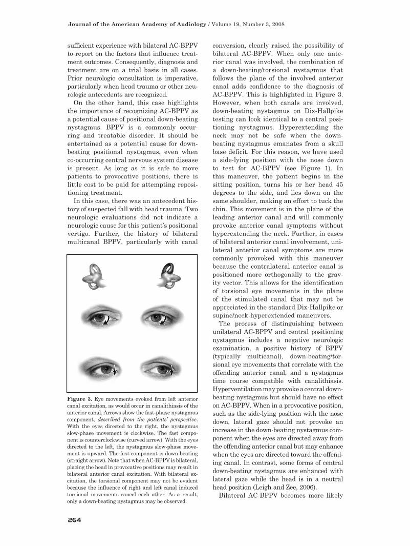
Figure 3. Eye movements evoked from left anterior canal excitation, as would occur in canalithiasis of the anterior canal. Arrows show the fast-phase nystagmus component, described from the patients’ perspective. With the eyes directed to the right, the nystagmus slow-phase movement is clockwise. The fast compo-nent is counterclockwise (curved arrow). With the eyes directed to the left, the nystagmus slow-phase move-ment is upward. The fast component is down-beating (straight arrow). Note that when AC-BPPV is bilateral, placing the head in provocative positions may result in bilateral anterior canal excitation. With bilateral ex-citation, the torsional component may not be evident because the influence of right and left canal induced torsional movements cancel each other. As a result, only a down-beating nystagmus may be observed.

265
Down-Beating Nystagmus in Anterior BPPV / Zapala
in the setting of bilateral multicanal BPPV with a negative neurologic examination (in our setting, performed by a neurologist, neuro-ophthalmologist, or neuro-otologist). Likelihood increases when the time course of evoked nystagmus is compatible with canalithiasis. However, persisting down-beating nystagmus does not necessarily preclude AC-BPPV canalithiasis, as this case shows. Confidence in the presence of AC-BPPV can be increased when the evoked nystagmus can be shown to follow the plane of the dependent anterior canal in the side-lying position with the nose down. Confirmation occurs when the symptoms respond to canalith repositioning.
Repositioning may be performed using a standard Epley maneuver, so long as hyper-extension of the neck is not considered a factor. When AC-BPPV occurs in isolation or is the primary provocative canal, we prefer to use a reversed Epley technique to clear the canal. This maneuver will often work without hyperextending the neck, which is an advantage when canalith repo-sitioning is attempted as a diagnostic trial.
In the case presented in this article, AC-BPPV symptoms appeared recalcitrant to canalith repositioning efforts. One pos-sible explanation for this would be that the orientation of the anterior semicircular canals would favor packing of canalith debris into the ampulla when the head is held upright. However, a simpler explana-tion is that this patient repeatedly moved her head into an orientation that promoted anterior canalithiasis. In other AC-BPPV cases in our clinic, a tendency to sleep on the stomach or in the fetal position with the nose down and practicing yoga have all been implicated in loading the anterior semi-circular canals. Because experience with AC-BPPV is still evolving, we cannot be certain of the importance of these historical antecedents or whether postrepositioning instructions to avoid these movements are important in the treatment of AC-BPPV. This case suggests they may be factors. If so, evoking this history not only is helpful in increasing awareness of AC-BPPV dur-ing the clinical exam but also may help in postrepositioning instructions.
In summary, AC-BPPV can mimic down-beating positional nystagmus of central origin, particularly when it is bilateral. Factors that increase the probability of bilateral AC-BPPV
include a history of bilateral multicanal BPPV, transient down-beating/torsional nystagmus that follows the plane of the provoked canal, the absence of neurologic symptoms, and neu-rologic clearance for canalith repositioning. Exploration for AC-BPPV using the side-lying position helps to isolate anterior canal symp-toms and minimizes neck hyperextension. When it is safe to do so, a diagnostic trial of canalith repositioning for possible anterior canal involvement may alleviate symptoms. The reversed Epley is helpful in clearing the anterior canal without hyperextending the neck, which can be useful when the diagnosis is uncertain.
NOTES
Eye movements in this article are described 1. from the patient’s perspective. So, from the perspective of the patient, a right-beating horizontal nystagmus has a quick phase that is directed to the patient’s right side. A clock-wise torsional nystagmus, in this system, means that the upper pole of the eye rolls to the patient’s right side—just like the hands of a clock. It is important to understand that the observer (audiologist, physical therapist, or physician) will see the upper pole of the eye roll to the left (the patient’s right side). So, from the perspective of the observer, the movement appears to be counterclockwise. In this article, all eye movements are described from the patient’s perspective.
In some cases, after a strong burst of 2. BPPV-induced nystagmus, a brief reversal nystagmus may be seen. This is thought to be neurogenic in origin. This reversal nystagmus may explain some but not all of the nystagmus noted in this case. That is, a reversal nystagmus from a left PC-BPPV may appear similar to right anterior canal stimulation. However, this so-called neuro-genic reversal nystagmus is less intense and briefer than the initial BPPV-induced eye movements. In the present case, some nys-tagmus referable to the anterior canal had no antecedent posterior canal stimulation—as would be expected in the case of AC-BPPV.
REFERENCES
Alpini D, Caputo D, Pugnetti L, et al. (2001) Vertigo and multiple sclerosis: aspects of differential diagno-sis. Neurol Sci 22(Suppl 2):S84–S87.
Baloh RW, Baringer JR. (1998) Dizzy patients: the varieties of vertigo. Hosp Pract 33(6):55–58.

Journal of the American Academy of Audiology / Volume 19, Number 3, 2008
266
Bertholon P, Bronstein AM, Davies RA, et al. (2002a) Positional down beating nystagmus in 50 patients: cerebellar disorders and possible anterior semicir-cular canalithiasis. J Neurol Neurosurg Psychiatry 72(3):366–372.
Bertholon P, Faye MB, Tringali S, Martin C. (2002b) Le vertige positionnel paroxystique benin du canal horizontal. Ann Otolaryngol Chir Cervicofac 119(2):73–80.
Bertholon P, Tringali S, Faye MB, et al. (2006) Prospec-tive study of positional nystagmus in 100 consecutive patients. Ann Otol Rhinol Laryngol 115(8):587–594.
Brandt T. (1990) Positional and positioning vertigo and nystagmus. J Neurol Sci 95(1):3–28.
Brantberg K, Bergenius J. (2002) Treatment of anterior benign paroxysmal positional vertigo by canal plugging: a case report. Acta Otolaryngol 122(1):28–30.
Bronstein AM, Miller DH, Rudge P, Kendall BE. (1987) Down beating nystagmus: magnetic resonance imaging and neuro-otological findings. J Neurol Sci 81(2–3):173–184.
Buttner U, Helmchen C, Brandt T. (1999) Diagnos-tic criteria for central versus peripheral positioning nystagmus and vertigo: a review. Acta Otolaryngol 119(1):1–5.
Califano L, Capparuccia PG, Di Maria D, et al. (2003) Treatment of benign paroxysmal positional vertigo of posterior semicircular canal by “Quick Liberatory Rotation Manoeuvre.” Acta Otorhinolaryngol Ital 23(3):161–167.
Cohen HS, Kimball KT. (2004) Treatment on Epley maneuver for benign paroxysmal positional vertigo. Am J Otolaryngol 25(1):33–37.
Crevits L. (2004) Treatment of AC-BPPV by a pro-longed forced position procedure. J Neurol Neurosurg Psychiatry 75(5):779–781.
Crevits L. (2005) Different canalith repositioning procedures for horizontal canal benign paroxysmal positional vertigo [comment]. Arch Otolaryngol Head Neck Surg 133(5):817.
Ekvall Hansson E, Mansson NO, Hakansson A. (2005) Benign paroxysmal positional vertigo among elderly patients in primary health care. Gerontology 51(6):386–389.
Epley JM. (1992) The canalith repositioning procedure: for treatment of benign paroxysmal positional vertigo. Arch Otolaryngol Head Neck Surg 107(3):399–404.
Epley JM. (1995) Positional vertigo related to semicir-cular canalithiasis. Arch Otolaryngol Head Neck Surg 112(1):154–161.
Furman JM, Cass SP. (1999) Benign paroxysmal posi-tional vertigo. New Engl J Med 341(21):1590–1596.
Gacek RR. (1985). Pathophysiology and management of cupulolithiasis. Am J Otolaryngol 6(2):66–74.
Han BI, Oh HJ, Kim JS. (2006) Nystagmus while recumbent in horizontal canal benign paroxysmal positional vertigo. Neurology 66(5):706–710.
Herdman SJ, Blatt PJ, Schubert MC. (2000) Ves-tibular rehabilitation of patients with vestibular hypofunction or with benign paroxysmal positional vertigo. Curr Opin Neurol 13(1):39–43.
Honrubia V, Baloh RW, Harris MR, Jacobson KM. (1999) Paroxysmal positional vertigo syndrome. Am J Otolaryngol 20(4):465–470.
House MG, Honrubia V. (2003) Theoretical models for the mechanisms of benign paroxysmal positional ver-tigo. (Erratum appears in Audiol Neurootol 8[5]:303.) Audiol Neurootol 8(2):91–99.
Jackson LE, Morgan B, Fletcher JC, Krueger WW. (2007) Anterior canal benign paroxysmal positional vertigo: an underappreciated entity. Otol Neurotol 28(2):218–222.
Katsarkas AKT. (1978) Paroxysmal positional vertigo: a study of 255 cases. J Otolaryngol 7:320–330.
Kerber KA, Jen JC, Perlman S, Baloh RW. (2005) Late-onset pure cerebellar ataxia: differentiating those with and without identifiable mutations. J Neu-rol Sci 238(1–2):41–45.
Korres S, Balatsouras DG, Kaberos A, et al. (2002) Occurrence of semicircular canal involvement in benign paroxysmal positional vertigo. Otol Neurotol 23(6):926–932.
Leigh RJ, Zee DS. (2006) The Neurology of Eye Move-ments. New York: Oxford University Press.
Lopez-Escamez JA, Molina MI, Gamiz M, et al. (2005) Multiple positional nystagmus suggests multiple canal involvement in benign paroxysmal vertigo. Acta Otolaryngol 125(9):954–961.
Marciano E, Marcelli V. (2002) Postural restrictions in labyrintholithiasis. Eur Arch Otorhinolaryngol 259(2):262–265.
Massoud EA, Ireland DJ. (1996) Post-treatment instructions in the nonsurgical management of benign paroxysmal positional vertigo. J Otolaryngol 25(1):121–125.
Prokopakis EP, Chimona T, Tsagournisakis M, et al. (2005) Benign paroxysmal positional vertigo: 10-year experience in treating 592 patients with canalith repo-sitioning procedure. Laryngoscope 115(9):1667–1671.
Semont A, Freyss G, Vitte E. (1989) Vertige position-nel paroxystique benin et manoeuvre liberatoire. Ann Otolaryngol Chir Cervicofac 106(7):473–476.
Walker MF, Zee DS. (1999) The effect of hyperventila-tion on downbeat nystagmus in cerebellar disorders. Neurol 53(7):1576–1579.
Walker MF, Zee DS. (2000) Bedside vestibular exami-nation. Otolaryngol Clin North Am 33(3):495–506.