Embed Size (px)
Citation preview
A Imagiologia do CHLC marcou presença no 30Th European Congress of Radiology, que decor-
reu no Áustria Center Vienna de 28 de Fevereiro a 4 de Março em Viena de Áustria.
Portugal foi um dos países organizadores com a China e Suíça.
O evento que marca a reunião anual da Sociedade Europeia de Radiologia, conhecida como
uma das reuniões mais inovadoras dentro da comunidade científica, contou com a presença
de mais de 75.502 membros dos 5 continentes de 157 países - Europa 50%, Ásia 23%,
América 7%, Brasil 16%, África 3% e Oceânia 1%, num total de mais de 25.000 participan-
tes, dos quais 296 portugueses, 105 Membros de Sociedades Institucionais, 300 exposito-
res num espaço com 26.000m2. Contou com 6,757abstracts, 3,475 posters, mais de 600
Sessões, 52 dedicadas em exclusivo a Radiographers.
Técnicos e Médicos Neurorradiologistas e Radiologistas do CHLC, assinalaram a sua presen-
ça com apresentação de comunicações e posters em representação Institucional.
Atendendo à qualidade do programa científico e à mais-valia que foi a frequência de cursos
e a participação em sessões científicas e Workshops, não poderíamos deixar de partilhar
este momento que foi para todos nós mais que uma simples aprendizagem, um complemen-
to que contribuirá para ampliar o conhecimento das diferentes vertentes que constituem a
Instituição Hospitalar numa perspetiva de otimizar e melhorar as nossas práticas.
A formação apesar do seu retorno personalizado é sempre institucional, pois as Instituições
são as pessoas “pessoas certas” e o valor acrescido que estas representam para a organiza-
ção e se reflete nos serviços que se presta.
Nesta Edição Especial ECR 2018, apresentamos uma seleção de alguns dos muitos temas
abordados, esperando que espelhe desde logo a reflexão da prática do que foi aprendido na
Academia.
Cristina Almeida
Coordenadora Técnica
Radiodiagnóstico e Neurorradiologia
Março 2018
Especial ECR2018
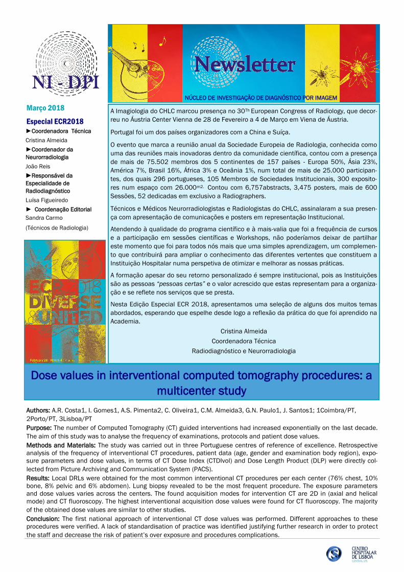
Authors: A.R. Costa1, I. Gomes1, A.S. Pimenta2, C. Oliveira1, C.M. Almeida3, G.N. Paulo1, J. Santos1; 1Coimbra/PT,
2Porto/PT, 3Lisboa/PT
Purpose: The number of Computed Tomography (CT) guided interventions had increased exponentially on the last decade.
The aim of this study was to analyse the frequency of examinations, protocols and patient dose values.
Methods and Materials: The study was carried out in three Portuguese centres of reference of excellence. Retrospective
analysis of the frequency of interventional CT procedures, patient data (age, gender and examination body region), expo-
sure parameters and dose values, in terms of CT Dose Index (CTDIvol) and Dose Length Product (DLP) were directly col-
lected from Picture Archiving and Communication System (PACS).
Results: Local DRLs were obtained for the most common interventional CT procedures per each center (76% chest, 10%
bone, 8% pelvic and 6% abdomen). Lung biopsy revealed to be the most frequent procedure. The exposure parameters
and dose values varies across the centers. The found acquisition modes for intervention CT are 2D in (axial and helical
mode) and CT fluoroscopy. The highest interventional acquisition dose values were found for CT fluoroscopy. The majority
of the obtained dose values are similar to other studies.
Conclusion: The first national approach of interventional CT dose values was performed. Different approaches to these
procedures were verified. A lack of standardisation of practice was identified justifying further research in order to protect
the staff and decrease the risk of patient’s over exposure and procedures complications.
►Coordenadora Técnica
Cristina Almeida
►Coordenador da
Neurorradiologia
João Reis
►Responsável da
Especialidade de
Radiodiagnóstico
Luísa Figueiredo
► Coordenação Editorial
Sandra Carmo
(Técnicos de Radiologia)
NÚCLEO DE INVESTIGAÇÃO DE DIAGNÓSTICO POR IMAGEM
Dose values in interventional computed tomography procedures: a
multicenter study
Página 2
Authors: N. Costa1, N. Bastati2, S. Pötter-Lang2, Z. Guengoern2, Y. Bican2, A. Ba-Ssalamah2; 1Lisbon/PT 2Vienna/AT
Purpose: The aim of the study was to create a scoring system based upon gadoxetic acid-enhanced magnetic resonance
imaging (gaMRI) features to predict the treatment response (TR) to chemotherapy.
Methods and Materials: This was a retrospective study of 30 consecutive patients (65.2±11.2years) with CRCLM, who
underwent gaMRI after chemotherapy and before hepatic resection. Metastases were classified according to a suggested
scoring system (0-6 points) in three groups of response: optimal (≤2 points); partial (2-4 points); and no-response (≥4
points). The scoring system comprised three features: overall homogeneity (homogeneous=0, mixed=1, heteroge-
neous=2); tumour-liver interface (sharp=0, mixed=1, ill defined=2); and peripheral rim enhancement (≤2 mm=0, 2-4
mm=1, ≥4 mm=2). Apparent diffusion coefficient (ADC) values were measured.The primary outcome was residual vital
tumour (RVT). The scoring system, response groups, and ADC values were calculated and compared with the RVT percen-
tage. Demographic, laboratory, and imaging findings were included in a multivariate statistical analysis. The three groups
of response were correlated with patient survival and the log-rank test was used to compare two survival distributions
(optimal/partial response vs no-response groups).
Results: Forty-one CRCLM showed good inter-observer agreement (κ=0.86). Multiple regression demonstrated an associa-
tion between RVT (32.9±11.2) and the scoring system (p<0.001), the response group (p<0.001), and the ADC values
(p<0.021). The survival distributions between optimal/partial response and no-response showed a trend to be different
(p=0.066).
Conclusion: gaMRI correlated well with our scoring system, different response groups, and ADC values in patients with
CRCLM treated with chemotherapy, and may be used to assess the RVT percentage.
Predicting the response of colorectal cancer liver metastases
(CRCLM) to preoperative chemotherapy using gadoxetic acid-
enhanced MRI
Prostate artery embolisation (PAE) for benign prostate obstruction
(BPO): the paradigm shift
Authors: Tiago Bilhim, Lisbon/PT
Learning Objectives:
1. To learn about the anatomy of male pelvic arteries relevant for selective embolisation of the prostate.
2. To understand what imaging modalities can be used for guidance inside the pelvis to find the prostate arteries.
3. To become familiar with clinical outcome and predictors of treatment response after prostate artery embolisation.
Abstract:
Prostate artery embolisation (PAE) has been proven to be safe and effective to relieve lower urinary tract symptoms
(LUTS) related to benign prostate obstruction (BPO). Knowledge of the anatomy of the prostate arteries (PAs) is cornersto-
ne for PAE. Pre-procedural CT-Angiography (CTA) and intraprocedural cone-beam CT (CBCT) have been proven to be relia-
ble tools to study the PAs. Identification of the PAs is generally performed under fluoroscopy. Developing alternatives are
the use of fusion imaging with the pre-procedural CTA images, overlay with the intra-procedural CBCT images and the use
of vessel-tracking software that automatically identifies the PAs. Up to 25% of patients may have clinical failure after PAE.
Most of these patients are nonresponders, with a minority being relapsers. Thus, it is very important to define predictors
of clinical outcome to help exclude those patients less likely to respond to PAE. Predictors of better clinical outcome that
have been identified include: younger age; lower baseline LUTS severity; patients under acute urinary retention; and ade-
nomatous-dominant BPO. Bilateral PAE has been proven to be better than unilateral PAE. MR-detected ischemia after
PAE, PSA values 24 hours after PAE, prostate volume reduction and clinical outcome have been shown to be correlated.
Prognostic quantification with perfusion imaging of the prostate, developing catheters and embolic agents/sizes are some
of the future directions in the investigative field of PAE on the path to inclusion in the guidelines for the management of
patients with symptomatic BPO.
Página 3 Especial ECR2018
Dr. João Reis Learn from my mistakes
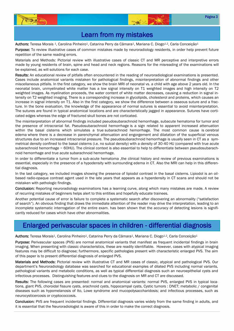
Authors: Teresa Morais 1, Carolina Pinheiro1, Catarina Perry da Câmara1, Mariana C. Diogo1,2, Carla Conceição1
Purpose: To review illustrative cases of common mistakes made by neuroradiology residents, in order help prevent future
repetition of the same reading errors.
Materials and Methods: Pictorial review with illustrative cases of classic CT and MR perceptive and interpretive errors
made by young residents of brain, spine and head and neck regions. Reasons for the misreading of the examinations will
be explained, as will solutions for each case.
Results: An educational review of pitfalls often encountered in the reading of neuroradiological examinations is presented.
Cases include anatomical variants mistaken for pathological findings, misinterpretation of abnormal findings and other
miscellaneous pitfalls. In the first category, we show the brain MRI of neonatal vs. a child with age above 2 years old. In the
neonatal brain, unmyelinated white matter has a low signal intensity on T1 weighted images and high intensity on T2
weighted images. As myelination proceeds, the water content of white matter decreases, causing a reduction in signal in-
tensity on T2 weighted imaging. There is a corresponding increase in glycolipids, cholesterol and proteins, which causes an
increase in signal intensity on T1. Also in the first category, we show the difference between a osseous suture and a frac-
ture. In the bone evaluation, the knowledge of the appearance of normal sutures is essential to avoid misinterpretation.
The sutures are found in typical anatomical locations and are characteristically jagged in appearance. Sutures have corti-
cated edges whereas the edge of fractured skull bones are not corticated.
The misinterpretation of abnormal findings included pseudosubarachnoid hemorrhage, subacute hematoma for tumor and
the presence of intracranial fat. Pseudosubarachnoid hemorrhage is a sign related to apparent increased attenuation
within the basal cisterns which simulates a true subarachnoid hemorrhage. The most common cause is cerebral
edema where there is a decrease in parenchymal attenuation and engorgement and dilatation of the superficial venous
structures due to an increased intracranial pressure. The pseudosubarachnoid hemorrhage is usually seen in CT as a sym-
metrical density confined to the basal cisterns (i.e. no sulcal density) with a density of 30-40 HU (compared with true acute
subarachnoid hemorrhage ~ 60HU). The clinical context is also essential to help to differentiate between pseudosubarach-
noid hemorrhage and true acute subarachnoid hemorrhage.
In order to differentiate a tumor from a sub-acute hematoma ,the clinical history and review of previous examinations is
essential, especially in the presence of a hypodensity with surrounding edema in CT. Also the MRI can help in this differen-
tial diagnosis.
In the last category, we included images showing the presence of lipiodol contrast in the basal cisterns. Lipiodol is an oil-
based radio-opaque contrast agent used in the late years that appears as a hyperdensity in CT scans and should not be
mistaken with pathologic findings.
Conclusion: Reporting neuroradiology examinations has a learning curve, along which many mistakes are made. A review
of recurring mistakes of beginners helps alert to this entities and hopefully educate trainees.
Another potential cause of error is failure to complete a systematic search after discovering an abnormality (“satisfaction
of search”). An obvious finding that draws the immediate attention of the reader may drive the interpretation, leading to an
incomplete systematic interrogation of the entire exam. has been shown that the accuracy of detecting lesions is signifi-
cantly reduced for cases which have other abnormalities.
Authors: Teresa Morais1, Carolina Pinheiro1, Catarina Perry da Câmara1, Mariana C. Diogo1,2, Carla Conceição1
Purpose: Perivascular spaces (PVS) are normal anatomical variants that manifest as frequent incidental findings in brain
imaging. When presenting with classic characteristics, these are readily identifiable. However, cases with atypical imaging
features may be difficult to diagnose; furthermore, specific pathologies present with characteristic enlarged PVS. The aim
of this paper is to present differential diagnosis of enlarged PVS.
Materials and Methods: Pictorial review with illustrative CT and MR cases of classic, atypical and pathological PVS. Our
department’s Neuroradiology database was searched for educational examples of dilated PVS including normal variants,
pathological variants and metabolic conditions, as well as typical differential diagnosis such an neuroepithelial cysts and
infectious processes. Distinguishing features and clues to the diagnosis on MR and CT are discussed.
Results: The following cases are presented: normal and anatomical variants: normal PVS, enlarged PVS in typical loca-
tions, giant PVS, choroidal fissure cysts, arachnoid cysts, hippocampal cysts, Cystic tumors : DNET; metabolic / congenital
diseases such as hypomelanosis of Ito, Lowe syndrome and mucopolysaccharidosis; and infectious processes, such as
neurocysticercosis or cryptococcosis.
Conclusion: PVS are frequent incidental findings. Differential diagnosis varies widely from the same finding in adults, and
it is essential that the Neuroradiologist is aware of this in order to make the correct diagnosis.
Enlarged perivascular spaces in children - differential diagnosis
Página 4
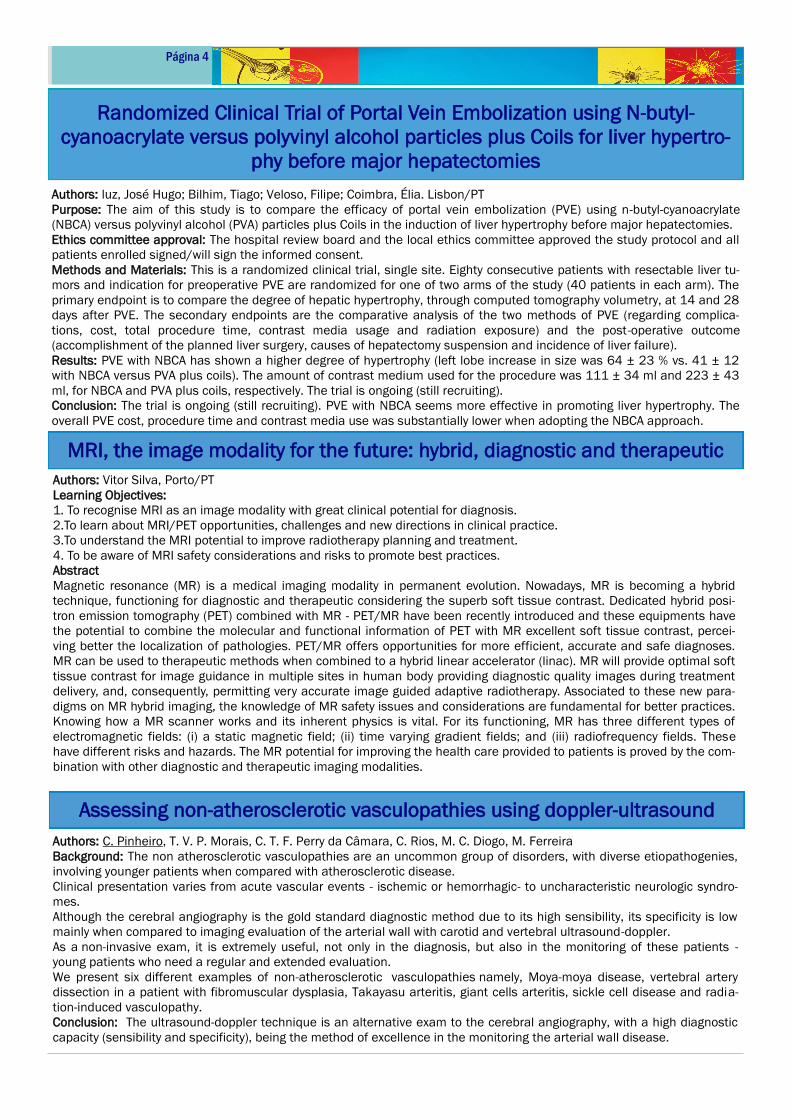
Authors: luz, José Hugo; Bilhim, Tiago; Veloso, Filipe; Coimbra, Élia. Lisbon/PT
Purpose: The aim of this study is to compare the efficacy of portal vein embolization (PVE) using n-butyl-cyanoacrylate
(NBCA) versus polyvinyl alcohol (PVA) particles plus Coils in the induction of liver hypertrophy before major hepatectomies.
Ethics committee approval: The hospital review board and the local ethics committee approved the study protocol and all
patients enrolled signed/will sign the informed consent.
Methods and Materials: This is a randomized clinical trial, single site. Eighty consecutive patients with resectable liver tu-
mors and indication for preoperative PVE are randomized for one of two arms of the study (40 patients in each arm). The
primary endpoint is to compare the degree of hepatic hypertrophy, through computed tomography volumetry, at 14 and 28
days after PVE. The secondary endpoints are the comparative analysis of the two methods of PVE (regarding complica-
tions, cost, total procedure time, contrast media usage and radiation exposure) and the post-operative outcome
(accomplishment of the planned liver surgery, causes of hepatectomy suspension and incidence of liver failure).
Results: PVE with NBCA has shown a higher degree of hypertrophy (left lobe increase in size was 64 ± 23 % vs. 41 ± 12
with NBCA versus PVA plus coils). The amount of contrast medium used for the procedure was 111 ± 34 ml and 223 ± 43
ml, for NBCA and PVA plus coils, respectively. The trial is ongoing (still recruiting).
Conclusion: The trial is ongoing (still recruiting). PVE with NBCA seems more effective in promoting liver hypertrophy. The
overall PVE cost, procedure time and contrast media use was substantially lower when adopting the NBCA approach.
Randomized Clinical Trial of Portal Vein Embolization using N-butyl-
cyanoacrylate versus polyvinyl alcohol particles plus Coils for liver hypertro-
phy before major hepatectomies
Authors: C. Pinheiro, T. V. P. Morais, C. T. F. Perry da Câmara, C. Rios, M. C. Diogo, M. Ferreira
Background: The non atherosclerotic vasculopathies are an uncommon group of disorders, with diverse etiopathogenies,
involving younger patients when compared with atherosclerotic disease.
Clinical presentation varies from acute vascular events - ischemic or hemorrhagic- to uncharacteristic neurologic syndro-
mes.
Although the cerebral angiography is the gold standard diagnostic method due to its high sensibility, its specificity is low
mainly when compared to imaging evaluation of the arterial wall with carotid and vertebral ultrasound-doppler.
As a non-invasive exam, it is extremely useful, not only in the diagnosis, but also in the monitoring of these patients -
young patients who need a regular and extended evaluation.
We present six different examples of non-atherosclerotic vasculopathies namely, Moya-moya disease, vertebral artery
dissection in a patient with fibromuscular dysplasia, Takayasu arteritis, giant cells arteritis, sickle cell disease and radia-
tion-induced vasculopathy.
Conclusion: The ultrasound-doppler technique is an alternative exam to the cerebral angiography, with a high diagnostic
capacity (sensibility and specificity), being the method of excellence in the monitoring the arterial wall disease.
MRI, the image modality for the future: hybrid, diagnostic and therapeutic
Assessing non-atherosclerotic vasculopathies using doppler-ultrasound
Authors: Vitor Silva, Porto/PT
Learning Objectives:
1. To recognise MRI as an image modality with great clinical potential for diagnosis.
2.To learn about MRI/PET opportunities, challenges and new directions in clinical practice.
3.To understand the MRI potential to improve radiotherapy planning and treatment.
4. To be aware of MRI safety considerations and risks to promote best practices.
Abstract
Magnetic resonance (MR) is a medical imaging modality in permanent evolution. Nowadays, MR is becoming a hybrid
technique, functioning for diagnostic and therapeutic considering the superb soft tissue contrast. Dedicated hybrid posi-
tron emission tomography (PET) combined with MR - PET/MR have been recently introduced and these equipments have
the potential to combine the molecular and functional information of PET with MR excellent soft tissue contrast, percei-
ving better the localization of pathologies. PET/MR offers opportunities for more efficient, accurate and safe diagnoses.
MR can be used to therapeutic methods when combined to a hybrid linear accelerator (linac). MR will provide optimal soft
tissue contrast for image guidance in multiple sites in human body providing diagnostic quality images during treatment
delivery, and, consequently, permitting very accurate image guided adaptive radiotherapy. Associated to these new para-
digms on MR hybrid imaging, the knowledge of MR safety issues and considerations are fundamental for better practices.
Knowing how a MR scanner works and its inherent physics is vital. For its functioning, MR has three different types of
electromagnetic fields: (i) a static magnetic field; (ii) time varying gradient fields; and (iii) radiofrequency fields. These
have different risks and hazards. The MR potential for improving the health care provided to patients is proved by the com-
bination with other diagnostic and therapeutic imaging modalities.
Página 5 Especial ECR2018
Dr. João Reis
APRENDIZAGEM - O congresso apresentou mini-cursos
“hands on it”, simpósios sobre o estado da arte em várias
áreas e muitas outras formas de aquisição de novos
conhecimentos.
BERND HAMM – Presidente do ECR 2018, é Professor de
Radiologia e presidente dos 3 departamentos de Radiolo-
gia Charité, Humboldt-Universität zu Berlin e Freie Univer-
sität, é também Diretor Clínico de Centro Charité que inclui
Radiologia, Neurorradiologia, Medicina Nuclear e Física
Médica
CASEIRO ALVES - Primeiro português a ser distinguido com
a Medalha de Ouro da Sociedade Europeia de Radiologia
Gastrointestinal e Abdominal. Professor Catedrático da
Faculdade de Medicina da Universidade de Coimbra e
Diretor da Clínica Universitária de Imagiologia dos Hospi-
tais da Universidade de Coimbra. Entre outros cargos foi
Vice-Presidente da FNS, Presidente da Direção da Associa-
ção Nacional de Unidades de Diagnóstico por Imagem
(ANAUDI), Vice-presidente da Direção da Sociedade Portu-
guesa de Radiologia e Medicina Nuclear (SPRMN) e Presi-
dente do Colégio da Especialidade de Radiodiagnóstico da
Ordem dos Médicos.
DAILY Newspaper Congress ECR
ESTUDO- Espaço de estudo e partilha
FIGLMÜLLER - Com o famoso Schnitzel, uma receita com
mais de 100 anos.
GESTÃO - Um dos temas abordados contribuindo para
ampliar o conhecimento das diversas vertentes que consti-
tuem a Instituição Hospitalar numa perspectiva de optimi-
zar e racionalizar recursos.
HEALTH CARE - Diagnostico e terapêutica, cuidar e tratar
constituíram grande enfoque nas mais variadas áreas e
métodos de Imagem.
INVESTIGAÇÃO - O ECR espelha todo o trabalho de investi-
gação realizado pelos profissionais da Imagiologia em todo
o mundo, no rigor, determinação e tentativa de contribuir
para o desenvolvimento científico e em última análise para
a prestação de melhores cuidados de saúde aos pacien-
tes.
KAISERINE Elisabeth - Imperatriz consorte do Império Aus-
tríaco e a rainha consorte da Hungria devido ao seu casa-
mento com o imperador Francisco José I. Era conhecida
como "Sissi da Áustria e Hungria".
LECTURE - 3.000 “Honorary Lectures” dedicadas à Ciência
e Investigação
MULTIDISCIPLINARY SESSIONS- O conceito destas sessões
é promover uma abordagem multidisciplinar de deteção e
tratamento, integrando radiologistas e outros clínicos para
partilha de experiências. Os tópicos abordados foram: Neu-
roimagem, Realidade virtual, Realidade aumentada, Distúr-
bios de saúde mental, Tumores ósseos primários, Implanta-
ção da válvula aórtica transcatéter
NETWORKING - Redes, parceiros, partilha de experiências,
momentos de aprendizagem e crescimento pessoal foram
evidentes durante todo o ECR.
ORGANIZAÇÃO— Portugal, China e Suíça, 75.502 membros
de 157 países, mais de 25.000 participantes.
PORTUGAL– Um dos países organizadores
QUALIDADE - Auditoria às práticas médicas, segurança no
planeamento do design dos departamentos de Imagiologia,
segurança dos campos magnéticos, protecção radiológica,
utilização de novas tecnonologias para melhorar o workflow,
eficiência e qualidade dos serviços, foram temas abordados
nas diferentes sessões com enfoque na Qualidade.
REFRESHER COURSES - Cursos de atualização organizados
pelos vários subcomités científicos. Com base no tema da
sessão, alguns cursos foram apresentados em formato
"integrado" com um painel de discussão
SACHERTORTR - Bolo de chocolate, criado por Franz Sacher
em 1832 para o príncipe Metternich em Viena, capital da
Áustria. É uma das mais famosas especialidades gastronó-
micas da cidade.
TECNOLOGIA - A Exposição Técnica, alojada num espaço
com 26.000m2 acolheu cerca de 300 empresas, dando a
conhecer as últimas novidades em hardware e software
Imagiológico.
UPDATE - Update your skill, através de vários cursos com
componente prática e enfoque nas indicações, vantagens,
limitações e controvérsias das diferentes práticas. Foram
utilizados fantomas para aprender / treinar aspectos técni-
cos bem como a escolha do método adequado.
VIENA— Viena, capital mundial da música, onde se realizam
anualmente 15.000 concertos de todo o tipo e ordem de
grandeza. Não existe outra cidade onde tantos composito-
res exerceram influência: Mozart, Mahler, Haydn, Beethoven
e o rei das valsas, Johann Strauß. A Wiener Philharmoniker
e os Wiener Sängerknaben são destaques mundiais, a Ópe-
ra Vienense (Staatsoper) e a Sala Dourada (Goldene Musik-
vereinssaal) estão entre os melhores locais de encenação.
X – RAY - 8 de Novembro dia internacional da Radiologia
Z...
Cristina Almeida– Técnica Coordenadora DPI
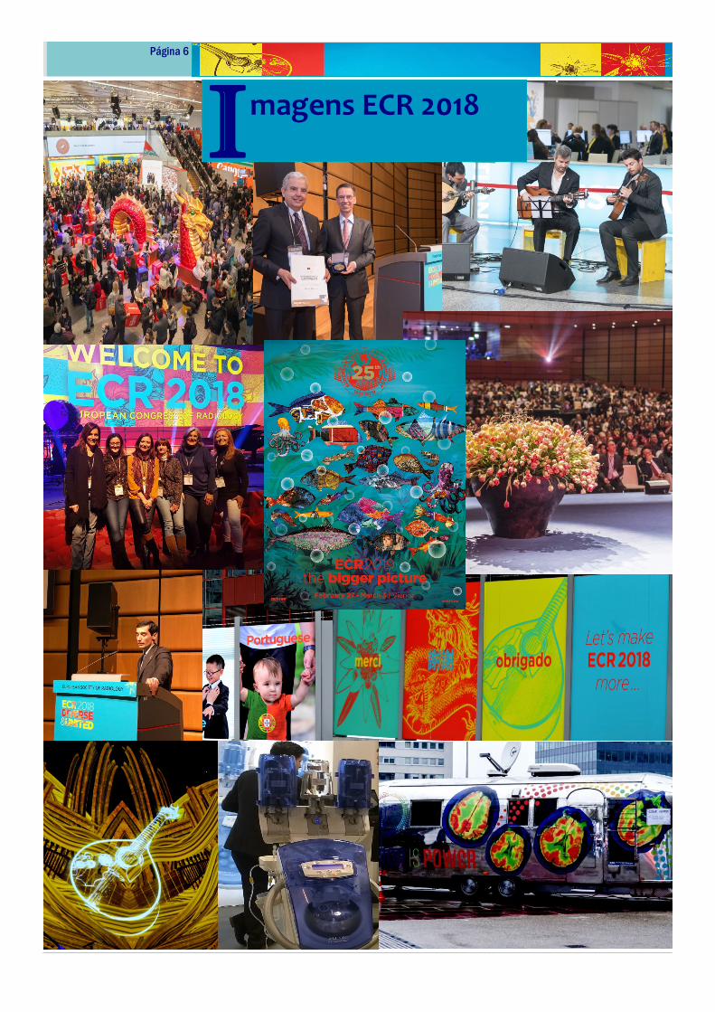
Página 6
I magens ECR 2018