Embed Size (px)
Citation preview
Zurich Open Repository andArchiveUniversity of ZurichMain LibraryStrickhofstrasse 39CH-8057 Zurichwww.zora.uzh.ch
Year: 2015
Doppler sonographic examination of uterine and placental perfusion in cowsin the last month of gestation and effects of epidural anesthesia and
isoxsuprine
Kim, Cornelia
Posted at the Zurich Open Repository and Archive, University of ZurichZORA URL: https://doi.org/10.5167/uzh-120738DissertationPublished Version
Originally published at:Kim, Cornelia. Doppler sonographic examination of uterine and placental perfusion in cows in the lastmonth of gestation and effects of epidural anesthesia and isoxsuprine. 2015, University of Zurich, VetsuisseFaculty.
Klinik für Reproduktionsmedizin
Abteilung für Grosstierreproduktion
der Vetsuisse-Fakultät Universität Zürich
Direktor Prof. Dr med. vet., Dipl. ECBHM Heinrich Bollwein
Arbeit unter wissenschaftlicher Betreuung von
Prof. Dr. med. vet., Dipl. ECBHM Ulrich Bleul
Doppler sonographic examination of uterine and placental perfusion in cows in the last
month of gestation and effects of epidural anesthesia and isoxsuprine
Inaugural-Disseration
zur Erlangung der Doktorwürde der
Vetsuisse-Fakultät Universität Zürich
vorgelegt von
Cornelia Kim
Tierärztin von Wettingen, Aargau
genehmigt auf Antrag von
Prof. Dr. med. vet., Dipl. ECBHM Ulrich Bleul, Referent
2015

1
Inhaltsverzeichnis
Summary............................................................................................................................................................................... 2
Zusammenfassung ............................................................................................................................................................ 3
Publikationsmanuskript ................................................................................................................................................. 4
Danksagung ............................................................................................................................................................................
Circulum Vitae .......................................................................................................................................................................
2
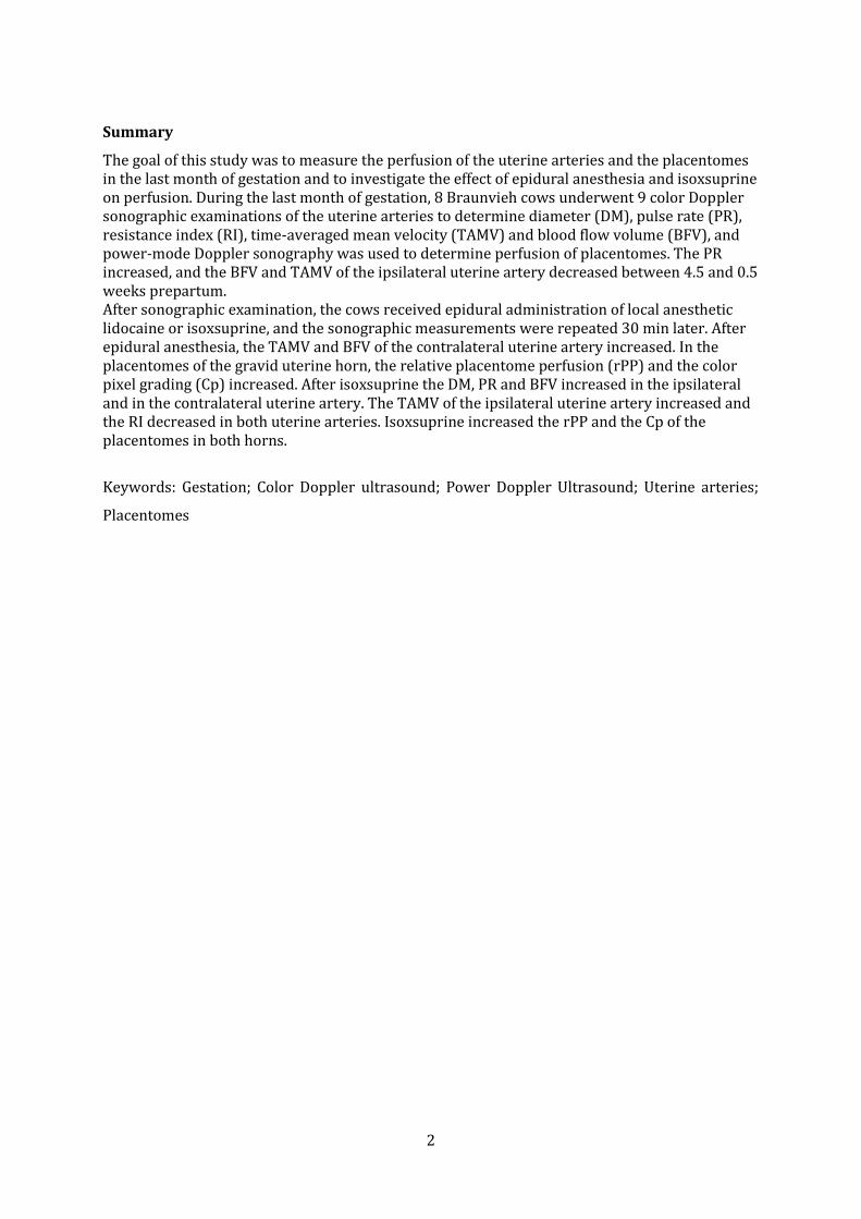
Summary
The goal of this study was to measure the perfusion of the uterine arteries and the placentomes in the last month of gestation and to investigate the effect of epidural anesthesia and isoxsuprine on perfusion. During the last month of gestation, 8 Braunvieh cows underwent 9 color Doppler sonographic examinations of the uterine arteries to determine diameter (DM), pulse rate (PR), resistance index (RI), time-averaged mean velocity (TAMV) and blood flow volume (BFV), and power-mode Doppler sonography was used to determine perfusion of placentomes. The PR increased, and the BFV and TAMV of the ipsilateral uterine artery decreased between 4.5 and 0.5 weeks prepartum. After sonographic examination, the cows received epidural administration of local anesthetic lidocaine or isoxsuprine, and the sonographic measurements were repeated 30 min later. After epidural anesthesia, the TAMV and BFV of the contralateral uterine artery increased. In the placentomes of the gravid uterine horn, the relative placentome perfusion (rPP) and the color pixel grading (Cp) increased. After isoxsuprine the DM, PR and BFV increased in the ipsilateral and in the contralateral uterine artery. The TAMV of the ipsilateral uterine artery increased and the RI decreased in both uterine arteries. Isoxsuprine increased the rPP and the Cp of the placentomes in both horns.
Keywords: Gestation; Color Doppler ultrasound; Power Doppler Ultrasound; Uterine arteries;
Placentomes
3
Zusammenfassung
Das Ziel dieser Studie war es, die Durchblutung in den Uterusgefässen und Plazentomen während des letzten Trächtigkeitsmonat zu quantifizieren und den Einfluss einer Epiduralanästhesie und Isoxsuprine auf die plazentäre Blutversorgung zu untersuchen. Im letzten Trächtigkeitsmonat wurden bei 8 Schweizer Braunviehkühen 9 Untersuchungen durchgeführt, wobei in den Aa. uterinae mittels Dopplersonographie der Durchmesser (DM), die Pulsfrequenz (PF), der Widerstandsindex (RI), die mittlere Blutflussgeschwindigkeit (TAMV) und das Blutflussvolumen (BFV) gemessen wurden. Der Blutfluss in Plazentomen wurde im Power Doppler Modus bestimmt. In der ipsilateralen A. uterina war 4.5 Wochen vor der Geburt die PF höher, das BFV sowie die TAMV tiefer als 0.5 Wochen vor der Geburt. Anschliessend an die sonographische Untersuchung erhielten die Kühe eine Epiduralanästhesie mit Lidocain oder Isoxsuprine intravenös, und die Messungen wurden 30 Minuten später wiederholt. Durch die Epiduralanästhesie erhöhte sich in der kontralateralen A. uterina die TAMV und das BFV und im graviden Horn die relative plazentomale Durchblutung (rPP) und die Blutflussmenge in den Plazentomen (Cp). Durch Isoxsuprine wurden der DM, die PF sowie das BFV in beiden Ae. uterinae erhöht. Zudem stieg die TAMV in der ipsilateralen A. uterina an und der Widerstandsindex sank in beiden Aa. uterinae. Der rPP und auch die Cp in den Plazentomen beider Hörner wurde erhöht.
Stichworte: Trächtigkeit; Color Doppler Ultraschall; Power Doppler Ultraschall; Uterine Arterien; Plazentome
4
Doppler sonographic examination of uterine and placental perfusion in cows in the last
month of gestation and effects of epidural anesthesia and isoxsuprine
C. Kim-Egloff 1, M. Hässig 2, R. Bruckmaier 3, U. Bleul1,*
1 Large Animal Reproduction Unit, Clinic for Reproductive Medicine, Vetsuisse-Faculty,
University of Zurich, Switzerland
2 Ambulatory and Stationary Clinic, Vetsuisse-Faculty, University of Zurich, Switzerland
3 Veterinary Physiology, Vetsuisse-Faculty, University of Bern, Switzerland
* Corresponding author. Tel.: +41 44 6358253; fax: +41 44 6358904. E-mail address:
[email protected] (U. Bleul).
Abstract
The massive increase in size of the fetus and uterus in the last trimester is accompanied by an
increasing demand for nutrients and oxygen and it is assumed that this demand is met by
increasing uterine and fetal perfusion. The goal of this study was to measure the perfusion of the
uterine arteries and the placentomes in the last month of gestation and to investigate the effect
of epidural anesthesia and isoxsuprine on perfusion. During the last month of gestation, 8
Braunvieh cows underwent 9 color Doppler sonographic examinations of the uterine arteries to
determine diameter (DM), pulse rate, resistance index (RI), time-averaged mean velocity
(TAMV) and blood flow volume (BFV), and power-mode Doppler sonography was used to
determine perfusion of placentomes. The pulse rate increased (P < 0.001), and the BFV and
TAMV of the ipsilateral uterine artery decreased between 4.5 and 0.5 weeks prepartum (BFV,
236.8 ± 65.80 and 208 ± 41.52 cm3/s, P < 0.01; TAMV, 140.0 ± 26.53 cm/s and 125.2 ± 18.46
cm/s, P < 0.05).
After sonographic examination, the cows received epidural administration of local anesthetic
(100 mg lidocaine) in the sacrococcygeal space or isoxsuprine (200 mg/cow, iv), and the
sonographic measurements were repeated 30 min later. After epidural anesthesia, the TAMV
and BFV of the contralateral uterine artery increased by 5.4 % (P < 0.05) and 7.9 % (P < 0.01). In
the placentomes of the gravid uterine horn, the relative placentome perfusion (rPP) and the
color pixel grading (Cp) increased by 10.1 % (P < 0.05) and 11.5 % (P < 0.01) after epidural
anesthesia. After isoxsuprine the DM, pulse rate and BFV increased by 4.7 %, 49.3 % and 16.9 %
in the ipsilateral uterine artery and by 10.8 %, 48.7 % and 22.8 % in the contralateral uterine
artery. The TAMV of the ipsilateral uterine artery increased by 7.1 % (P < 0.01) and the RI
decreased in both uterine arteries (ipsilateral 24.2 %, contralateral 14.9 %, both P < 0.00001).

5
Isoxsuprine increased the rPP and the Cp of the placentomes by 18.1 % and 18.3% in the gravid
horn and by 10.2 % and 24.2 % in the non-gravid horn.
Blood flow variables changed little in the last month of gestation. However, epidural anesthesia
and isoxsuprine caused changes in uterine and placentome perfusion that suggest improvement
of placental nutrient and oxygen supply to the fetus.
Keywords: Gestation; Color Doppler ultrasound; Power Doppler Ultrasound; Uterine arteries;
Placentomes; Intrauterine resuscitation
6
Introduction
The increasing fetal demand for nutrients and oxygen in the last trimester of gestation
are met mainly through increased blood flow to the uterus, which in turn increases perfusion in
the uterus [1]. In the past, invasive techniques were used to measure blood flow in the uterine
arteries, which are largely responsible for uterine perfusion in cattle and horses [2, 3], but these
techniques have been replaced by color Doppler sonography for transrectal measurement of
uterine blood flow [4]. Color Doppler sonography has been used to measure blood flow variables
in cycling cows as well as for quantification of uterine blood flow in pregnant cows and mares
[4-7]. The resistance index (RI) decreased and time-averaged maximum velocity (TAMV), blood
flow volume (BFV) and vessel diameter increased with advancing pregnancy in cows [4].
Another study showed that the birth weight of calves was positively correlated with blood flow
volume [8].
The uterine arteries are mainly responsible for blood supply to the maternal placenta,
and the umbilical arteries supply the fetal placenta. In addition to uterine blood flow, which was
measured transrectally, fetal blood flow was measured transabdominally in an umbilical artery
during the last few weeks of gestation [9], but no measurable changes in umbilical blood flow
were detected in the last month of gestation. Another study measured umbilical blood flow
during parturition using ultrasonographic transducers placed transvaginally on an umbilical
artery and vein after rupture of the allantochorion [10]. Mean total blood flow was lower during
the 60 min before delivery compared with the 30-min period before, and blood flow decreased
transiently during uterine contractions. Furthermore, calves with a blood pH ≥ 7.2 immediately
postnatum had a higher total blood flow in the last hour of gestation than calves with a pH < 7.2,
which was interpreted as a direct effect of blood flow on placental gas exchange.
We are not aware of any reports on the direct measurement of placental circulation and
specifically on blood flow in the placentomes, which prompted us to measure placental blood
flow at the end of gestation using power-mode Doppler sonography. Color Doppler sonography
describes blood flow based on the frequency shift of a flow volume, whereas power-mode
Doppler sonography displays the strength of the Doppler signal in color by determining all
moving particles in the blood, which allows recording of blood flow independent of blood flow
velocity and direction. This is crucial for the quantitative and semiquantitative assessment of
blood flow in tissues with low blood flow velocity and numerous blood vessels [11] such as
placentomes. The goal of this study was to investigate whether blood flow in placentomes can be
assessed quantitatively based on the size of the perfused area, and to what extent the color
differences in the power-mode Doppler image are suitable for the semiquantitative
determination of the number of all cells in the blood per unit of area.
Recently studies showed the effects of drugs on uterine and placental blood flow in cows
[7, 9]. In human medicine, intrauterine resuscitative measures are employed to improve
7
placental perfusion when abnormal fetal heart rate patterns are recognized with the goal of
preventing fetal hypoxia and subsequent acidosis [12]. Two of these measures are the
administration of a uterine relaxant drug and epidural anesthesia. We are not aware of any
studies investigating the effect of such measures on placental perfusion in cows except for one
investigation of changes in uterine perfusion induced by various drugs in the last month of
gestation [9]. The latter study showed that the uterine relaxant drug isoxsuprine increased
uterine blood flow volume by 5 % and epidural anesthesia by 6 %. Our hypothesis was that
these two treatments also affect perfusion of the placentomes.
The purpose of the study was to increase our knowledge of Doppler sonographic changes
in semiquantitative and quantitative blood flow variables in the uterine arteries and
placentomes in the last month of gestation, and to determine the effect of isoxsuprine and
epidural anesthesia on these variables.
1. Materials and methods
1.1. Cows
Eight Braunvieh cows, which ranged in age from 4 to 14 years and had normal singleton
pregnancies, were used. Lactation numbers ranged from 3 to 11. The cows were admitted to our
clinic one month before the calculated due date. They were kept in tie stalls, bedded with straw,
and fed hay, grass silage and water ad libitum and had daily access to an exercise yard.
The use of animals for this study was approved by the cantonal veterinary office of
Zurich (permit number 213/2010).
1.2. Study design, B-mode, and Doppler sonography of the uterine arteries
The cows were examined twice a week by the same investigator (CK) starting one month
before the calculated date of birth. In total 9 examinations per cow were included into the final
analysis. After the clinical examination, both uterine arteries and the placentomes were
examined sonographically using a portable ultrasound machine (LOGIQ e; General Electric
Medical System, Glattbrugg, Switzerland). Transabdominal B-mode sonographic examination of
the placentomes was carried out followed by transabdominal power-mode Doppler sonography
using a 4-MHz convex probe (4C-RS, General Electric Medical System). Two placentomes in the
gravid horn and one in the non-gravid horn were examined from the left and right ventral flanks
cranial to the udder. The sites of examination were marked on the skin and kept constant for
each cow. Scanning of the placentomes was repeated several times and the images were saved,
and four measurements per placentome were then selected for analysis.
8
Transrectal B-mode and Color Doppler sonography of the ipsilateral (pregnant horn) and
contralateral uterine arteries was then carried out using a 10-MHz linear probe (I739-RS,
General Electric Medical System). Both uterine arteries were examined and identified as
described [4, 5]. First, the external iliac arteries were identified at the point where they branch
off the aorta. Following the external iliac artery ventrally, the uterine artery was identified
crossing the external iliac artery. To confirm its identity, the uterine artery was followed
dorsally toward the internal iliac artery until the ligamentum teres vesicae was encountered.
This is the atrophied stump of the umbilical artery and allowed definitive identification of the
uterine artery. The uterine artery was then traced back toward the uterus, and the point of
measurement was located immediately cranial to the point where the uterine artery crossed the
external iliac artery and vein. Measurements were repeated 3 to 5 times by temporarily
removing the probe from the site of measurement and repositioning it. Transverse images of the
uterine arteries were taken in B-mode for measuring the arterial diameter and Doppler
sonographic images for blood flow analysis.
After these measurements, the cows received either the tocolytic drug isoxsuprine (200
mg/cow intravenously, Graeub Veterinary Products, Bern, Switzerland) or epidural anesthesia
using 5 mL 2 % lidocaine (100 mg/cow injected into the sacrococcygeal space, Streuli Pharma
Uznach, Switzerland). Location of the sacrococcygeal space was facilitated by up-and-down
movements of the tail and the use of the hanging drop technique. B-mode and power-mode
Doppler sonographic examinations were repeated after 30 min. One complete examination took
2 hours and was accompanied by collection of jugular blood into a heparinized blood tube (16
I.E. heparin/mL blood, Sarstedt, Nürnbrecht, Germany) for measurement of progesterone
concentration.
1.3. Progesterone measurement
Blood was centrifuged at 4,000 rpm for 10 min and the plasma stored at -22°C.
Progesterone was measured with the RIA kit IM1188 from Beckman Coulter GmbH (Krefeld,
Germany) according to the producer's protocol. The intra- and interassay variation coefficients
were 4.5 % and 8.5 %.
9
1.4. Data collection, analysis, and statistics
1.4.1. Blood flow measurement in the uterine arteries
The software program PixelFlux (Chameleon Software, Münster, Germany) was used to
analyze ultrasound images and to determine vessel diameter (DM), pulse rate (PR), resistance
index (RI), time-averaged mean velocity (TAMV) and blood flow volume (BFV). The diameter
was measured in four B-mode images that showed a perfectly circular artery and no motion
artifacts, and the mean was calculated from the four measurements. Pulse rate, RI, TAMV and
BFV were calculated from Doppler sonographic images. For each artery, four images were
selected that showed at least two representative and uniform Doppler waves. The mean was
calculated from the four measurements. Pulse rate was defined as number of spectral waves per
unit of time [min-1]. The RI describes the resistance to flow distal to the point of measurement
and is calculated using the formula RI=(D-S)/S, where D is the end-diastolic blood flow velocity
and S the peak-systolic velocity. The TAMV of the arterial blood during a cardiac cycle is derived
from the formula TAMV [cm/sec]=(TAMF x c)/(2F x cos α), where TAMF is the time-averaged
maximum rate shift over the cardiac cycle, c is the ultrasound propagation velocity, F the
transmitted wave rate and α the angle between the ultrasound beam and the blood flow
direction [8]. The BFV was calculated using the formula BFV [cm3/sec]=TAMV x A, where A is the
arterial circumference, derived from the DM and using A= π x (DM/2)2.
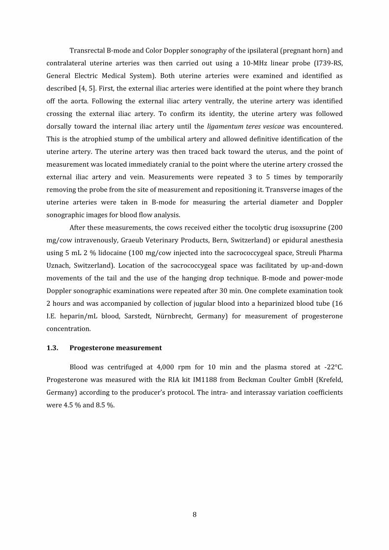
1.4.2. Blood flow measurement in placentomes
The same software program (PixelFlux, Chameleon Software) was used for analysis of
perfusion of placentomes. The transducer was positioned to image the maximum size of the
placentomes. Slight adjustments in location of the transducer relative to the placentome were
made to achieve a maximum number of color pixels in the placentome before freezing the
images. The area of the placentome that appeared in color in the power-mode Doppler
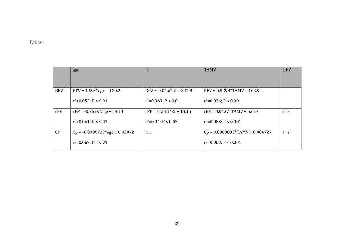
sonogram, representing the area with measurable blood flow (Fig. 1), as well as the entire area
of the placentome, which was defined as region of interest, were traced and the areas measured.
The colored area was expressed as a proportion of the entire region of interest, and the
proportions calculated from the four measurements per placentome were averaged, generating
the measure referred to as relative placentome perfusion (rPP), which was used for analysis
[13].
In power-mode Doppler sonography, the amount of moving cells in the blood
corresponds with the color of pixels, and therefore the RGB scale, included in all power Doppler
sonograms (Fig. 1), was used to assess semiquantitatively the amount of moving cells per unit
area (pixel). The RGB scale was divided into 143 color grades. Grade 1 corresponded to the color
10
black and grade 143 corresponded to light yellow. Black pixels represent no movement and
yellow pixels represent maximum amount of moving cells in the blood.
A software program was developed for quantification of blood flow. The RGB color
values of all pixels in the region of interest were expressed as a ratio of all pixels and calculated
as
Where x is the color grade on the RGB scale and pixel is the number of pixels for one of the 143
color grades.
In a second step, the analysis was limited to pixels representing a high amount of blood flow.
Pixels with color grades from orange to yellow (Cp84) corresponding to color grades 84 to 143
of the RGB scale were evaluated.
Pixel orange (Cp 84)
The calculated Cp and Cp84 values from the same four measurements per placentome used for
rPP were averaged and used for further analyses.
1.4.3. Statistics
The calculated values were entered into an Excel file (Microsoft, Wallisellen,
Switzerland) and the add-in statistic program StatEL (ad Science Company, Paris, France) was
used for analysis of the sonographic variables. The program Stata (StataCorp, College Station, TX,
USA) was used for the regression model and the multivariate regression. The Shapiro-Wilk test
was used to test for normality. Normal data with homogeneity of variance (DM, TAMV, BFV)
were analyzed using analysis of variance, and a post hoc test, i.e. Bonferroni, was used for
comparison between different time points. The paired t-test was used for comparison of
variables before and after treatment. Not normal distributed data (RI, HR, rPP, CP, Cp84) were
analyzed using the Friedman and the Wilcoxon signed-rank test. The combined TAMV and BFV
were calculated as mean values of measurements from the arteries of the pregnant and the non-
pregnant horn. Results were expressed as mean ± standard deviation. A P-value ≤ 0.05 was
considered significant. Correlations between progesterone concentrations, the age of the cows
and all sonographic variables, as well as between the variables measured in the uterine arteries
(RI, TAMV and BFV) and in the placentomes (rPP, CP) were calculated. Univariate linear
regression models with the week prepartum as dependent variable and the sonographic
parameter as well as the progesterone concentration as independent variables were calculated.
The independent variables with a P < 0.2 were integrated into a multivariate regression model.
Stepwise backward elimination regression was carried out. The final model consisted only
variables with a P ≤ 0.01.
11
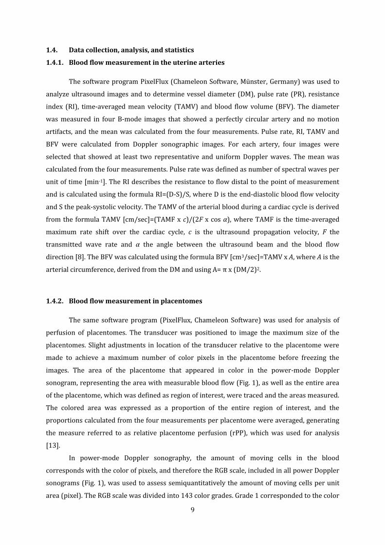
2. Results
2.1. Changes in uterine and placentome perfusion at the end of gestation
The changes in diameter of the ipsilateral (P = 0.07) and contralateral uterine arteries (P
= 0.08) in the last month of gestation missed the significance level narrowly. The pulse rate in
the uterine arteries increased from 75.78 ± 10.78 bpm at 4.5 weeks ap to 84.20 ± 9.85 bpm at
0.5 weeks ante partum (ap) (P < 0.001). The RI remained unchanged during the study period.
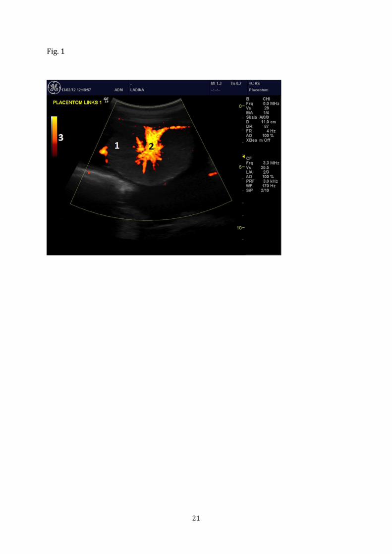
The TAMV and the BFV (Fig. 2) in the arteries of the gravid and the non-gravid horn combined
changed (TAMV P < 0.05, BFV P < 0.01). TAMV and BFV decreased in the ipsilateral uterine
artery between 4.5 and 0.5 weeks ap by 10.8 % and 12.1 %, but remained unchanged in the
contralateral uterine artery.
The rPP in both uterine horns did not change significantly during the study period.
Similar to the rPP, the quotient Cp of the gravid and non-gravid horns combined missed the
significance level narrowly (Fig. 2, P = 0.09). The placentome blood flow assessed by pixel
grading in the yellow-to-orange color spectrum (Cp84) did not change significantly during the
study period.
2.2. Correlations of sonographic variables
2.2.1. Age of the dam
Except RI and TAMV the sonographic variables showed a significant, but very weak
correlation to the age of the dam (Table 1).
2.2.2. Correlation between sonographic variables
The RI showed a good correlation to TAMV (TAMV = -254.3*RI + 250.5; r2=0.38; P <
0.0001). All other correlations were weak or not significant (Table 1).
2.2.3. Progesterone concentration
The progesterone concentration decreased in the last month of gestation (P < 0.001), but
correlations with sonographic variables were weak or did not exist (r² ranged from 0.00005 to
0.3). The variables pulse rate, DM and BFV of the contralateral uterine artery remained in the
model after backward elimination in stepwise regressions. In the multivariate regression
analysis the highest correlation coefficient was calculated between BFV in the contralateral
uterine artery, progesterone concentration and week ap (r2=0.4; P < 0.0001).
12
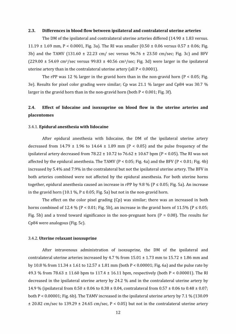
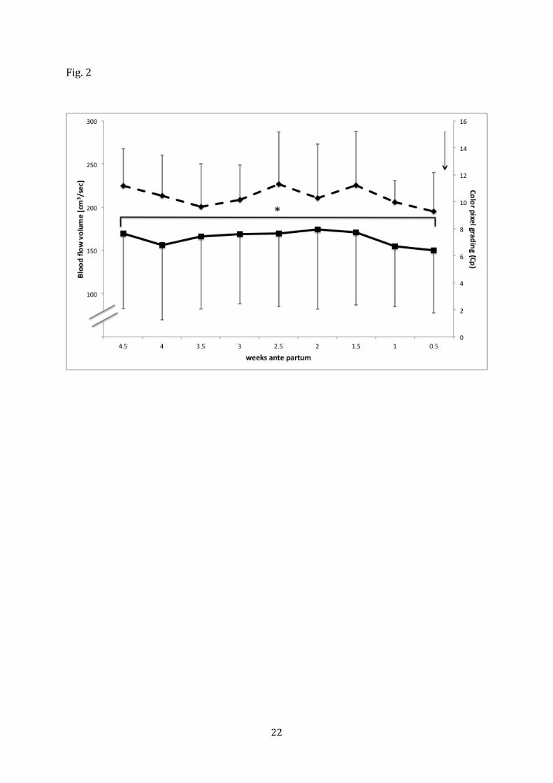
2.3. Differences in blood flow between ipsilateral and contralateral uterine arteries
The DM of the ipsilateral and contralateral uterine arteries differed (14.90 ± 1.83 versus.
11.19 ± 1.69 mm, P < 0.0001, Fig. 3a). The RI was smaller (0.50 ± 0.06 versus 0.57 ± 0.06; Fig.
3b) and the TAMV (131.60 ± 22.23 cm/ sec versus 96.76 ± 23.50 cm/sec; Fig. 3c) and BFV
(229.00 ± 54.69 cm3/sec versus 99.83 ± 40.56 cm3/sec; Fig. 3d) were larger in the ipsilateral
uterine artery than in the contralateral uterine artery (all P < 0.0001).
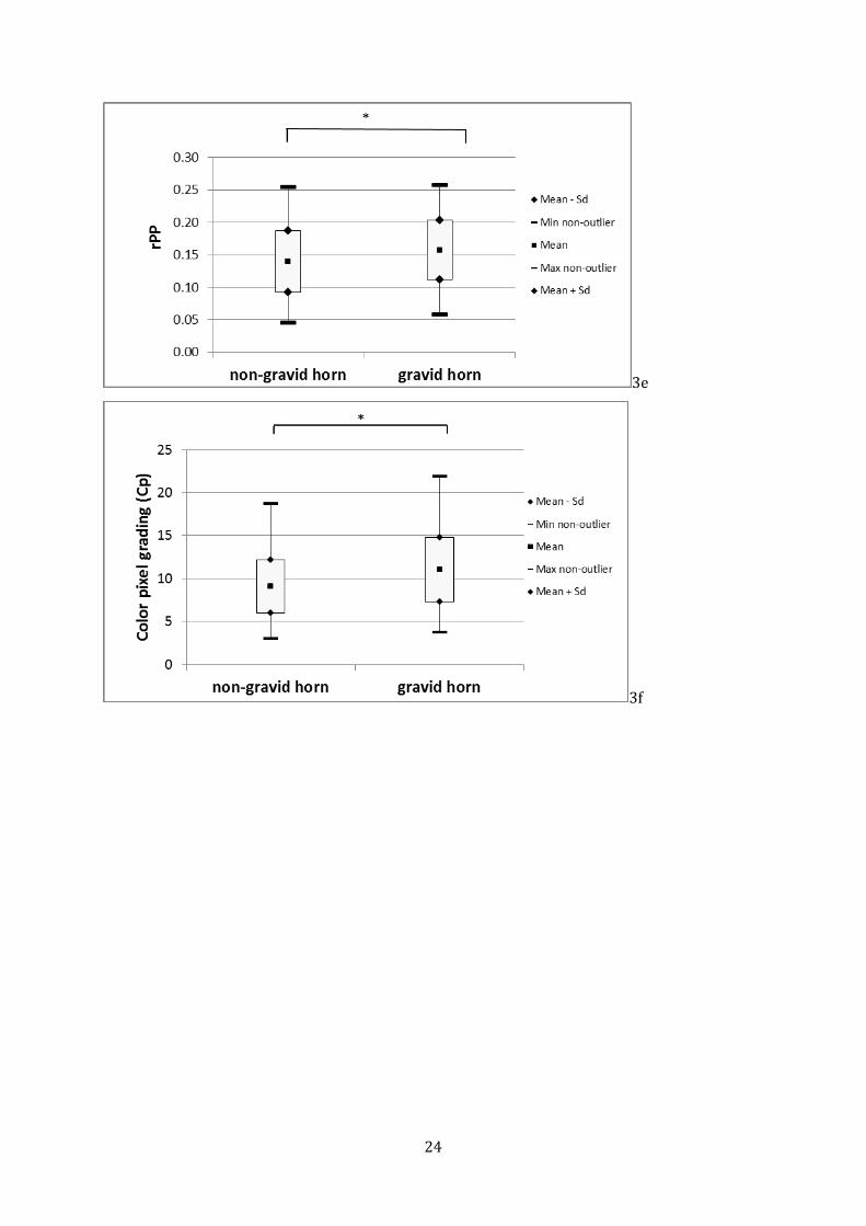
The rPP was 12 % larger in the gravid horn than in the non-gravid horn (P < 0.05; Fig.
3e). Results for pixel color grading were similar; Cp was 21.1 % larger and Cp84 was 30.7 %
larger in the gravid horn than in the non-gravid horn (both P < 0.001; Fig. 3f).
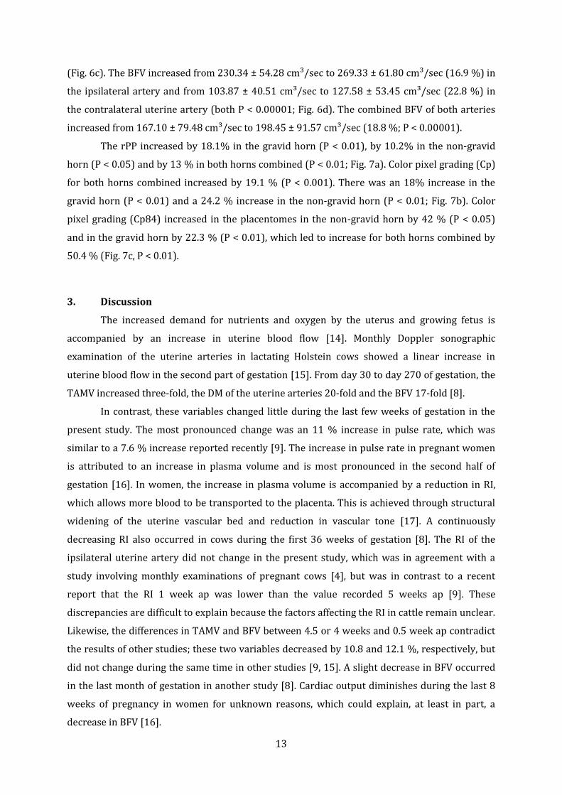
2.4. Effect of lidocaine and isoxsuprine on blood flow in the uterine arteries and
placentomes
3.4.1. Epidural anesthesia with lidocaine
After epidural anesthesia with lidocaine, the DM of the ipsilateral uterine artery
decreased from 14.79 ± 1.96 to 14.64 ± 1.89 mm (P < 0.05) and the pulse frequency of the
ipsilateral artery decreased from 78.22 ± 10.72 to 76.62 ± 10.67 bpm (P < 0.05). The RI was not
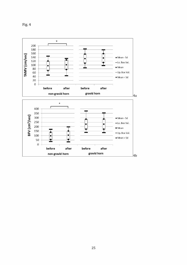
affected by the epidural anesthesia. The TAMV (P < 0.05; Fig. 4a) and the BFV (P < 0.01; Fig. 4b)
increased by 5.4% and 7.9% in the contralateral but not the ipsilateral uterine artery. The BFV in
both arteries combined were not affected by the epidural anesthesia. For both uterine horns
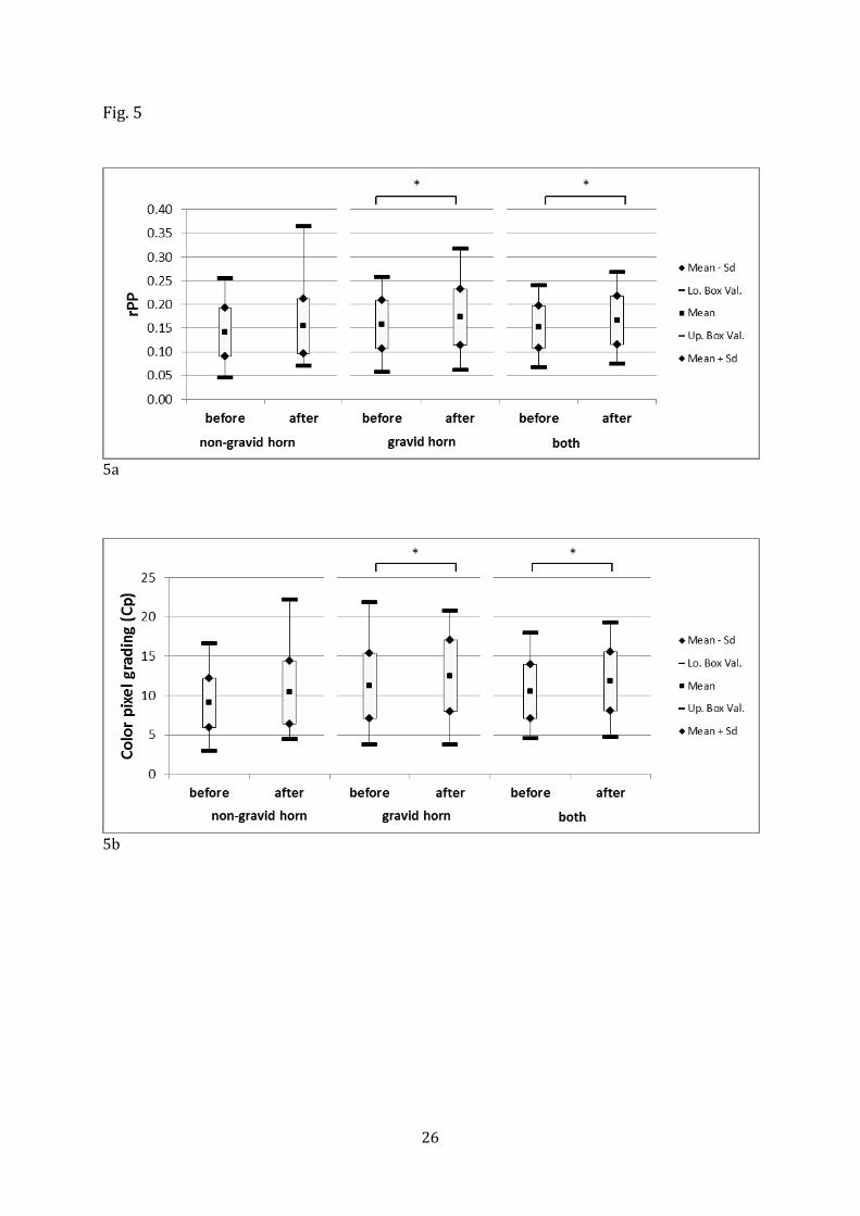
together, epidural anesthesia caused an increase in rPP by 9.8 % (P ≤ 0.05; Fig. 5a). An increase
in the gravid horn (10.1 %, P ≤ 0.05; Fig. 5a) but not in the non-gravid horn.
The effect on the color pixel grading (Cp) was similar; there was an increased in both
horns combined of 12.4 % (P < 0.01; Fig. 5b), an increase in the gravid horn of 11.5% (P ≤ 0.05;
Fig. 5b) and a trend toward significance in the non-pregnant horn (P = 0.08). The results for
Cp84 were analogous (Fig. 5c).
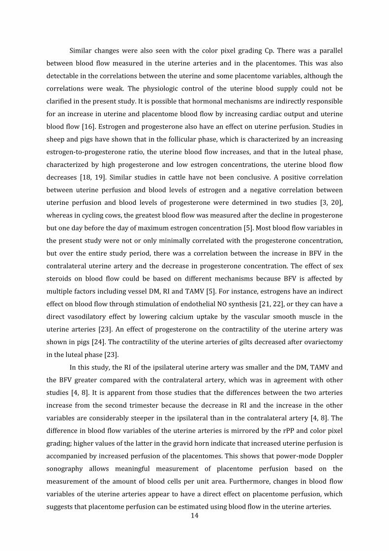
3.4.2. Uterine relaxant isoxsuprine
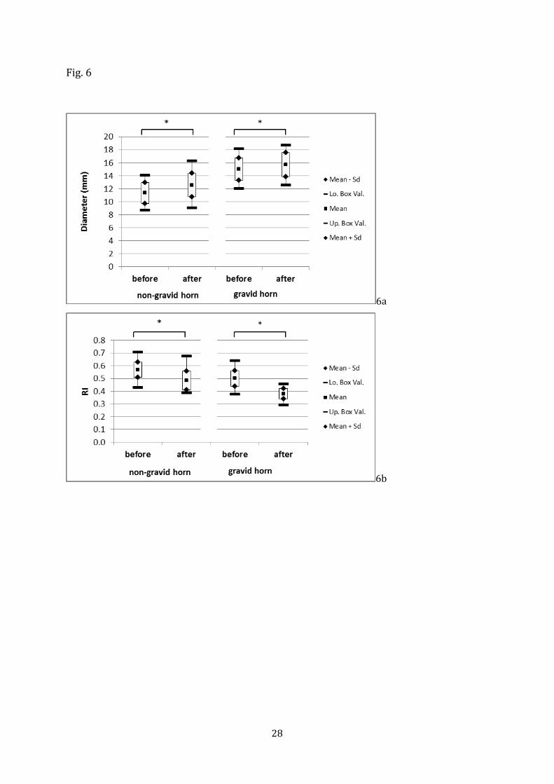
After intravenous administration of isoxsuprine, the DM of the ipsilateral and
contralateral uterine arteries increased by 4.7 % from 15.01 ± 1.73 mm to 15.72 ± 1.86 mm and
by 10.8 % from 11.34 ± 1.61 to 12.57 ± 1.81 mm (both P < 0.00001; Fig. 6a) and the pulse rate by
49.3 % from 78.63 ± 11.60 bpm to 117.4 ± 16.11 bpm, respectively (both P < 0.00001). The RI
decreased in the ipsilateral uterine artery by 24.2 % and in the contralateral uterine artery by
14.9 % (ipsilateral from 0.50 ± 0.06 to 0.38 ± 0.04, contralateral from 0.57 ± 0.06 to 0.48 ± 0.07;
both P < 0.00001; Fig. 6b). The TAMV increased in the ipsilateral uterine artery by 7.1 % (130.09
± 20.82 cm/sec to 139.29 ± 24.65 cm/sec, P < 0.05) but not in the contralateral uterine artery
13
(Fig. 6c). The BFV increased from 230.34 ± 54.28 cm³/sec to 269.33 ± 61.80 cm³/sec (16.9 %) in
the ipsilateral artery and from 103.87 ± 40.51 cm³/sec to 127.58 ± 53.45 cm³/sec (22.8 %) in
the contralateral uterine artery (both P < 0.00001; Fig. 6d). The combined BFV of both arteries
increased from 167.10 ± 79.48 cm³/sec to 198.45 ± 91.57 cm³/sec (18.8 %; P < 0.00001).
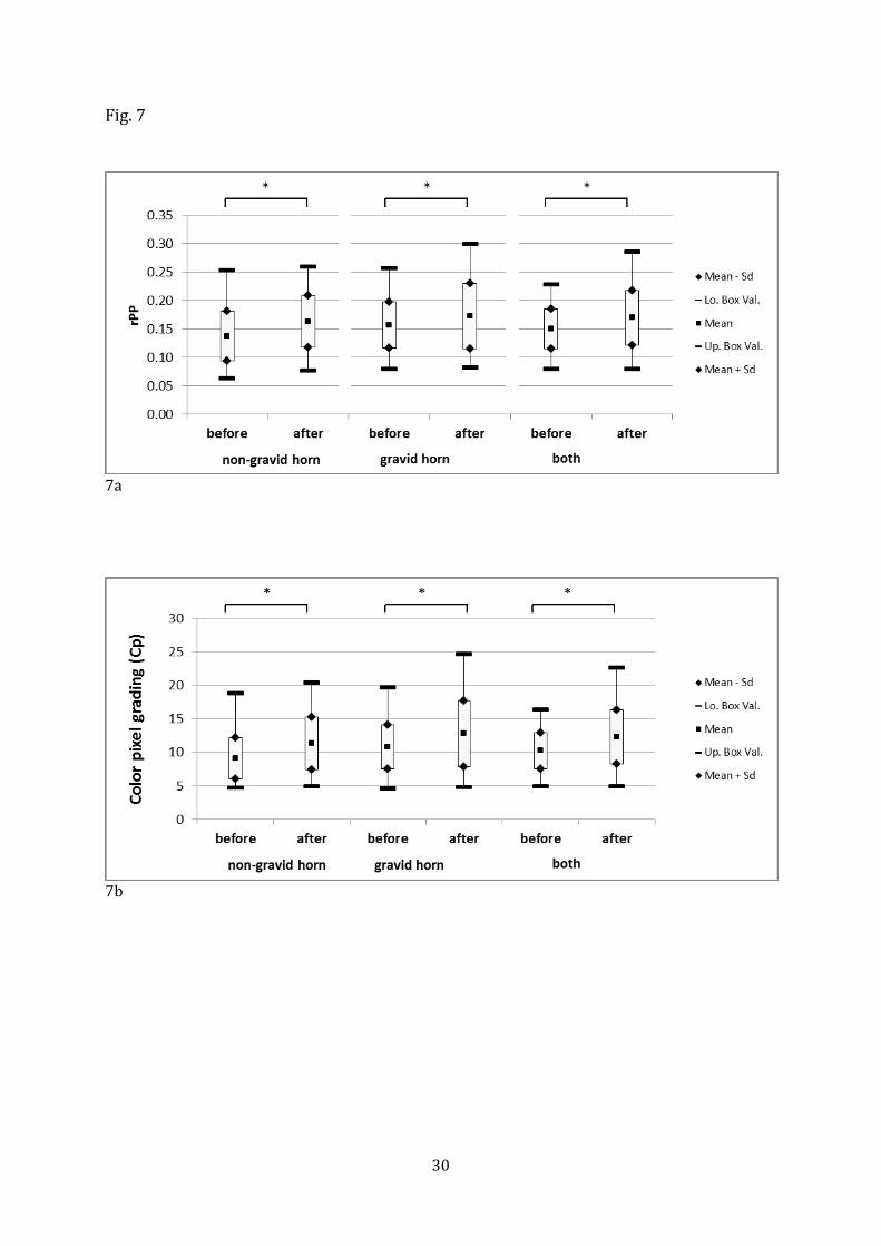
The rPP increased by 18.1% in the gravid horn (P < 0.01), by 10.2% in the non-gravid
horn (P < 0.05) and by 13 % in both horns combined (P < 0.01; Fig. 7a). Color pixel grading (Cp)
for both horns combined increased by 19.1 % (P < 0.001). There was an 18% increase in the
gravid horn (P < 0.01) and a 24.2 % increase in the non-gravid horn (P < 0.01; Fig. 7b). Color
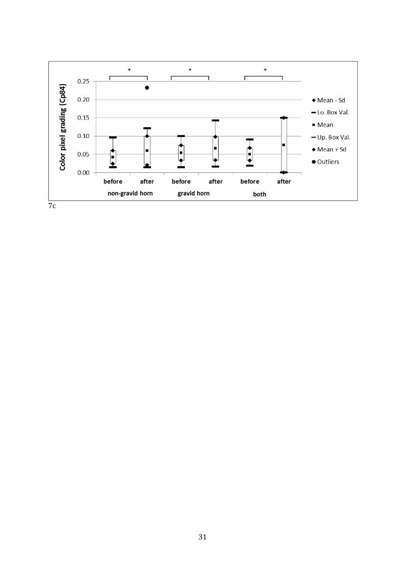
pixel grading (Cp84) increased in the placentomes in the non-gravid horn by 42 % (P < 0.05)
and in the gravid horn by 22.3 % (P < 0.01), which led to increase for both horns combined by
50.4 % (Fig. 7c, P < 0.01).
3. Discussion
The increased demand for nutrients and oxygen by the uterus and growing fetus is
accompanied by an increase in uterine blood flow [14]. Monthly Doppler sonographic
examination of the uterine arteries in lactating Holstein cows showed a linear increase in
uterine blood flow in the second part of gestation [15]. From day 30 to day 270 of gestation, the
TAMV increased three-fold, the DM of the uterine arteries 20-fold and the BFV 17-fold [8].
In contrast, these variables changed little during the last few weeks of gestation in the
present study. The most pronounced change was an 11 % increase in pulse rate, which was
similar to a 7.6 % increase reported recently [9]. The increase in pulse rate in pregnant women
is attributed to an increase in plasma volume and is most pronounced in the second half of
gestation [16]. In women, the increase in plasma volume is accompanied by a reduction in RI,
which allows more blood to be transported to the placenta. This is achieved through structural
widening of the uterine vascular bed and reduction in vascular tone [17]. A continuously
decreasing RI also occurred in cows during the first 36 weeks of gestation [8]. The RI of the
ipsilateral uterine artery did not change in the present study, which was in agreement with a
study involving monthly examinations of pregnant cows [4], but was in contrast to a recent
report that the RI 1 week ap was lower than the value recorded 5 weeks ap [9]. These
discrepancies are difficult to explain because the factors affecting the RI in cattle remain unclear.
Likewise, the differences in TAMV and BFV between 4.5 or 4 weeks and 0.5 week ap contradict
the results of other studies; these two variables decreased by 10.8 and 12.1 %, respectively, but
did not change during the same time in other studies [9, 15]. A slight decrease in BFV occurred
in the last month of gestation in another study [8]. Cardiac output diminishes during the last 8
weeks of pregnancy in women for unknown reasons, which could explain, at least in part, a
decrease in BFV [16].
14
Similar changes were also seen with the color pixel grading Cp. There was a parallel
between blood flow measured in the uterine arteries and in the placentomes. This was also
detectable in the correlations between the uterine and some placentome variables, although the
correlations were weak. The physiologic control of the uterine blood supply could not be
clarified in the present study. It is possible that hormonal mechanisms are indirectly responsible
for an increase in uterine and placentome blood flow by increasing cardiac output and uterine
blood flow [16]. Estrogen and progesterone also have an effect on uterine perfusion. Studies in
sheep and pigs have shown that in the follicular phase, which is characterized by an increasing
estrogen-to-progesterone ratio, the uterine blood flow increases, and that in the luteal phase,
characterized by high progesterone and low estrogen concentrations, the uterine blood flow
decreases [18, 19]. Similar studies in cattle have not been conclusive. A positive correlation
between uterine perfusion and blood levels of estrogen and a negative correlation between
uterine perfusion and blood levels of progesterone were determined in two studies [3, 20],
whereas in cycling cows, the greatest blood flow was measured after the decline in progesterone
but one day before the day of maximum estrogen concentration [5]. Most blood flow variables in
the present study were not or only minimally correlated with the progesterone concentration,
but over the entire study period, there was a correlation between the increase in BFV in the
contralateral uterine artery and the decrease in progesterone concentration. The effect of sex
steroids on blood flow could be based on different mechanisms because BFV is affected by
multiple factors including vessel DM, RI and TAMV [5]. For instance, estrogens have an indirect
effect on blood flow through stimulation of endothelial NO synthesis [21, 22], or they can have a
direct vasodilatory effect by lowering calcium uptake by the vascular smooth muscle in the
uterine arteries [23]. An effect of progesterone on the contractility of the uterine artery was
shown in pigs [24]. The contractility of the uterine arteries of gilts decreased after ovariectomy
in the luteal phase [23].
In this study, the RI of the ipsilateral uterine artery was smaller and the DM, TAMV and
the BFV greater compared with the contralateral artery, which was in agreement with other
studies [4, 8]. It is apparent from those studies that the differences between the two arteries
increase from the second trimester because the decrease in RI and the increase in the other
variables are considerably steeper in the ipsilateral than in the contralateral artery [4, 8]. The
difference in blood flow variables of the uterine arteries is mirrored by the rPP and color pixel
grading; higher values of the latter in the gravid horn indicate that increased uterine perfusion is
accompanied by increased perfusion of the placentomes. This shows that power-mode Doppler
sonography allows meaningful measurement of placentome perfusion based on the
measurement of the amount of blood cells per unit area. Furthermore, changes in blood flow
variables of the uterine arteries appear to have a direct effect on placentome perfusion, which
suggests that placentome perfusion can be estimated using blood flow in the uterine arteries.
15
Drugs that are often used in bovine obstetrics may affect the perfusion of the uterine and
umbilical arteries [9]. Epidural anesthesia with 2% lidocaine is commonly used in cows to
suppress the Ferguson reflex during treatment of dystocia, and also is used in protracted labor
in women to improve oxygenation of the fetus [12]. Uterine perfusion of cows is affected by
epidural anesthesia in several ways [9]. The pulse rate of the contralateral uterine artery, but
not the ipsilateral artery, was reduced after epidural anesthesia, but this difference was thought
to be due to the successive rather than concurrent measuring protocol. In the present study, a
decrease in pulse rate was observed only in the ipsilateral uterine artery. Epidural anesthesia
most likely reduces stress caused by the examination, which in turn results in a lower pulse rate
[9].
In contrast to a recent study from our clinic [9], we only detected an increase in TAMV
and BFV in the contralateral but not ipsilateral uterine artery; the reason for this is not clear.
The BFV of the contralateral uterine artery is considerably smaller and therefore the unilateral
increase did not translate into an overall increase in uterine BFV. Sample sizes were relatively
small in both studies and it is therefore possible that large individual variation, which is
common in cycling cows [5], was responsible for the discrepancy.
In agreement with another study [9], epidural anesthesia did not affect the RI. Studies in
human obstetrics generated inconsistent results concerning the effect of epidural anesthesia on
RI but most did not show an effect [25-29]. Despite the unchanged RI, the rPP and
measurements of color pixel grading Cp and Cp84 increased significantly in the gravid horn,
whereas in the non-gravid horn, there was a smaller increase with a trend toward significance
albeit accompanied by increased TAMV and BFV. This may reflect a weakness of the study
because only one placentome was examined in the non-gravid horn. Because the BFV did not
increase in the ipsilateral uterine artery, presumably because of the decreasing pulse rate and
the smaller DM compared to the pre-treatment values, a reduction in vascular resistance in the
placentomes is a reasonable explanation for the measured increase in perfusion. However, the
RI, defined to reflect this reduction, is an unreliable measure [25]; in the case of proportional
unidirectional changes in end-diastolic blood flow velocity and peak systolic velocity, which are
used to calculate the RI, this variable does not indicate a change.
Isoxsuprine is a β sympathomimetic drug and binds to β2 receptors, which are abundant
in the myometrium at the time of parturition, causing relaxation of the smooth musculature [30,
31]. It also has positive chronotropic and inotropic effects and causes peripheral vasodilation
[30]. There was an increase in the pulse rate by 50 %, which was similar to the effect seen in
another study [9], and we suspected that the increase in BFV by 19 % was due, at least in part, to
the inotropic properties of isoxsuprine. Unlike an earlier study, in which the increase in BFV was
limited to the contralateral uterine artery [9], we observed an increase in both arteries, albeit
larger in the contralateral artery. This could have been due to the vasodilatory effect of
16
isoxsuprine; the DM of the ipsilateral and contralateral uterine arteries increased by 5 % and 11
%, respectively. The unilateral increase in BFV in the contralateral artery in the former study
also was accompanied by unilateral increase in DM in that vessel [9]. Individual variations
related to vascular fibrosis from previous pregnancies have been described in cycling cows and
could explain these differences [5]. The cows used in our study varied considerably in the
number of previous pregnancies and with increasing age the blood flow in the placentomes
decreased. However, RI and TAMV in the uterine arteries did not show any correlation to the age
of the cows and BFV even increased with increasing age. The TAMV did not increase in the
contralateral uterine artery as would have been expected based on the positive inotropic effect
of isoxsuprine. This finding was similar to the results of another study, in which there even was
a decrease in TAMV of the contralateral uterine artery [9]. According to the continuity equation,
the decrease in TAMV can be explained by the larger increase in DM in the contralateral artery.
The decrease in RI by 24 % in the ipsilateral uterine artery and by 15 % in the
contralateral uterine artery also can be attributed to the vasodilatory effect of isoxsuprine
because the RI reflects the vascular resistance distal to the point of measurement. These results
agree with previously published reductions in RI by 20 % and 11 %, respectively [8]. Similar
findings were reported in pregnant women [32, 33].
An increase in placental perfusion after the administration of isoxsuprine in cows has
been suspected based on the increase in BFV and reduction in RI in the uterine arteries [9,12].
This could be confirmed in the present study because direct measurement showed an increase
in the perfused area of the placentomes in the gravid and non-gravid uterine horns. Thus,
isoxsuprine has a direct positive effect on placental perfusion. So-called intrauterine
resuscitative measures are used in human medicine to combat fetal hypoxia and improve fetal
oxygen supply [12]. One such measure is the use of uterine relaxant drugs to reduce excessive
uterine contractions and to improve uterine perfusion. In cows, it is also known that uterine
contractions are accompanied by a lowered blood supply to the fetus [34], and it is therefore
plausible that isoxsuprine given during dystocia improves placental perfusion through the
elimination of contractions as well as its direct effect on perfusion of placentomes. This could be
advantageous in delayed birth, for instance when a cow is prepared for Caesarean section [12].
However, isoxsuprine also has positive chronotropic and inotropic effects in the fetus [9], which
could increase fetal oxygen consumption and further compromise an already hypoxic fetus.
Epidural anesthesia might be preferable in these situations; although there was no improvement
in uterine perfusion in contrast to an earlier study [9], perfusion of placentomes was
nevertheless increased. This means that the advantages provided by isoxsuprine could also be
achieved with epidural anesthesia without the risk of increasing fetal heart rate [9]. When the
discussed limitations are taken into account, isoxsuprine as well as epidural anesthesia seem
suitable for improving placental perfusion in cows in advanced pregnancy. This conclusion is
17
based on results from a novel approach of direct assessment of placental perfusion. The
determination of the rPP and the amount of moving cells in the blood (Cp) were useful for the
direct assessment of placental perfusion. The selective measurement of the relative area with a
very large amount of moving cells in the blood (Cp 84) did not appear to provide an advantage
for assessing placental perfusion. The direct measurement of placental perfusion using power-
mode sonography makes it possible to assess the effects of pharmacological measures to
improve fetal well-being during parturition.
Acknowledgements
We thank Marco Hutter, ETH Zürich, Institute of Robotics and Intelligent Systems for developing
the software for quantitation of blood flow in placentomes and Dr. med. vet. Penelope A. Baloi,
Resident ECVDI, Division of Diagnostic Imaging, Vetsuisse Faculty, University of Zurich, for
technical support.
References
[1] Ferrell CL, Ford SP. Blood flow steroid secretion and nutrient uptake of the gravid bovine
uterus. J of Anim Sci. 1980;50:1113-21.
[2] Ford SP, Chenault JR. Blood flow to the corpus luteum-bearing ovary and ipsilateral uterine
horn of cows during the oestrous cycle and early pregnancy. J Reprod Fertil. 1981;62:555-62.
[3] Waite LR, Ford SP, Young DF, Conley AJ. Use of ultrasonic Doppler waveforms to estimate
changes in uterine artery blood flow and vessel compliance. J Anim Sci. 1990;68:2450-8.
[4] Bollwein H, Baumgartner U, Stolla R. Transrectal Doppler sonography of uterine blood flow
in cows during pregnancy. Theriogenology. 2002;57:2053-61.
[5] Bollwein H, Meyer HH, Maierl J, Weber F, Baumgartner U, Stolla R. Transrectal Doppler
sonography of uterine blood flow. Theriogenology. 2000;53:1541-52.
[6] Bollwein H, Maierl J, Mayer R, Stolla R. Transrectal color Doppler sonography of the A.
uterina in cyclic mares. Theriogenology. 1998;49:1483-8.
[7] Dzięcioł M, Stańczyk E, Noszczyk-Nowak A, Michlik K, Kozdrowski R, Niżański W, et al. The
influence of Sildenafil citrate on uterine tissue perfusion and the cardiovascular system during
the luteal phase of the ovarian cycle in cows. Acta Histochemica. 2014;116:377-81.
[8] Panarace M, Garnil C, Marfil M, Jauregui G, Lagioia J, Luther E, et al. Transrectal Doppler
sonography for evaluation of uterine blood flow throughout pregnancy in 13 cows.
Theriogenology. 2006;66:2113-9.
18
[9] Waldvogel D, Bleul U. Effect of xylazine, isoxsuprine, and lidocaine on Doppler sonographic
uterine and umbilical blood flow measurements in cows during the last month of pregnancy.
Theriogenology. 2014;81:993-1003.
[10] Bleul U, Lejeune B, Schwantag S, Kahn W. Ultrasonic transit-time measurement of blood
flow in the umbilical arteries and veins in the bovine fetus during stage II of labor.
Theriogenology. 2007;67:1123-33.
[11] Bude RO, Rubin JM. Power Doppler sonography. Radiology. 1996;200:21-3.
[12] Garite TJ, Simpson KR. Intrauterine resuscitation during labor. Clin Obstet Gynecol.
2011;54:28-39.
[13] Luttgenau J, Beindorff N, Ulbrich SE, Kastelic JP, Bollwein H. Low plasma progesterone
concentrations are accompanied by reduced luteal blood flow and increased size of the
dominant follicle in dairy cows. Theriogenology. 2011;76:12-22.
[14] Satterfield MC, Bazer FW, Spencer TE, Wu G. Sildenafil Citrate Treatment Enhances Amino
Acid Availability in the Conceptus and Fetal Growth in an Ovine Model of Intrauterine Growth
Restriction. The Journal of Nutrition. 2010;140:251-8.
[15] Herzog K, Koerte J, Flachowsky G, Bollwein H. Variability of uterine blood flow in lactating
cows during the second half of gestation. Theriogenology. 2011;75:1688-94.
[16] Hall Ga. Textbook of Medical Physiology (11 ed). Philadelphia: Saunders; 2005. p. 1034.
[17] Thornburg KL, Jacobson SL, Giraud GD, Morton MJ. Hemodynamic changes in pregnancy.
Semin Perinatol. 2000;24:11-4.
[18] Ford SP. Control of uterine and ovarian blood flow throughout the estrous cycle and
pregnancy of ewes, sows and cows. J Anim Sci. 1982;55 Suppl 2:32-42.
[19] Rupnow HL, Phernetton TM, Shaw CE, Modrick ML, Bird IM, Magness RR. Endothelial
vasodilator production by uterine and systemic arteries. VII. Estrogen and progesterone effects
on eNOS. Am J Physiol-Heart C. 2001;280:H1699-705.
[20] Ford SP, Chenault JR, Echternkamp SE. Uterine blood flow of cows during the oestrous cycle
and early pregnancy: effect of the conceptus on the uterine blood supply. J Reprod Fertil.
1979;56:53-62.
[21] Magness RR, Rosenfeld CR. Local and systemic estradiol-17 beta: effects on uterine and
systemic vasodilation. Am J Physiol. 1989;256:E536-42.
[22] Kim KH, Bender JR. Rapid, estrogen receptor-mediated signaling: why is the endothelium so
special? Science Signaling. 2005:pe28.
[23] Stice SL, Ford SP, Rosazza JP, Van Orden DE. Role of 4-hydroxylated estradiol in reducing
Ca2+ uptake of uterine arterial smooth muscle cells through potential-sensitive channels. Biol
Reprod. 1987;36:361-8.
19
[24] Ford SP, Reynolds LP, Farley DB, Bhatnagar RK, Van Orden DE. Interaction of ovarian
steroids and periarterial alpha 1-adrenergic receptors in altering uterine blood flow during the
estrous cycle of gilts. Am J Obstet Gynecol. 1984;150:480-4.
[25] Valentin M, Ducarme G, Ceccaldi PF, Bougeois B, Luton D. Uterine artery, umbilical, and fetal
cerebral Doppler velocities after epidural analgesia during labor. Int J Gynecol Obstet.
2012;118:145-8.
[26] Hughes AB, Devoe LD, Wakefield ML, Metheny WP. The effects of epidural anesthesia on the
Doppler velocimetry of umbilical and uterine arteries in normal term labor. Obstet Gynecol.
1990;75:809-12.
[27] Patton DE, Lee W, Miller J, Jones M. Maternal, uteroplacental, and fetoplacental
hemodynamic and Doppler velocimetric changes during epidural anesthesia in normal labor.
Obstet Gynecol. 1991;77:17-9.
[28] Alahuhta S, Rasanen J, Jouppila P, Jouppila R, Hollmen AI. Epidural sufentanil and
bupivacaine for labor analgesia and Doppler velocimetry of the umbilical and uterine arteries.
Anesthesiology. 1993;78:231-6.
[29] Ramos-Santos E, Devoe LD, Wakefield ML, Sherline DM, Metheny WP. The effects of
epidural anesthesia on the Doppler velocimetry of umbilical and uterine arteries in normal and
hypertensive patients during active term labor. Obstet Gynecol. 1991;77:20-6.
[30] Plumb D. Veterinary drug handbook. Pharma Vet Publishing; White Bear Lake. 1999. 113-4.
[31] Windholz M. Budavari S BR, Otterbein E. The Merck Index. Merck, Rahway, NJ. 1983. 2194.
[32] Deutinger J, Rudelstorfer R, Pattermann A, Bernaschek G. Vaginosonographic velocimetry in
uterine arteries before and after administration of beta-mimetics. Brit J Obstet Gynaecol.
1992;99:417-21.
[33] Wright JW, Patterson RM, Ridgway LE, 3rd, Berkus MD. Effect of tocolytic agents on fetal
umbilical velocimetry. Am J Obstet Gynecol. 1990;163:748-50.
[34] Bleul U, Schwantag S, Kahn W. Blood gas analysis of bovine fetal capillary blood during
stage II labor. Theriogenology. 2008;69:245-51.
20
Table 1
age
RI TAMV BFV
BFV BFV = 4.594*age + 120.2
r2=0.052; P < 0.01
BFV = -304.6*RI + 327.8
r2=0.069; P < 0.01
BFV = 0.5298*TAMV + 103.9
r2=0.036; P < 0.001
rPP rPP = -0.2599*age + 14.11
r2=0.061; P < 0.01
rPP = -12.21*RI + 18.15
r2=0.04; P < 0.05
rPP = 0.0437*TAMV + 6.617
r2=0.088; P < 0.001
n. s.
CP Cp = -0.0006729*age + 0.02072
r2=0.067; P < 0.01
n. s. Cp = 0.0000833*TAMV + 0.004727
r2=0.088; P < 0.001
n. s.
21
Fig. 1
22
Fig. 2
23
Fig. 3
3a
3b
3c
3d
24
3e
3f
25
Fig. 4
4a
4b
26
Fig. 5
5a
5b
27
5c
28
Fig. 6
6a
6b

29
6c
6d
30
Fig. 7
7a
7b
31
7c
32
Legends to the Tables and Figures
Table 1: Correlation equation and coefficient of linear comparison of age and blood flow
variables measured in the uterine arteries using Color Doppler sonography and placentomes
using power-mode Doppler sonography in 8 cows, which ranged in age from 4 to 14 years.
Fig. 1: Placentome (1) imaged in power Doppler mode. The perfused portion appears in color
(2) and is demarcated from the rest of the placentome. (3) Scale used to determine the RGB
values.
Fig. 2: Mean and standard deviation of blood flow volume of the uterine arteries (■) and the
color pixel grading in the placentomes (♦) in 8 cows in the last month of gestation. The arrow
indicates parturition.
* indicates significant difference of blood flow volume of the uterine arteries between 4.5 and
0.5 weeks ante partum.
Fig. 3 a-f: Box and whisker plots of DM, RI, TAMV, BFV, relative placentome perfusion (rPP) and
of the color pixel grading (Cp) of the ipsilateral and contralateral uterine arteries in 8 cows in
the last month of gestation.
* indicates significant difference.
Fig. 4 a-b: Box and whisker plots of the TAMV and BFV in the contralateral and ipsilateral
uterine arteries before and after epidural anesthesia with lidocaine in 8 cows in the last month
of gestation.
* indicates a significant difference.
Fig. 5 a-c: Box and whisker plots of the relative placentome perfusion (rPP), color pixel grading
(Cp) and color pixel grading from orange to yellow (Cp84) in the non-gravid and gravid uterine
horns and for both horns combined before and after epidural anesthesia in 8 cows in the last
month of gestation.
* indicates a significant difference.
Fig. 6 a-d: Box and whisker plots of the DM, RI, TAMV and BFV in the ipsilateral and
contralateral uterine arteries before and after administration of isoxsuprine in 8 cows in the last
month of gestation.
* indicates a significant difference.

33
Fig. 7 a-c: Box and whisker plots of the relative placentome perfusion (rPP), color pixel grading
(Cp) and color pixel grading from orange to yellow (Cp84) in the non-gravid and gravid horns
and both horns combined before and after the administration of isoxsuprine in 8 cows in the last
month of gestation.
* indicates a significant difference.

Danksagung
An dieser Stelle möchte ich mich bei allen, die zum Gelingen dieser Arbeit beigetragen haben,
ganz herzlich danken, insbesondere
Herrn Prof. Dr. U. Bleul für die Überlassung des Themas, die Übernahme des Referats, die stets
grosse Hilfsbereitschaft und die unzähligen Ratschläge und Anregungen bis zur Fertigstellung
der Arbeit.
Prof. Dr. M. Hässig und Prof. Dr. R. Bruckmaier für die Übernahme der Koautorenschaft.
Prof. Dr. Marco Hutter für die Erstellung des Auswertungsprogrammes.
Dr. Penelope Baloi für die Unterstützung bei ultrasonographischen Fragen.
Den Tierpflegerinnen und Tierpfleger des Departements für Nutztiere für die Betreuung der
Kühe.
Meinen Kollegen aus dem Departement für Nutztiere für ihre Unterstützung, ihr Interesse, ihre
wertvollen Tipps und Aufmunterungen.
Meinem Ehemann Stephan Kim, meiner Mutter Anna Egloff und meinen Freunden für ihre
Unterstützung, die ermutigenden Worte und ihren Glauben an mich.
Circulum Vitae
Vorname, Name Kim, Cornelia
Geburtsdatum 06. Mai 1982
Geburtsort Baden AG
Nationalität CH
Heimatort Wettingen, AG
1989-1994 Primarschule Wettingen
1994-1997 Sekundarschule Wettingen
1997-1999 Bezirksschule Wettingen
1999- 2003 Kantonsschule Baden,CH,
2. Juli 2003 Maturitätsabschluss, Kantonsschule Baden, CH
September 2003- Studium der Veterinärmedizin, Universität Zürich, CH
Oktober 2008
20.Oktober 2008 Abschlussprüfung vet.med., Universität Zürich, CH
Mai 2010- Anfertigung der Dissertation
November 2015 unter der Leitung von Prof. Dr. med. vet. U. Bleul
am Departement für Nutztiere
der Vetsuisse-Fakultät, Universität Zürich
Direktor: Prof. Dr. Dr. h.c. U. Braun)
Oktober 2008 – Arbeitsstelle in der Tierarztpraxis Rüdiger Endingen, AG
April 2010
Mai 2010- Assistenztierärztin am Departement für Nutztiere, Reproduktionsmedizin
September 2013
Seit März 2014 Teilhaberin Tierarztpraxis Aesch GmbH, Wettingen AG


![Synthetic PreImplantation Factor (PIF) prevents … pathway leads to activa-tion of the uterine and placental endothelium and the release of embryotoxic cytokines [9, 10]. We hypothesize](https://img.pdfslide.us/doc/110x75/5d3f20cc88c993860c8caa18/synthetic-preimplantation-factor-pif-prevents-pathway-leads-to-activa-tion-of.jpg)