Embed Size (px)
Citation preview
Donor Kidney Evaluation
Nasreen Mohamed, MD, FRCPAa, Lynn D. Cornell, MDb,*KEYWORDS
� Kidney transplant � Donor biopsy � Expanded criteria donor � Zero time biopsy
ABSTRACT
I n patients with end-stage renal disease, kidneytransplantation is the best means to extendsurvival and offer a better quality of life. The
current shortage of organs available for transplan-tation has led to an effort to expand the kidneydonor pool, including the use of nonideal donorkidneys. Assessment of the quality of the donatedkidney is essential, and would facilitate the deci-sion to transplant a potential organ or discard it.Multiple clinical and histologic parameters havebeen examined to evaluate the donor kidney andrelate the findings to the graft outcome, butclear-cut criteria are yet to be defined.
OVERVIEW
Kidney transplantation is the best choice of treat-ment for most patients with end-stage renal dis-ease. Patients who do not have an acceptableliving kidney donor may turn to the deceaseddonor waiting list. The shortage of organs availablefor donation with the growing waiting list has beena major challenge in the field. Clinicians started toturn their focus toward expanding the kidneydonor pool by using nonideal kidneys, and inrecent years, there has been an increasing use ofkidneys from older donors and from donors withcomorbidities, such as hypertension.1,2
Expandedcriteria donor (ECD) kidneys are gener-ally defined as those frompatients 60 years or older,or age 50 to 59 with 2 of 3 of the following factors:death due to cerebrovascular cause, history of hy-pertension, or serum creatinine greater than1.5 mg/dL (or creatinine clearance<60 mL/min).3
Kidneys retrieved fromECDsare at higher risk ofde-layed graft function and primary nonfunction, and
Disclosure and Conflict of Interest: None.a Department of Pathology and Laboratory Medicine, Kinbet Street-mbc035, PO Box 15215, Dammam 31444, Kingogy, Department of Laboratory Medicine and PathologyMN 55905, USA* Corresponding author.E-mail address: [email protected]
Surgical Pathology 7 (2014) 357–365http://dx.doi.org/10.1016/j.path.2014.04.0021875-9181/14/$ – see front matter � 2014 Elsevier Inc. All
have decreased graft survival compared with kid-neys from standard criteria donors (SCDs).4 ECDkidneys are typified by having reduced creatinineclearance with a higher percentage of scleroticglomeruli as a sequel to aging or other coexistingdiseases. The remaining viable nephrons might nothave thecapacity towithstandadditional stressdur-ing the transplantation procedure, nephrotoxicimmunosuppression, and requirement to functioninstead of 2 kidneys. Thus, the quality of the donororgan is a critically important matter, that is, thenumber of surviving nephrons having the ability towithstand the injury will determine the long-termfunction and survival. One study found that donorfactors accounted for 64% of the variability in allo-graft function at 6 months posttransplant.5,6 Thegreater risk of delayed graft function and poorlong-term function might compel the surgeon toerr to the side of discarding the organ.
The increasing evidence of reduced graft sur-vival rate from deceased donors necessitatesassessment of the quality of the donated kidneybefore transplantation. Many institutes have ac-quired the practice of biopsy evaluation beforetransplantation. Evaluation of the donor biopsycan help determine the functional reserve ofa donor kidney under consideration for trans-plantation and to improve both short-term andlong-term graft function. In the United States,approximately 75% of kidneys from ECDs arebiopsied and 41% of those are discardedbecause of the histopathologic characteristics.7
Azancot and colleagues showed poor reproduc-ibility of donor biopsy evaluation between on-call(non-renal) pathologists and renal pathologists.8
Recipient outcomes (one-year eGFR and deathcensored graft survival) correlated with scoresby the renal pathologists but not by the on-call
g Fahad Specialist Hospital-Dammam, Amer Bin Tha-dom of Saudi Arabia; b Division of Anatomic Pathol-, Mayo Clinic, 200 First Street Southwest, Rochester,
rights reserved. surgpath.th
eclinics.com
Mohamed & Cornell358
pathologists. In addition, a subset of discardedkidneys deemed unsuitable by the on-callpathologist was considered by the renal patholo-gist to be adequate for transplantation. These re-sults suggest that donor biopsies be evaluatedby a renal pathologist. One possibility toconsider is whole-slide imaging and evaluationby a renal pathologist on-call at a distant site, ifa local renal pathologist is not available, particu-larly as these biopsies are not used for primarypatient diagnosis.9 Although ECD kidneys arenot ideal, they can be used for elderly recipientsto offer a better quality of life and survival. Sur-geons might also consider using dual kidneytransplantation to improve the nephron massand increase the GFR in the recipient.
CLASSIFICATION OF DONORS
The deceased donor kidneys are classified into 2broad groups, reflecting the quality of the organand driven by the risk of graft loss: SCDs andECDs.4,10
ECD
A donor at the time of death is 60 years or older ora donor aged 50 to 59 years and has any of thefollowing 3 criteria: (1) cause of death is cerebro-vascular accident, (2) history of hypertension, (3)terminal serum creatinine greater than 1.5 mg/dL.3,10,11 Other categories of deceased donor kid-neys are donation after cardiac death (DCD) anddonation after brain death (DBD).
SCD
Any donor who does not fulfill the criteria of ECD.10
DBD
A donor who has primarily brain death with main-tained cardiac and respiratory circulation by med-ical measures. The donor can be SCD or ECD.10
DCD
DCD includes donors who do not fit the braindeath category but who have cardiac standstill orcessation of cardiac function before organ pro-curement. They can be divided into controlledDCD and uncontrolled DCD. Controlled DCD in-cludes donors whose life support will be with-drawn in the controlled environment of theoperating room. The donor hemodynamic and res-piratory functions are maintained. UncontrolledDCD includes candidates who die in the emer-gency room before consent is obtained for organ
donation and a catheter is placed in the femoralvessels and peritoneum to cool the organs. Italso includes candidates who consented for organdonation, but suffer a cardiac arrest requiring car-diopulmonary resuscitation during procurement oforgans.10
CLINICAL AND LABORATORY EVALUATION
OF DONORS
Clinical history and laboratory results of the poten-tial donor should be reviewed before consideringthat patient’s organs for donation. Patients with ahistory of diabetes, hypertension, and other condi-tions are considered as potential donors, as wellas patients with perimortal disseminated intravas-cular coagulation and acute tubular injury.1,12 Inpatients with traumatic death, a complete clinicalhistory may not be available, and so some centershave used donor age as a surrogate marker forrenal function and a guide to suitability of the organfor transplantation. We know that global glomeru-losclerosis and other chronic histopathologicchanges increase with age on the whole innormal-functioning kidneys,13,14 but there isconsiderable variability among individual kidneys,and so age alone is not a satisfactory factor.Even among healthy living donors, glomeruloscle-rosis occurs with increasing age that is not ex-plained by chronic kidney disease risk factors.13
The calculated donor glomerular filtration rate(GFR) may be a better clinical indicator of qualityof a kidney for transplantation than age alone;one study suggested a calculated donor GFR ofless than 85 mL per minute as an indicator ofpoor donor kidney quality.15 Some clinicianshave used a creatinine clearance of greater than60 to 70 mL per minute as the acceptable cutoffvalue for accepting organs for single kidney dona-tion, whereas others recommend dual kidneytransplantation with a lower creatinine clear-ance.16–18 Rao and colleagues19 put forth the Kid-ney Donor Profile Index (KDPI), which is a scorethat estimates the risk of graft failure and takesinto account donor age, race/ethnicity, heightand weight, history of hypertension and diabetes,serum creatinine, cerebrovascular cause of death,DCD, and hepatitis C status. KDRI values can helpallocate kidneys and determine whether to trans-plant a single kidney or 2 kidneys.20,21
DONOR BIOPSY ASSESSMENT
Renal biopsy is performed after procurement andbefore kidney transplantation, and serves as animportant tool for evaluation of the kidney, partic-ularly with ECD. In approximately 85% of potential
Donor Kidney Evaluation 359
donors older than 65, the organ undergoes histo-logic assessment pretransplantation.11 Approxi-mately 40% of ECD kidneys are discarded. Themain reason for kidneys to be discarded in theUnited States was the biopsy findings, as reportedto the United Network for Organ Sharing.11 Donorbiopsies may also be performed to evaluate for asuspected neoplasm. The donor biopsy, per-formed before transplantation for rapid evaluationof suitability of an organ for transplantation, is tobe distinguished from the implantation or “time-zero” biopsy. The time-zero biopsy is generallyperformed after perfusion of the transplanted graftand is done for the purpose of evaluating baselinechronic changes in a graft that is already deemedacceptable for transplantation.
The preimplantation biopsy may be a wedge bi-opsy or a needle-core biopsy. Most surgeons pre-fer wedge biopsies to reduce the risk of damaginglarge arteries and result in uncontrolled bleeding.22
The wedge biopsy, however, may overrepresentthe degree of global glomerulosclerosis, whichtends to affect disproportionally the subcapsularglomeruli in patients with vascular disease. Wedgebiopsies also may be superficial and not samplearteries, compared with needle-core biopsies,which are more likely to contain arteries.23
Urgent assessment of the donor biopsy isessential when the decision to transplant a kidneyis contingent with the extent of the chronicchanges, and so rapid evaluation is important.The biopsy tissue can undergo rapid processing,with a permanent section available within 2 to3 hours; the other most widely used alternative isfrozen section. The frozen section will provide
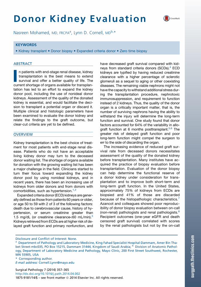
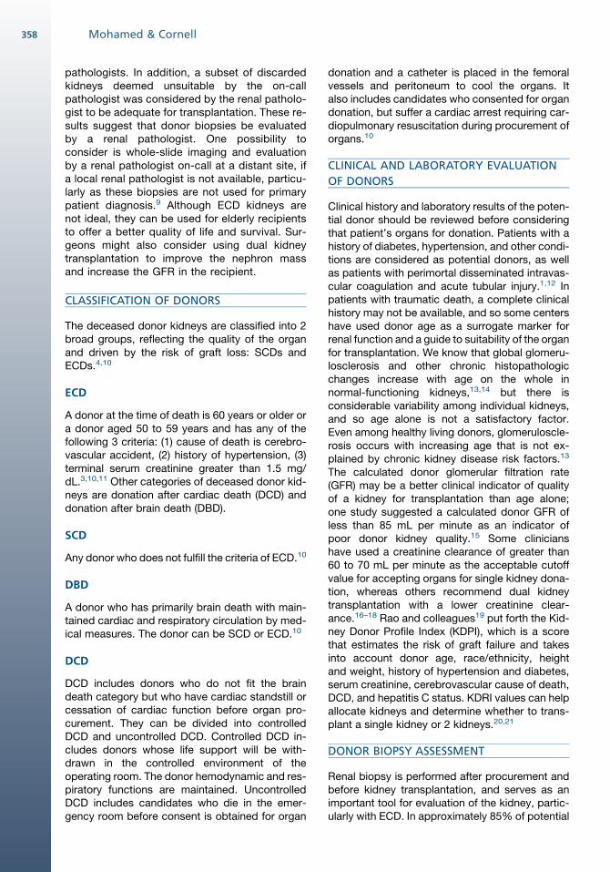
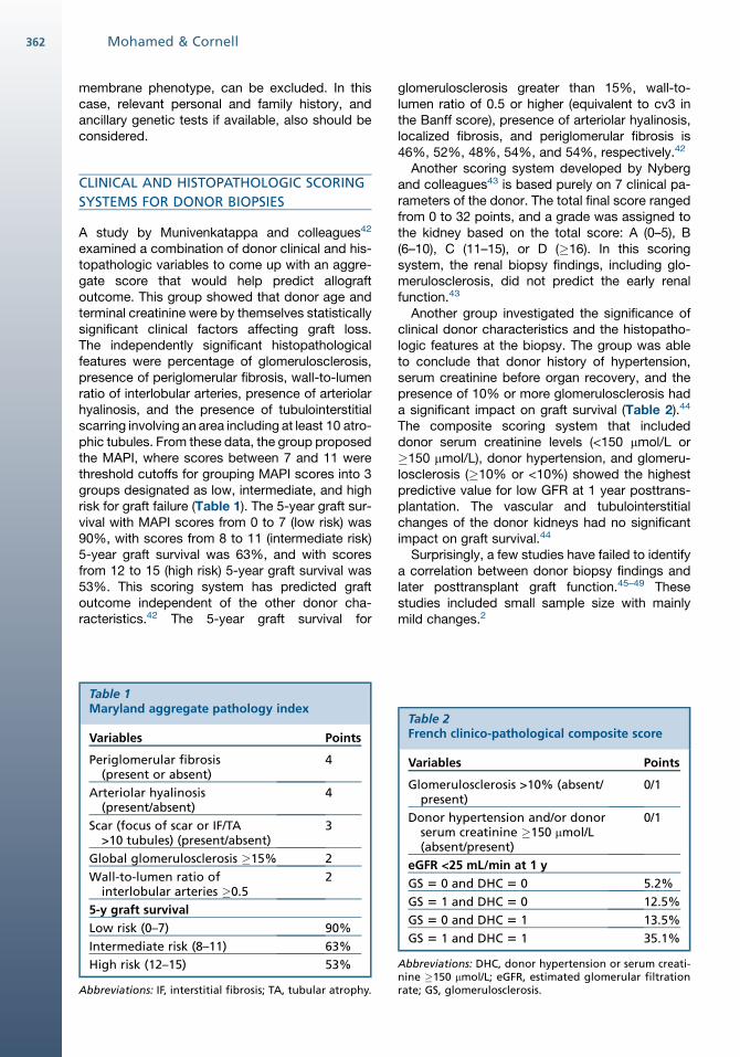
Fig. 1. Mild fibrous intimal thickening (arrow, left panel)particularly in older donors. There is focal interstitial fibrtubular epithelial flattening, a feature of acute tubularedema, shown here, is typically seen on frozen sections.frozen section (arrow, right panel) (Hematoxylin and eosmagnification �200, right).
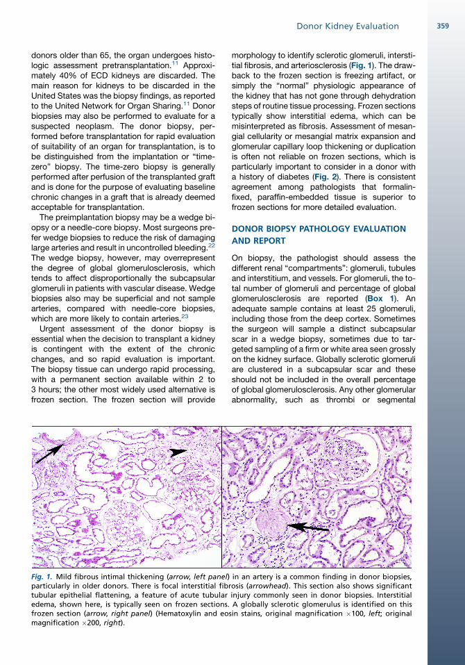
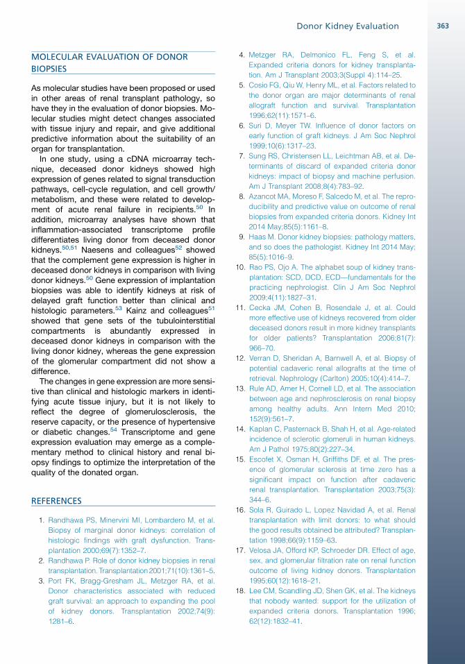
morphology to identify sclerotic glomeruli, intersti-tial fibrosis, and arteriosclerosis (Fig. 1). The draw-back to the frozen section is freezing artifact, orsimply the “normal” physiologic appearance ofthe kidney that has not gone through dehydrationsteps of routine tissue processing. Frozen sectionstypically show interstitial edema, which can bemisinterpreted as fibrosis. Assessment of mesan-gial cellularity or mesangial matrix expansion andglomerular capillary loop thickening or duplicationis often not reliable on frozen sections, which isparticularly important to consider in a donor witha history of diabetes (Fig. 2). There is consistentagreement among pathologists that formalin-fixed, paraffin-embedded tissue is superior tofrozen sections for more detailed evaluation.
DONOR BIOPSY PATHOLOGY EVALUATION
AND REPORT
On biopsy, the pathologist should assess thedifferent renal “compartments”: glomeruli, tubulesand interstitium, and vessels. For glomeruli, the to-tal number of glomeruli and percentage of globalglomerulosclerosis are reported (Box 1). Anadequate sample contains at least 25 glomeruli,including those from the deep cortex. Sometimesthe surgeon will sample a distinct subcapsularscar in a wedge biopsy, sometimes due to tar-geted sampling of a firm or white area seen grosslyon the kidney surface. Globally sclerotic glomeruliare clustered in a subcapsular scar and theseshould not be included in the overall percentageof global glomerulosclerosis. Any other glomerularabnormality, such as thrombi or segmental
in an artery is a common finding in donor biopsies,osis (arrowhead). This section also shows significantinjury commonly seen in donor biopsies. InterstitialA globally sclerotic glomerulus is identified on thisin stains, original magnification �100, left; original
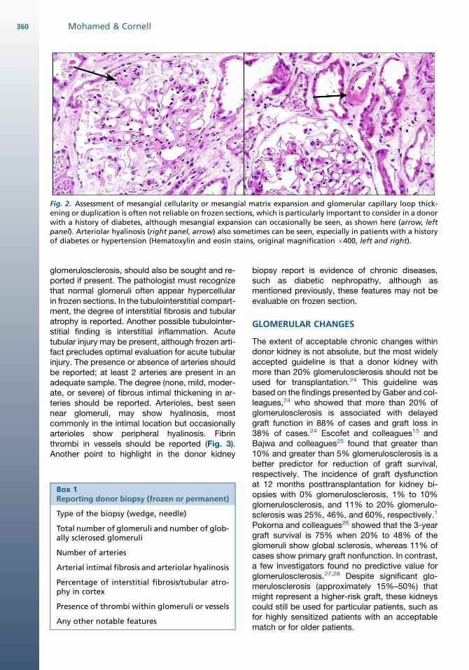
Fig. 2. Assessment of mesangial cellularity or mesangial matrix expansion and glomerular capillary loop thick-ening or duplication is often not reliable on frozen sections, which is particularly important to consider in a donorwith a history of diabetes, although mesangial expansion can occasionally be seen, as shown here (arrow, leftpanel). Arteriolar hyalinosis (right panel, arrow) also sometimes can be seen, especially in patients with a historyof diabetes or hypertension (Hematoxylin and eosin stains, original magnification �400, left and right).
Mohamed & Cornell360
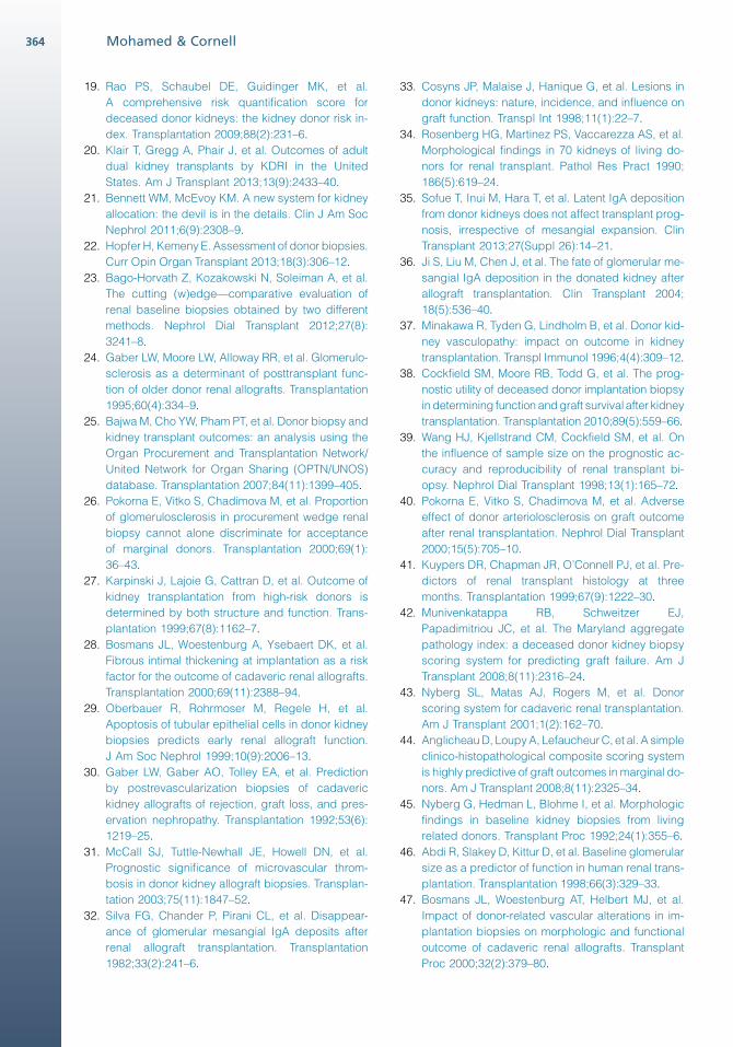
glomerulosclerosis, should also be sought and re-ported if present. The pathologist must recognizethat normal glomeruli often appear hypercellularin frozen sections. In the tubulointerstitial compart-ment, the degree of interstitial fibrosis and tubularatrophy is reported. Another possible tubulointer-stitial finding is interstitial inflammation. Acutetubular injury may be present, although frozen arti-fact precludes optimal evaluation for acute tubularinjury. The presence or absence of arteries shouldbe reported; at least 2 arteries are present in anadequate sample. The degree (none, mild, moder-ate, or severe) of fibrous intimal thickening in ar-teries should be reported. Arterioles, best seennear glomeruli, may show hyalinosis, mostcommonly in the intimal location but occasionallyarterioles show peripheral hyalinosis. Fibrinthrombi in vessels should be reported (Fig. 3).Another point to highlight in the donor kidney
Box 1Reporting donor biopsy (frozen or permanent)
Type of the biopsy (wedge, needle)
Total number of glomeruli and number of glob-ally sclerosed glomeruli
Number of arteries
Arterial intimal fibrosis and arteriolar hyalinosis
Percentage of interstitial fibrosis/tubular atro-phy in cortex
Presence of thrombi within glomeruli or vessels
Any other notable features
biopsy report is evidence of chronic diseases,such as diabetic nephropathy, although asmentioned previously, these features may not beevaluable on frozen section.
GLOMERULAR CHANGES
The extent of acceptable chronic changes withindonor kidney is not absolute, but the most widelyaccepted guideline is that a donor kidney withmore than 20% glomerulosclerosis should not beused for transplantation.24 This guideline wasbased on the findings presented by Gaber and col-leagues,24 who showed that more than 20% ofglomerulosclerosis is associated with delayedgraft function in 88% of cases and graft loss in38% of cases.24 Escofet and colleagues15 andBajwa and colleagues25 found that greater than10% and greater than 5% glomerulosclerosis is abetter predictor for reduction of graft survival,respectively. The incidence of graft dysfunctionat 12 months posttransplantation for kidney bi-opsies with 0% glomerulosclerosis, 1% to 10%glomerulosclerosis, and 11% to 20% glomerulo-sclerosis was 25%, 46%, and 60%, respectively.1
Pokorna and colleagues26 showed that the 3-yeargraft survival is 75% when 20% to 48% of theglomeruli show global sclerosis, whereas 11% ofcases show primary graft nonfunction. In contrast,a few investigators found no predictive value forglomerulosclerosis.27,28 Despite significant glo-merulosclerosis (approximately 15%–50%) thatmight represent a higher-risk graft, these kidneyscould still be used for particular patients, such asfor highly sensitized patients with an acceptablematch or for older patients.
Fig. 3. Thrombi are seen in a glomerulus (arrow, left panel) or in an artery (arrowhead, right panel) in these ex-amples (Hematoxylin and eosin, original magnification �400, left; periodic acid-Schiff, original magnification�200, right).
Donor Kidney Evaluation 361
The presence of scattered fibrin thrombi in a fewglomeruli does not contraindicate transplantation;these thrombi will likely dissolve by an intact fibri-nolytic system.29 Gaber and colleagues30 found noeffect on graft outcome in donor biopsies thatshowed thrombi in fewer than 50% of glomeruli.In another study, Pokorna and colleagues26 docu-mented fibrin thrombi within glomerular capillariesin 5 donor kidneys, which were transplanted into10 patients. In 33% there was a nonfunctioninggraft, 56% had delayed graft function, and 11%had immediate graft function. Although theglomerular thrombi did not affect graft survival,63% of recipients had delayed graft function.31
Occasionally, pretransplant or posttransplantbiopsies show donor-derived glomerulonephritis.The most frequent of these is immunoglobulin (Ig)A nephropathy or mesangial IgA-dominant de-posits,which tends to have no impact on graft func-tion and usually resolves with time.32 Indeed,IgA-dominant immune complex deposits havebeen reported in approximately 10% of normaldonor kidneys,33,34 and so IgA deposits in theabsence of clinically apparent diseaseormesangialhypercellularity may not represent the disease ofIgA nephropathy. A recent study concluded thateven donor biopsies that showed mesangial ex-pansion with IgA deposits did not affect the trans-plant prognosis.35 Subsequent biopsies of graftswith donor-derived IgA deposits show progressivelessening and eventual disappearance of the de-posits.36 Other documented glomerular lesions indonor kidneys include membranous glomerulone-phritis and focal segmental glomerulosclerosis.
VASCULAR CHANGES
Besides glomerulosclerosis, at least moderatearteriosclerosis (>25% luminal narrowing) is
another predictor of worse graft outcome and is acommon reason for discarding organs.2,12 Gener-ally, significant vascular changes have been asso-ciated with delayed graft function and the graftoutcome,27,28,37–39 although scoring arteries indonor biopsies might not be possible because oflimited sample size in most cases.40 Vascular pa-thology was independently associated with risk ofgraft failure in the Maryland Aggregate PathologyIndex (MAPI) study (see later in this article).
TUBULOINTERSTITIAL CHANGES
Acute tubular injury is often present in donor bi-opsies; studies have shown that histologic scoringof acute tubular necrosis26,41 and apoptosis29 pre-dicts delayed graft function. The value of interstitialfibrosis and tubular atrophy has not been exam-ined extensively, some suggested its adverse ef-fect on graft outcome,1 whereas others foundlittle value of scoring them.26
LIVING DONOR BIOPSIES
The presence of microscopic hematuria and/ormild proteinuria in a potential living donor warrantsfurther investigation, including consideration of arenal biopsy. Biopsy assessment, including useof immunofluorescence and electron microscopy,will aid in the decision to proceed with organdonation or not. Relevant pathology is generallyconsidered a contraindication for donation to pro-tect the donor from developing progressive kidneydisease aggravated further by reduced nephronmass. Kidneys with thin glomerular basementmembrane nephropathy may be considered fortransplantation, however, so long as Alportsyndrome, including carrier status of Alport syn-drome and Alport syndrome with a thin basement
Mohamed & Cornell362
membrane phenotype, can be excluded. In thiscase, relevant personal and family history, andancillary genetic tests if available, also should beconsidered.
CLINICAL AND HISTOPATHOLOGIC SCORING
SYSTEMS FOR DONOR BIOPSIES
A study by Munivenkatappa and colleagues42
examined a combination of donor clinical and his-topathologic variables to come up with an aggre-gate score that would help predict allograftoutcome. This group showed that donor age andterminal creatinine were by themselves statisticallysignificant clinical factors affecting graft loss.The independently significant histopathologicalfeatures were percentage of glomerulosclerosis,presence of periglomerular fibrosis, wall-to-lumenratio of interlobular arteries, presence of arteriolarhyalinosis, and the presence of tubulointerstitialscarring involving an area including at least 10 atro-phic tubules. From these data, the group proposedthe MAPI, where scores between 7 and 11 werethreshold cutoffs for grouping MAPI scores into 3groups designated as low, intermediate, and highrisk for graft failure (Table 1). The 5-year graft sur-vival with MAPI scores from 0 to 7 (low risk) was90%, with scores from 8 to 11 (intermediate risk)5-year graft survival was 63%, and with scoresfrom 12 to 15 (high risk) 5-year graft survival was53%. This scoring system has predicted graftoutcome independent of the other donor cha-racteristics.42 The 5-year graft survival for
Table 1Maryland aggregate pathology index
Variables Points
Periglomerular fibrosis(present or absent)
4
Arteriolar hyalinosis(present/absent)
4
Scar (focus of scar or IF/TA>10 tubules) (present/absent)
3
Global glomerulosclerosis �15% 2
Wall-to-lumen ratio ofinterlobular arteries �0.5
2
5-y graft survival
Low risk (0–7) 90%
Intermediate risk (8–11) 63%
High risk (12–15) 53%
Abbreviations: IF, interstitial fibrosis; TA, tubular atrophy.
glomerulosclerosis greater than 15%, wall-to-lumen ratio of 0.5 or higher (equivalent to cv3 inthe Banff score), presence of arteriolar hyalinosis,localized fibrosis, and periglomerular fibrosis is46%, 52%, 48%, 54%, and 54%, respectively.42
Another scoring system developed by Nybergand colleagues43 is based purely on 7 clinical pa-rameters of the donor. The total final score rangedfrom 0 to 32 points, and a grade was assigned tothe kidney based on the total score: A (0–5), B(6–10), C (11–15), or D (�16). In this scoringsystem, the renal biopsy findings, including glo-merulosclerosis, did not predict the early renalfunction.43
Another group investigated the significance ofclinical donor characteristics and the histopatho-logic features at the biopsy. The group was ableto conclude that donor history of hypertension,serum creatinine before organ recovery, and thepresence of 10% or more glomerulosclerosis hada significant impact on graft survival (Table 2).44
The composite scoring system that includeddonor serum creatinine levels (<150 mmol/L or�150 mmol/L), donor hypertension, and glomeru-losclerosis (�10% or <10%) showed the highestpredictive value for low GFR at 1 year posttrans-plantation. The vascular and tubulointerstitialchanges of the donor kidneys had no significantimpact on graft survival.44
Surprisingly, a few studies have failed to identifya correlation between donor biopsy findings andlater posttransplant graft function.45–49 Thesestudies included small sample size with mainlymild changes.2
Table 2French clinico-pathological composite score
Variables Points
Glomerulosclerosis >10% (absent/present)
0/1
Donor hypertension and/or donorserum creatinine �150 mmol/L(absent/present)
0/1
eGFR <25 mL/min at 1 y
GS 5 0 and DHC 5 0 5.2%
GS 5 1 and DHC 5 0 12.5%
GS 5 0 and DHC 5 1 13.5%
GS 5 1 and DHC 5 1 35.1%
Abbreviations: DHC, donor hypertension or serum creati-nine �150 mmol/L; eGFR, estimated glomerular filtrationrate; GS, glomerulosclerosis.
Donor Kidney Evaluation 363
MOLECULAR EVALUATION OF DONOR
BIOPSIES
As molecular studies have been proposed or usedin other areas of renal transplant pathology, sohave they in the evaluation of donor biopsies. Mo-lecular studies might detect changes associatedwith tissue injury and repair, and give additionalpredictive information about the suitability of anorgan for transplantation.
In one study, using a cDNA microarray tech-nique, deceased donor kidneys showed highexpression of genes related to signal transductionpathways, cell-cycle regulation, and cell growth/metabolism, and these were related to develop-ment of acute renal failure in recipients.50 Inaddition, microarray analyses have shown thatinflammation-associated transcriptome profiledifferentiates living donor from deceased donorkidneys.50,51 Naesens and colleagues52 showedthat the complement gene expression is higher indeceased donor kidneys in comparison with livingdonor kidneys.50 Gene expression of implantationbiopsies was able to identify kidneys at risk ofdelayed graft function better than clinical andhistologic parameters.53 Kainz and colleagues51
showed that gene sets of the tubulointerstitialcompartments is abundantly expressed indeceased donor kidneys in comparison with theliving donor kidney, whereas the gene expressionof the glomerular compartment did not show adifference.
The changes in gene expression are more sensi-tive than clinical and histologic markers in identi-fying acute tissue injury, but it is not likely toreflect the degree of glomerulosclerosis, thereserve capacity, or the presence of hypertensiveor diabetic changes.54 Transcriptome and geneexpression evaluation may emerge as a comple-mentary method to clinical history and renal bi-opsy findings to optimize the interpretation of thequality of the donated organ.
REFERENCES
1. Randhawa PS, Minervini MI, Lombardero M, et al.
Biopsy of marginal donor kidneys: correlation of
histologic findings with graft dysfunction. Trans-
plantation 2000;69(7):1352–7.
2. Randhawa P. Role of donor kidney biopsies in renal
transplantation. Transplantation2001;71(10):1361–5.
3. Port FK, Bragg-Gresham JL, Metzger RA, et al.
Donor characteristics associated with reduced
graft survival: an approach to expanding the pool
of kidney donors. Transplantation 2002;74(9):
1281–6.
4. Metzger RA, Delmonico FL, Feng S, et al.
Expanded criteria donors for kidney transplanta-
tion. Am J Transplant 2003;3(Suppl 4):114–25.
5. Cosio FG, Qiu W, Henry ML, et al. Factors related to
the donor organ are major determinants of renal
allograft function and survival. Transplantation
1996;62(11):1571–6.
6. Suri D, Meyer TW. Influence of donor factors on
early function of graft kidneys. J Am Soc Nephrol
1999;10(6):1317–23.
7. Sung RS, Christensen LL, Leichtman AB, et al. De-
terminants of discard of expanded criteria donor
kidneys: impact of biopsy and machine perfusion.
Am J Transplant 2008;8(4):783–92.
8. Azancot MA, Moreso F, Salcedo M, et al. The repro-
ducibility and predictive value on outcome of renal
biopsies from expanded criteria donors. Kidney Int
2014 May;85(5):1161–8.
9. Haas M. Donor kidney biopsies: pathology matters,
and so does the pathologist. Kidney Int 2014 May;
85(5):1016–9.
10. Rao PS, Ojo A. The alphabet soup of kidney trans-
plantation: SCD, DCD, ECD—fundamentals for the
practicing nephrologist. Clin J Am Soc Nephrol
2009;4(11):1827–31.
11. Cecka JM, Cohen B, Rosendale J, et al. Could
more effective use of kidneys recovered from older
deceased donors result in more kidney transplants
for older patients? Transplantation 2006;81(7):
966–70.
12. Verran D, Sheridan A, Barnwell A, et al. Biopsy of
potential cadaveric renal allografts at the time of
retrieval. Nephrology (Carlton) 2005;10(4):414–7.
13. Rule AD, Amer H, Cornell LD, et al. The association
between age and nephrosclerosis on renal biopsy
among healthy adults. Ann Intern Med 2010;
152(9):561–7.
14. Kaplan C, Pasternack B, Shah H, et al. Age-related
incidence of sclerotic glomeruli in human kidneys.
Am J Pathol 1975;80(2):227–34.
15. Escofet X, Osman H, Griffiths DF, et al. The pres-
ence of glomerular sclerosis at time zero has a
significant impact on function after cadaveric
renal transplantation. Transplantation 2003;75(3):
344–6.
16. Sola R, Guirado L, Lopez Navidad A, et al. Renal
transplantation with limit donors: to what should
the good results obtained be attributed? Transplan-
tation 1998;66(9):1159–63.
17. Velosa JA, Offord KP, Schroeder DR. Effect of age,
sex, and glomerular filtration rate on renal function
outcome of living kidney donors. Transplantation
1995;60(12):1618–21.
18. Lee CM, Scandling JD, Shen GK, et al. The kidneys
that nobody wanted: support for the utilization of
expanded criteria donors. Transplantation 1996;
62(12):1832–41.
Mohamed & Cornell364
19. Rao PS, Schaubel DE, Guidinger MK, et al.
A comprehensive risk quantification score for
deceased donor kidneys: the kidney donor risk in-
dex. Transplantation 2009;88(2):231–6.
20. Klair T, Gregg A, Phair J, et al. Outcomes of adult
dual kidney transplants by KDRI in the United
States. Am J Transplant 2013;13(9):2433–40.
21. Bennett WM, McEvoy KM. A new system for kidney
allocation: the devil is in the details. Clin J Am Soc
Nephrol 2011;6(9):2308–9.
22. Hopfer H, KemenyE. Assessment of donor biopsies.
Curr Opin Organ Transplant 2013;18(3):306–12.
23. Bago-Horvath Z, Kozakowski N, Soleiman A, et al.
The cutting (w)edge—comparative evaluation of
renal baseline biopsies obtained by two different
methods. Nephrol Dial Transplant 2012;27(8):
3241–8.
24. Gaber LW, Moore LW, Alloway RR, et al. Glomerulo-
sclerosis as a determinant of posttransplant func-
tion of older donor renal allografts. Transplantation
1995;60(4):334–9.
25. Bajwa M, Cho YW, Pham PT, et al. Donor biopsy and
kidney transplant outcomes: an analysis using the
Organ Procurement and Transplantation Network/
United Network for Organ Sharing (OPTN/UNOS)
database. Transplantation 2007;84(11):1399–405.
26. Pokorna E, Vitko S, Chadimova M, et al. Proportion
of glomerulosclerosis in procurement wedge renal
biopsy cannot alone discriminate for acceptance
of marginal donors. Transplantation 2000;69(1):
36–43.
27. Karpinski J, Lajoie G, Cattran D, et al. Outcome of
kidney transplantation from high-risk donors is
determined by both structure and function. Trans-
plantation 1999;67(8):1162–7.
28. Bosmans JL, Woestenburg A, Ysebaert DK, et al.
Fibrous intimal thickening at implantation as a risk
factor for the outcome of cadaveric renal allografts.
Transplantation 2000;69(11):2388–94.
29. Oberbauer R, Rohrmoser M, Regele H, et al.
Apoptosis of tubular epithelial cells in donor kidney
biopsies predicts early renal allograft function.
J Am Soc Nephrol 1999;10(9):2006–13.
30. Gaber LW, Gaber AO, Tolley EA, et al. Prediction
by postrevascularization biopsies of cadaveric
kidney allografts of rejection, graft loss, and pres-
ervation nephropathy. Transplantation 1992;53(6):
1219–25.
31. McCall SJ, Tuttle-Newhall JE, Howell DN, et al.
Prognostic significance of microvascular throm-
bosis in donor kidney allograft biopsies. Transplan-
tation 2003;75(11):1847–52.
32. Silva FG, Chander P, Pirani CL, et al. Disappear-
ance of glomerular mesangial IgA deposits after
renal allograft transplantation. Transplantation
1982;33(2):241–6.
33. Cosyns JP, Malaise J, Hanique G, et al. Lesions in
donor kidneys: nature, incidence, and influence on
graft function. Transpl Int 1998;11(1):22–7.
34. Rosenberg HG, Martinez PS, Vaccarezza AS, et al.
Morphological findings in 70 kidneys of living do-
nors for renal transplant. Pathol Res Pract 1990;
186(5):619–24.
35. Sofue T, Inui M, Hara T, et al. Latent IgA deposition
from donor kidneys does not affect transplant prog-
nosis, irrespective of mesangial expansion. Clin
Transplant 2013;27(Suppl 26):14–21.
36. Ji S, Liu M, Chen J, et al. The fate of glomerular me-
sangial IgA deposition in the donated kidney after
allograft transplantation. Clin Transplant 2004;
18(5):536–40.
37. Minakawa R, Tyden G, Lindholm B, et al. Donor kid-
ney vasculopathy: impact on outcome in kidney
transplantation. Transpl Immunol 1996;4(4):309–12.
38. Cockfield SM, Moore RB, Todd G, et al. The prog-
nostic utility of deceased donor implantation biopsy
in determining function andgraft survival after kidney
transplantation. Transplantation 2010;89(5):559–66.
39. Wang HJ, Kjellstrand CM, Cockfield SM, et al. On
the influence of sample size on the prognostic ac-
curacy and reproducibility of renal transplant bi-
opsy. Nephrol Dial Transplant 1998;13(1):165–72.
40. Pokorna E, Vitko S, Chadimova M, et al. Adverse
effect of donor arteriolosclerosis on graft outcome
after renal transplantation. Nephrol Dial Transplant
2000;15(5):705–10.
41. Kuypers DR, Chapman JR, O’Connell PJ, et al. Pre-
dictors of renal transplant histology at three
months. Transplantation 1999;67(9):1222–30.
42. Munivenkatappa RB, Schweitzer EJ,
Papadimitriou JC, et al. The Maryland aggregate
pathology index: a deceased donor kidney biopsy
scoring system for predicting graft failure. Am J
Transplant 2008;8(11):2316–24.
43. Nyberg SL, Matas AJ, Rogers M, et al. Donor
scoring system for cadaveric renal transplantation.
Am J Transplant 2001;1(2):162–70.
44. AnglicheauD, LoupyA, LefaucheurC, et al. A simple
clinico-histopathological composite scoring system
is highly predictive of graft outcomes in marginal do-
nors. Am J Transplant 2008;8(11):2325–34.
45. Nyberg G, Hedman L, Blohme I, et al. Morphologic
findings in baseline kidney biopsies from living
related donors. Transplant Proc 1992;24(1):355–6.
46. Abdi R, Slakey D, Kittur D, et al. Baseline glomerular
size as a predictor of function in human renal trans-
plantation. Transplantation 1998;66(3):329–33.
47. Bosmans JL, Woestenburg AT, Helbert MJ, et al.
Impact of donor-related vascular alterations in im-
plantation biopsies on morphologic and functional
outcome of cadaveric renal allografts. Transplant
Proc 2000;32(2):379–80.
Donor Kidney Evaluation 365
48. Curschellas E, Landmann J, Durig M, et al.
Morphologic findings in “zero-hour” biopsies of
renal transplants. Clin Nephrol 1991;36(5):215–22.
49. Sund S, Reisaeter AV, Fauchald P, et al. Living
donor kidney transplants: a biopsy study 1 year af-
ter transplantation, compared with baseline
changes and correlation to kidney function at 1
and 3 years. Nephrol Dial Transplant 1999;14(10):
2445–54.
50. Hauser P, Schwarz C, Mitterbauer C, et al. Genome-
wide gene-expression patterns of donor kidney bi-
opsies distinguish primary allograft function. Lab
Invest 2004;84(3):353–61.
51. Kainz A, Mitterbauer C, Hauser P, et al. Alterations
in gene expression in cadaveric vs. live donor
kidneys suggest impaired tubular counterbalance
of oxidative stress at implantation. Am J Transplant
2004;4(10):1595–604.
52. Naesens M, Li L, Ying L, et al. Expression of com-
plement components differs between kidney allo-
grafts from living and deceased donors. J Am
Soc Nephrol 2009;20(8):1839–51.
53. Mueller TF, Reeve J, Jhangri GS, et al. The tran-
scriptome of the implant biopsy identifies donor
kidneys at increased risk of delayed graft function.
Am J Transplant 2008;8(1):78–85.
54. Mueller TF, Solez K, Mas V. Assessment of kidney
organ quality and prediction of outcome at time of
transplantation. Semin Immunopathol 2011;33(2):
185–99.