Embed Size (px)
Citation preview
Exp. Pathol. 29, 147-151 (1986) VEB Gustav Fischer Verlag Jena
1) Department of Pathophysiology, Medical Academy, Poznall, Poland; 2) Department of :\Iathematics, Agricultural and Technical Academy, Olsztyn-Kortowo, Poland
"Dome-curve" - three size classes of domes of MDCK epithelial monolayer
By P. POPOWICZ1), J. KURZYCA1) and S. POPOWICZ2)
With 2 figures
(Reeeived August 29, 1984)
Address for correspondence: Dr. P. POPOWICZ, Department of Pathophysiology, Medical Academy, Swit(cickiego 6, 60-781 Poznarl, Poland
Key war d s: kidney epithelial cells; blister-like hemicysts; transcellular transport: water, ions; domes; NIDCK monolayer; cell culture
Summary
Borders between monolayer of lVIDCK epithelial cells and domes were made visible by fixation and staining with 1 % silver nitrate in 4 % aqueous solution of mannitol. If the lengths of circumferences of domes are plotted against the frequency of their occurrence a "dome-curve" can be obtained. It was found that three size classes of domes exist in the lVIDCK monolayer: small with 0.08ll mm of circumference, medium with 0.1449 mm of circumference and large with 0.2439 nun of circumference.
Introduction
The blister-like hemicysts, defined as domes have been observed in a cultured transporting epithelium. They appear in the established cell lines derived from mammalian (15, 12) or amphibian kidney (6) as well as in the primary culture of papillary colleeting tubule cells (8). Domes are noticed not only in renal epithelium but also in epithelium from various organs like urinary bladder (9), mammary gland (5), oral tissues (3), endometrial carcinoma (18), ventral prostate (13) andretinal pigment epithelium (2). It has been shown that dome forIllation, noted when epithelial cells are grown on an impermeable support, can be used as evidence of transcellular ions and water transport (10). The potential of kidney epithelial cell cultures for experimental models in studies of renal function and disease has been reviewed (11). One of the best characterized established cell lines from mammalian kidney is the lYIDCK epithelial line (14) and in the last few years this culture received considerable attention. The MDCK cell line is distinguished by a relatively large number of domes per field of culture area.
M aierials and iV! ethods
lYIDCK cells were maintained in Dulbeeco's modified Eagle's medium supplemented with 10°;;) fetal bovine serum (Flow Lab.) and containing 200 Itg/ml gentamicin. Cells were plated at 105/cm2
and grown to eonfluency in a 50-mm plastic culture dish. The cultures were incubated in a humidified atmosphere of 5 % cal'bon dioxide and 95 % air at B7 DC. Plates were used for domes determination in 5 days after plating. The growth medium was removed from the culture dish and the lYIDCK monolayer was washed gently and quickly with distilled water to eliminate serum. 5 ml of 1 % silver nitrate in 4 % aqueous solution of mannitol was added to the dish. Afterwards the cell culture was kept in darkness for 10 min. After removing silver nitrate, the monolayer was washed with distilled
10* 147
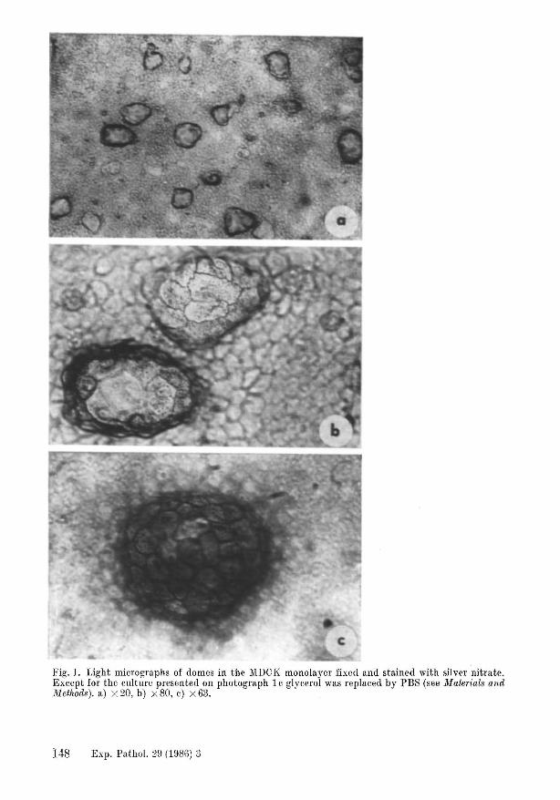
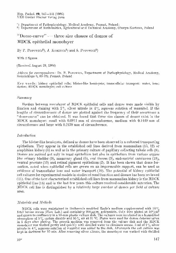
Fig. 1. Light micrographs of domes in the MDCK monolayer fixed and stained with silver nitrate. Except for the culture presented on photograph 1 c glycerol was replaced by PBS (see Materials and Method.s). a) x 20, b) x 80, c) x 63.
148 Exp. Pathol. 29 (1986) 3
water and 5 ml of 2.76 % aqueous solution of glycerol was added to the pl ate. The cell eulture was exposed to light t ill it became brown. At that very moment glycerol was replaced by phosphate buffered saline (PBS). The lengths of circumferences of domes wore measured using the Kontron :MOP AM 02 electronic sample analyzer (Reichert) of photographi c nega tive pi ctUTes.
Results
The living monolayers and domes are generally observed by light microsco py but ordinary histological methods, mostly dehydrations, give occasion t o destruction of domes. Isoosmotic solutions of silver nitrate for fixati on, st aining and maintaining th e unchanged monolayer during some time prevent the domes from co ll apse and devastation (fig. 1 c) . Borders between monolayer and domes are vi sibly marked as the black lines determining the shape and circumference of observed blist ers (fig. 1 a, b). Using t he t est of goodn ess of fit },-Kolmogoroy (7) on significance level (X = 0.10 it was demonstrated that the di stribution of lengths of circumferences was: F(x) = 0.25 FI(x) + 0.25 F2(x) + 0.15 Fa(x) + 0.35 F4(X) where
Fl (x) was normal distribution with mean 0.063 and variance 0.020 F 2(x) was normal distributi on with mean 0.091 and variance 0.017 F3(X) was normal distribution with 1l1ean 0.145 and variance 0.020 F1(x) was normal distribution with mean 0.230 and variance 0.033
Ta.bl c 1. Domes of :vIDCK epith elial monolayer (from one 50 mm culture Petri dish)
Class Xl ) SD2) number of domes
small 0.0811 0.020 93 medium 0.1449 0.020 28 large 0.2439 0.035 65
Total number 186 Mode 0.088
Statistical analyses were carried out using the maximuIU likelihood mothod 1) lUean value in millimetres 2) standard deviation
20
8
· 6 • E 0 1 ~
ow
"0 2
• 0 X 8
6
, 2
0.05 0.10 0,5 0,20 0,25 0,30 0,35
mllll .1 •••
Fig. 2. Dot ted line (observed frc([u cney) is the histogram relating the numb er of analysed domes to their lengths of circumferen ces . Continuous lin e (expected freqnency) is the "dome-curve" as the graphic, representation of studied rc la tiol1ship.
Exp . Pathol. 29 (188G) 3 149
The hypothetiral distribution was a mixture of 4 normal di stributions and thi s suggested that different groups of domes existed in the lVIDCK monolayer. Using the test for perrentage coefficient it \vas ascertained that the analysed sample was made up of 50 '!o of small, 15 u ~
of medium and 35 °0 of large domes (table 1). The lenghts of circumfr renees are plotted against the frequency of their occurrence (fig. 2) and a new parameter obtained: " dom ecurve" may help to characterize the gro ups of domes in the lVIDCK epithelial monol ayer. Three peaks of the continuous line eorrespond exactly with classified groups of blisters : the first peak includes the small, second the medium and third the large domes.
Discuss-ion
F ntil now there is no evidence that "dome" cells are different from " monolayer" celk RABITO et al. (17) demonstrated that transepithelial ion transport by domes did not differ from transport by monolayer. Domes revea l particular behaviour conditioned by rulture or experimental medium. Removal of Ca2+ and the addition of ethylene glycol-bis(p-aminoethyl ether)-N,N-tetraaeetic aci d (EGTA) caused the blisters to vanish completely within a few minutes (4). Ouabain, an inhibitor of active solute transport abolished domes formation (1) and based on experiments with the lVIDCK l11onolayers LEVER (16) has demonstrated that certain low molecular weight compo unds have been inducers of dome formation. The methods of dome determination in these inv estigations have been based on calculation of the Humber of domes per field without their classification. We conclude that lVlDCK cells in a confluent monolayer give evidenee of dome development. Medium and large blisters can come into existence by regularly increasing the fluid capacity of small domes with various times of their formation. This swelling of small domes is interrelated with ageing of both monolayer and cystlike structures. Displacement of the " dome-curve" to the right side of the graph should indicate the ageing or swelling of domes but preponderance of the left part of the "domecurve" should indicate the dome formation. On the other hand , the lVIDCK established cell line contains cell subpopulati ons w'hich vary in the dom e-forming (16) phenotype, and this may suggest that the "dome-curve" consist s of different ~ize types of domes.
Literature
1. ABAZA, N. A., J. LEIGHTOX and S. G. SCHULTZ, Effeets of ouabain on the funetion and strnl'ture of a tell line (:UDCK) derived from canine kidney. In vitro 19, 642- 650 (1983).
2. AHo:-l so?\", J. F., Human retinal pigment cell culture. In Vitro 19, 642- 650 (1983). 3. BIllEK, C., .I. E. AUBIN, U. BIIAIl GAVA, D. )1. , BRl'XETTE and A. H. MELC HEH, Dome formation
by oral epithelia in vitro. In Vitro 18, 382- 392 (1982). 4. CEIlEl JIDO, ilI.,.J. EHHE NFELD, S. FEHI'ANDEZ-CASTELO and 1. MEZA, Fluxes, junction and blisters
in cultured monolayers of epitheloid ('('lIs (ilIDCK). Ann. N.Y. Ata d. Sci. 372, 422-441 (1981). 5. DAS, ~.K ., H. L. HOSICK ann S. NAXDI, Influence of seeding density on multicellular organi
zation and nuclear events in cultures of normal and neoplastie mOllse mammary epithelium. J. Nat\. Cancer Inst. 52, 849 - 861 (1974).
6. DI!AGSTEN, P R., J. S. HANDLEH and R. BLL\IEXTIIAL, Asymmetry in ppithelial cells: is the tight junction a barrier to lateral diffusion in the plasma membran e? In: Membranes in Growth and De\"l' lop ment. Alan R. Liss, Inc. 1982, pp. 525-536.
7. FISH, 1\1., Probability Theory and Mathematieal Statistic-s, 3]'(1 ('(1. John Wiley, Xew York 19G3. 8. GHENJEH,F. C., T. E. ROLLI~S and W. L. S:lIl'fII, Kinin-in(1.1H'ed prostaglandi;{ synthesis by renal
papillary eolleeting tllbule tells in culture. Am. J. Physiul 241 , F94- F104 (1981). 9. IhxDLJ;H, J. S., R. E. STEELE, iiI. SA HIB , .J. B. WADE, A. J. PJlESTO?\" , N. LAWSON and J. P.
J OHXSON, Toad urinary bladder epithelial tells in t ultllre: maintl'na nt (> of ppithclial strneture sodi um transport and response to hormones. Proe. Xatl. Atad. Sei. l:SA ill , 4151-4155 (1979).
10. HA XDLER, J. S. , F. l\L PEHK1 NS and .1. P. JOHNSOl": , Studips of rl'lIaJ ("ell fUlldioll using tell ('ll iture techniqnes. Am . J . PhysioJ. 238, 1'1- 1'9 (1980).
11. I:!OH STEll, M., Tissue cnlture in nephrology: potential and li mits for the study of renal disease. El in . Wo el!ensehr. 38, 9G5- 973 (1980).
12. HL'LL, R. N. , W. R. CHERny and G. WL\VEH, The origin and t huraeteristie of a pig kidney tell strain LLC-PK1• In Vitro 12, 670- 677 (197G).
150 Exp. Pathol. 29 (1986) 3
13. KeBOTA, Y., E. B. GEHLY, K. H. LINK and Cu. HEIDELBEHGEH, Development of two dOlled epithelial cell lines from normal adult mouse and rat ventral prostate. In Vitro 17, 965-978 (1981).
14. LEIGHTON, J., Brief history of active transport in culture. Ann. N.Y. Acad. Sei. 372, 352-353 (1981).
15. -, 1. W. ESTES, S. MANSlJKIIANI and Z. BHADA, A cell line derived from normal dog kidney (MDCK) exhibiting qualities of papillary adenocarcinoma and renal tubular epithelium. Cancer 26, 1022-1028 (1970).
Ill. LEVEH, J. E., Regulation of dome formation in kidney epithelial cell cultures. Ann. N.Y. Acad. Sci. 372,371-383 (1981).
17. R~IBITo, CA., R. TeHAo, J. VALENTICH and J. LEIGHTON, Distribution and characteristics of the occluding junctions in a monolayer of a cell line (MDCK) derived from canine kidney. J. Membrane Bio!. 43, 351-365 (1978).
18. W"IT, D. L., D. S. GHOSSO, J. R. DAVIS, E. A. SUHWIT and C. D. CUI\ISTIAN, Charaeterization of a new human endometrial carcinoma (RL95-2) established in tissue culture. In Vitro 19, 147-158 (1983).
Exp. Patho!. 29, 151 (1986) VEB Gustav Fischer Verlag Jena
Book Review
Illustrated Encyclopcdin of Human IJistology by Prof. Dr. RADIVOJ V. KRSTIC; 450 pages, with 1576 figures, DlH 89.-. Springer-Verlag Berlin, Heidelberg, New York, Tokyo 1984.
This book is much more than only an "Encyclopedia of hnman histology", although it is such an eneyelopedia. - The alphabetically arranged items come partly from the macroscopical, light- and electronmicroscopical morphology (anatomy, histology, cytology), but also from the biochemistry and physiology, immunology etc. as well as from the histological, histochemical and instrumental techniques. So the levels of the light- and electron microscopic preparation, the histometric-stereological methods, the fundamentals of histo- and cytochemistry, immunocytochemistry, immunofiuoreseence etc. are Tepresented.
This is in correspondence with the idea of the book which is outlined by the author in his preface: Normal and pathological morphology have been an "enormous progress" in the last thTee decades by new technical instrum(]nts and methods. Therefore it became possibl(] to elucidate not only new structmal components and their complexity on all levels of observation but also their interrelations with metabolism and function.
This is of importance not only for research workers in all fields of medicine, biology and pharmaceutics, but also for the clinicians and last not least the students of medicine, stomatology, veterinary medieine, biology and pharmaceuties. For all of them it is impossible to follow the widespread modern literature in those fields to find an adequate detailed as well as comprehensive survey whieh is of importance for their work. - This is the aim of the present new book by R. V. KRSTlC.
The reader finds the name of the item and any synonyms and the Latin term, if one exists (according to Nomina Anatomica 4th ed. 1977), followed by a definition of the item and a comprehensive description of the most important features; in many eases one or more illustrations are added: halftone illustrations, pen drawings and diagrams, mostly newly drawn by the author for this book and a certain number reproduced from his well-known former books. - Cross references between
related items arc indicated by anows and the reader will find additional cross references pertaining to the item as well as a list of suggested further reading. A list of the most frequently llsed symbols and abbreviations is added.
By those methods of representation the author succeeds in not only giving an enormous amount of detailed and integrated information but he also succeeds in enabling the reader to get rapid and up-to-date orientation. Especially the book is directed to the interrelations of modern morphology with biochemistry and physiology with regard to the needs of clinical and experimental medicine. So it is extremely useful for physicians of all specialities who need a rapid, clear and comprehensive information in the fields ('overed by this boole
The most important quality is the connection of a short and informative tl'xt including synonyme and cross references with illustrations (1576 figures!). They show the high didactic and technical quality which is well-known from the books by R. V. l\HSTIC. We may congratulate the author but also the publishing house to this fascinating and extremely useful book.
F. BOLCI(, Jena
Exp. Pathol. 29 (1986) 3 151