Embed Size (px)
Citation preview
LETTERS
Identification of a serotonin/glutamate receptorcomplex implicated in psychosisJavier Gonzalez-Maeso1,2, Rosalind L. Ang1, Tony Yuen1, Pokman Chan1, Noelia V. Weisstaub5,6,Juan F. Lopez-Gimenez8, Mingming Zhou5, Yuuya Okawa1, Luis F. Callado9,10, Graeme Milligan8, Jay A. Gingrich5,6,7,Marta Filizola3, J. Javier Meana9,10 & Stuart C. Sealfon1,4
The psychosis associated with schizophrenia is characterized byalterations in sensory processing and perception1,2. Some anti-psychotic drugs were identified by their high affinity for serotonin5-HT2A receptors (2AR)3,4. Drugs that interact with metabotropicglutamate receptors (mGluR) also have potential for the treatmentof schizophrenia5–7. The effects of hallucinogenic drugs, such aspsilocybin and lysergic acid diethylamide, require the 2AR8–10 andresemble some of the core symptoms of schizophrenia10–12. Herewe show that the mGluR2 interacts through specific transmem-brane helix domains with the 2AR, a member of an unrelatedG-protein-coupled receptor family, to form functional complexesin brain cortex. The 2AR–mGluR2 complex triggers uniquecellular responses when targeted by hallucinogenic drugs, andactivation of mGluR2 abolishes hallucinogen-specific signallingand behavioural responses. In post-mortem human brain fromuntreated schizophrenic subjects, the 2AR is upregulated andthe mGluR2 is downregulated, a pattern that could predisposeto psychosis. These regulatory changes indicate that the 2AR–mGluR2 complex may be involved in the altered cortical processesof schizophrenia, and this complex is therefore a promising newtarget for the treatment of psychosis.
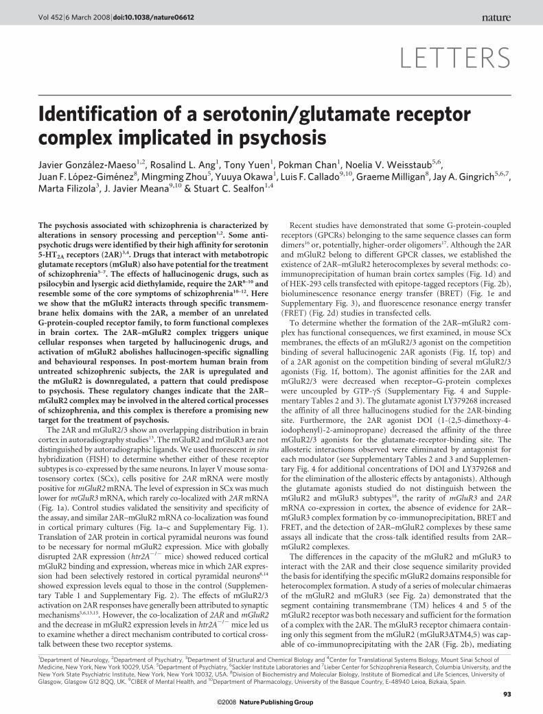
The 2AR and mGluR2/3 show an overlapping distribution in braincortex in autoradiography studies13. The mGluR2 and mGluR3 are notdistinguished by autoradiographic ligands. We used fluorescent in situhybridization (FISH) to determine whether either of these receptorsubtypes is co-expressed by the same neurons. In layer V mouse soma-tosensory cortex (SCx), cells positive for 2AR mRNA were mostlypositive for mGluR2 mRNA. The level of expression in SCx was muchlower for mGluR3 mRNA, which rarely co-localized with 2AR mRNA(Fig. 1a). Control studies validated the sensitivity and specificity ofthe assay, and similar 2AR–mGluR2 mRNA co-localization was foundin cortical primary cultures (Fig. 1a–c and Supplementary Fig. 1).Translation of 2AR protein in cortical pyramidal neurons was foundto be necessary for normal mGluR2 expression. Mice with globallydisrupted 2AR expression (htr2A2/2 mice) showed reduced corticalmGluR2 binding and expression, whereas mice in which 2AR expres-sion had been selectively restored in cortical pyramidal neurons8,14
showed expression levels equal to those in the control (Supplemen-tary Table 1 and Supplementary Fig. 2). The effects of mGluR2/3activation on 2AR responses have generally been attributed to synapticmechanisms5,6,13,15. However, the co-localization of 2AR and mGluR2and the decrease in mGluR2 expression levels in htr2A2/2 mice led usto examine whether a direct mechanism contributed to cortical cross-talk between these two receptor systems.
Recent studies have demonstrated that some G-protein-coupledreceptors (GPCRs) belonging to the same sequence classes can formdimers16 or, potentially, higher-order oligomers17. Although the 2ARand mGluR2 belong to different GPCR classes, we established theexistence of 2AR–mGluR2 heterocomplexes by several methods: co-immunoprecipitation of human brain cortex samples (Fig. 1d) andof HEK-293 cells transfected with epitope-tagged receptors (Fig. 2b),bioluminescence resonance energy transfer (BRET) (Fig. 1e andSupplementary Fig. 3), and fluorescence resonance energy transfer(FRET) (Fig. 2d) studies in transfected cells.
To determine whether the formation of the 2AR–mGluR2 com-plex has functional consequences, we first examined, in mouse SCxmembranes, the effects of an mGluR2/3 agonist on the competitionbinding of several hallucinogenic 2AR agonists (Fig. 1f, top) andof a 2AR agonist on the competition binding of several mGluR2/3agonists (Fig. 1f, bottom). The agonist affinities for the 2AR andmGluR2/3 were decreased when receptor–G-protein complexeswere uncoupled by GTP-cS (Supplementary Fig. 4 and Supple-mentary Tables 2 and 3). The glutamate agonist LY379268 increasedthe affinity of all three hallucinogens studied for the 2AR-bindingsite. Furthermore, the 2AR agonist DOI (1-(2,5-dimethoxy-4-iodophenyl)-2-aminopropane) decreased the affinity of the threemGluR2/3 agonists for the glutamate-receptor-binding site. Theallosteric interactions observed were eliminated by antagonist foreach modulator (see Supplementary Tables 2 and 3 and Supplemen-tary Fig. 4 for additional concentrations of DOI and LY379268 andfor the elimination of the allosteric effects by antagonists). Althoughthe glutamate agonists studied do not distinguish between themGluR2 and mGluR3 subtypes18, the rarity of mGluR3 and 2ARmRNA co-expression in cortex, the absence of evidence for 2AR–mGluR3 complex formation by co-immunoprecipitation, BRET andFRET, and the detection of 2AR–mGluR2 complexes by these sameassays all indicate that the cross-talk identified results from 2AR–mGluR2 complexes.
The differences in the capacity of the mGluR2 and mGluR3 tointeract with the 2AR and their close sequence similarity providedthe basis for identifying the specific mGluR2 domains responsible forheterocomplex formation. A study of a series of molecular chimaerasof the mGluR2 and mGluR3 (see Fig. 2a) demonstrated that thesegment containing transmembrane (TM) helices 4 and 5 of themGluR2 receptor was both necessary and sufficient for the formationof a complex with the 2AR. The mGluR3 receptor chimaera contain-ing only this segment from the mGluR2 (mGluR3DTM4,5) was cap-able of co-immunoprecipitating with the 2AR (Fig. 2b), mediating
1Department of Neurology, 2Department of Psychiatry, 3Department of Structural and Chemical Biology and 4Center for Translational Systems Biology, Mount Sinai School ofMedicine, New York, New York 10029, USA. 5Department of Psychiatry, 6Sackler Institute Laboratories and 7Lieber Center for Schizophrenia Research, Columbia University, and theNew York State Psychiatric Institute, New York, New York 10032, USA. 8Division of Biochemistry and Molecular Biology, Institute of Biomedical and Life Sciences, University ofGlasgow, Glasgow G12 8QQ, UK. 9CIBER of Mental Health, and 10Department of Pharmacology, University of the Basque Country, E-48940 Leioa, Bizkaia, Spain.
Vol 452 | 6 March 2008 | doi:10.1038/nature06612
93Nature Publishing Group©2008
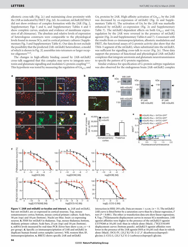
allosteric cross-talk (Fig. 2c) and maintaining close proximity withthe 2AR as indicated by FRET (Fig. 2d). In contrast, mGluR2DTM4,5did not show evidence of complex formation with the 2AR (Fig. 2,Supplementary Figs 5 and 6, and Supplementary Tables 4 and 5show complete curves, analysis and evidence of membrane expres-sion of all chimaeras). The absolute and relative levels of expressionof heterologous constructs were comparable to the physiologicallevels found in mouse SCx, and in cortical primary cultures (Supple-mentary Fig. 5 and Supplementary Table 4). Our data do not excludethe possibility that the predicted 2AR–mGluR2 heterodimer, a modelof which is shown in Fig. 2f, assembles into tetramers or larger recep-tor oligomers19,20.
The changes in high-affinity binding caused by 2AR–mGluR2cross-talk suggested that this complex may serve to integrate sero-tonin and glutamate signalling and modulate G-protein coupling21,22.This hypothesis was tested by measuring the regulation of Gaq/11 and
Gai proteins by 2AR. High-affinity activation of Gaq/11 by the 2ARwas decreased by co-expression of mGluR2 (Fig. 2e and Supple-mentary Table 6). The activation of Gai by the 2AR was markedlyenhanced by mGluR2 co-expression (Fig. 2e and SupplementaryTable 7). The mGluR2-dependent effects on both Gaq/11 and Gai
regulation by the 2AR were reversed in the presence of mGluR2agonist (Fig. 2e and Supplementary Tables 6 and 7). Consonant withthe results from co-immunoprecipitation, allosteric modulation andFRET, the functional assays of G-protein activity also show that theTM4–5 segment of the mGluR2, when substituted into the mGluR3,was sufficient for signalling cross-talk to occur (Fig. 2e). These datasupport the presence of functional and physiological 2AR–mGluR2complexes that integrate serotonin and glutamate neurotransmissionto specify the pattern of G-protein regulation.
Similar evidence for specification of G-protein subtype regulationwas also observed for the endogenous brain 2AR–mGluR2 complex
Num
ber
of c
opie
sp
er µ
g R
NA
40,000
30,000
20,000
10,000
Somatosensorycortex
Thalamus
0.6
mGluR2-Rluc + 5HT2A–GFP2
mGluR2-Rluc + pGFP2
mGluR2-Rluc + 5HT2C–GFP2mGluR3-Rluc + 5HT2A–GFP2
BR
ET2 0.4
0.2
0
0
100
75
500 –10 –9 –8 –7 0 –10 –9 –8 –7 0 –10 –9 –8 –7
0 –10 –9 –8 –7 0 –10 –9 –8 –6–7 0 –7 –6 –5 –4
100
75
50
100
75
50
100
75
50
100100
75
50
25
0
75
50
0.5 1.0Fluorescence/luminescence
log([LY379268] (M))
log([DOI] (M))
log([DCG-IV] (M))
log([DOM] (M))
log([L-CCG-I] (M))
log([DOB] (M))
[3 H]L
Y34
1495
bou
nd (%
)[3 H
]Ket
anse
rin b
ound
(%)
1.5 2.0
0
mGluR2mGluR3
118
47
118
47
kDa kDa
118
118
5-HT2AR mGluR2
5-HT2AR mGluR3
mGluR3
mGluR3
Overlay
Overlay
5-HT2AR mGluR2
5-HT2AR mGluR3
Overlay
Overlay
a d
e
f
IP 2ARWB mGluR2
IP 2ARWB mGluR3
WB mGluR2
WB mGluR3
47
WB 2ARV kDa
b
c
VI
V
VI
Figure 1 | 2AR and mGluR2 co-localize and interact. a, 2AR and mGluR2,but not mGluR3, are co-expressed in cortical neurons. Top, mousesomatosensory cortex; bottom, mouse cortical primary culture. Scale bars,50 mm (top) and 10mm (bottom). Nuclei are blue. Inset: co-expressingneuron. b, FISH for mGluR3 in thalamus. Top, mouse thalamus; bottom,thalamic primary culture. Scale bars, 25 mm (top) and 10 mm (bottom).c, mRNA levels measured by real-time PCR (Error bars show s.e.m.; n 5 6per group). d, Specific co-immunoprecipitation of 2AR and mGluR2 induplicate human frontal cortex samples (arrows). WB, western blot; IP,immunoprecipitation. e, BRET2 shows specific 2AR and mGluR2
interaction in HEK-293 cells. Data are means 6 s.e.m. (n 5 3). The mGluR2/2AR curve is fitted better by a saturation curve than by a linear regression, Ftest (P , 0.001). The other co-transfection data sets show linear regressions.f, Top, [3H]ketanserin displacement curves in mouse SCx membranes. 2ARagonist affinities were higher in the presence of the mGluR2/3 agonistLY379268 at 10mM (red) than in vehicle alone (black). [3H]LY341495displacement curves (bottom panels). mGluR2/3 agonist affinities werelower in the presence of the 2AR agonist DOI at 10mM (red) than in vehiclealone (black). DCG-IV, (2S,29R,39R)-2-(29,39-dicarboxycyclopropyl)-glycine; L-CCG-I, (2S,19S,29S)-2-(carboxycyclopropyl)-glycine.
LETTERS NATURE | Vol 452 | 6 March 2008
94Nature Publishing Group©2008
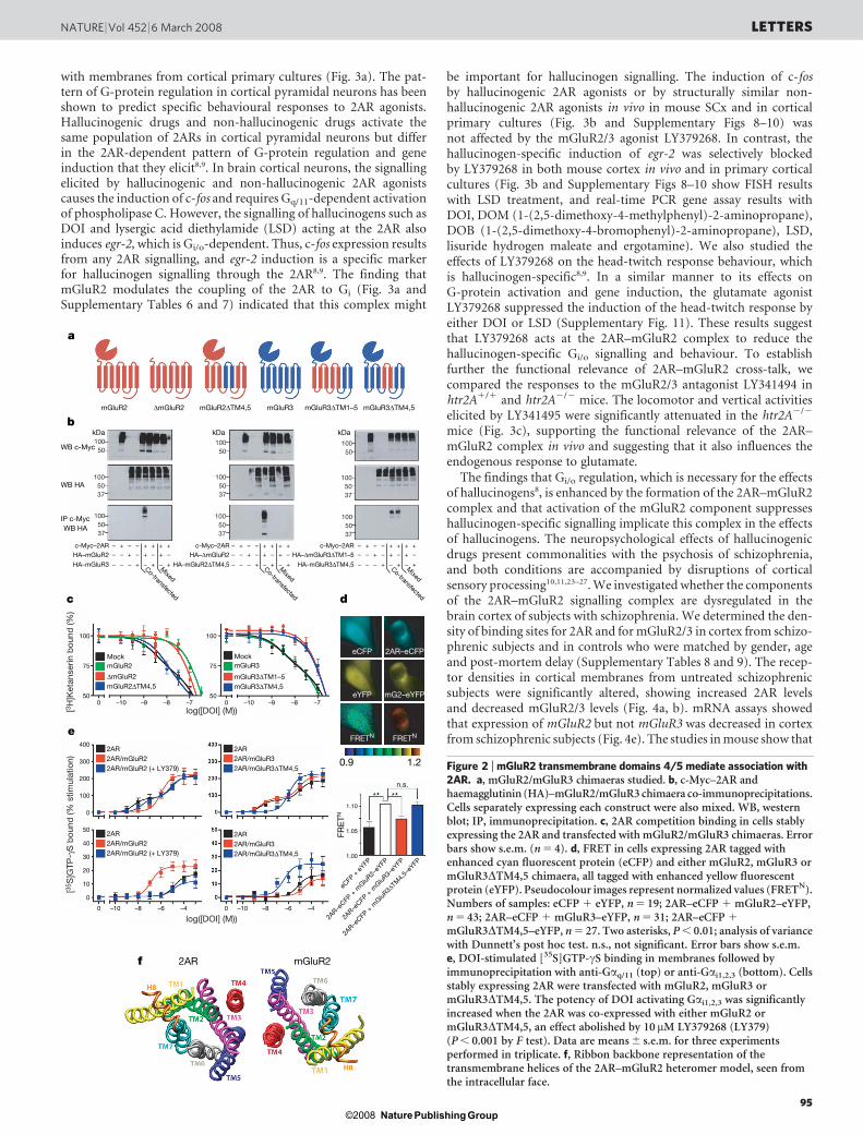
with membranes from cortical primary cultures (Fig. 3a). The pat-tern of G-protein regulation in cortical pyramidal neurons has beenshown to predict specific behavioural responses to 2AR agonists.Hallucinogenic drugs and non-hallucinogenic drugs activate thesame population of 2ARs in cortical pyramidal neurons but differin the 2AR-dependent pattern of G-protein regulation and geneinduction that they elicit8,9. In brain cortical neurons, the signallingelicited by hallucinogenic and non-hallucinogenic 2AR agonistscauses the induction of c-fos and requires Gq/11-dependent activationof phospholipase C. However, the signalling of hallucinogens such asDOI and lysergic acid diethylamide (LSD) acting at the 2AR alsoinduces egr-2, which is Gi/o-dependent. Thus, c-fos expression resultsfrom any 2AR signalling, and egr-2 induction is a specific markerfor hallucinogen signalling through the 2AR8,9. The finding thatmGluR2 modulates the coupling of the 2AR to Gi (Fig. 3a andSupplementary Tables 6 and 7) indicated that this complex might
be important for hallucinogen signalling. The induction of c-fosby hallucinogenic 2AR agonists or by structurally similar non-hallucinogenic 2AR agonists in vivo in mouse SCx and in corticalprimary cultures (Fig. 3b and Supplementary Figs 8–10) wasnot affected by the mGluR2/3 agonist LY379268. In contrast, thehallucinogen-specific induction of egr-2 was selectively blockedby LY379268 in both mouse cortex in vivo and in primary corticalcultures (Fig. 3b and Supplementary Figs 8–10 show FISH resultswith LSD treatment, and real-time PCR gene assay results withDOI, DOM (1-(2,5-dimethoxy-4-methylphenyl)-2-aminopropane),DOB (1-(2,5-dimethoxy-4-bromophenyl)-2-aminopropane), LSD,lisuride hydrogen maleate and ergotamine). We also studied theeffects of LY379268 on the head-twitch response behaviour, whichis hallucinogen-specific8,9. In a similar manner to its effects onG-protein activation and gene induction, the glutamate agonistLY379268 suppressed the induction of the head-twitch response byeither DOI or LSD (Supplementary Fig. 11). These results suggestthat LY379268 acts at the 2AR–mGluR2 complex to reduce thehallucinogen-specific Gi/o signalling and behaviour. To establishfurther the functional relevance of 2AR–mGluR2 cross-talk, wecompared the responses to the mGluR2/3 antagonist LY341494 inhtr2A1/1 and htr2A2/2 mice. The locomotor and vertical activitieselicited by LY341495 were significantly attenuated in the htr2A2/2
mice (Fig. 3c), supporting the functional relevance of the 2AR–mGluR2 complex in vivo and suggesting that it also influences theendogenous response to glutamate.
The findings that Gi/o regulation, which is necessary for the effectsof hallucinogens8, is enhanced by the formation of the 2AR–mGluR2complex and that activation of the mGluR2 component suppresseshallucinogen-specific signalling implicate this complex in the effectsof hallucinogens. The neuropsychological effects of hallucinogenicdrugs present commonalities with the psychosis of schizophrenia,and both conditions are accompanied by disruptions of corticalsensory processing10,11,23–27. We investigated whether the componentsof the 2AR–mGluR2 signalling complex are dysregulated in thebrain cortex of subjects with schizophrenia. We determined the den-sity of binding sites for 2AR and for mGluR2/3 in cortex from schizo-phrenic subjects and in controls who were matched by gender, ageand post-mortem delay (Supplementary Tables 8 and 9). The recep-tor densities in cortical membranes from untreated schizophrenicsubjects were significantly altered, showing increased 2AR levelsand decreased mGluR2/3 levels (Fig. 4a, b). mRNA assays showedthat expression of mGluR2 but not mGluR3 was decreased in cortexfrom schizophrenic subjects (Fig. 4e). The studies in mouse show that
kDa kDa kDa
a
c
b
d
e
f
mGluR2
WB c-Myc
WB HA
WB HAIP c-Myc
c-Myc–2AR
100
0 –10 –9 –8 –7 0 –10 –9 –8 –7
75
400
300
200
100
0
50
40
30
20
10
0
0 –10 –8 –6 –4 0 –10 –8 –6 –4
50
100
75
50
– + + + + +– –
– – + – + –+ –
– – – + – +– +
– + + + + +– –
– – + – + –+ –
– – – + – +– +
– + + + + +– –
– – + – + –+ –
– – – + – +– +
1.10
n.s.
1.05
1.00
HA–mGluR2
HA–mGluR3
Mock
2AR2AR/mGluR22AR/mGluR2 (+ LY379)
2AR2AR/mGluR32AR/mGluR3∆TM4,5
2AR2AR/mGluR32AR/mGluR3∆TM4,5
2AR2AR/mGluR22AR/mGluR2 (+ LY379)
mGluR2MockmGluR3
∆mGluR2mGluR2∆TM4,5
mGluR3∆TM1–5mGluR3∆TM4,5
c-Myc–2ARHA–∆mGluR2
HA–mGluR2∆TM4,5
c-Myc–2ARHA–∆mGluR3∆TM1–5
HA–mGluR3∆TM4,5
∆mGluR2 mGluR2∆TM4,5 mGluR3 mGluR3∆TM1–5 mGluR3∆TM4,5
0.9 1.2
Co-transfected
Mixed
eCFP
+ e
YFP
2AR–e
CFP +
mGluR
2–eY
FP
2AR–e
CFP +
mGluR
3–eY
FP
2AR–e
CFP +
mGluR
3∆TM4,
5–eY
FP
2AR–eCFP
mG2–eYFP
eCFP
eYFP
FRETN FRETN
FRE
TN
2AR mGluR2
Co-transfected
Mixed
Co-transfected
Mixed
log([DOI] (M))
log([DOI] (M))
[35 S
]GTP
-γS
bou
nd (%
stim
ulat
ion)
[3H
]Ket
anse
rin b
ound
(%)
10050
10050
10050
1005037
1005037
1005037
1005037
1005037
1005037
Figure 2 | mGluR2 transmembrane domains 4/5 mediate association with2AR. a, mGluR2/mGluR3 chimaeras studied. b, c-Myc–2AR andhaemagglutinin (HA)–mGluR2/mGluR3 chimaera co-immunoprecipitations.Cells separately expressing each construct were also mixed. WB, westernblot; IP, immunoprecipitation. c, 2AR competition binding in cells stablyexpressing the 2AR and transfected with mGluR2/mGluR3 chimaeras. Errorbars show s.e.m. (n 5 4). d, FRET in cells expressing 2AR tagged withenhanced cyan fluorescent protein (eCFP) and either mGluR2, mGluR3 ormGluR3DTM4,5 chimaera, all tagged with enhanced yellow fluorescentprotein (eYFP). Pseudocolour images represent normalized values (FRETN).Numbers of samples: eCFP 1 eYFP, n 5 19; 2AR–eCFP 1 mGluR2–eYFP,n 5 43; 2AR–eCFP 1 mGluR3–eYFP, n 5 31; 2AR–eCFP 1
mGluR3DTM4,5–eYFP, n 5 27. Two asterisks, P , 0.01; analysis of variancewith Dunnett’s post hoc test. n.s., not significant. Error bars show s.e.m.e, DOI-stimulated [35S]GTP-cS binding in membranes followed byimmunoprecipitation with anti-Gaq/11 (top) or anti-Gai1,2,3 (bottom). Cellsstably expressing 2AR were transfected with mGluR2, mGluR3 ormGluR3DTM4,5. The potency of DOI activating Gai1,2,3 was significantlyincreased when the 2AR was co-expressed with either mGluR2 ormGluR3DTM4,5, an effect abolished by 10mM LY379268 (LY379)(P , 0.001 by F test). Data are means 6 s.e.m. for three experimentsperformed in triplicate. f, Ribbon backbone representation of thetransmembrane helices of the 2AR–mGluR2 heteromer model, seen fromthe intracellular face.
NATURE | Vol 452 | 6 March 2008 LETTERS
95Nature Publishing Group©2008
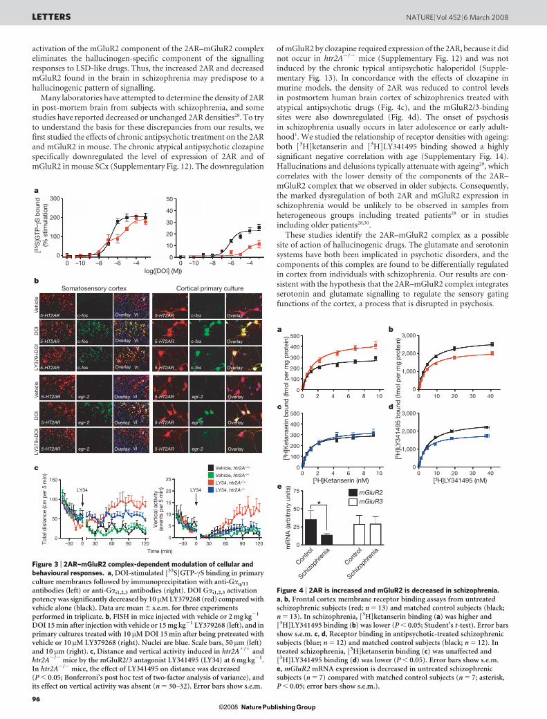
activation of the mGluR2 component of the 2AR–mGluR2 complexeliminates the hallucinogen-specific component of the signallingresponses to LSD-like drugs. Thus, the increased 2AR and decreasedmGluR2 found in the brain in schizophrenia may predispose to ahallucinogenic pattern of signalling.
Many laboratories have attempted to determine the density of 2ARin post-mortem brain from subjects with schizophrenia, and somestudies have reported decreased or unchanged 2AR densities28. To tryto understand the basis for these discrepancies from our results, wefirst studied the effects of chronic antipsychotic treatment on the 2ARand mGluR2 in mouse. The chronic atypical antipsychotic clozapinespecifically downregulated the level of expression of 2AR and ofmGluR2 in mouse SCx (Supplementary Fig. 12). The downregulation
of mGluR2 by clozapine required expression of the 2AR, because it didnot occur in htr2A2/2 mice (Supplementary Fig. 12) and was notinduced by the chronic typical antipsychotic haloperidol (Supple-mentary Fig. 13). In concordance with the effects of clozapine inmurine models, the density of 2AR was reduced to control levelsin postmortem human brain cortex of schizophrenics treated withatypical antipsychotic drugs (Fig. 4c), and the mGluR2/3-bindingsites were also downregulated (Fig. 4d). The onset of psychosisin schizophrenia usually occurs in later adolescence or early adult-hood1. We studied the relationship of receptor densities with ageing:both [3H]ketanserin and [3H]LY341495 binding showed a highlysignificant negative correlation with age (Supplementary Fig. 14).Hallucinations and delusions typically attenuate with ageing29, whichcorrelates with the lower density of the components of the 2AR–mGluR2 complex that we observed in older subjects. Consequently,the marked dysregulation of both 2AR and mGluR2 expression inschizophrenia would be unlikely to be observed in samples fromheterogeneous groups including treated patients28 or in studiesincluding older patients28,30.
These studies identify the 2AR–mGluR2 complex as a possiblesite of action of hallucinogenic drugs. The glutamate and serotoninsystems have both been implicated in psychotic disorders, and thecomponents of this complex are found to be differentially regulatedin cortex from individuals with schizophrenia. Our results are con-sistent with the hypothesis that the 2AR–mGluR2 complex integratesserotonin and glutamate signalling to regulate the sensory gatingfunctions of the cortex, a process that is disrupted in psychosis.
300 50
40
30
20
10
0
200
100
00 –10 –8 –6 –4 0 –10 –8 –6 –4
5-HT2AR c-fos
c-fos
c-fos
Overlay
5-HT2AR Overlay
5-HT2AR Overlay
5-HT2AR egr-2
egr-2
egr-2
Overlay
5-HT2AR Overlay
5-HT2AR Overlay
5-HT2AR c-fos
c-fos
c-fos
Overlay
5-HT2AR Overlay
5-HT2AR Overlay
5-HT2AR egr-2
egr-2
egr-2
Overlay
5-HT2AR Overlay
5-HT2AR Overlay
Veh
icle
DO
ILY
379+
DO
IV
ehic
le
150
LY34 LY34
25
Vehicle, htr2A+/+
Vehicle, htr2A–/–
LY34, htr2A+/+
LY34, htr2A–/–20
15
10
5
0
100
50
0–30 0 30 60
Time (min)
Tota
l dis
tanc
e (c
m p
er 5
min
)
Ver
tical
act
ivity
(eve
nts
per
5 m
in)
90 120 –30 0 30 60 90 120
DO
ILY
379+
DO
I
a
b
c
Somatosensory cortex Cortical primary culture
V
VI
V
VI
V
VI
V
VI
V
VI
V
VI
[35 S
]GTP
-γS
bou
nd(%
stim
ulat
ion)
log([DOI] (M))
Figure 3 | 2AR–mGluR2 complex-dependent modulation of cellular andbehavioural responses. a, DOI-stimulated [35S]GTP-cS binding in primaryculture membranes followed by immunoprecipitation with anti-Gaq/11
antibodies (left) or anti-Gai1,2,3 antibodies (right). DOI Gai1,2,3 activationpotency was significantly decreased by 10mM LY379268 (red) compared withvehicle alone (black). Data are mean 6 s.e.m. for three experimentsperformed in triplicate. b, FISH in mice injected with vehicle or 2 mg kg21
DOI 15 min after injection with vehicle or 15 mg kg21 LY379268 (left), and inprimary cultures treated with 10mM DOI 15 min after being pretreated withvehicle or 10mM LY379268 (right). Nuclei are blue. Scale bars, 50mm (left)and 10mm (right). c, Distance and vertical activity induced in htr2A1/1 andhtr2A2/2 mice by the mGluR2/3 antagonist LY341495 (LY34) at 6 mg kg21.In htr2A2/2 mice, the effect of LY341495 on distance was decreased(P , 0.05; Bonferroni’s post hoc test of two-factor analysis of variance), andits effect on vertical activity was absent (n 5 30–32). Error bars show s.e.m.
500 3,000
2,000
1,000
0
3,000
2,000
1,000
0
[3H
]Ket
anse
rin b
ound
(fm
ol p
er m
g p
rote
in)
[3H
]LY
3414
95 b
ound
(fm
ol p
er m
g p
rote
in)
[3H]LY341495 (nM)[3H]Ketanserin (nM)
Contro
l
Schizo
phren
ia
Contro
l
Schizo
phren
ia
mR
NA
(arb
itrar
y un
its)
400
300
200
100
00 2 4 6 8 10 0 10 20 30 40
0 10
mGluR2mGluR3
20 30 400 2 4 6 8 10
500
400
300
200
100
0
75
50
25
0
a
c
b
d
e
Figure 4 | 2AR is increased and mGluR2 is decreased in schizophrenia.a, b, Frontal cortex membrane receptor binding assays from untreatedschizophrenic subjects (red; n 5 13) and matched control subjects (black;n 5 13). In schizophrenia, [3H]ketanserin binding (a) was higher and[3H]LY341495 binding (b) was lower (P , 0.05; Student’s t-test). Error barsshow s.e.m. c, d, Receptor binding in antipsychotic-treated schizophrenicsubjects (blue; n 5 12) and matched control subjects (black; n 5 12). Intreated schizophrenia, [3H]ketanserin binding (c) was unaffected and[3H]LY341495 binding (d) was lower (P , 0.05). Error bars show s.e.m.e, mGluR2 mRNA expression is decreased in untreated schizophrenicsubjects (n 5 7) compared with matched control subjects (n 5 7; asterisk,P , 0.05; error bars show s.e.m.).
LETTERS NATURE | Vol 452 | 6 March 2008
96Nature Publishing Group©2008
METHODS SUMMARYAll reagents were purchased from commercial vendors except for LY379268 (Eli
Lilly and Co.). Mouse lines, treatment protocols, behavioural studies, dissec-
tions, and primary neuronal cultures, approved by Institutional Use and Care
Committees, have been described previously8,9. Protocols used for FISH8, bind-
ing assays8, real-time PCR8, FRET17 and co-immunoprecipitation17 were per-
formed as described previously or with minor modifications. Epitope-tagged,
BRET2, FRET and chimaera receptor constructs were generated by using stand-
ard cloning techniques and were confirmed by sequencing. BRET2 using Renilla
luciferase and green fluorescent protein (GFP2) was performed in HEK-293 cells.
Matched schizophrenia and control human brains were obtained from autopsies
performed in the Basque Institute of Legal Medicine, Bilbao, Spain, in compli-
ance with policies of research and ethical review boards for post-mortem brain
studies.
Full Methods and any associated references are available in the online version ofthe paper at www.nature.com/nature.
Received 2 November; accepted 20 December 2007.Published online 24 February 2008.
1. Freedman, R. Schizophrenia. N. Engl. J. Med. 349, 1738–1749 (2003).2. Sawa, A. & Snyder, S. H. Schizophrenia: diverse approaches to a complex disease.
Science 296, 692–695 (2002).3. Lieberman, J. A. et al. Serotonergic basis of antipsychotic drug effects in
schizophrenia. Biol. Psychiatry 44, 1099–1117 (1998).4. Miyamoto, S., Duncan, G. E., Marx, C. E. & Lieberman, J. A. Treatments for
schizophrenia: a critical review of pharmacology and mechanisms of action ofantipsychotic drugs. Mol. Psychiatry 10, 79–104 (2005).
5. Aghajanian, G. K. & Marek, G. J. Serotonin model of schizophrenia: emerging roleof glutamate mechanisms. Brain Res. Brain Res. Rev. 31, 302–312 (2000).
6. Marek, G. J. Metabotropic glutamate 2/3 receptors as drug targets. Curr. Opin.Pharmacol. 4, 18–22 (2004).
7. Patil, S. T. et al. Activation of mGlu2/3 receptors as a new approach to treatschizophrenia: a randomized Phase 2 clinical trial. Nature Med. 13, 1102–1107(2007).
8. Gonzalez-Maeso, J. et al. Hallucinogens recruit specific cortical 5-HT2A receptor-mediated signaling pathways to affect behavior. Neuron 53, 439–452 (2007).
9. Gonzalez-Maeso, J. et al. Transcriptome fingerprints distinguish hallucinogenicand nonhallucinogenic 5-hydroxytryptamine 2A receptor agonist effects inmouse somatosensory cortex. J. Neurosci. 23, 8836–8843 (2003).
10. Vollenweider, F. X., Vollenweider-Scherpenhuyzen, M. F., Babler, A., Vogel, H. &Hell, D. Psilocybin induces schizophrenia-like psychosis in humans via aserotonin-2 agonist action. Neuroreport 9, 3897–3902 (1998).
11. Gouzoulis-Mayfrank, E. et al. Psychological effects of (S)-ketamine and N,N-dimethyltryptamine (DMT): a double-blind, cross-over study in healthyvolunteers. Pharmacopsychiatry 38, 301–311 (2005).
12. Colpaert, F. C. Discovering risperidone: the LSD model of psychopathology.Nature Rev. Drug Discov. 2, 315–320 (2003).
13. Marek, G. J., Wright, R. A., Schoepp, D. D., Monn, J. A. & Aghajanian, G. K.Physiological antagonism between 5-hydroxytryptamine2A and group IImetabotropic glutamate receptors in prefrontal cortex. J. Pharmacol. Exp. Ther.292, 76–87 (2000).
14. Weisstaub, N. V. et al. Cortical 5-HT2A receptor signaling modulates anxiety-likebehaviors in mice. Science 313, 536–540 (2006).
15. Benneyworth, M. A. et al. A selective positive allosteric modulator ofmetabotropic glutamate receptor subtype 2 blocks a hallucinogenic drug modelof psychosis. Mol. Pharmacol. 72, 477–484 (2007).
16. Angers, S., Salahpour, A. & Bouvier, M. Dimerization: an emerging concept for Gprotein-coupled receptor ontogeny and function. Annu. Rev. Pharmacol. Toxicol.42, 409–435 (2002).
17. Lopez-Gimenez, J. F., Canals, M., Pediani, J. D. & Milligan, G. The a1b-adrenoceptor exists as a higher-order oligomer: effective oligomerization is
required for receptor maturation, surface delivery, and function. Mol. Pharmacol.71, 1015–1029 (2007).
18. Wright, R. A., Arnold, M. B., Wheeler, W. J., Ornstein, P. L. & Schoepp, D. D.[3H]LY341495 binding to group II metabotropic glutamate receptors in rat brain.J. Pharmacol. Exp. Ther. 298, 453–460 (2001).
19. Palczewski, K. et al. Crystal structure of rhodopsin: A G protein-coupled receptor.Science 289, 739–745 (2000).
20. Fotiadis, D. et al. Atomic-force microscopy: Rhodopsin dimers in native discmembranes. Nature 421, 127–128 (2003).
21. Kenakin, T. Efficacy at G-protein-coupled receptors. Nature Rev. Drug Discov. 1,103–110 (2002).
22. Gonzalez-Maeso, J., Rodriguez-Puertas, R. & Meana, J. J. Quantitativestoichiometry of G-proteins activated by m-opioid receptors in postmortemhuman brain. Eur. J. Pharmacol. 452, 21–33 (2002).
23. Carlsson, A. The neurochemical circuitry of schizophrenia. Pharmacopsychiatry 39(Suppl. 1), S10–S14 (2006).
24. Vollenweider, F. X. & Geyer, M. A. A systems model of altered consciousness:integrating natural and drug-induced psychoses. Brain Res. Bull. 56, 495–507(2001).
25. Vollenweider, F. X. et al. Positron emission tomography andfluorodeoxyglucose studies of metabolic hyperfrontality and psychopathologyin the psilocybin model of psychosis. Neuropsychopharmacology 16, 357–372(1997).
26. Umbricht, D. et al. Effects of the 5-HT2A agonist psilocybin on mismatchnegativity generation and AX-continuous performance task: implications for theneuropharmacology of cognitive deficits in schizophrenia.Neuropsychopharmacology 28, 170–181 (2003).
27. Gouzoulis-Mayfrank, E. et al. Inhibition of return in the human 5HT2A agonist andNMDA antagonist model of psychosis. Neuropsychopharmacology 31, 431–441(2006).
28. Dean, B. The cortical serotonin2A receptor and the pathology of schizophrenia: alikely accomplice. J. Neurochem. 85, 1–13 (2003).
29. Davidson, M. et al. Severity of symptoms in chronically institutionalized geriatricschizophrenic patients. Am. J. Psychiatry 152, 197–207 (1995).
30. Gurevich, E. V. & Joyce, J. N. Alterations in the cortical serotonergic system inschizophrenia: a postmortem study. Biol. Psychiatry 42, 529–545 (1997).
Supplementary Information is linked to the online version of the paper atwww.nature.com/nature.
Acknowledgements We thank L. Devi and L. Ivic for critiquing the manuscript;S. Morgello and the Manhattan HIV Brain Bank for providing control brain cortex;I. Rodil, L. Uriguen and B. Lin for assistance with biochemical assays; the MountSinai Microscopy and Microarray, Real-Time PCR and Bioinformatics SharedResearch Facilities; the staff members of the Basque Institute of Legal Medicine fortheir cooperation in the study; J. H. Prather for a gift of LY379268; and J.-P. Pin forproviding the signalling peptide sequence of rat mGluR5. This study was supportedby the National Institutes of Health, UPV/EHU and the Basque Government, theSpanish Ministry of Health, the REM-TAP Network, the Whitehall Foundation, theGatsby Foundation and the American Foundation for Suicide Prevention.
Author Contributions J.G.M. and S.C.S. designed experiments, supervisedresearch and wrote the manuscript. J.G.M. performed experiments. R.L.A.performed BRET experiments. T.Y. designed and cloned receptor chimaeras. Y.O.assisted with experiments. P.C. performed FISH studies. N.V.W. and M.Z.,supervised by J.A.G., performed behaviour experiments and developed mutantmouse lines. J.L.G., supervised by G.M., performed FRET experiments. M.F.performed computer modelling. L.F.C. and J.J.M. performed schizophreniapost-mortem human brain studies. All authors discussed the results andcommented on the manuscript.
Author Information Reprints and permissions information is available atwww.nature.com/reprints. Correspondence and requests for materials should beaddressed to J.G.M. ([email protected]) or S.C.S.([email protected]).
NATURE | Vol 452 | 6 March 2008 LETTERS
97Nature Publishing Group©2008
METHODSMaterials and drug administration. DOI, DOM, DOB, LSD and lisuride hydro-
gen maleate were purchased from Sigma-Aldrich. (1R,4R,5S,6R)-4-Amino-2-
oxabicyclo[3.1.0]hexane-4,6-dicarboxylic acid (LY379268) was obtained from
Eli Lilly and Co. 2S-2-amino-2-(1S,2S-2-carboxycyclopropan-1-yl)-3-(xanth-9-
yl)-propionic acid (LY341495), (2S,29R,39R)-2-(29,39-dicarboxycyclopropyl)-
glycine (DCG-IV), (2S,19S,29S)-2-(carboxycyclopropyl)-glycine (L-CCG-I),
clozapine and haloperidol were obtained from Tocris Cookson Inc.
[3H]Ketanserin and [35S]GTP-cS were purchased from PerkinElmer.
[3H]LY341495 was purchased from American Radiolabelled Chemicals, Inc.
The injected doses (intraperitoneal) were: DOI, 2 mg kg21; DOM, 4 mg kg21;
DOB, 1 mg kg21; LSD, 0.24 mg kg21; lisuride hydrogen maleate, 0.4 mg kg21;
ergotamine, 0.5 mg kg21; LY379268, 15 mg kg21; LY341495, 6 mg kg21; cloza-
pine, 25 mg kg21; and haloperidol, 1 mg kg21, unless otherwise indicated.
Transient transfection of HEK-293 cells. HEK-293 cells were maintained
in DMEM medium supplemented with 10% (v/v) fetal bovine serum at 37 uCin a humidified 5% CO2 atmosphere. Transfection was performed with
Lipofectamine 2000 reagent (Invitrogen) in accordance with the manufacturer’s
instructions. HEK-293 cells stably expressing human 2AR have been described
previously9,31.
Co-immunoprecipitation studies. Co-immunoprecipitation studies in post-
mortem human brain, and co-immunoprecipitation studies using an
N-terminally c-Myc-tagged form of 2AR, and N-terminally haemagglutinin
(HA)-tagged forms of mGluR2, mGluR3 or mGluR2/mGluR3 chimaeras in
HEK-293 cells were performed as described previously, with minor modifica-
tions17. In brief, the samples were incubated overnight with protein A/G beads
and anti-2AR (post-mortem human brain) or anti-c-Myc antibody (HEK-293
cells) at 4 uC on a rotating wheel. Equal amounts of proteins were resolved by
SDS–PAGE. Detection of proteins by immunoblotting with anti-2AR (Santa
Cruz Biotechnology), anti-mGluR2 and anti-mGluR3 (Abcam Inc.) in post-
mortem human brain, or anti-c-Myc and anti-HA antibodies (Santa Cruz
Biotechnology) in HEK-293 cells was conducted with an enhanced chemilumin-
escence system in accordance with the manufacturer’s recommendations.
Bioluminescence resonance energy transfer (BRET2) in HEK-293 live cells.The human 2AR, serotonin 5-HT2C (2CR), mGluR2 and mGluR3 receptors with
mutated stop codons were subcloned into the pRluc and pGFP2 plasmids
(PerkinElmer), such that Renilla luciferase (Rluc) and green fluorescent protein
(GFP2) were present at the carboxy termini of the receptors. All sequences were
confirmed by DNA sequencing. After 48 h, transfected cells were washed with
PBS, suspended to (1–2) 3 106 cells ml21 and were treated with DeepBlueC
Coelenterazine Substrate (5 mM final concentration; PerkinElmer ). Equivalent
amounts of total DNA composed of various ratios of the Rluc- or GFP2-tagged
receptors were transfected32. Light emission was monitored with a Fusion
Universal Microplate Analyser (PerkinElmer). A BRET2 signal is defined as
the light emitted by GFP2 at 515 nm in response to the light emitted at
410 nm by Rluc on catalysis of DeepBlueC. The values were corrected by sub-
tracting the background BRET2 signal detected when the receptor–Rluc con-
struct was expressed alone (see Supplementary Fig. 3 for luminescence and
fluorescence values). The specificities of mGluR2–Rluc and 2AR–GFP2 interac-
tions were assessed by comparison with co-expression of mGluR2–Rluc and
2CR–GFP2, mGluR3–Rluc and 2AR–GFP2 and mGluR2–Rluc and GFP2.
Data from a single experiment, which was replicated three times, are shown as
means 6 s.e.m. (Fig. 1e).
FRET. Forms of the 2AR and mGluR2 C-terminally fused to eCFP and eYFP
were generated, and FRET microscopy in living cells was conducted as reported
previously17. Results from a single experiment, representative of two or three
independent studies, are shown in Fig. 2d.
[3H]Ketanserin, [3H]LY341495 and [35S]GTP-cS binding. Membrane prepara-
tions and [3H]ketanserin binding assays were performed as reported previously8.
[3H]LY341495 binding was performed as described previously, with minor
modifications18. In brief, membrane preparations were incubated for 60 min at
4 uC. Non-specific binding was determined in the presence of 1 mM L-glutamate.
[35S]GTP-cS binding experiments were initiated by the addition of membranes
containing 35 mg of protein to an assay buffer (20 mM HEPES, 3 mM MgCl2,
100 mM NaCl, 0.2 mM ascorbic acid and 0.5 nM [35S]GTP-cS) supplemented
with 0.1mM or 10mM GDP for Gaq/11 and Gai, respectively, and containing the
indicated concentration of ligands. Non-specific binding was determined in the
presence of 100mM GTP-cS. Reactions were incubated for 30 min at 30 uC, and
were terminated by the addition of 0.5 ml of ice-cold buffer, containing 20 mM
HEPES, 3 mM MgCl2, 100 mM NaCl and 0.2 mM ascorbic acid. The samples
were centrifuged at 16,000g for 15 min at 4 uC, and the resulting pellets were
resuspended in solubilization buffer (100 mM Tris, 200 mM NaCl, 1 mM EDTA,
1.25% Nonidet P40) plus 0.2% SDS. Samples were precleared with Pansorbin
(Calbiochem), followed by immunoprecipitation with antibody against Gaq/11
or Gai1,2,3 (Santa Cruz Biotechnology). Finally, the immunocomplexes were
washed twice with solubilization buffer, and bound [35S]GTP-cS was measured
by liquid-scintillation spectrometry.
Construction of receptor chimaeras. All PCR reactions were performed with
PfuTurbo Hotstart DNA polymerase (Stratagene). Cycling conditions were 30
cycles of 94 uC for 30 s, 55 uC for 30 s and 72 uC for 1 min per kilobase of ampli-
con, with an initial denaturation/activation of 94 uC for 2 min and a final exten-
sion of 72 uC for 7 min.
HA-tagged wild-type human mGluR2 and mGluR3 constructs: the rat
mGluR5 signal peptide (SP)33 along with an HA epitope tag was amplified by
PCR with primers NheI-HA_SP/S (59-TTTTgctagcGAATTCCTTTCCTAAA-
ATGG-39) and HA_SP-KpnI/A (59-TTTTggtaccACGCGTGGCGTAGTCGG-
GTA-39) with pRK5 as template. Wild-type human mGluR2 and mGluR3 were
amplified with primers MluI-hGRM2/S (59-agctacgcgtAAGAAGGTGCTGA-
CCCTGGA-39) hGRM2-XbaI/A (59-AAtctagaTCAAAGCGATGACGTTGTCG-
AG-39) and KpnI-hGRM3/S (59-acgtggtaccTTAGGGGACCATAACTTTCT-39)
hGRM3-XhoI/A (59-acgtctcgagTCACAGAGATGAGGTGGTGG-39), respec-
tively. The rat mGluR5 signal peptide/HA epitope fragment was digested with
NheI and MluI, the human mGluR2 fragment was digested with MluI and XbaI
and were simultaneously subcloned into the NheI and XbaI sites of pcDNA3.1
(Invitrogen) to yield the HA-tagged mGluR2 construct. Similarly, the rat
mGluR5 signal peptide/HA fragment was digested with NheI and KpnI, the
human mGluR2 PCR product was digested with KpnI and XhoI, and they were
subcloned simultaneously into the NheI and XhoI sites of pcDNA3.1 to give the
HA-tagged mGluR2 construct.
Chimaeric human mGluR2 with transmembrane domain 4 and 5 from human
mGluR3: a fragment of the transmembrane domain TM1 to the C terminus of
the second intracellular loop of the human mGluR2 was amplified with primers
hGRM2-1476/S (59-GGACACCAGCCTCATCCCAT-39) and hGRM2i2GRM3-
TM4/A (59-CAGATGAAAACCTGAGAACTAGGACTGATGAAGCGTGGCC-
39). A fragment of the TM4 to TM5 of the human mGluR3 was amplified with
primers hGRM2i2GRM3TM4/S (59-GGCCACGCTTCATCAGTCCTAGTTC-
TCAGGTTTTCATCTG-39) and hGRM3TM5GRM2i3/A (59-TTTTCGGGGC-
ACTTGCGAGTTTTGAAGGCGTACACAGTGC-39). The two fragments were
annealed and reamplified with primers hGRM2-1476/S and hGRM3TM5-
GRM2i3/A. The third intracellular loop to the C terminus of the human
mGluR2 was amplified with primers hGRM3TM5GRM2i3/S (59-GCACTGTG-
TACGCCTTCAAAACTCGCAAGTGCCCCGAAAA-39) and hGRM2-XbaI/A.
This fragment was then annealed with the previous PCR product and reamplified
with primers hGRM2-1476/S and hGRM2-XbaI/A. To reconstitute the complete
chimaeric receptor, the N-terminal domain of the HA-tagged wild-type human
mGluR2 was released with NheI and BstBI, the final PCR product was digested
with BstBI and XbaI, and the two fragments were simultaneously subcloned into
the NheI and XbaI sites of pcDNA3.1.
Chimaeric human mGluR3 with transmembrane domain 4 and 5 from human
mGluR2: a fragment of the transmembrane domain TM1 to the C terminus of
the second intracellular loop of the human mGluR3 was amplified with primers
hGRM3-2541/S (59-TGAAAGTTGGTCACTGGGCA-39) and hGRM3i2GRM2-
TM4/A (59-CAGATGGCCACCTGTGAGGCGGGGCTGATGAATTTTGGCC-
39). A fragment of the TM4 to TM5 of the human mGluR2 was amplified with
primers hGRM3i2GRM2TM4/S (59-GGCCAAAATTCATCAGCCCCGCCTC-
ACAGGTGGCCATCTG-39) and hGRM2TM5GRM3i3/A (59-TTTTCTGGGC-
ACTTCCGCGTCTTGAAGGCATAAAGCGTGC-39). The two fragments were
annealed and reamplified with primers hGRM3-2541/S and hGRM2TM5-
GRM3i3/A. The third intracellular loop to the C terminus of the human
mGluR3 was amplified with primers hGRM2TM5GRM3i3/S (59-GCACGC-
TTTATGCCTTCAAGACGCGGAAGTGCCCAGAAAA-39) and hGRM3-XhoI/
A. This fragment was then annealed with the previous PCR product and ream-
plified with primers hGRM3-2541/S and hGRM3-XhoI/A. To reconstitute the
complete chimaeric receptor, the N-terminal domain of the HA-tagged wild-
type human mGluR3 was released with NheI and PstI, the final PCR product was
digested with PstI and XhoI, and the two fragments were simultaneously sub-
cloned into the NheI and XhoI sites of pcDNA3.1.
Chimaeric human mGluR3 with transmembrane domains 1 to 5 from human
mGluR2: a small fragment of the N-terminal domain to the beginning of TM1 of
the human mGluR3 was amplified with primers hGRM3-2541/S and hGRM3-
NGRM2TM1/A (59-ACAGCCCAGGCATCGCCCCAGCGGATGTAGTCCT-
CAGGAAGGT-39). A fragment of the TM1 to TM5 of the human mGluR2
was amplified with primers hGRM3NGRM2TM1/S (59-ACCTTCCTGAG-
GACTACATCCGCTGGGGCGATGCCTGGGCTGT-39) and hGRM2TM5-
GRM3i3/A. The two fragments were annealed and reamplified with primers
hGRM3-2541/S and hGRM2TM5GRM3i3/A. The third intracellular loop to
the C terminus of the human mGluR3 was amplified with primers
doi:10.1038/nature06612
Nature Publishing Group©2008
hGRM2TM5GRM3i3/S and hGRM3-XhoI/A. This fragment was then annealedwith the previous PCR product and reamplified with primers hGRM3-2541/S
and hGRM3-XhoI/A. To reconstitute the complete chimaeric receptor, the
N-terminal domain of the HA-tagged wild-type human mGluR3 was released
with NheI and PstI, the final PCR product was digested with PstI and XhoI, and
the two fragments were simultaneously subcloned into the NheI and XhoI sites of
pcDNA3.1.
Molecular modelling. Three-dimensional molecular models of the seven TM
regions of 2AR and mGluR2 were built by using the crystal structures of b2-
adrenergic receptor34 and rhodopsin35, respectively, as structural templates, and
the latest version of the homology-modelling program MODELLER36. The use of
the very recent crystal structure of b2-adrenergic receptor to build a model of
2AR is justified by the higher sequence identity between these two receptors than
to rhodopsin; in addition, the suitability of the rhodopsin template to build
models of family C GPCRs, which includes the mGluR2, has recently been
discussed37. The sequence alignment between the transmembrane helices of
b2-adrenergic receptor and 2AR was obtained with BLAST38. For mGluR2 we
used the same alignment with rhodopsin as described in ref. 37. A multiple
alignment of available mGluR2 and mGluR3 sequences was performed withthe CLUSTALW program, version 1.81 (ref. 39). Supplementary Fig. 7 shows
the details of these sequence alignments in the transmembrane regions. To build
a reasonable configuration of the 2AR–mGluR2, we used the TM4,5–TM4,5
configuration deriving from atomic force microscopy of rhodopsin in native
disc membranes40 as a template for the heteromer interface between 2AR and
mGluR2. This modelling was obtained with the assistance of the Insight II User
Graphical Interface (Accelrys Inc.) on a graphics workstation.
Neuronal primary culture. Primary cultures of cortical and thalamic neurons
were prepared as described previously8.
Mouse brain samples. Experiments were performed as described previously8.
FISH. Synthesis of modified DNA oligonucleotide probes, probe labelling and
FISH were performed as described previously8,41. See Supplementary Table 10 for
oligonucleotide probe sequences.
Quantitative real-time PCR. Quantitative real-time PCR experiments were
performed as described previously8. See Supplementary Tables 11 and 12 for
primer pair sequences.
Behavioural studies. Behavioural studies were performed as described pre-
viously8,14. Motor function was assessed with a computerized three-dimensionalactivity monitoring system (AccuScan Instruments). The activity monitor has 32
infrared sensor pairs, with 16 along each side spaced 2.5 cm apart. The system
determines motor activity on the basis of the frequency of interruptions to
infrared beams traversing the x, y and z planes. Total distance (cm) travelled
and vertical activity were determined automatically from the interruptions of
beams in the horizontal and vertical planes, respectively.
Brain samples. Human brains were obtained at autopsies performed in the
Forensic Anatomical Institute, Bilbao, Spain. The study was developed in com-
pliance with policies of research and ethical review boards for post-mortem brain
studies (Basque Institute of Legal Medicine, Spain). Deaths were subjected to
retrospective searching for previous medical diagnosis and treatment, using the
examiner’s information and records of hospitals and mental health centres. After
searching of ante-mortem information had been fulfilled, 25 subjects who had
met criteria of schizophrenia according to the Diagnostic and Statistical Manual
of Mental Disorders (DSM-IV)42 were selected. A toxicological screening for
antipsychotics, other drugs and ethanol was performed on samples of blood,
urine, liver and gastric contents. All subjects who were drug-free before death (as
revealed by the absence of prescriptions in medical histories) also gave negativeresults in the toxicological screening. The toxicological assays were performed at
the National Institute of Toxicology, Madrid, Spain, using a variety of standard
procedures including radioimmunoassay, enzymatic immunoassay, high-
performance liquid chromatography and gas chromatography–mass spectro-
metry. Controls for the present study were chosen among the collected brains
on the basis, whenever possible, of the following cumulative criteria: negative
medical information on the presence of neuropsychiatric disorders or drug
abuse; appropriate gender, age and post-mortem delay to match each subject
in the schizophrenia group; sudden and unexpected death (motor vehicle acci-
dents); and toxicological screening for psychotropic drugs with negative results
except for ethanol. Tissue pH was assumed to be an indicator of agonal status43.
Thus, prolonged terminal hypoxia resulted in low tissue pH. It has been shown
that gene expression patterns are strongly dependent on tissue pH. Rapid deaths,
associated with accidents, cardiac events or asphyxia, generally resulted in a
normal tissue pH with a minor influence on gene expression patterns44. All
schizophrenic and control subjects showed a sudden and rapid death without
a long agonal phase. The tissue storage period before assays did not differ sig-nificantly between schizophrenic cases (82 6 9 months (mean 6 s.e.m.)) and
controls (85 6 10 months (mean 6 s.e.m.)). Two independent groups of schizo-
phrenic subjects were selected depending on the presence of antipsychotics in the
toxicological screening (Supplementary Tables 8 and 9).
31. Ebersole, B. J., Visiers, I., Weinstein, H. & Sealfon, S. C. Molecular basis of partialagonism: orientation of indoleamine ligands in the binding pocket of the humanserotonin 5-HT2A receptor determines relative efficacy. Mol. Pharmacol. 63,36–43 (2003).
32. James, J. R., Oliveira, M. I., Carmo, A. M., Iaboni, A. & Davis, S. J. A rigorousexperimental framework for detecting protein oligomerization usingbioluminescence resonance energy transfer. Nature Methods 3, 1001–1006(2006).
33. Blahos, J. et al. Extreme C terminus of G protein a-subunits contains a site thatdiscriminates between Gi-coupled metabotropic glutamate receptors. J. Biol.Chem. 273, 25765–25769 (1998).
34. Cherezov, V. et al. High-resolution crystal structure of an engineered human b2-adrenergic G protein-coupled receptor. Science 318, 1258–1265 (2007).
35. Li, J., Edwards, P. C., Burghammer, M., Villa, C. & Schertler, G. F. Structure ofbovine rhodopsin in a trigonal crystal form. J. Mol. Biol. 343, 1409–1438 (2004).
36. Sali, A. & Blundell, T. L. Comparative protein modelling by satisfaction of spatialrestraints. J. Mol. Biol. 234, 779–815 (1993).
37. Binet, V. et al. Common structural requirements for heptahelical domain functionin class A and class C G protein-coupled receptors. J. Biol. Chem. 282,12154–12163 (2007).
38. Altschul, S. F., Gish, W., Miller, W., Myers, E. W. & Lipman, D. J. Basic localalignment search tool. J. Mol. Biol. 215, 403–410 (1990).
39. Thompson, J. D., Higgins, D. G. & Gibson, T. J. CLUSTAL W: improving thesensitivity of progressive multiple sequence alignment through sequenceweighting, position-specific gap penalties and weight matrix choice. Nucleic AcidsRes. 22, 4673–4680 (1994).
40. Liang, Y. et al. Organization of the G protein-coupled receptors rhodopsin andopsin in native membranes. J. Biol. Chem. 278, 21655–21662 (2003).
41. Chan, P., Yuen, T., Ruf, F., Gonzalez-Maeso, J. & Sealfon, S. C. Method formultiplex cellular detection of mRNAs using quantum dot fluorescent in situhybridization. Nucleic Acids Res. 33, e161 (2005).
42. American Psychiatric Association. Diagnostic and Statistical Manual of MentalDisorders 4th edn (American Psychiatric Association, Washington DC, 1994).
43. Preece, P. & Cairns, N. J. Quantifying mRNA in postmortem human brain:influence of gender, age at death, postmortem interval, brain pH, agonal state andinter-lobe mRNA variance. Brain Res. Mol. Brain Res. 118, 60–71 (2003).
44. Li, J. Z. et al. Systematic changes in gene expression in postmortem human brainsassociated with tissue pH and terminal medical conditions. Hum. Mol. Genet. 13,609–616 (2004).
doi:10.1038/nature06612
Nature Publishing Group©2008


![The identification of cortical pyramidal neurons using a ... · Differential Algorithm (LDA) [2] and Regularization Preserving Estimates (RPP) [3] algorithm have been proved to be](https://img.pdfslide.us/doc/110x75/5b37e92e7f8b9abd438cb758/the-identification-of-cortical-pyramidal-neurons-using-a-differential-algorithm.jpg)