Embed Size (px)
Citation preview
Manuscript type: Original Article
DOI : 10.5152/EurJTher.2019.190108
Title : Malignant Transformation of Mature Cystic Teratomas: A Retrospective Analysis of 181 Cases
Running Title: Malignancy Potential in Mature Cystic Teratomas
Zehra Bozdağ1, Neslihan Bayramoğlu Tepe2
1Department of Pathology, University of Gaziantep, Gaziantep, Turkey
2Department of Obstetrics and Gynecology, University of Gaziantep, Gaziantep, Turkey
Corresponding Author: Neslihan Bayramoglu Tepe
E-mail: [email protected]
Received: 08.10.2019
Accepted: 11.11.2019
How to cite: Bozdağ Z, Bayramoğlu Tepe N. Malignant Transformation of Mature Cystic Teratomas: A
Retrospective Analysis of 181 Cases. Eur J Ther 2019; DOI: 10.5152/EurJTher.2019.190108
ABSTRACT
Introduction: Mature cystic teratoma (MCT) comprises 20% of all ovarian tumors and is the most
prevalent ovarian germ cell tumor. Any of its component tissues may undergo a malignant
transformation (MT). This study aims to retrospectively analyze the malignancies that arise from MCT of
the ovary.
Materials and Methods: Histopathological diagnoses of resection materials obtained from adult
patients who were operated for an ovarian mass and diagnosed by our laboratory between January
2012 and December 2018 were reviewed.
Results: Of our one-hundred-and-eighty-one (181) cases, seven (3.86%) were detected to have MT. Of
our cases with MT; 5 had papillary thyroid carcinoma, 1 had squamous cell carcinoma, and 1 had a
strumal carcinoid.
Conclusion: The diagnosis of malignancies arising from MCT is important for the identification of follow-
up and treatment options for the patients. Reporting the data obtained from cases who demonstrate
MT will be informative for the pre/postoperative management of the patients.
Keywords: Mature cystic teratoma, malignancy, ovarian tumor
INTRODUCTION
Mature cystic teratoma of the ovary (MCT) is the most prevalent ovarian germ cell tumor and originates
from two or three germ layers (ectoderm, mesoderm, and endoderm). It comprises 20% of all ovarian
tumors (1).
The prevalence rates of these tumors, which may be encountered in patients of ages ranging from
childhood to post-menopause, typically peak around ages 20-40 (2). MCTs are usually clinically
asymptomatic and are detected either incidentally or due to the pressure from the mass during
gynecological examination. Transvaginal USG is the primary diagnostic tool in the diagnosis of MCT (3).
Tumor markers have limited role in the diagnosis (4).
MCTs have a typical macroscopic appearance; most are cystic and contain sebaceous material. Tumor
components may include various tissues including hair, fat, bone, cartilage, glial tissue, gastrointestinal
epithelium, respiratory epithelium, and thyroid tissue (3).
MCT is a benign tumor; however, it may rarely undergo a malignant transformation (at a rate of 1-2%)
and this is usually encountered at more advanced ages (5). Malignant transformations may arise from
any of the tissue components of the MCT. The most common malignant transformation reported in case
series is squamous cell carcinoma (SCC) and the literature contains reports of various types of sarcoma
and adenocarcinoma, melanoma, and basal cell carcinoma arising from MCT (6-14).
This study aims to retrospectively evaluate 181 adult patients with mature cystic teratoma who were
analyzed and diagnosed by our laboratory between 2012-2018 with regard to their histopathological
findings and malignant transformations.
MATERIALS and METHODS
Histopathological diagnoses of resection materials obtained from patients who had been operated for
ovarian masses between January 2012 and December 2018 were reviewed. Data belonging to 181 of
these patients who were diagnosed with MCT or dermoid cysts were evaluated with regard to patient
age, tumor location, tumor diameter, macroscopic properties of the tumor, and malignant
transformations seen after histopathological diagnosis. The evaluation was based on hospital file
records as well as pathology reports. Where necessary, archived pathology slides were re-evaluated.
RESULTS
The ages of our patients ranged from 17 to 72 (mean: 30.47) and 62.43% of MCT patients were aged
between 20-40 years. The tumor was localized in the right ovary in 85 cases (46.96%), left ovary in 77
cases (42.54%), and bilaterally in 19 cases (10.49%). An evaluation of macroscopic tumor characteristics
revealed that the diameter of the tumor was smaller than 10 cm in 124 cases (68.5%), between 10-20
cm in 53 cases (29.28%), and greater than 20 cm in 4 cases (2.2%). Macroscopically, the surface of the
section was cystic in 68 cases, solid in 41, and solid-cystic in 72. Macroscopically, the solid component
was observed to increase at greater tumor diameters.
In our series, 7 (3.86%) of the 181 cases manifested malignant transformations. Of our cases with MT, 5
had papillary thyroid carcinoma, 1 had squamous cell carcinoma, and 1 had a strumal carcinoid (Figure
1,2,3,4). Three of the papillary thyroid carcinoma cases had microcarcinomas (<1 cm). Clinical
complaints included pelvic pain in 5 cases, pelvic tenderness in one case, and a clinically asymptomatic
mass that was incidentally detected during routine check-up. We could access the tumor marker levels
of only 2 cases and both of these cases were determined to have high CA125 levels. Three cases were
operated for a malignant preliminary diagnosis, while the rest were operated for a preliminary diagnosis
of dermoid cyst. The majority of the cases demonstrated unilateral localizations and solid cut surfaces.
Clinicopathological findings of our malignant transformation cases have been presented in Table 1.
DISCUSSION
Mature cystic teratoma (MCT) is the most prevalent ovarian germ cell tumor and comprises 20% of all
ovarian tumors (1). Common associated complications include torsion, infection, and rupture. Malignant
transformation of MCT is a rare (1-2%) but serious complication (5, 15). In the literature, a 35-year study
by Ayhan and colleagues that included 501 patients found a malignant transformation rate of 1.4% (15).
Rathore et al. determined this rate as 3.5% in their 230-patient series, while Bal et al. determined a
malignant transformation rate of 6.6% in their 75-patient series (16, 17). In our series, 7 of the 181 cases
were found to have malignant transformations and the malignant transformation rate was 3.8%.
Clinical prediction of MT is difficult. Studies did not find a correlation between MT and serum tumor
markers, however, more than 70% of the cases were reported to have high Ca 125 or Ca 19-9 levels (4).
In our case series, of the three cases suspected of having malignancies, tumor marker levels of only two
could be accessed and both of these cases were found to have high Ca 125 levels.
These cases are typically unilateral (17). In our series, three cases underwent total abdominal
hysterectomy and bilateral salpingo-oophorectomy, while others underwent unilateral excision for
unilateral masses. Patients who underwent bilateral salpingo-oophorectomy also had unilateral tumors.
The most common malignant transformation of MCT is squamous cell carcinoma. Among the 7 MT cases
in our series, only 1 had SCC (0.55%), while the most common MT was papillary thyroid carcinoma (PTC)
arising from struma ovarii with 5 cases (2.76%). One of our cases (0.55%) had a strumal carcinoid.
Struma ovarii is a monodermal teratoma of the ovary and constitutes 2% of all germ cell tumors. 5-10%
of struma ovarii cases are malignant. They usually become manifest as unilateral masses in the
reproductive period. However, their incidence increases with age. The most prevalent MT in struma
ovarii is papillary thyroid carcinoma, followed respectively by follicular carcinoma and follicular variant
of papillary carcinoma (18). Five of our cases were found to have follicular variant of papillary
carcinoma, while one case had a poorly differentiated thyroid carcinoma arising from PTC. All of our
cases were unilateral; two were in the second decade, one in the fourth decade, and the other two were
in the sixth and seventh decades. Two cases of advanced age were detected to have PTC and poorly
differentiated thyroid carcinomas arising from PTC, while three cases had tumors in the form of a
microcarcinoma. Presence of microcarcinoma cases caused the most prevalent MT in our series to be
PTC, contradicting the literature and our expectations with a higher rate. We reasoned that the
detection of microcarcinoma cases was linked to the high number of macroscopic samples.
Malignant struma ovarii cases may clinically manifest thyrotoxicosis and studies have reported cases of
hypothyroidism after resection (18). Clinical data of our cases were inspected with regard to thyroid
states and it was found that none had history of any thyroid-related clinical complaints.
There is no consensus regarding the clinical approach to malignant struma ovarii cases. According to the
various approaches in the literature that have been described for cases and case series, patients must
be screened for metastasis after diagnosis and their serum thyroglobulin and iodine 131 levels must be
monitored. Thyroidectomy has been reported in certain cases (18, 19). Postop follow-up data of three
cases (cases 5 and 6) could be accessed. Thyroid examinations of these cases did not indicate any
pathologies.
The most common malignant transformation of MCT is squamous cell carcinoma (20), which was the
second most prevalent MT in our series. In a study by Kikkawa et al. that investigated a series of 37
patients with SCC arising from MCT over a period of 17 years, the mean age was determined as 55.2
years (21). Our case was 47-years old. In the epidermal component, malignant transformations may
arise from squamous, ciliated, and non-ciliated columnar epithelia. In ovarian squamous cell carcinomas,
metastatic carcinomas must certainly be eliminated. In ovarian metastases, the most common primary
cancers are cervical or vaginal squamous cell carcinomas. Immunohistochemically, SCCs arising from
MCTs were reported to show HPV and strong P16 positivity. Based on these findings, HPV was thought
to be a risk factor for this malignancy (20). On gynecological examination, our case did not have any
cervical or vaginal pathologies. Our patient was not immunohistochemically evaluated for HPV and P16
expression. Standart treatment for SCCs arising from MCTs are total hysterectomy, bilateral salpingo-
oophorectomi, surgical staging (omentectomy, appendectomy, peritoneal biopsies, pelvic and paraaortic
lymphadenectomy) in early cases and optimal cytoreductive surgery in advanced stage cases.
Chemotherapy is corresponded for those with more advanced disease but the efficacy of radiotheraphy
is uncorroborated. When the cases spread outside the ovary, the prognosis is poor (20).
Strumal carcinoid tumor of the ovary is a rare form of ovarian teratoma composed of carcinoid and
thyroid tissue (22). Strumal carcinoid was first described in 1970 by Robboy and Scully (23). Clinically,
the symptoms associated with the mass may be accompanied by symptoms of carcinoid syndrome. Our
case did not clinically manifest symptoms of carcinoid syndrome. Strumal carcinoids can present an
admixture of thyroid tissue and the carcinoid tumor component. The thyroid component is composed of
micro and macrofollicles containing colloid. The carcinoid component demonstrates a pure trabecular or
a mixed trabecular-insular pattern in most cases. The thyroid and carcinoid components can be
differentiated based on immunohistochemical markers as well as cellular properties (22). Our case
showed an admixture of thyroid tissue and the carcinoid component presenting trabecular and solid
patterns. As also determined in our case, the carcinoid component shows synaptophysin and
chromogranin positivity, and the thyroid component shows follicular nuclear TTF-1, cytoplasmic and
colloidal thyroglobulin positivity. In the differential diagnosis of ovarian stromal carcinoid tumors,
thyroid carcinomas, ovarian granulosa, and Sertoli Leydig cell tumors arising from stromal carcinoids
must be eliminated. Strumal carcinoids almost always have a benign manifestation (24). Kurabayesi et
al. reported detecting bone and breast metastases in cases who had shown marked cellular atypia, high
mitotic activity, and focal necrosis 3.5 years earlier (25). Our case did not have cellular atypia, high
mitotic activity, or necrosis, and is currently undergoing the first year of postoperative follow-up.
In conclusion, the diagnosis of malignancies that arise from mature cystic teratomas is important for the
follow-up and treatment options of the patients. These tumors are encountered quite frequently in the
pathological routine and must be inspected thoroughly for malignant transformations. Reporting the
data obtained from cases of MT will be informative for the pre/postoperative management of the
patients.
Ethics Committee Approval: Ethical committee approval was obtained from University School of
Medicine ethical board (Approval No: 2019/323).
Author Contribution: Concept- Z.B., N.B.T.; Design- Z.B; Supervision- N.B.T.; Resources-Z.B., N.B.T;
Materials-Z.B; Data Collection and/or Processing- Z.B., N.B.T.; Analysis and/or Interpretation- Z.B.;
Literature Search- N.B.T.; Writing Manuscript- Z.B., N.B.T.; Critical Review-Z.B., N.B.T.
Conflict of İnterest: The authors have no conflicts of interest to declare.
Financial Disclosure: The authors declared that this study has received no financial support.
References:
1. R. J. Kurman, M. L. Carcangiu, C. S. Herrington, and R. H. Young, Eds. WHO Classification of Tumours of
Female Reproductive Organs, IARC, Lyon. 2014.
2. Gadducci A, Pistolesi S, Guerrieri ME, Cosio S, Carbone FG, Naccarato AG. Malignant Transformation in
Mature Cystic Teratomas of the Ovary: Case Reports and Review of the Literature. Anticancer Res. 2018
Jun;38(6):3669-3675.
3. Sahin H, Abdullazade S, Sanci M. Mature cystic teratoma of the ovary: a cutting edge overview on
imaging features. Insights Imaging 2017 Apr;8(2):227-241.
4. Emin U, Tayfun G, Cantekin I, Özlem UB, Ümit B, Leyla M. Tumor markers in mature cystic teratomas
of the ovary. Arch Gynecol Obstet 2009; 279:145-7.
5. Li C, Zhang Q, Zhang S, Dong R, Sun C, Qiu C, Zhang Z, Yang X, Kong B. Squamous cell carcinoma
transformation in mature cystic teratoma of the ovary: a systematic review. BMC Cancer 2019 Mar
11;19(1):217.
6. Allam Nandyana P, Bui MM, Caracciolo JT, Hakam A. Squamous cell carcinoma
and osteosarcoma arising from a dermoid cyst--a case report and review of literature. Int J Clin Exp
Pathol 2010 Jan 10;3(3):313-8.
7. Takahashi H, Chaopotong P, Kajita S, Hashimura M, Yamazaki H, Saegusa M. Mixed angiosarcoma,
clear cell adenocarcinoma and mature teratoma elements in an ovarian tumor: a case report and
literature review. Pathol Int 2012 Aug;62(8):538-42.
8. Takahashi H, Chaopotong P, Kajita S, Hashimura M, Yamazaki H, Saegusa M. Mixed angiosarcoma,
clear cell adenocarcinoma and mature teratoma elements in an ovarian tumor: a case report and
literature review. Pathol Int 2012 Aug; 62(8):538-42.
9. Clark ME, Will MD. Intestinal-Type Adenocarcinoma Arising in a Mature Cystic Teratoma of the Ovary.
Int J Gynecol Pathol 2016 Jul;35(4):352-6.
10. Halabi M, Oliva E, Mazal PR, Breitenecker G, Young RH. Prostatic tissue in mature cystic teratomas of
the ovary: a report of four cases, including one with features of prostatic adenocarcinoma, and
cytogenetic studies. Int J Gynecol Pathol 2002 Jul;21(3):261-7.
11. Song YJ, Ryu SY, Choi SC, Lee ED, Lee KH, Cho SY. Adenocarcinoma arising from the respiratory
ciliated epithelium in a benign cystic teratoma of the ovary. Arch Gynecol Obstet 2009 Oct;280(4):659-
62.
12. Moghaddam Y, Lindsay R, Tolhurst J, Millan D, Siddiqui N. A case of sebaceous carcinoma arising in a
benign cystic teratoma of the ovary and review of the literature. Scott Med J 2013 May;58(2):e18-22.
13. Shen X, Fan Y, Cao S. Primary malignant melanoma arising in an ovarian cystic teratoma.
Melanoma Res 2017 Dec;27(6):601-606.
14. Yoder N, Marks A, Hui P, Litkouhi B, Cron J. Low-Grade Astrocytoma within a Mature
Cystic Teratoma in an Adolescent Patient. J Pediatr Adolesc Gynecol 2018 Jun;31(3):325-327.
15. Ayhan A, Bukulmez O, Genc C, Karamursel BS, Ayhan A. Mature cystic teratomas of the ovary: case
series from one institution over 34 years. Eur Obstet Gynecol Reprod Biol 2000;88(2):153-7.
16. Rathore R, Sharma D, Agarwal S. Malignant transformation in mature cystic teratoma of the ovary: a
retrospective study of eight cases and review of literature. Prz Menopauzalny 2018 Jun;17(2):63-68.
17. Bal A, Mohan H, Singh SB, Sehgal A. Malignant trans- formation in mature cystic teratoma of the
ovary: report of five cases and review of the literature Arch Gynecol Obstet 2007; 275:179-82.
18. Bağlan Z, Demirtaş GS, Ulukuş M, Zekioğlu O, Yılmaz H. Struma Ovaride Tiroid Papiller Kanser: Olgu
Sunumu. Türk Jinekolojik Onkoloji Dergisi 2012-4/ Ek Sayı, 20-23
19. Pineyro MM, Pereda J, Schou P, de Los Santos K, de la Pena S, Caserta B, Pisabarro R. Papillary
Thyroid Microcarcinoma Arising Within a Mature Ovarian Teratoma: Case Report and Review of the
Literature Clinical Medicine Insights: Endocrinology and DiabetesVolume 10: 1–3.
20. Gadducci A, Guerrieri ME, Cosio S. Squamous cell carcinoma arising from mature cystic teratoma of
the ovary: A challenging question for gynecologic oncologists. Critical Reviews in Oncology/Hematology.
Volume 133, January 2019, Pages 92-98.
21. Kikkawa F, Ishikawa H, Tamakoshi K, Nawa A, Suganuma N, Tomoda Y. Squamous cell
carcinoma arising from mature cystic teratoma of the ovary: a clinicopathologic analysis. Obstet
Gynecol.1997 Jun;89(6):1017-22.
22. Kurt S, Doğan ÖE, Ulukuş EÇ, Timur HT, Saygılı U. Ovaryum Matür Kistik Teratomdan Gelişen Strumal
Karsinoid Tümör. Turkiye Klinikleri J Case Rep 2017;25(4):193-6.
23. Robboy SJ, Scully RE, Norris HJ. Primary trabecular carcinoid of the ovary. Obstet Gynecol
1977;49(2):202-207.
24. Küçükzeybek BB, Vatansever A, Caylı AO, Rezanko T. Strumal Carcinoid of the Ovary: A Case Report.
Meandros Medical and Dental Journal 2016; 17(2): 116-119.
25. Kurabayashi T, Minamikawa T, Nishijima S, Tsuneki I, Tamura M, Yanase T, Hashidate H, Shibuya H,
Motoyama T. Primary strumal carcinoid tumor of the ovary with multiple bone and breast metastases. J
Obstet Gynaecol Res 2010 Jun;36(3):567-71.
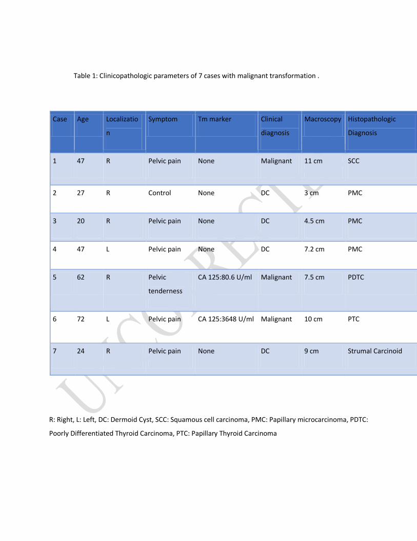
Table 1: Clinicopathologic parameters of 7 cases with malignant transformation .
Case Age Localizatio
n
Symptom Tm marker Clinical
diagnosis
Macroscopy Histopathologic
Diagnosis
1 47 R Pelvic pain None Malignant 11 cm SCC
2 27 R Control None DC 3 cm PMC
3 20 R Pelvic pain None DC 4.5 cm PMC
4 47 L Pelvic pain None DC 7.2 cm PMC
5 62 R Pelvic
tenderness
CA 125:80.6 U/ml Malignant 7.5 cm PDTC
6 72 L Pelvic pain CA 125:3648 U/ml Malignant 10 cm PTC
7 24 R Pelvic pain None DC 9 cm Strumal Carcinoid
R: Right, L: Left, DC: Dermoid Cyst, SCC: Squamous cell carcinoma, PMC: Papillary microcarcinoma, PDTC:
Poorly Differentiated Thyroid Carcinoma, PTC: Papillary Thyroid Carcinoma
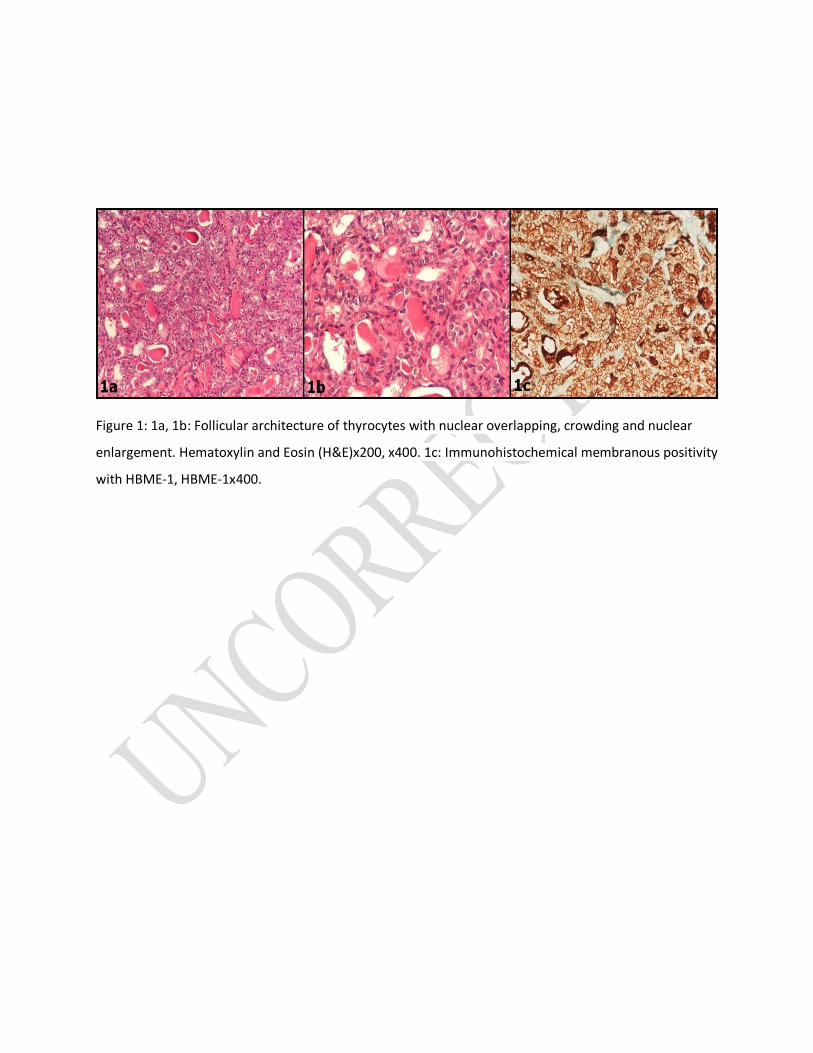
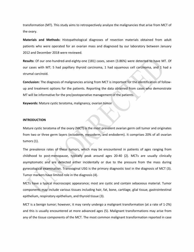
Figure 1: 1a, 1b: Follicular architecture of thyrocytes with nuclear overlapping, crowding and nuclear
enlargement. Hematoxylin and Eosin (H&E)x200, x400. 1c: Immunohistochemical membranous positivity
with HBME-1, HBME-1x400.
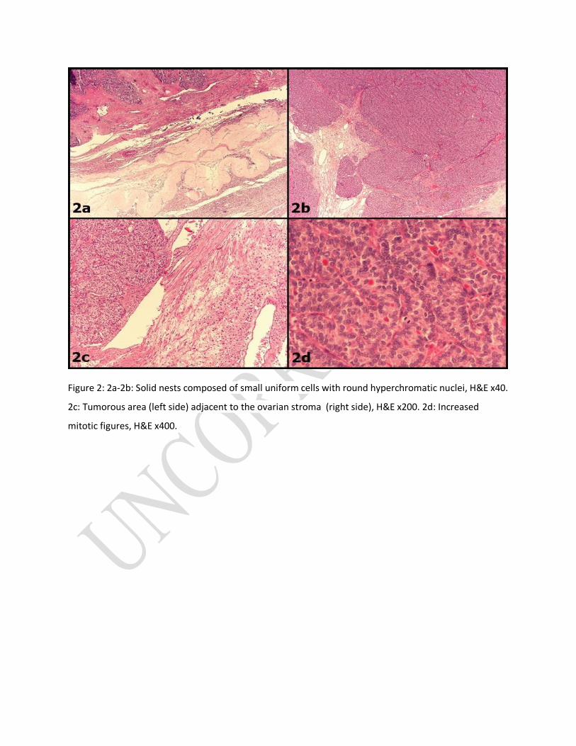
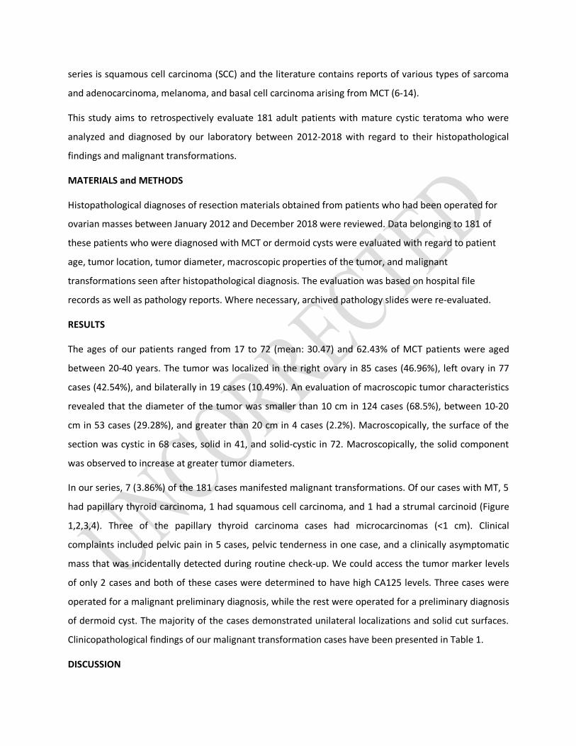
Figure 2: 2a-2b: Solid nests composed of small uniform cells with round hyperchromatic nuclei, H&E x40.
2c: Tumorous area (left side) adjacent to the ovarian stroma (right side), H&E x200. 2d: Increased
mitotic figures, H&E x400.
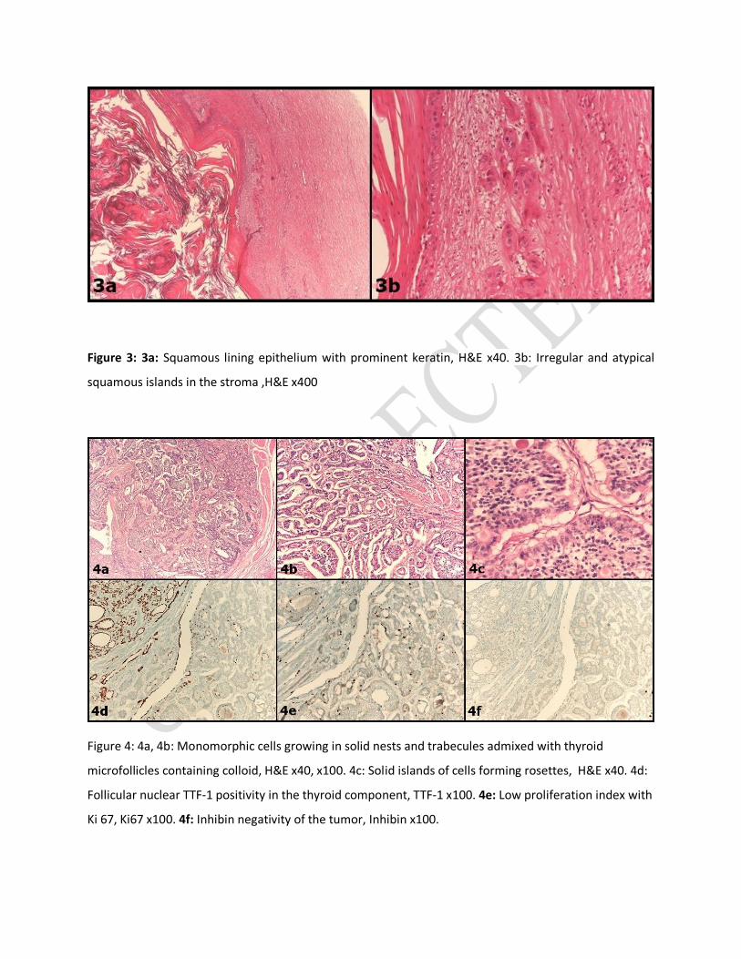
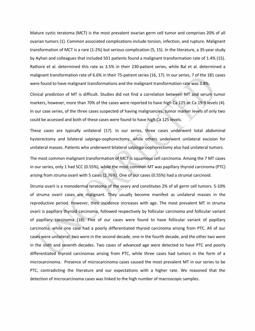
Figure 3: 3a: Squamous lining epithelium with prominent keratin, H&E x40. 3b: Irregular and atypical
squamous islands in the stroma ,H&E x400
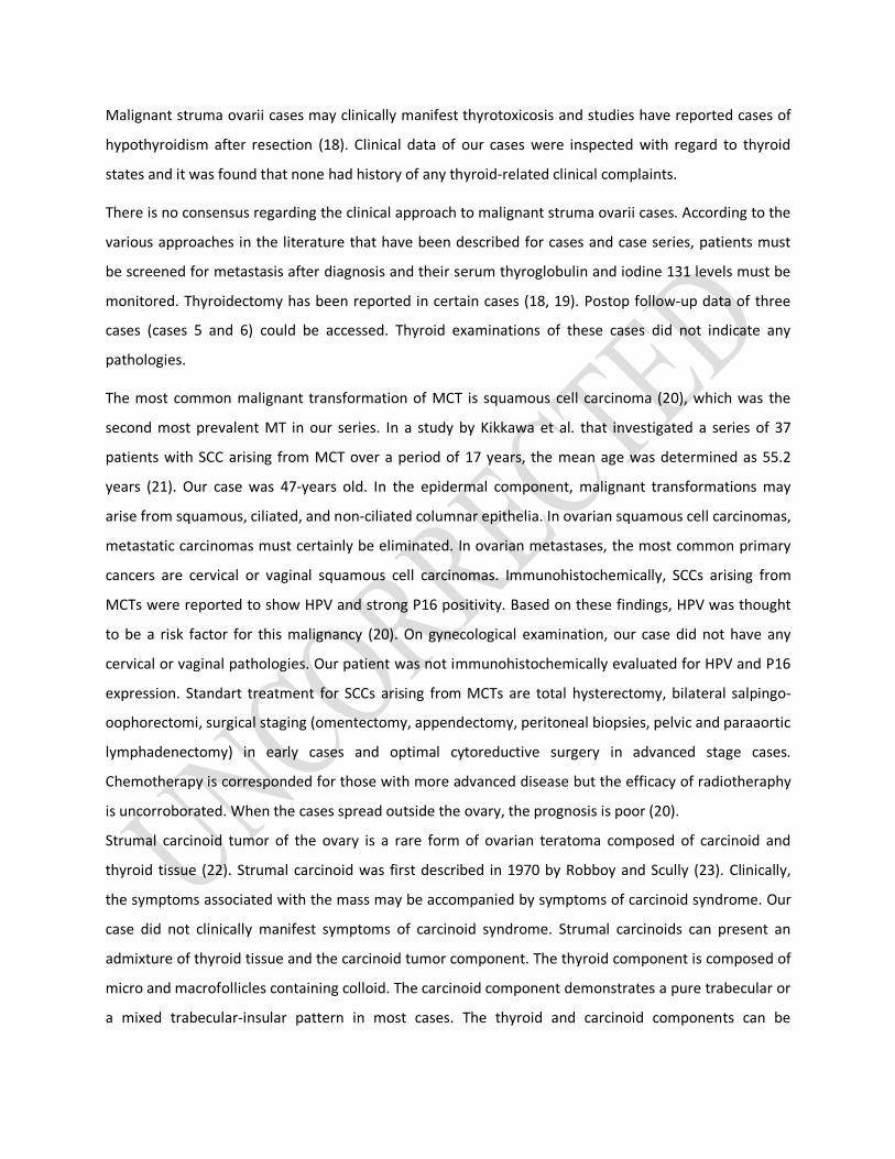
Figure 4: 4a, 4b: Monomorphic cells growing in solid nests and trabecules admixed with thyroid
microfollicles containing colloid, H&E x40, x100. 4c: Solid islands of cells forming rosettes, H&E x40. 4d:
Follicular nuclear TTF-1 positivity in the thyroid component, TTF-1 x100. 4e: Low proliferation index with
Ki 67, Ki67 x100. 4f: Inhibin negativity of the tumor, Inhibin x100.