Embed Size (px)
Citation preview
a
bMDDIFTQCREGNAVAVRLWLDNTENDLNQGDDHGFSPLHWACREGRSAVVEMLIMRGARINVMNRGDDTP
LHLAASHGHRDIVQKLLQYKADINAVNEHGNVPLHYACFWGQDQVAEDLVANGALVSICNKYGEMPVDKA
KAPLRELLRERAEKMGQNLNRIPYKDTFWKGTTRTRPRNGTLNKHSGIDFKQLNFLTKLNENHSGELWKG
RWQGNDIVVKVLKVRDWSTRKSRDFNEECPRLRIFSHPNVLPVLGACQSPPAPHPTLITHWMPYGSLYNV
LHEGTNFVVDQSQAVKFALDMARGMAFLHTLEPLIPRHALNSRSVMIDEDMTARISMADVKFSFQCPGRM
YAPAWVAPEALQKKPEDTNRRSADMWSFAVLLWELVTREVPFADLSNMEIGMKVALEGLRTIPPGISPHV
CKLMKICMNEDPAKRPKFDMIVPILEKMQDK
50kDa50
75100150250
37
25
20
15
HeL
a ly
sate
GS
T+ly
sate
Pull-down
GS
T-O
spE
GS
T-O
spE
+lys
ate
GS
T
y12
y13
y11
y4 y7y3 y5 y8 y9 b14 + H2Oy1y6
b8 b9
b10b7
50
70
100
60
30
20
90
80
40
10
Inte
nsity
%
69.0 389.6 710.2 1030.8 1351.4 1672.0
Mass (m/z)
3743.4
G D D T P L H L A A S H G H Rb8
y7 y6 y5 y4 y3y12y13
b9 b10b7
y8y9y11
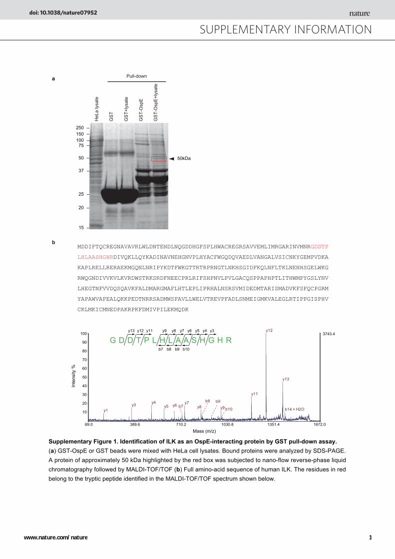
Supplementary Figure 1. Identification of ILK as an OspE-interacting protein by GST pull-down assay. (a) GST-OspE or GST beads were mixed with HeLa cell lysates. Bound proteins were analyzed by SDS-PAGE. A protein of approximately 50 kDa highlighted by the red box was subjected to nano-flow reverse-phase liquid chromatography followed by MALDI-TOF/TOF (b) Full amino-acid sequence of human ILK. The residues in red belong to the tryptic peptide identified in the MALDI-TOF/TOF spectrum shown below.
SUPPLEMENTARY INFORMATIONdoi: 10.1038/nature07952
www.nature.com/nature 1
c
a
d
b
ILK
OspE
ILK-/-/ WT-ILK
ILK-/-/ K220M-ILK
ILK-/-/ S343D-ILK
OspE
Whole-cell Lysate IP: IgG IP: ILK
- + +
+ +
++ +
- -
- - -
- -+
- --
- - -
+ +- -- - -- --
- + +
+ +
++ +
- -
- - -
- -+
- --
- - -
+ +- -- - -- --
- + +
+ +
++ +
- -
- - -
- -+
- --
- - -
+ +- -- - -- --
ILK
OspE
ILK-/-/ WT-ILK
OspE
MBP
- + +
+ ++ +
-
- + +-
32P-MBPAutoradiogram
Autoradiogram
CBB staining GST-ILK
GST-ILK
MBP
GST
GST-OspE
- + +
- +
+ - -
- -
+
-
- - -+
32P-MBP
GST-OspE
GST
MBP
pAkt(S473)
Akt
pGSK3β(S9)
GSK3β
Moc
k
Moc
k
Moc
k
Moc
k
Osp
E
Osp
E
Osp
E
Osp
E
ILK-/- WT-ILK K220M-ILK S343D-ILK
ILK-/- + - -+
ILK-/- + - - - - - -+ + - - - - - -+ + - - - - - -+
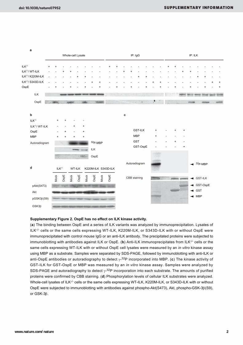
Supplementary Figure 2. OspE has no effect on ILK kinase activity.(a) The binding between OspE and a series of ILK variants was analyzed by immunoprecipitation. Lysates of ILK-/- cells or the same cells expressing WT-ILK, K220M-ILK, or S343D-ILK with or without OspE were immunoprecipitated with control mouse IgG or an anti-ILK antibody. The precipitated proteins were subjected to immunoblotting with antibodies against ILK or OspE. (b) Anti-ILK immunoprecipitates from ILK-/- cells or the same cells expressing WT-ILK with or without OspE cell lysates were measured by an in vitro kinase assay using MBP as a substrate. Samples were separated by SDS-PAGE, followed by immunoblotting with anti-ILK or anti-OspE antibodies or autoradiography to detect γ-32P incorporated into MBP. (c) The kinase activity of GST-ILK for GST-OspE or MBP was measured by an in vitro kinase assay. Samples were analyzed by SDS-PAGE and autoradiography to detect γ-32P incorporation into each substrate. The amounts of purified proteins were confirmed by CBB staining. (d) Phosphorylation levels of cellular ILK substrates were analyzed. Whole-cell lysates of ILK-/- cells or the same cells expressing WT-ILK, K220M-ILK, or S343D-ILK with or without OspE were subjected to immunoblotting with antibodies against phospho-Akt(S473), Akt, phospho-GSK-3β(S9), or GSK-3β.
doi: 10.1038/nature07952 SUPPLEMENTARY INFORMATION
www.nature.com/nature 2
b
c
d
ILK
α-Parvin
β-Parvin
PINCH
OspE
IP:ILK
ILK
-/-/ M
ock
ILK
flox/
flox /
Moc
k
ILK
-/-/ O
spE
ILK
flox/
flox /
Osp
E
Input
ILK
α-Parvin
β-Parvin
PINCH
OspE
ILK
-/-/ M
ock
ILK
flox/
flox /
Moc
k
ILK
-/-/ O
spE
ILK
flox/
flox /
Osp
E
wild
type
N-te
rm
C-te
rm
C-te
rm E
359K
wild
type
N-te
rm
C-te
rm
C-te
rm E
359K
wild
type
N-te
rm
C-te
rm
C-te
rm E
359K
GST-OspE GST
Pull down
Input
IB:MycIB:Myc
KINASEANKILK wide type
ILK N-term
ILK C-term
ILK C-term E359K E359K
OspE binding
+
-
+
+
6 12 180
OspE
ILK
Actin
ILK
Actin
CHX h
Mock
6 12 180CHX h
a
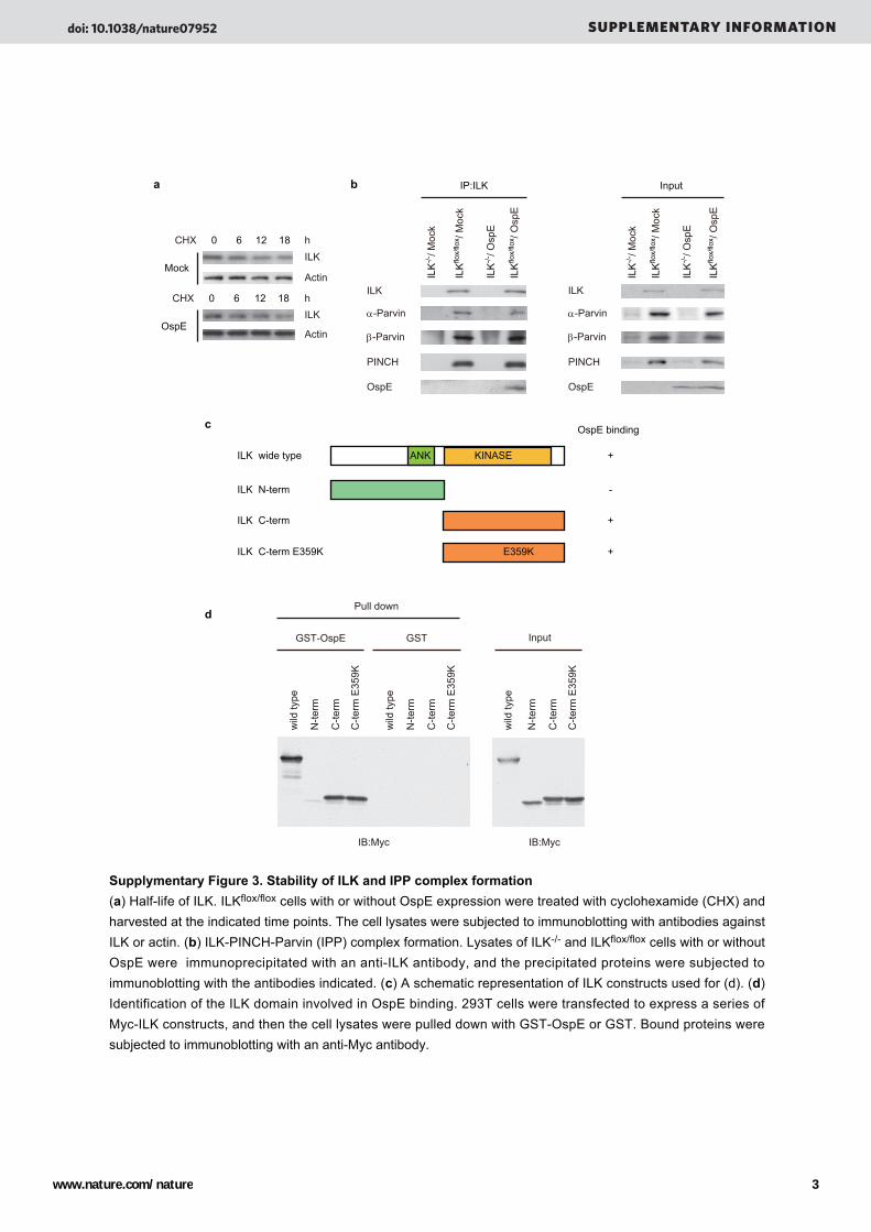
Supplymentary Figure 3. Stability of ILK and IPP complex formation(a) Half-life of ILK. ILKflox/flox cells with or without OspE expression were treated with cyclohexamide (CHX) and harvested at the indicated time points. The cell lysates were subjected to immunoblotting with antibodies against ILK or actin. (b) ILK-PINCH-Parvin (IPP) complex formation. Lysates of ILK-/- and ILKflox/flox cells with or without OspE were immunoprecipitated with an anti-ILK antibody, and the precipitated proteins were subjected to immunoblotting with the antibodies indicated. (c) A schematic representation of ILK constructs used for (d). (d) Identification of the ILK domain involved in OspE binding. 293T cells were transfected to express a series of Myc-ILK constructs, and then the cell lysates were pulled down with GST-OspE or GST. Bound proteins were subjected to immunoblotting with an anti-Myc antibody.
doi: 10.1038/nature07952 SUPPLEMENTARY INFORMATION
www.nature.com/nature 3
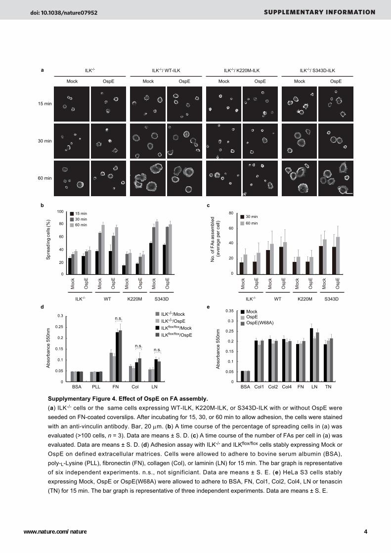
a
b c
15 min
30 min
60 min
Spre
adin
g ce
lls (%
)
100
80
60
40
20
0
30 min15 min
60 min
ILK-/- WT K220M S343D
ILK-/- ILK-/-/ WT-ILK ILK-/-/ K220M-ILK ILK-/-/ S343D-ILK
Osp
E
Moc
k
Moc
k
Moc
k
Moc
k
Osp
E
Osp
E
Osp
E
No.
of F
As a
ssem
bled
(a
vera
ge p
er c
ell)
30 min
60 min
80
60
40
20
0
ILK-/- WT K220M S343D
Osp
E
Moc
k
Moc
k
Moc
k
Moc
k
Osp
E
Osp
E
Osp
Ed e
0
0.05
0.1
0.15
0.2
0.25
0.3
BSA PLL FN Col LN
ILK-/-/MockILK-/-/OspEILKflox/flox/MockILKflox/flox/OspE
n.s.
n.s.n.s.
0
0.05
0.1
0.15
0.2
0.25
0.3
0.35
BSA Col1 Col2 Col4 FN LN TN
MockOspEOspE(W68A)
Abs
orba
nce
550n
m
Abs
orba
nce
550n
m
Supplymentary Figure 4. Effect of OspE on FA assembly.(a) ILK-/- cells or the same cells expressing WT-ILK, K220M-ILK, or S343D-ILK with or without OspE were seeded on FN-coated coverslips. After incubating for 15, 30, or 60 min to allow adhesion, the cells were stained with an anti-vinculin antibody. Bar, 20 μm. (b) A time course of the percentage of spreading cells in (a) was evaluated (>100 cells, n = 3). Data are means ± S. D. (c) A time course of the number of FAs per cell in (a) was evaluated. Data are means ± S. D. (d) Adhesion assay with ILK-/- and ILKflox/flox cells stably expressing Mock or OspE on defined extracellular matrices. Cells were allowed to adhere to bovine serum albumin (BSA), poly-L-Lysine (PLL), fibronectin (FN), collagen (Col), or laminin (LN) for 15 min. The bar graph is representative of six independent experiments. n.s., not significiant. Data are means ± S. E. (e) HeLa S3 cells stably expressing Mock, OspE or OspE(W68A) were allowed to adhere to BSA, FN, Col1, Col2, Col4, LN or tenascin (TN) for 15 min. The bar graph is representative of three independent experiments. Data are means ± S. E.
Mock OspE Mock OspE Mock OspE Mock OspE
doi: 10.1038/nature07952 SUPPLEMENTARY INFORMATION
www.nature.com/nature 4
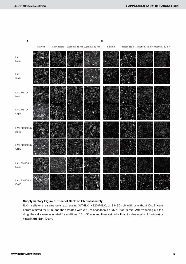
a b
Starved Nocodazole Washout, 15 min Washout, 30 min Starved Nocodazole Washout, 15 min Washout, 30 min
ILK-/-
/Mock
ILK-/-
/OspE
ILK-/-/ WT-ILK
/Mock
ILK-/-/ WT-ILK
/OspE
ILK-/-/ K220M-ILK
/Mock
ILK-/-/ K220M-ILK
/OspE
ILK-/-/ S343D-ILK
/Mock
ILK-/-/ S343D-ILK
/OspE
Supplymentary Figure 5. Effect of OspE on FA disassembly.ILK-/- cells or the same cells expressing WT-ILK, K220M-ILK, or S343D-ILK with or without OspE were serum-starved for 48 h, and then treated with 2.5 μM nocodazole at 37 ºC for 30 min. After washing out the drug, the cells were incubated for additional 15 or 30 min and then stained with antibodies against tubulin (a) or vinculin (b). Bar, 10 μm.
doi: 10.1038/nature07952 SUPPLEMENTARY INFORMATION
www.nature.com/nature 5

a
5 15 300 5 15 300
Mock OspE
IP: β1 integrin
IB: β1 integrin
IB: Streptavidin-HRP
c
b
0
0.5
1
1.5
2
Mock OspE OspE(W68A)
*
n.s.
β1 in
tegr
in, a
ctiv
e st
ate
leve
l
MockOspE
β1 in
tegr
in, a
ctiv
e st
ate
leve
l
0
0.5
1
1.5
ILKflox/floxILK-/-ILKflox/floxILK-/-
β1 in
tegr
in, t
otal
leve
l
0
0.5
1
1.5
ILKflox/floxILK-/-
β1 in
tegr
in, s
urfa
ce le
vel
0
1
0.5
1.5
0
0.5
1
1.5
2
Mock OspE OspE(W68A)
β1 in
tegr
in, t
otal
leve
l
0
0.5
1
1.5
2
Mock OspE OspE(W68A)
*
n.s.
β1 in
tegr
in, s
urfa
ce le
vel
Supplymentary Figure 6. β1 Integrin internalization was delayed by OspE expression.(a) The expression of β1 integrin in ILK-/- and ILKflox/flox cells stably expressing Mock or OspE was analyzed by FACS with an anti-β1 integrin (Ha2/5) antibody under non-permeabilizing (left) or permeabilizing (middle) conditions. The active state integrin was detected with an anti-β1 integrin (9EG7) antibody (right). The bar graphs are representative of three independent experiments. Data are means ± S. D. (b) The expression of β1 integrin in HeLa S3 cells expressing Mock, OspE, or OspE(W68A) was analyzed by FACS with an anti-β1 integrin antibody under non-permeabilizing (left) or permeabilizing (middle) conditions. The active state integrin was detected with binding of FITC labeled-FN7-10 fragments to HeLa S3 cells (right). The bar graphs are representative of three independent experiments. Data are means ± S. E. *p<0.01. n.s., not significant. (c) Internalization of β1 integrin. The cell surface proteins were labeled with biotin at 4 °С for 30 min. After labelling the cells were incubated at 37 °С for the indicated time points (min), and then the cells were subjected to immunoprecipitation with an anti-β1 integrin antibody. Precipitated proteins were analyzed by probing with peroxidase-conjugated streptavidin or an anti-β1 integrin antibody.
doi: 10.1038/nature07952 SUPPLEMENTARY INFORMATION
www.nature.com/nature 6
Inpu
t
a b
c
d e
f g
ILK
CBB
GS
T
Osp
E 1
-58
Osp
E 1
-44
Osp
E 2
9-88
Osp
E 4
4-88
Osp
E 2
9-58
Osp
E 1
-65
Osp
E 1
-75
Osp
E 1
-80
Osp
E 4
A
Osp
E Q
67A
Osp
E W
68A
Osp
E L
69A
Osp
E T
70A
Osp
E
Pull down
Mock
OspE
OspE(W68A)
Actin MergeVinculin
GFP OspE 1-58 OspE 1-44 OspE 29-88 OspE 44-88
OspE 29-58 OspE 1-65 OspE 1-75 OspE 1-80 OspE 4A
OspE Q67A OspE W68A OspE L69A OspE T70A OspE
1 88
58
44
65
75
80
29
AAAA
Q67A
W68A
L69A
T70A
OspE
OspE 1-58
OspE 1-44
OspE 29-88
OspE 44-88
OspE 29-58
OspE 1-65
OspE 1-75
OspE 1-80
OspE 4A
OspE Q67A
OspE W68A
OspE L69A
OspE T70A
88
8844
29 58
FA n
umbe
r/cel
l
β1 in
tegr
in, t
otal
leve
l
β1 in
tegr
in, s
urfa
ce le
vel
ILK
OspE
pFAK(Y397)
FAK
Moc
k
Osp
E
Osp
E(W
68A
)
β1 integrin
Actin
**250
200
150
100
50
0
Moc
k
Osp
E
Osp
E(W
68A
)
2.0
1.5
1.0
0.5
0
Moc
k
Osp
E
Osp
E(W
68A
)
* *2.0
1.5
1.0
0.5
0
Moc
k
Osp
E
Osp
E(W
68A
)
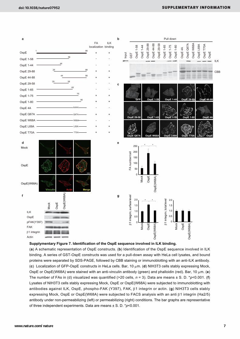
Supplymentary Figure 7. Identification of the OspE sequence involved in ILK binding. (a) A schematic representation of OspE constructs. (b) Identification of the OspE sequence involved in ILK binding. A series of GST-OspE constructs was used for a pull-down assay with HeLa cell lysates, and bound proteins were separated by SDS-PAGE, followed by CBB staining or immunoblotting with an anti-ILK antibody. (c) Localization of GFP-OspE constructs in HeLa cells. Bar, 10 μm. (d) NIH3T3 cells stably expressing Mock, OspE or OspE(W68A) were stained with an anti-vinculin antibody (green) and phalloidin (red). Bar, 10 μm. (e) The number of FAs in (d) visualized was quantified (>20 cells, n = 3). Data are means ± S. D. *p<0.001. (f) Lysates of NIH3T3 cells stably expressing Mock, OspE or OspE(W68A) were subjected to immunoblotting with antibodies against ILK, OspE, phospho-FAK (Y397), FAK, β1 integrin or actin. (g) NIH3T3 cells stably expressing Mock, OspE or OspE(W68A) were subjected to FACS analysis with an anti β1 integrin (Ha2/5) antibody under non-permeabilizing (left) or permeabilizing (right) conditions. The bar graphs are representative of three independent experiments. Data are means ± S. D. *p<0.001.
FAlocalization
ILKbinding
+
-
+
+
-
-
-
+
+
-
+
-
+
+
+
-
+
+
-
-
-
+
+
-
+
-
+
+
doi: 10.1038/nature07952 SUPPLEMENTARY INFORMATION
www.nature.com/nature 7
b
ILK
CBB
Pull down
Inpu
t
GS
T-O
spE
GS
T-O
spO
1-2
GS
T
GS
T-O
spO
1-1
GS
T-O
sp1 S
TYM
c
a
OspE
OspO1-1
OspO1-2
OspO1STYM
GFP Vinculin Actin Merge
S. flexneri OspE1 --MLTQTIFPCLPQKQENIILEVS------NPVLLSSTVTTDGYTVFNKKAAIYELQIP-AASRTKTLKFTATEMQWLTKINEAGIDEKQSQRYSDF
S. flexneri OspE2 --MLTQTIFPCLPQKQENIILEVS------NPVLLSSTVTTDGYTVFNKKAAIYELQIP-AANRTKTLKFTATEMQWLTKINEAGIDEKQSQRYSDF
S. dysenteriae OspE1 --MLTQTIFPCLPQKQENIILEVS------NPVLLSSTVTTDGYTVFNKKAAIYELQIP-AANRTKTLKFTATEMQWLTKINEAGIDEKQSQRYSDF
S. sonnei OspE2 --MLTQTIFPCLPQKQENIILEVS------NPVLLSSTVTTDGYTVFNKKAAIYELQIP-AANRTKTLKFTATEMQWLTKINEVGIVEKQSQRHSNI
S. boydii OspE1 --MLTQTIFPCLPQKQENIILEVS------NPVLLSSTVTTDGYTVFNKKAAIYELQIP-ATNRTKTLKFTATEMQWLTKINEVGIVEKQSQRHSNI
EHEC OspO1-1 MPFSIKNRFSSSQVHYPEISGPIKDKPASKNCILTSTTCNVDSYTVYQKKACSFDMRPPGAGERTPKLKLSVTEMTWLSKTIETEIHNTKE------
EHEC OspO1-2 MPFSIKSIFSGHTWHQPEISRPIADKSSTKNCILDSTTCNVDGFTVFNRRSCSFDMRPPGSADRTPQLRLSISEVAWMSKIIETETNNTNKS-----
S. typhimurium Osp1STYM MPFSIKNICSGPKGHCPEISSPIQDKPVPRNCTLTSTTCDIQSYTVFSRWSCSYEMRPPGAEERTPRLKFSATELSWLSKTIETERRNTKE------
S. enteritidis Osp1SENT MPFSIKNICSGPQGHYPEISRPIQNKPVPRNCTLISTTCKIQEYTVFSRCSCSFEMRPPGTEERTPRLKLSATELSWLSKTIETEMRNTKE------
EHEC EspO1O103:H2 MPLSIGSCFSCHTGRRPEISDPIIDKPSTKNCELISTTCNVDGITVISRSSYGFDMKPPGAGERAPRLKLSASEAQWMAAIIDAEGHNISNT-----
EPEC EspO1EPE22 MPFSIKNIFSNSKGSYPEISGPVQDKPVSKNCTLTSTTCSMNDYTVFSRKSCTFDMRPPGAGDRTPQLKLSASELIWLSKTIDTERNNIKE------
AEPEC EspO1AEPEC MPFSIKNIFSNSKGNYPEISGPVQDKPVSKNCTLTSTTCPMNDYTVFSRKSCTFDMRPPGAGDRTPKLKLAATEMTWLSKTIETEIHNTKE------
C. rodentium EspO1CIROD MPLSIRNIFSRASTHRPEISGPVIDKPIPKNCTLISSTCNLDGIMVINRRTSFYDIKPPGAGERQPSLKISASEAQWMCKIIETEINNSNK------
*
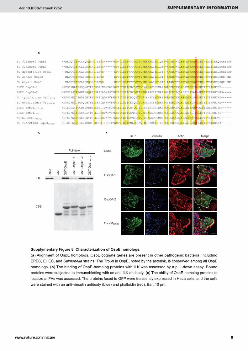
Supplymentary Figure 8. Characterization of OspE homologs.(a) Alignment of OspE homologs. OspE cognate genes are present in other pathogenic bacteria, including EPEC, EHEC, and Salmonella strains. The Trp68 in OspE, noted by the asterisk, is conserved among all OspE homologs. (b) The binding of OspE-homolog proteins with ILK was assessed by a pull-down assay. Bound proteins were subjected to immunoblotting with an anti-ILK antibody. (c) The ability of OspE-homolog proteins to localize at FAs was assessed. The proteins fused to GFP were transiently expressed in HeLa cells, and the cells were stained with an anti-vinculin antibody (blue) and phalloidin (red). Bar, 10 μm.
doi: 10.1038/nature07952 SUPPLEMENTARY INFORMATION
www.nature.com/nature 8
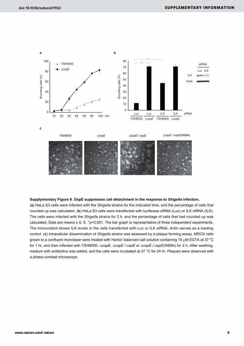
a b
c
ILK
Actin
Luc ILK
siRNA
0
10
20
30
40
50
60
70
80
siRNALuc Luc ILK ILK
YSH6000 ΔospE YSH6000 ΔospE
*
Rou
ndin
g ce
lls (%
)
*
Rou
ndin
g ce
lls (%
)
0
20
40
60
80
100
10 20 30 45 60 90 120
YSH6000
ΔospE
min
YSH6000 ΔospE ΔospE / ospE ΔospE / ospE(W68A)
Supplymentary Figure 9. OspE suppresses cell detachment in the response to Shigella infection.(a) HeLa S3 cells were infected with the Shigella strains for the indicated time, and the percentage of cells that rounded up was calculated. (b) HeLa S3 cells were transfected with luciferase siRNA (Luc) or ILK siRNA (ILK). The cells were infected with the Shigella strains for 2 h, and the percentage of cells that had rounded up was calculated. Data are means ± S. E. *p<0.001. The bar graph is representative of three independent experiments. The immunoblot shows ILK levels in the cells transfected with Luc or ILK siRNA. Actin serves as a loading control. (c) Intracellular dissemination of Shigella strains was assessed by a plaque forming assay. MDCK cells grown to a confluent monolayer were treated with Hanks' balanced salt solution containing 10 μM EGTA at 37 °С for 1 hr, and then infected with YSH6000, ΔospE, ΔospE / ospE or ΔospE / ospE(W68A) for 2 h. After washing, medium with antibiotics was added, and the cells were incubated at 37 °С for 24 hr. Plaques were observed with a phase-contrast microscope.
doi: 10.1038/nature07952 SUPPLEMENTARY INFORMATION
www.nature.com/nature 9
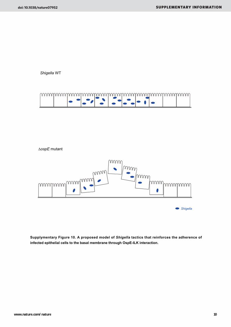
Shigella WT
ΔospE mutant
Shigella
Supplymentary Figure 10. A proposed model of Shigella tactics that reinforces the adherence of infected epithelial cells to the basal membrane through OspE-ILK interaction.
doi: 10.1038/nature07952 SUPPLEMENTARY INFORMATION
www.nature.com/nature 10