Embed Size (px)
Citation preview
Name _______________________________
DOGFISH SHARK
DISSECTION
Each individual person must complete this packet.
Completion of this packet will be your grade for this project.
Page 1
Name _______________________________
Dissection You will be dissecting a dogfish shark: Squalus acanthias. The equipment you will be using includes:
o a large dissection tray o surgical scissors o scalpel o probe o forceps
Dissection is a learned skill that takes practice and patience. Some general rules to remember are:
1. Do not make deep cuts with scissors or scalpels as you may damage tissue underneath.
2. Know the anatomical terms listed next so you can follow the directions. 3. Read the section you are working on before you start cutting. 4. Try to answer each other’s questions about anatomy before asking your
teacher for help. Use the notes from earlier this 6-weeks to help you identify organs.
Anatomical Terms Cranial - toward the head Caudal - toward the rear Dorsal - toward the spinal cord (back) Ventral - toward the belly Medial - toward the middle Distal - away from Lateral - to the side
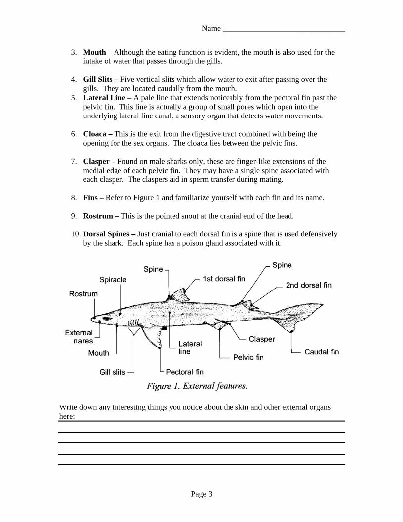
I. External Features (Figure 1) Familiarize yourself with the following external features:
1. External Nares – These are a pair of openings (nostrils) on each side of the head,
cranial from the eyes. Water is taken into the smaller of the two openings and expelled through the larger opening. The water passes by a sensory membrane allowing the shark to detect chemicals in the water.
2. Spiracles – These are small openings caudal from the eyes. These openings
allow water to pass through the gills even when the shark’s mouth is closed.
Page 2
Name _______________________________
3. Mouth – Although the eating function is evident, the mouth is also used for the intake of water that passes through the gills.
4. Gill Slits – Five vertical slits which allow water to exit after passing over the
gills. They are located caudally from the mouth. 5. Lateral Line – A pale line that extends noticeably from the pectoral fin past the
pelvic fin. This line is actually a group of small pores which open into the underlying lateral line canal, a sensory organ that detects water movements.
6. Cloaca – This is the exit from the digestive tract combined with being the
opening for the sex organs. The cloaca lies between the pelvic fins.
7. Clasper – Found on male sharks only, these are finger-like extensions of the medial edge of each pelvic fin. They may have a single spine associated with each clasper. The claspers aid in sperm transfer during mating.
8. Fins – Refer to Figure 1 and familiarize yourself with each fin and its name.
9. Rostrum – This is the pointed snout at the cranial end of the head.
10. Dorsal Spines – Just cranial to each dorsal fin is a spine that is used defensively
by the shark. Each spine has a poison gland associated with it.
Write down any interesting things you notice about the skin and other external organs here:
Page 3
Name _______________________________
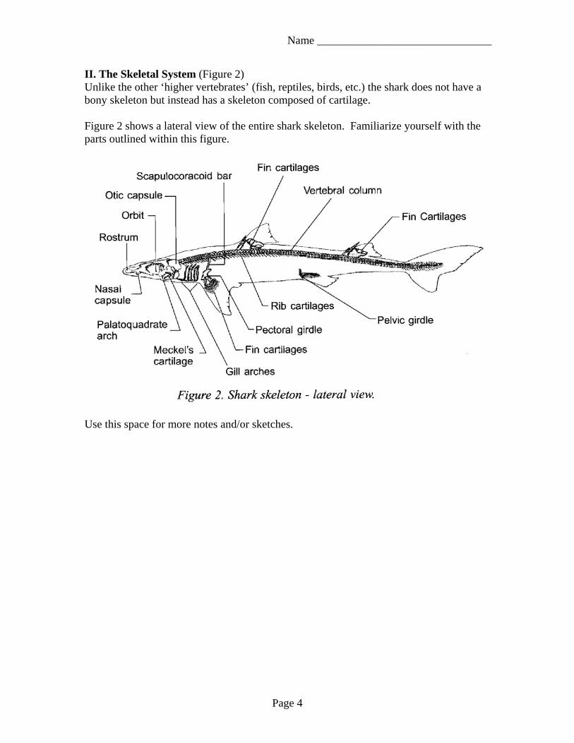
II. The Skeletal System (Figure 2) Unlike the other ‘higher vertebrates’ (fish, reptiles, birds, etc.) the shark does not have a bony skeleton but instead has a skeleton composed of cartilage. Figure 2 shows a lateral view of the entire shark skeleton. Familiarize yourself with the parts outlined within this figure.
Use this space for more notes and/or sketches.
Page 4
Name _______________________________
Observations: 1. How is the shark’s nose different from our own? 2. Why are the Spiracles important? 3. The mouth of the shark is part of which organ system(s)? 4. What is the function of the Gill Slits? 5. What does the Lateral Line do? 6. What two organ systems is the Cloaca a part of? 7. Since the Clasper is only present on male dogfish sharks, what gender is your shark? 8. How many fins does a dogfish shark have? 9. What’s another name for the Rostrum? 10. Where are the Dorsal Spines located?
Page 5
Name _______________________________
III. Beginning the Dissection: The Muscular System You will want to have Page 2 with the anatomical terms handy to help you translate. Place your shark ventral side down to begin. You will need to flip the shark over after step one to complete this section.
1. Remove each of the dorsal spines by cutting where it meets the body. This will prevent you from stabbing yourself unintentionally.
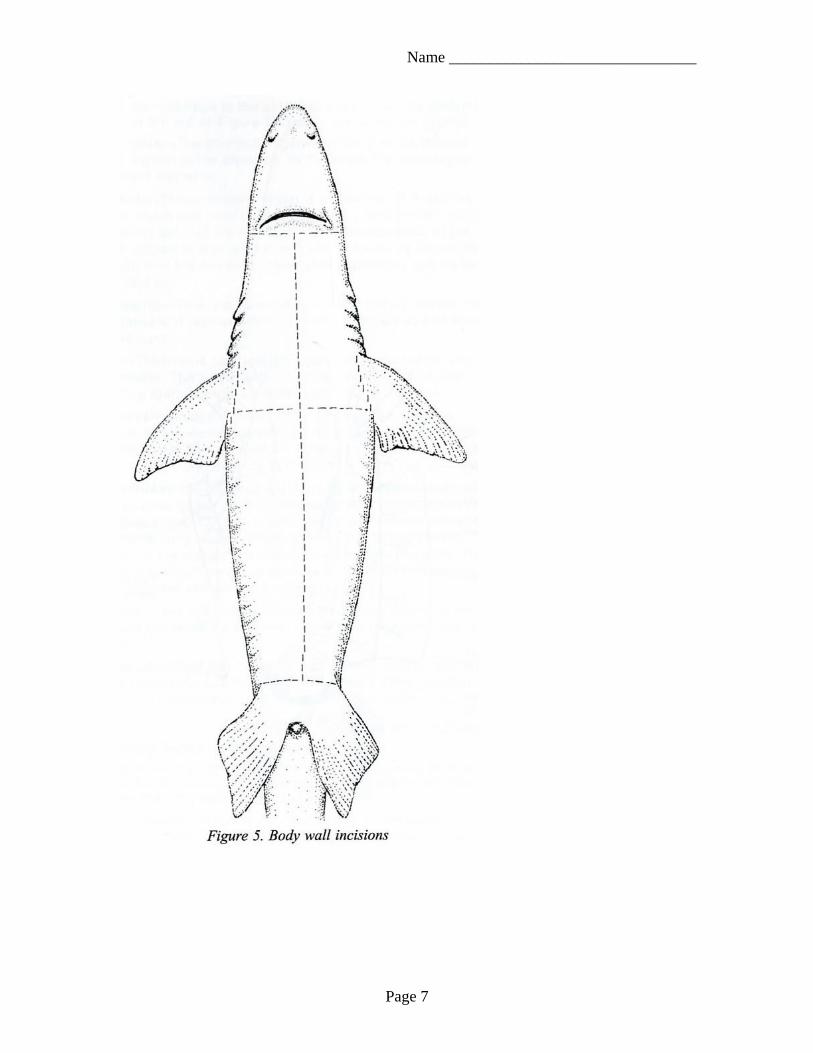
Flip your shark over onto its back. Be sure to refer to the diagram on the next page as you begin cutting into the skin.
2. Make a mid-ventral incision from the cloaca cranially to just below the jaw. Make your incisions shallow.
3. Cut around the head, around each fin, around the spircles, and around the cloaca.
4. From the cloaca cut dorsally around the shark – this will make a circle around the tail. Remember you are cutting through the skin only.
5. Using the handles of your scissors or your gloved fingers carefully peel off the skin to expose the muscles.
6. Compare your specimen with Figure 3 and Figure 4. 7. Try to identify as many of the structures listed as possible.
Make any notes you have in the area below.
Page 6
Name _______________________________
Page 7
Name _______________________________
Page 8
Name _______________________________
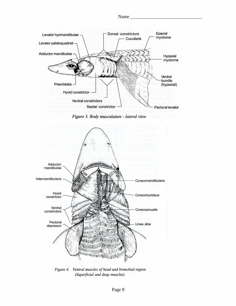
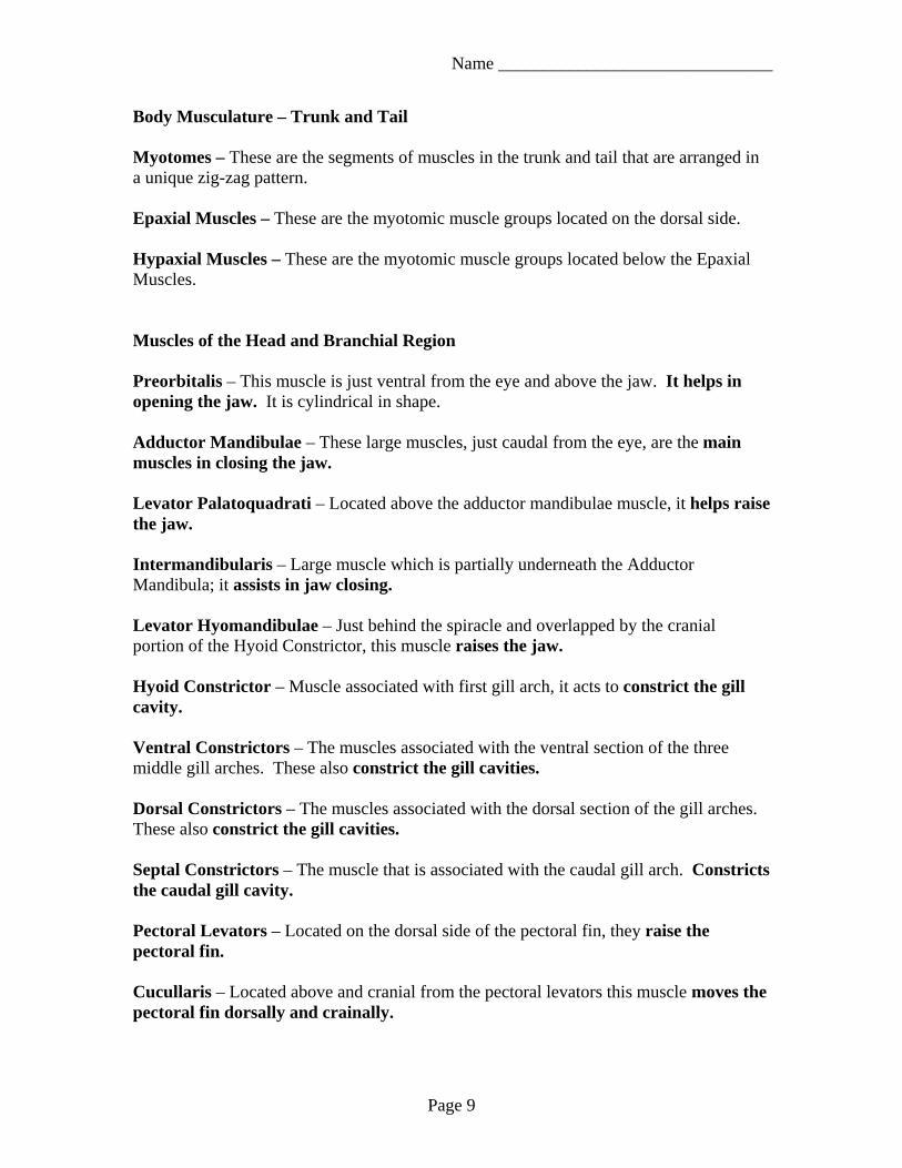
Body Musculature – Trunk and Tail Myotomes – These are the segments of muscles in the trunk and tail that are arranged in a unique zig-zag pattern. Epaxial Muscles – These are the myotomic muscle groups located on the dorsal side. Hypaxial Muscles – These are the myotomic muscle groups located below the Epaxial Muscles. Muscles of the Head and Branchial Region Preorbitalis – This muscle is just ventral from the eye and above the jaw. It helps in opening the jaw. It is cylindrical in shape. Adductor Mandibulae – These large muscles, just caudal from the eye, are the main muscles in closing the jaw. Levator Palatoquadrati – Located above the adductor mandibulae muscle, it helps raise the jaw. Intermandibularis – Large muscle which is partially underneath the Adductor Mandibula; it assists in jaw closing. Levator Hyomandibulae – Just behind the spiracle and overlapped by the cranial portion of the Hyoid Constrictor, this muscle raises the jaw. Hyoid Constrictor – Muscle associated with first gill arch, it acts to constrict the gill cavity. Ventral Constrictors – The muscles associated with the ventral section of the three middle gill arches. These also constrict the gill cavities. Dorsal Constrictors – The muscles associated with the dorsal section of the gill arches. These also constrict the gill cavities. Septal Constrictors – The muscle that is associated with the caudal gill arch. Constricts the caudal gill cavity. Pectoral Levators – Located on the dorsal side of the pectoral fin, they raise the pectoral fin. Cucullaris – Located above and cranial from the pectoral levators this muscle moves the pectoral fin dorsally and crainally.
Page 9
Name _______________________________
Post-Lab – Answer the following questions before continuing with your dissection: 1. How did you position your shark to remove the Dorsal Spines? 2. How did you position your shark once you remove the Dorsal Spines? 3. In step 2 you made an incision from where to where? 4. In steps 3 and 4, why did you cutting around all the fins? 5. Was the shark’s skin as thick as you expected it to be? Why or why not?
Page 10
Name _______________________________
IV. Dissecting the Abdominal Cavity Use the figure on the next page to show you where to cut through the muscles.
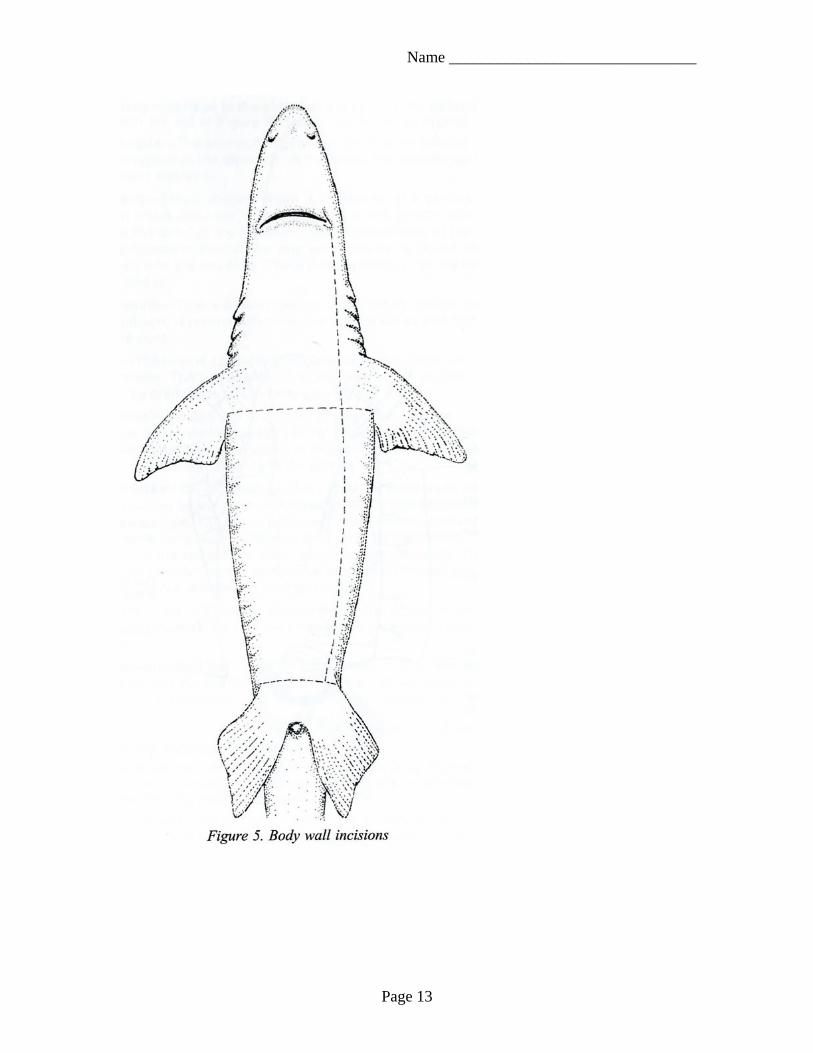
1. Place your shark ventral side up on the dissection tray. Refer to Figure 5 on the page 13
2. Using scissors – blunt tip inside the shark – make a cut from the left side of the jaw (the shark’s left) caudally down through the middle of the gill slits and through the pectoral girdle down to just above the cloaca. Cutting through the pectoral girdle may be difficult. Ask if you need help.
Refer to Figure 5 on the page 13 3. From the cloaca make transverse (side to side) cuts around the shark. 4. From the pectoral girdle, make transverse cuts around dorsally. 5. You may pin the flaps of muscle tissue to the dorsal sides of the shark or remove
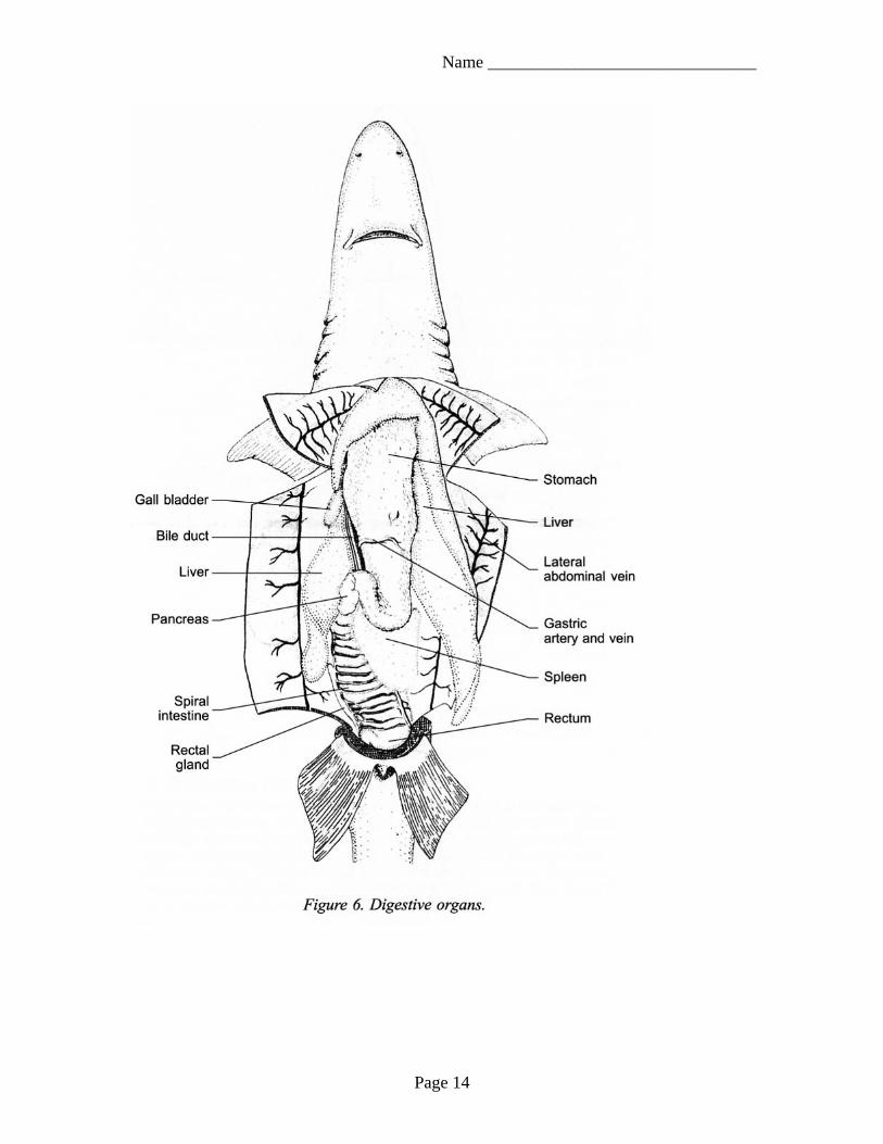
the tissue and place to the side so you can cover the internal organs overnight. At this point, with the help of the figure on page 14, you should be able to identify the organs in the list below. Esophagus – The connection between the pharynx to the stomach. In the shark the esophagus is very short and wide. Stomach – This J-shaped organ is composed of a cardiac portion which lies near to the heard and a limb portion which is after the bend of the stomach. The stomach ends at the pyloric sphincter – a muscular ring which opens or closes the stomach into the intestine. The pyloric sphincter can be felt if you choose to find it. Duodenum – This is a short section immediately caudal from the stomach. It receives liver secretions known as bile from the bile duct. Liver – The liver is composed of three lobes, two large and one smaller. The gall bladder is located within the smaller lobe. The bladder stores the bile secreted by the liver. Pancreas – Divided into two parts: The ventral pancreas, which is easily viewed on the ventral surface of the duodenum and the dorsal pancreas which is long and thin located behind the duodenum and extends to the spleen. Spiral Intestine – Located cranially from the duodenum and distinguished by the extensive network of arteries and veins over its surface. Rectum – This is the short end portion of the digestive tract between the intestine and the cloaca. The rectum stores solid wastes. Spleen – Located just caudal to the stomach and proximal (before) to the spiral intestine. This organ is not part of the digestive tract, but is associated with the circulatory system.
Page 11
Name _______________________________
Circulatory System
1. Lift the flaps over the area of the heart and pin them where they stay out of the way.
2. It may be necessary to cut some tissue that may be attached to the heart. 3. If you would like to cut open the chambers of the heart for a better look you may
do so. You should now be able to identify the some of the structures that are listed below. Sinus Venosus – Dorsal to the ventricle, this is a thin walled, non-muscular sac which acts as a collecting place for deoxygenated blood. Atrium – Similar to the atrium of a human. Ventricle – The main contracting chamber of the heart. Conus Arteriosus – A muscular reservoir that empties after the ventricle contracts. It gives the blood flow an added boost. Mouth Structures Teeth – These are derived from the scales which cover the shark’s body! They have been adapted to function as cutting structures. The teeth of a shark are replaced regularly as they wear out. Pharynx – The cavity caudal from the spiracles to the esophagus. The gill slits open on either side of the caudal region. The gill rakers are cartilaginous protrusions which prevent large particles of food from entering the gills. Tongue – The tongue of the shark is immovable.
Page 12
Name _______________________________
Page 13
Name _______________________________
Page 14
Name _______________________________
Post-Lab – Answer the following questions before continuing with your dissection: 1. How is the shark’s digestive system different from a human’s? 2. How is the shark’s circulatory system different from a human’s? 3. How did you position your shark to begin dissecting the abdominal cavity? 4. Why do you think cutting through the pectoral girdle may have been difficult? 5. Write at least one think you found interesting about the shark’s digestive or circulatory system.
Page 15
Name _______________________________
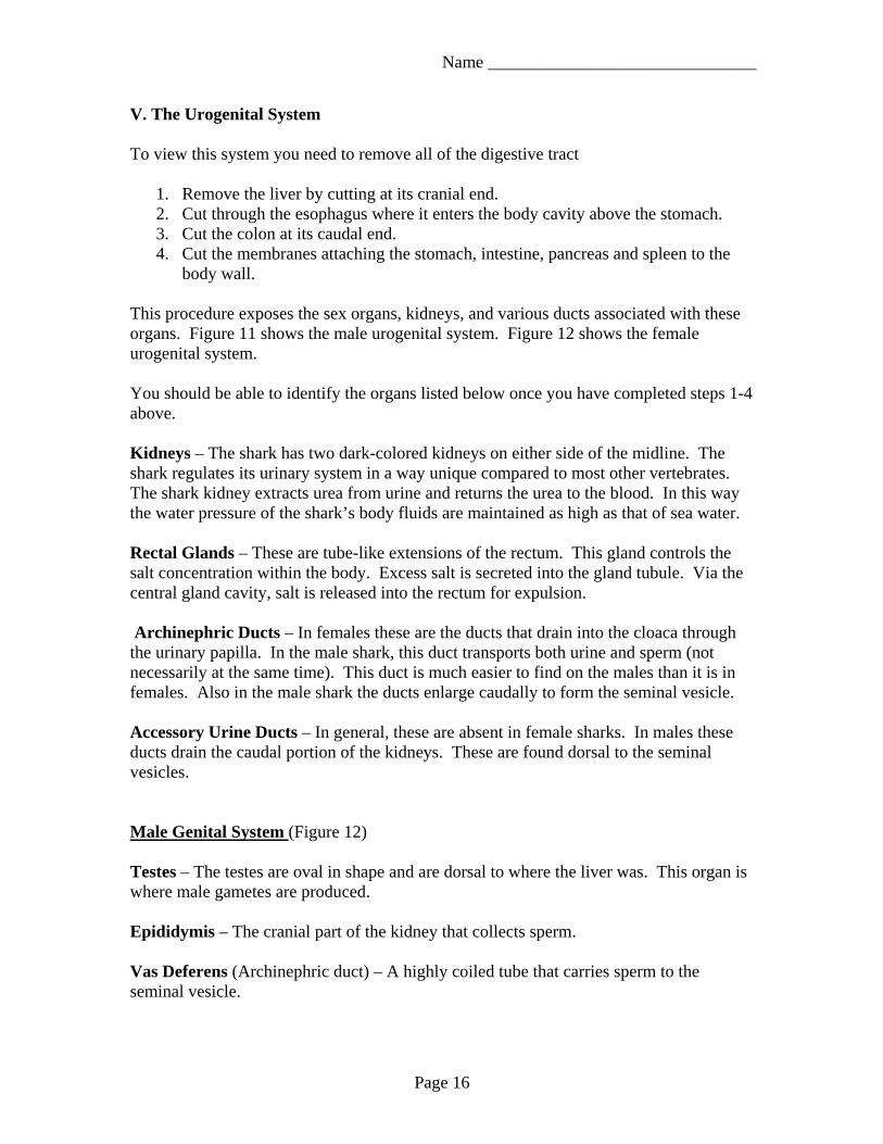
V. The Urogenital System To view this system you need to remove all of the digestive tract
1. Remove the liver by cutting at its cranial end. 2. Cut through the esophagus where it enters the body cavity above the stomach. 3. Cut the colon at its caudal end. 4. Cut the membranes attaching the stomach, intestine, pancreas and spleen to the
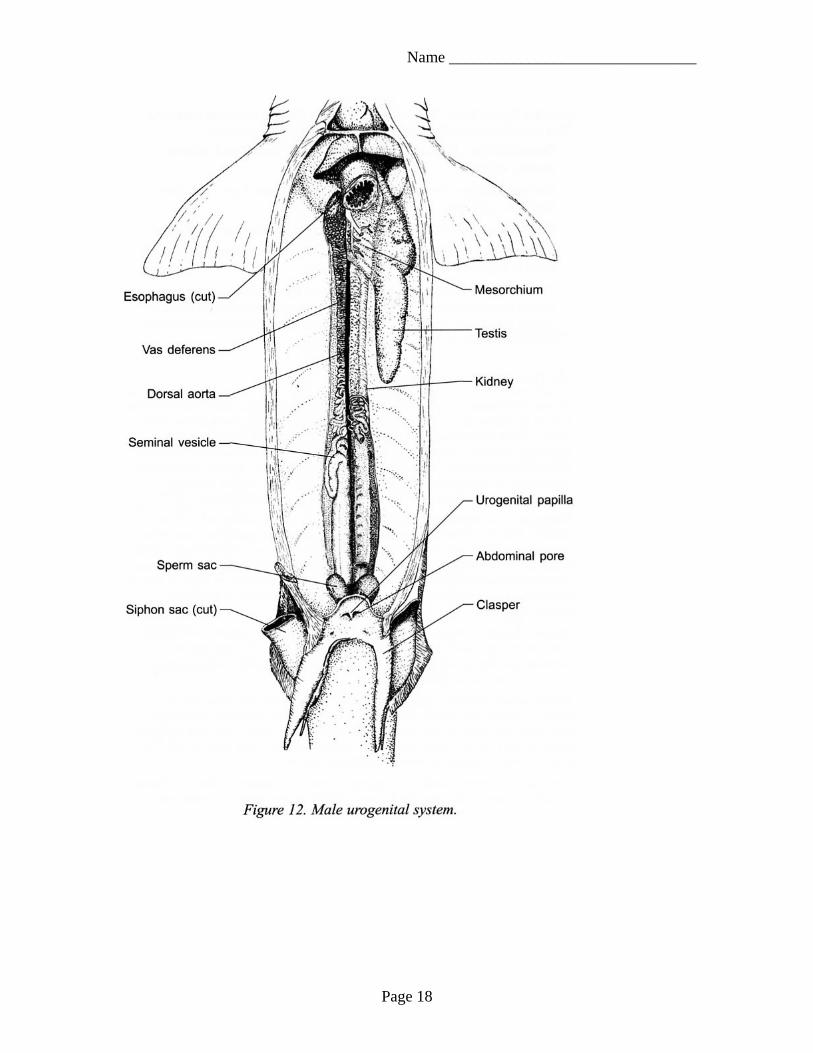
body wall. This procedure exposes the sex organs, kidneys, and various ducts associated with these organs. Figure 11 shows the male urogenital system. Figure 12 shows the female urogenital system. You should be able to identify the organs listed below once you have completed steps 1-4 above. Kidneys – The shark has two dark-colored kidneys on either side of the midline. The shark regulates its urinary system in a way unique compared to most other vertebrates. The shark kidney extracts urea from urine and returns the urea to the blood. In this way the water pressure of the shark’s body fluids are maintained as high as that of sea water. Rectal Glands – These are tube-like extensions of the rectum. This gland controls the salt concentration within the body. Excess salt is secreted into the gland tubule. Via the central gland cavity, salt is released into the rectum for expulsion. Archinephric Ducts – In females these are the ducts that drain into the cloaca through the urinary papilla. In the male shark, this duct transports both urine and sperm (not necessarily at the same time). This duct is much easier to find on the males than it is in females. Also in the male shark the ducts enlarge caudally to form the seminal vesicle. Accessory Urine Ducts – In general, these are absent in female sharks. In males these ducts drain the caudal portion of the kidneys. These are found dorsal to the seminal vesicles. Male Genital System (Figure 12) Testes – The testes are oval in shape and are dorsal to where the liver was. This organ is where male gametes are produced. Epididymis – The cranial part of the kidney that collects sperm. Vas Deferens (Archinephric duct) – A highly coiled tube that carries sperm to the seminal vesicle.
Page 16
Name _______________________________
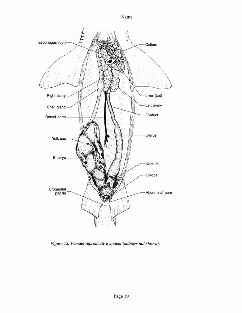
Seminal Vesicle – An enlarged section of the vas deferens that dds secretions to the sperm. Sperm Sacs – A pair of small sacs created by invaginations of the seminal vesicles that receives sperm and seminal secretions from the seminal vesicle. Siphon – Produces a secretion that is expelled with the aid of the clasper during mating. Female Genital System (Figure 13) Ovaries – Two cream colored organs that were dorsal to the liver and are on each side of the mid-dorsal line. Depending on the maturity of your specimen, it may or may not show eggs within each ovary. The eggs move into the body cavity and then into the oviducts when they are ready to be fertilized. Oviducts – Elongated tubes that lay dorsal and lateral along the body cavity. These structures are very prominent in mature sharks. Both oviducts share a common opening to the body cavity called the ostium. Shell Gland – Found at the cranial end of the oviducts. This gland secretes a thin shell around a group of eggs and is a reservoir for sperm storage. Eggs are fertilized in this gland as they pass through. Uterus – The enlarged caudal end of the oviduct. It is here that eggs develop.
Page 17
Name _______________________________
Page 18
Name _______________________________
Page 19
Name _______________________________
Post-Lab – Answer the following questions before continuing with your dissection: 1. What do the kidneys look like? 2. Where are the testes located in the male shark? 3. Where are the ovaries located in the female shark? 4. What is the function of the shell gland? 5. What is the function of the siphon?
Page 20
Name _______________________________
VI. The Nervous System: The Brain
1. Remove the skin from the dorsal section of the head. 2. With your scalpel, carefully shave the chondocranium (shark’s cranium) down to
expose the brain, the olfactory lobes, and the major brain nerves. Shave off thin sections so that you don’t cut into the brain or nerves.
3. Remove chips of cartilage with forceps. Remove the chondocranium from the tip of the rostrum back to the gill slits.
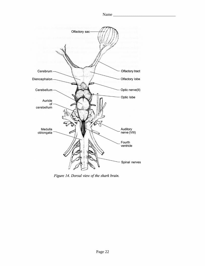
Now that you’ve exposed the nervous system, you should be able to identify the following organs. Olfactory Sacs – Two large bulbous nerve sensors that detect chemicals in the surrounding water. Olfactory Lobes – Area of the brain that receives nerve signals from the olfactory sacs and processes them. Cerebrum – The two hemispheres between the olfactory lobes and are associated with sight and smell. Diencephalon – The region just caudal from the cerebrum and separates the fore and mid-brain. Includes the thalamus and the hypothalamus. Optic Lobe – Large prominent lobes of the mid-brain that receive nerves from the eyes. Cerebellum – Just caudal from the optic lobes it controls muscular coordination and position. Auricle of Cerebellum (Restiform body) – A lateral extension of the cerebellum. Medulla Oblongata – The base of the brain, a widening of the spinal cord. Controls many of the spinal reflexes.
Page 21
Name _______________________________
Page 22
Name _______________________________
Post-Lab – Answer the following questions before continuing with your dissection: 1. What is the largest single part of the shark’s nervous system? 2. What does the optic lobe receive information from? 3. What does the medulla oblongata control? 4. What does the cerebellum control? 5. What did you find interesting about the shark’s nervous system?
Page 23
Name _______________________________
Dissection Clean-up Now that you have completed your dissection it is time to clean up!
1. Return all parts of the shark to the bag it came in. 2. Secure the bag with a rubber band – just like when we started the lab.
3. Place the secured bag in one of the boxes we removed them from at the beginning
of the lab.
4. Wash and dry all of your dissection tools.
5. Return all of the tools to your teacher.
6. Wash and dry your dissection tray.
7. Return your dissection tray to your teacher.
8. Make sure you have completed all parts of the dissection packet.
9. Turn in your dissection packet to your teacher in order to receive credit for the project!
Page 24