Embed Size (px)
Citation preview

DOES THE FUNCTIONAL REACH TEST REFLECT STABILITY LIMITS INELDERLY PEOPLE?
Erika Jonsson, Marketta Henriksson and Helga Hirschfeld
From the Motor Control and Physical Therapy Research Laboratory, Division of Physiotherapy, Neurotec Department,Karolinska Institutet, Stockholm, Sweden
Objective: To explore how the Functional Reach test corre-lates with the displacement of the centre of pressure andwhether the test is a measure of the stability limits in healthyelderly people. Also to explore the performance parametersduring the Functional Reach test.Design: Method comparison study.Subjects: Twenty-seven healthy elderly subjects.Methods: Whole body kinematics (ELITE systems), groundreaction forces (AMTI) and muscle activity (EMG) parallelwith clinical yardstick measure while performing theFunctional Reach test.Results: This study showed a low correlation (r = 0.38)between reach distance and displacement of centre ofpressure and a moderate correlation (r = 0.68) betweenforward rotation of the trunk and reach distance. Themovement during the Functional Reach test was character-ized by a large forward rotation of the trunk and a smallextension in the ankle. The latter constraining centre ofpressure forward displacement.Conclusions: The results suggest that the Functional Reachtest is a weak measure of the stability limits. Movement ofthe trunk seems to influence the test more than thedisplacement of the centre of pressure. When using theFunctional Reach test for assessing balance, compensatorymechanisms should be taken into account.
Key words: Functional Reach, balance, centre of pressure,elderly, kinetic, kinematics.
J Rehabil Med 2003; 35: 26–30
Correspondence address: Helga Hirschfeld, Motor Controland Physical Therapy Research Laboratory, NeurotecDepartment, 23100, SE-141 83 Huddinge, Sweden. E-mail:[email protected]
Submitted April 3, 2002; accepted July 22, 2002
INTRODUCTION
The Functional Reach test (FRT) is a well-known clinicalmeasure of balance developed by Duncan et al. (1), and testedfor both validity and reliability (1–4). FRT measures thedistance between the length of the arm and a maximal forwardreach in the standing position, while maintaining a fixed base ofsupport. It was developed as a dynamic measure of balance withno attempt to control for the movement strategy (1). FRT is usedseparately or as an item in Berg’s Balance Scale (5) and is usedin patients with diagnoses as different as stroke (6), Parkinson
(7), vestibular hypofunction (8), multiple sclerosis (9) and hipfractures (10). FRT has also been associated with an increasedrisk of fall and frailty in elderly people who are unable to reachmore than 15 cm (2, 4).
To make sound interpretations, a researcher must beconfident that the measurements are reproducible, or reliable.Although reliability is necessary for validity, it does notvalidate the meaning of the measure. The accuracy or validityof the measurement provided by an instrument can bedetermined by comparing the reading on the device with astandard measure (11). Laboratory measures of kinematics andkinetics can be regarded as standard measures. Clinical balancetests measure task parameters that reflect what is required fromthe subject to solve the task but not how the task is performed.Laboratory instruments for balance evaluation, such as analysisof ground reaction forces (kinetics) and of movement (kine-matics), measure performance parameters, i.e. how a task ormovement is performed and what exactly the subject is doing(12).
The FRT is suggested to be a clinical measure of the stabilitylimits and has been developed from a leaning task (1). Such atask involves displacement of the centre of pressure (CoP)forward by rotating around the ankle joints with maintained hipextension (13). A leaning task also has, like other forwardoriented movements, anticipatory muscle activation in thetibialis anterior prior to CoP displacement (14). One way toexplore the stability limit is to investigate the location and thepath of the CoP during task performance. CoP indicates thelocation where the resultant ground reaction force has its originand is directed towards the body (15). Duncan et al. (1) havereported a correlation of 0.71 between CoP displacement andreach distance during FRT. However, the correlation is based onthe sum of CoP displacements in anterior/posterior and medial/lateral direction together. FRT, however, assesses only theanterior stability. Duncan et al. (1) have also stated that areaching task is a more functional task than a leaning task. Yet,we believe that a reaching task may involve other factors thanjust those of balance.
As a first step towards understanding the relation between taskparameters and performance parameters in clinical balance tests,the purpose of this study was: (1) to explore how the inter-pretation of the FRT correlates with the displacement of CoPand whether FRT is a clinical measure of the stability limits inhealthy elderly people; and (2) to investigate performanceparameters during FRT with analysis of movement, groundreaction forces and anticipatory muscle activity.
2003 Taylor & Francis. ISSN 1650–1977 J Rehabil Med 35
J Rehabil Med 2002; 35: 26–30

METHODS
Subjects
Healthy elderly volunteers aged 65–80 years without any history ofneurological or musculoskeletal disorders were recruited from pen-sioners’ organizations in the vicinity of Stockholm county, Sweden.Exclusion criteria are listed in Table I. Thirty-three healthy elderlypeople were enrolled after having given their informed consent. Fourwere excluded during data analysis due to several missing markers, and 2subjects were excluded due to the fact that they stood on their toes in all 5trials. The remaining 27 healthy elderly subjects, 18 women and 9 men,participated in the study. The subjects’ mean age was 71.3 (SD 4.0) yearsand mean height and weight was 167.1 (SD 8.9) cm and 72.0 (SD12.6) kg, respectively. The subjects did not use any walking aids andwere active both indoors and outdoors.
Procedure
The FRT was examined simultaneously clinically by yardstick measureand experimentally by kinetic and kinematic measures. The subject wasstanding barefoot on 2 force plates and was allowed to make severalpractice trials before 5 consecutive trials were recorded.
Functional Reach test procedure
The ability of standing subjects to reach with the left hand horizontallyforward (90 degrees shoulder flexion and straight arm) while maintain-ing a fixed base of support was examined. Instructions were similar tothose of Duncan et al. (1). A 150-cm yardstick was horizontally mountedon the wall, at the height of the acromion. Reach distance was measuredas the displacement of the finger between initial position and endposition. In accordance with Duncan et al. (1) the reaching strategy wasnot otherwise controlled for.
Kinematics
A two-camera optoelectronic system (Elite BTS, Milan, Italy) was usedto record the kinematics in a three-dimensional reference system duringFRT. The cameras were placed 4 m from the force plates, at 35-degreeangles to the sagittal plane. The explored field was 2 � 2 m, giving anaccuracy of 0.78 mm. Spherical reflective markers (diameter 1 cm) wereplaced on 10 anatomical landmarks on the right side of the body; themandible joint, the cheek, spinal process C7, L1, L5, the lateral femoralcondyle, lateral malleolus, the heel, the fifth metatarsal bone and theacromion. Two markers were placed on landmarks on the left side of thebody; the lateral humeral epicondyle and the tip of the third phalanx. Inaddition, 2 markers were placed as spatial orientation on the wall and onthe back corner of the force plate beneath the right foot.
Force plates
The ground reaction forces were recorded on 2 equal force plates(AMTI, Advanced Mechanical Technology Incorporation, modelMc818-6-1000; size 457 � 203 mm; accuracy 0.25 N). Three orthogonalforces, anterior/posterior, medial/lateral and vertical were measured withstrain gauges mounted in the force plates.
Electromyography
Electromyography (EMG) of 4 ankle muscles were recorded with theBagnoli-8 system, Boston, MA with surface differential electrodes typeDE-02 (23 � 17 mm). The surface electrodes were attached withadhesive tape over the belly of the left and right tibialis anterior andleft and right lateral gastrocnemius. EMG data were stored together withthe forces on a SC/ZOOM flexible laboratory computer system(Department of Physiology, Umea University, Sweden) and thekinematics were simultaneously stored together with the forces on theELITE system computer for further analysis.
Data analysis
FRT was recorded for 5 seconds with a sampling frequency of 100 Hz forkinematic and force plate data and 800 Hz for the EMGs. The data wastransformed into ASCII files and analysed by means of Axograph (AxonInstruments), a MacIntosh based software package. Before analysis theforce and kinematic signals were digitally filtered for signal smoothing.
EMG signals were band-pass filtered between 10 Hz and 1 kHz. Fivetrials of FRT were analysed for each subject, except for subject 27 whohad 4 trials suitable for analysis due to missing markers (a total of 134trials). The peak amplitude of the finger marker displacement wasdefined from cursor read-outs and set as the time zero. All otherconsidered peak amplitudes were measured at that instant. Peakamplitudes of trunk and ankle angles were computed as changes relativeto the baseline of initial standing. The following angular displacementswere measured: trunk segment angle by joining markers on L5 and theright shoulder vs the vertical axis and ankle joint angle by measuring 2intersecting segments formed by joining markers between the knee andlateral malleolus and the heel and the fifth metatarsal bone. Theamplitudes of the force signals were normalized by body mass andexpressed as percentage of body weight (%BW). The net CoPdisplacement was calculated for anterior/posterior and medial/lateraldirection. For making comparisons between subjects possible thedisplacement of CoP was normalized to foot length, i.e. base of support,and presented in the text as a percentage of base of support. The positionof marker 14 on the lateral, rear corner of the force plate beneath the rightfoot was used for transforming the CoP co-ordinates relative to the forceplate to the CoP position relative to the foot. The EMG data wereanalysed in SC/ZOOM and the burst onset latencies were measuredrelative to the onset of the anterior/posterior force. The onset latency oftibialis anterior and lateral gastrocnemius was defined as EMG activity�2 SD above mean baseline activity and lasting longer than 30 ms. Todistinguish anticipatory from feedback activity, anticipatory muscleactivity was defined as onset between 40 and 100 ms prior to the onset ofthe anterior/posterior force.
Statistical methods and analysis
All statistical analyses were performed using STATISTICA forWindows (StatSoft Inc. 2000). Significance level was set at p � 0.01and t-test for dependent samples was used to compare the means ofdependent variables. Correlation was tested by means of the Pearsonproduct moment correlation. The strength of the correlation coefficientwas classified according to Munro (16).
RESULTS
FRT measurement
The mean value of the clinically measured distance (FRclin) was29.4 (SD 5.4) cm and the experimentally registered displace-ment of the finger marker (FRexp) was 27.9 (SD 5.6) cm. FRexp
was significantly larger (p � 0.01) than FRclin.
Displacement of CoP
At initial standing the CoP position in the anterior/posteriordirection was mean 36.6 (SD 7.8)% of the base of supportstarting with 0% at the heel edge and 100% at the toe edge.During FRT performance, CoP initially displaced posteriorly,
Table I. Exclusion criteria
Less than 65 years of ageLess than 90 degrees of shoulder flexionUnable to follow a 3-step commandUnable to stand unassisted for 60 secondsDiabetes with neuropathy in the lower extremitiesDementiaHip- or knee-joint replacementWeight bearing pain in the lower extremitiesDecreased hearing or sightPathological condition that can influence vestibulary or other
sensory systems
J Rehabil Med 35
Functional Reach test 27

mean 8.1 (SD 3.7)% or 2.0 (SD 0.9) cm, and thereafter movedanteriorly, mean 37.4 (SD 10.6)% or 9.2 (SD 2.6 cm), as shownin Fig. 1.
Thereby CoP displaced anteriorly 29 (SD 8.3%) or 7.2 (SD2) cm from the initial position, which was used in the correlationanalysis. The end location of CoP was at 65.8 (SD 5.6)%. Thepattern of the CoP displacement was similar for all subjects butdiffered between starting position and amplitude of the posteriorand anterior displacement. FRclin distance had a low correlationto the anterior displacement of CoP (r = 0.40) and the correlationwas still low (r = 0.38) when normalized to base of support (Fig.
2). There was also a low correlation between FRclin and theposterior displacement of CoP (r = 0.26).
Angular displacement during FRT
The mean forward rotation of the trunk during FRT was 32 (SD9.6) degrees. From initial standing, the ankle angle increasedtowards extension in 90% of the trials, mean 6.7 (SD 3.4)degrees, and for the remaining 10% it either remained similar ordecreased towards flexion, mean 2.0 (SD 1.3). Trunk forwardrotation and ankle joint angle both increased as FRclin increased.FRclin and trunk forward rotation showed a moderate correlation,r = 0.67 (Fig. 3) and FRclin and ankle joint angle showed a lowcorrelation, r = 0.39.
Age and height
The results showed no significant (p � 0.05) correlation betweenFRclin and age and height (r = �0.27 and r = 0.22, respectively).
Weight distribution and muscle activity
Analysis of the weight distribution normalized to body massduring FRT showed a larger (p � 0.001) weight beneath the left
Fig. 1. (A) Displacement of centre of pressure (CoP) expressed aspercentage of base of support (%BoS) in 1 trial in 1 subject. (B)Subject mean and SD of CoP initial position (Start), peak posteriordisplacement (Post) and peak anterior displacement (Ant) areexpressed as percentage of base of support during forwardreaching. The mean and SD is shown for 13 subjects, representingthe range of all subjects. For clarity, the posterior and anteriormeasures are plotted with a lateral offset.
Fig. 2. Relation of centre of pressure (CoP) anterior displacementexpressed as percentage of base of support (%BoS) and FRclindistance for all trials.
Fig. 3. Relation of forward rotation of the trunk and FRclin distancefor all trials.
J Rehabil Med 35
28 E. Jonsson et al.
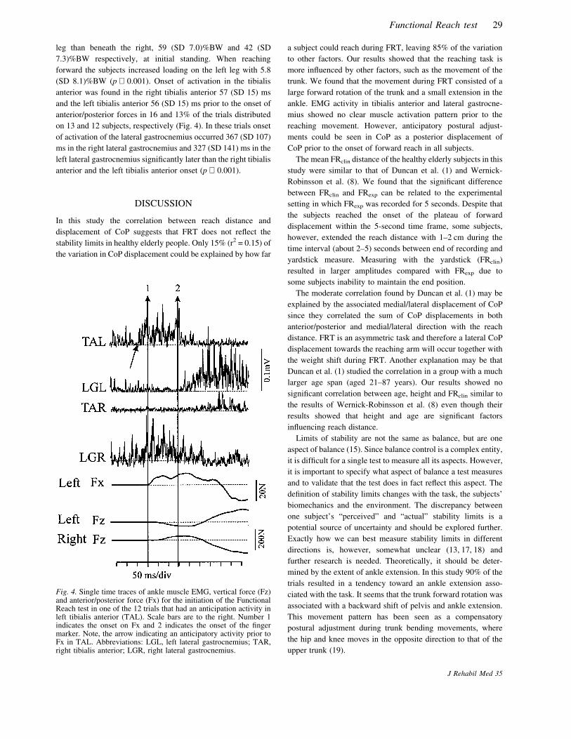
leg than beneath the right, 59 (SD 7.0)%BW and 42 (SD7.3)%BW respectively, at initial standing. When reachingforward the subjects increased loading on the left leg with 5.8(SD 8.1)%BW (p � 0.001). Onset of activation in the tibialisanterior was found in the right tibialis anterior 57 (SD 15) msand the left tibialis anterior 56 (SD 15) ms prior to the onset ofanterior/posterior forces in 16 and 13% of the trials distributedon 13 and 12 subjects, respectively (Fig. 4). In these trials onsetof activation of the lateral gastrocnemius occurred 367 (SD 107)ms in the right lateral gastrocnemius and 327 (SD 141) ms in theleft lateral gastrocnemius significantly later than the right tibialisanterior and the left tibialis anterior onset (p � 0.001).
DISCUSSION
In this study the correlation between reach distance anddisplacement of CoP suggests that FRT does not reflect thestability limits in healthy elderly people. Only 15% (r2 = 0.15) ofthe variation in CoP displacement could be explained by how far
a subject could reach during FRT, leaving 85% of the variationto other factors. Our results showed that the reaching task ismore influenced by other factors, such as the movement of thetrunk. We found that the movement during FRT consisted of alarge forward rotation of the trunk and a small extension in theankle. EMG activity in tibialis anterior and lateral gastrocne-mius showed no clear muscle activation pattern prior to thereaching movement. However, anticipatory postural adjust-ments could be seen in CoP as a posterior displacement ofCoP prior to the onset of forward reach in all subjects.
The mean FRclin distance of the healthy elderly subjects in thisstudy were similar to that of Duncan et al. (1) and Wernick-Robinsson et al. (8). We found that the significant differencebetween FRclin and FRexp can be related to the experimentalsetting in which FRexp was recorded for 5 seconds. Despite thatthe subjects reached the onset of the plateau of forwarddisplacement within the 5-second time frame, some subjects,however, extended the reach distance with 1–2 cm during thetime interval (about 2–5) seconds between end of recording andyardstick measure. Measuring with the yardstick (FRclin)resulted in larger amplitudes compared with FRexp due tosome subjects inability to maintain the end position.
The moderate correlation found by Duncan et al. (1) may beexplained by the associated medial/lateral displacement of CoPsince they correlated the sum of CoP displacements in bothanterior/posterior and medial/lateral direction with the reachdistance. FRT is an asymmetric task and therefore a lateral CoPdisplacement towards the reaching arm will occur together withthe weight shift during FRT. Another explanation may be thatDuncan et al. (1) studied the correlation in a group with a muchlarger age span (aged 21–87 years). Our results showed nosignificant correlation between age, height and FRclin similar tothe results of Wernick-Robinsson et al. (8) even though theirresults showed that height and age are significant factorsinfluencing reach distance.
Limits of stability are not the same as balance, but are oneaspect of balance (15). Since balance control is a complex entity,it is difficult for a single test to measure all its aspects. However,it is important to specify what aspect of balance a test measuresand to validate that the test does in fact reflect this aspect. Thedefinition of stability limits changes with the task, the subjects’biomechanics and the environment. The discrepancy betweenone subject’s “perceived” and “actual” stability limits is apotential source of uncertainty and should be explored further.Exactly how we can best measure stability limits in differentdirections is, however, somewhat unclear (13, 17, 18) andfurther research is needed. Theoretically, it should be deter-mined by the extent of ankle extension. In this study 90% of thetrials resulted in a tendency toward an ankle extension asso-ciated with the task. It seems that the trunk forward rotation wasassociated with a backward shift of pelvis and ankle extension.This movement pattern has been seen as a compensatorypostural adjustment during trunk bending movements, wherethe hip and knee moves in the opposite direction to that of theupper trunk (19).
Fig. 4. Single time traces of ankle muscle EMG, vertical force (Fz)and anterior/posterior force (Fx) for the initiation of the FunctionalReach test in one of the 12 trials that had an anticipation activity inleft tibialis anterior (TAL). Scale bars are to the right. Number 1indicates the onset on Fx and 2 indicates the onset of the fingermarker. Note, the arrow indicating an anticipatory activity prior toFx in TAL. Abbreviations: LGL, left lateral gastrocnemius; TAR,right tibialis anterior; LGR, right lateral gastrocnemius.
J Rehabil Med 35
Functional Reach test 29

CoP anterior displacement in this study is consistent with theresults of Wernick-Robinson et al. (8). Notably, similar anteriordisplacement of CoP was seen during a leaning task in elderlypeople (13) indicating that the CoP displacement during reach-ing is not larger than during leaning. We found a startingposition that ranged from 23 to 49% of base of support, similarto the results of Crenna & Frigo (14) who found the startingposition to be slightly anterior of the lateral malleolus duringupright stance. Another study (18) has reported a mean positionof CoP in double limb standing as 11.5 (SD 2) cm forward fromthe heel edge, which should be approximately 40–50% of baseof support in our subjects.
Anticipatory postural changes are associated with voluntarymovements. The posterior displacement of CoP seen prior to thereaching movement is an anticipatory adjustment to create adistance between CoP and centre of mass location (moment arm)(14). Prior to the beginning of a voluntary movement and CoPposterior displacement, muscle activation can be recorded inmuscles other than the primary movers. Crenna & Frigo (14)reported anticipatory postural adjustments in the tibialis anteriorand soleus before the initiation of a forward oriented armmovement. Surprisingly, we only found anticipatory tibialisanterior activity in 13% (right leg) and 16% (left leg) of thetrials. The lack of an anticipatory postural adjustment pattern inthe tibialis anterior as well as the initial weight distributiontowards the left leg may be due to the fact that the initial positionplaces the subject in a already prepared position or that it isspeed-related and due to the slow movement during FRT (20).This may question whether this reaching task does in factrepresent a functional volitional movement. Further research isneeded to investigate if other muscles have anticipatory activityduring FRT.
Although, FRT has proven to be reliable and valid (1–4) thereare major factors influencing the evaluation. Wernick-Robinsonet al. (8) found that the movement strategy influenced FRT andrecommend to assess movement strategy during FRT. Otherresearchers (21, 22) implicate that a reduced spinal flexibilityresults in reduced reach distance, while still others questionFRTs ability to differentiate elderly non-fallers from fallers (17).Our results suggest that FRT is a weak measure of the anteriorstability limits in standing. Yet this study was limited to dealingwith correlation. However, the quality of the displacement ofCoP needs further investigation.
If FRT is to be used as a balance test one has to considercompensatory mechanisms for decreased flexibility and strengthinterfering with the evaluation. We believe that a leaning taskmay be more valuable for measuring the limits of stability andrecommend further research to develop a clinical measure thatreflects the stability limits in both anterior/posterior and medial/lateral direction.
ACKNOWLEDGEMENTS
We are grateful to Ing-Marie Apel for technical assistance, Ake Seiger
for critical reading and our colleagues at the research laboratory forvaluable discussions. This study was supported by the LegitimeradeSjukgymnasters Riksforbund Minnesfond, Board of Research for Healthand caring sciences, National research school in health and caringscience, Gun and Bertil Stohnes Foundation and KP ’s Jubileumsfond.
REFERENCES
1. Duncan PW, Weiner DK, Chandler J, Studenski S. Functional reach:a new clinical measure of balance. J Gerontol 1990; 45: M192–M197.
2. Duncan PW, Studenski S, Chandler J, Prescott B. Functional reach:predictive validity in a sample of elderly male veterans. J Gerontol1992; 47: M93–M98.
3. Weiner DK, Bongiorni DR, Studenski SA, Duncan PW, Kochers-berger GG. Does functional reach improve with rehabilitation? ArchPhys Med Rehabil 1993; 74: 796–800.
4. Weiner DK, Duncan PW, Chandler J, Studenski SA. Functionalreach: a marker of physical frailty. J Am Geriatr Soc 1992; 40: 203–207.
5. Berg K, Wood-Dauphinnee S, Williams JI, Gayton D. Measuringbalance in the elderly: Preliminary development of an instrument.Physiother Can 1989; 41: 304–311.
6. Fishman MN, Colby LA, Sachs LA, Nichols DS. Comparison ofupper-extremity balance tasks and force platform testing in personswith hemiparesis. Phys Ther 1997; 77: 1052–1062.
7. Smithson F, Morris ME, Iansek R. Performance on clinical tests ofbalance in Parkinson’s disease. Phys Ther 1998; 78: 577–592.
8. Wernick-Robinson M, Krebs DE, Giorgetti MM. Functional reach:does it really measure dynamic balance? Arch Phys Med Rehabil1999; 80: 262–269.
9. Frzovic D, Morris ME, Vowels L. Clinical tests of standing balance:performance of persons with multiple sclerosis. Arch Phys MedRehabil 2000; 81: 215–221.
10. Ingemarsson AH, Frandin K, Hellstrom K, Rundgren A. Balancefunction and fall-related efficacy in patients with newly operated hipfracture. Clin Rehabil 2000; 14: 497–505.
11. Domholt E. Physical therapy research; principles and applications.2nd edn. Philadelphia, USA: WB Saunders; 2000, p. 244–247.
12. Latash ML. Control of human movement. Illinois, USA: HumanKinetics Publishers; 1993, p. 106–108.
13. King MB, Judge JO, Wolfson L. Functional base of supportdecreases with age. J Gerontol 1994; 49: M258–M263.
14. Crenna P, Frigo C. A motor programme for the initiation of forward-oriented movements in humans. J Physiol 1991; 437: 635–653.
15. Shumway-Cook A, Woollacott M. Motor control: theory andpractical applications. Maryland, USA: Lippincott Williams &Wilkins; 2000, p. 163–193.
16. Munro BH. Statistical methods for health and care research.Baltimore, USA: Lippincott Williams & Wilkins; 2001, p. 223–243.
17. Wallmann HW. Comparison of elderly nonfallers and fallers onperformance measures of functional reach, sensory organization, andlimits of stability. J Gerontol A Biol Sci Med Sci 2001; 56: M580–M583.
18. Murray MP, Seireg AA, Sepic SB. Normal postural stability andsteadiness: quantitative assessment. J Bone Joint Surg Am 1975; 57:510–516.
19. Crenna P, Frigo C, Massion J, Pedotti A. Forward and backwardaxial synergies in man. Exp Brain Res 1987; 65: 538–548.
20. Rogers MW, Pai YC. Dynamic transitions in stance supportaccompanying leg flexion movements in man. Exp Brain Res1990; 81: 398–402.
21. Cavanaugh JT, Shinberg M, Ray L, Shipp KM, Kuchibhatla M,Schenkman M. Kinematic characterization of standing reach:comparison of younger vs. older subjects. Clin Biomech (Bristol,Avon) 1999; 14: 271–279.
22. Schenkman M, Morey M, Kuchibhatla M. Spinal flexibility andbalance control among community-dwelling adults with and withoutParkinson’s disease. J Gerontol A Biol Sci Med Sci 2000; 55: M441–M445.
J Rehabil Med 35
30 E. Jonsson et al.