Embed Size (px)
Citation preview
1
Does prosopagnosia take the eyes out of face representations?
Evidence for a defect in representing diagnostic facial information
following brain damage
Roberto Caldara1, Philippe Schyns1, Eugène Mayer2, Marie L. Smith1, Frédéric
Gosselin3 and Bruno Rossion4
1 Department of Psychology, University of Glasgow, United Kingdom
2 Hôpitaux Universitaires de Genève, Geneva, Switzerland
3 Département de Psychologie, Université de Montréal, Canada
4 Unité de Neurosciences Cognitives et Laboratoire de Neurophysiologie, University
of Louvain, Belgium
Correspondence: [email protected]
2
Abstract
One of the most impressive disorders following brain damage to the ventral occipito-
temporal cortex is prosopagnosia, or the inability to recognize faces. Although acquired
prosopagnosia with preserved general visual and memory functions is rare, several cases have
been described in the neuropsychological literature and studied at the functional and neural
level over the last decades. Here we tested a brain-damaged patient presenting a deficit
restricted to the category of faces to clarify the nature of the missing and preserved
components of the face processing system when it is selectively damaged. Following learning
to identify 10 neutral and happy faces through extensive training, we tested patient PS’
recognition of faces using Bubbles, a response classification technique that sampled facial
information across the faces in different bandwidths of spatial frequencies (see Gosselin &
Schyns, 2001). Although PS gradually used less information to identify the faces over testing,
the total information required (i.e. the number of bubbles) to identify faces was much larger
than for normal controls, and decreased less steeply with practice. Most importantly, the
facial information used to identify individual faces differed between PS and controls.
Specifically, in marked contrast to controls, PS did not use the optimal eye information to
identify familiar faces, but instead the mouth and the external features, as normal observers
typically do when processing unfamiliar faces. Together, the findings reported here suggest
that damage to the face processing system is characterized with an inability to use the
information that is optimal to judge identity (the eyes), focusing instead on suboptimal
information.
Keywords: prosopagnosia, face processing, recognition task, response classification
technique.
3
INTRODUCTION
In humans, faces convey at a glance a great deal of information (e.g., person’s
identity, gender, mood, ethnical origin, age) which is crucial for efficient social interactions.
A long-standing goal of the face processing research agenda has been to identify which cues
are extracted from a face in order to categorize it (e.g., according to its gender, expression,
identity, race and so forth). In other words, what is the diagnostic (i.e. most useful)
information that is extracted from faces, and how does it vary according to the task at hand?
To address this issue, various methods have been used, such as categorizing faces presented
with masked or isolated facial features (e.g., Bruce, Burton, Hanna, Healey, & et al., 1993),
with surface and shape properties separated (Hill, Bruce, & Akamatsu, 1995), with only
information from the principal components of a face set (e.g., Calder, Burton, Miller, Young,
& Akamatsu, 2001), or, more recently, by finely sampling information from face stimuli and
deriving the information subset associated with the categorization judgments (Gosselin &
Schyns, 2001; Schyns, Bonnar, & Gosselin, 2002; Sekuler, Gaspar, Gold, & Bennett, 2004).
Humans are generally considered face processing experts because they efficiently
extract the diagnostic cues allowing face categorization, identification and generalization
(Diamond & Carey, 1986; Tanaka, 2001). Yet, several observations illustrate the complexity
and difficulty of face categorization. First, ‘expert’ human face processing system of the
adults undergoes quite a long development, not reaching full maturity before puberty
(Campbell et al., 1999; Carey, 1992; Taylor, McCarthy, Saliba, & Degiovanni, 1999).
Secondly, the face processing system is not so efficient with unfamiliar faces, as
demonstrated by the striking difference between our excellent ability to generalize across
different images of familiar faces, and our relatively poor performance when performing the
same task on unfamiliar faces (e.g., Bruce et al., 1999; Burton, Wilson, Cowan, & Bruce,
4
1999; Young, McWeeny, Hay, & Ellis, 1986), suggesting that the efficiency of face
processing arise from robust long-term representations of others’ faces.
Possibly the most striking evidence that the extraction of diagnostic information from
faces is non trivial is the observation of patients who have lost this ability, despite no other
obvious impairments of the visual system (at least as far as neuropsychological tests
demonstrate) and a preserved ability to recognize people through other modalities (e.g.,
voice). Bodamer (1947) (Bodamer, 1947; English translation by Ellis & Florence, 1990), who
coined the term “prosopagnosia”, attributed the first clinical observation of this disturbance to
Quaglino (1867). However, it became apparent that Wigan (1844) had most likely been the
first to report on a case of prosopagnosia (see Grüsser & Landis, 1991). This condition,
generally follows brain damage to bilateral or unilateral right occipito-temporal areas (e.g.,
Damasio, Damasio, & Van Hoesen, 1982; Farah, 1990; Landis, Regard, Bliestle, & Kleihues,
1988; Sergent & Signoret, 1992). While being able to detect a face amongst objects (‘face
detection’), prosopagnosic patients typically loose the ability to identify familiar faces,
including famous persons, friends and relatives, or even their own face (Damasio, 1985).
Despite the rarity of prosopagnosic patients with well-preserved visual perception and
memory, a number of such cases have been described over the last decades (e.g., Farah, 1990
- for more recent cases, see; Gauthier, Behrmann, & Tarr, 1999; Laeng & Caviness, 2001;
Sergent & Signoret, 1992).
The clinical and anatomical conditions of prosopagnosia have been of great interest to
Cognitive Neuroscientists willing to clarify the neuro-functional mechanisms of normal face
processing. Several key findings have been made. Anatomical descriptions of prosopagnosia
support the critical role of the right hemisphere in the occipito-temporal pathway of face
processing (Bouvier & Engel, 2004; Damasio, 1985; Landis et al., 1988; Sergent & Signoret,
1992). The double dissociations reported between the ability to perceive unfamiliar and
5
familiar faces (Malone, Morris, Kay, & Levin, 1982), between the recognition of facial
expression and facial identity (e.g., Bruyer et al., 1983; Tranel, Damasio, & Damasio, 1988),
or between lip-reading and face identification (Campbell, Landis, & Regard, 1986), have
helped in isolating the different sub-functions in a cognitive architecture of face processing
(Bruce & Young, 1986). The study of prosopagnosia at the functional level has probably
initiated (Bodamer, 1947) and largely contributed to the ‘never-ending’ debate about the
modularity of face processing (e.g., Damasio et al., 1982; Farah, Levinson, & Klein, 1995;
Gauthier et al., 1999).
Despite the theoretical importance of prosopagnosic cases, an important area of
research remains largely unexplored. In descriptions of clinical cases, it is usually reported
that prosopagnosic patients, who rely on non-facial cues to recognize people (e.g., gait, voice,
clothes …), still attend to and extract information from faces that is used to recognize people.
Yet, these patients appear to have lost the ability to extract and/or build diagnostic
representations of other people’s faces. What is then the nature of the facial information that
brain-damaged prosopagnosic patients extract when processing faces? Or, to put it
differently, what is the nature of the missing and preserved components of the face
processing system when it is selectively damaged? Answering this question would
undoubtedly contribute to functionally characterize the selective face impairment that is
prosopagnosia, and from there on provide a better understanding of functional aspects of the
normal, ‘expert’, face processing system.
Several limitations that hamper a better understanding of the functional aspects of
face processing in prosopagnosia. First, the prosopagnosic patients described in the literature
usually suffer from many low-level and high-level visual deficits besides their face
impairments: loss of visual acuity, visual field defects (hemianopia, upper visual field
problems, or left quadrantopsia generally - for reviews see Barton, 2003; Goldsmith & Liu,
6
2001), achromatopsia (about 60% of overlap, Bouvier & Engel, 2004), or difficulties at
general configural processing and object recognition (e.g., Gauthier et al., 1999; Levine &
Calvanio, 1989; Sergent & Signoret, 1992). Consider for instance LH, a patient initially
presented with a disproportionately large deficit with face compared to object processing,
performing even in the normal range for subtle discrimination tasks on non-face objects such
as pairs of glasses (Farah et al., 1995). Further studies revealed that LH had clear deficits at
performing visual discrimination tasks on complex patterns and at recognizing non-face
objects (e.g., Farah, McMullen, & Meyer, 1991; Levine & Calvanio, 1989; Levine, Calvanio,
& Wolf, 1980). Crucially, his recognition failures proved to be more pronounced for certain
categories than others (e.g., living vs. non-living; Farah et al., 1990), when previous studies
had concluded that the deficits were due to general factors such as the loss of configural
information processing (Levine & Calvanio, 1989). Even though in such cases these deficits
may not explain fully the prosopagnosia, they are likely to affect and modify the strategies
that the patient uses when processing faces. A trivial example is that of a prosopagnosic
suffering from achromatopsia, who will be unable to use eyes or hair color to recognize or
discriminate people. For this reasons it is inherently difficult to establish the functional
aspects that are specific to the face impairment in of prosopagnosia. Associated deficits are
likely to present confounding factors.
Any attempt at clarifying the components (missing and preserved) of prosopagnosia
faces the problem of clarifing the information cues to perform various (face) categorization
tasks. A typical strategy is to hypothesize a priori that certain facial cues are not properly
processed in prosopagnosia, and test this hypothesis by selectively manipulating these cues.
This approach may provide interesting outcomes, but is inherently limited because faces vary
according to a large number of dimensions (Valentine, 1991). By selecting a priori a subset
of this information for testing, one could miss important components of the prosopagnosic
7
deficits, or disclose processing differences between the damaged and normal face processing
systems that are not central to prosopagnosia. Moreover, by selectively manipulating the type
of information that is diagnostic for the task at hand over a number of trials, subjects may be
forced to rely on this information (e.g., the mouth) and learn to improve dramatically their
performance (Barton, Cherkasova, & O'Connor, 2001; Barton, Press, Keenan, & O'Connor,
2002). Thus, a better approach to determine the information used and not used is to bias as
little as possible the tested information. To this end, we trained and tested a single patient –
PS - suffering from a remarkably selective deficit at processing faces following brain damage
(Rossion et al., 2003) with Bubbles (Gosselin & Schyns, 2001) a non biased sampling
technique that can determine the specific visual information used to categorize a visual
stimulus.
PS is a 53-year-old female who sustained a head-close injury in 1991. After several
months of spontaneous recovery and neuropsychological reeducation, she was left with a
massive prosopagnosic deficit, being unable to recognize famous and familiar people.
Despite large occipital and occipito-temporal lesions (see Figure 1 in Rossion et al., 2003),
PS’s low-level vision is almost perfect, her visual acuity being 8/10 in both eyes (august
2003), with a full visual field, apart from a small right paracentral scotoma. She reads
normally (although being slowed down) and, crucially, does not present any problem at
object perception and recognition, even for subordinate-level discriminations (Caldara et al.,
submitted; Rossion et al., 2003). Her deficit truly appears to be restricted to the category of
faces. With faces, PS is able to categorize a face as a face, discriminate faces from objects
and from a complex scene background, even at brief presentations (100 ms - Schiltz et al.,
submitted). Her gender and expression performances are relatively well preserved, although
below normal range (Rossion et al., 2003). This is in stark contrast with her inability to
recognize previously seen or familiar faces, and to match unfamiliar faces (see Table 1 and
8
methods section). In sum, PS is unable to derive an individual representation of a face that is
both selective and invariant (robust) enough so that it can be discriminated from other faces,
and be associated with the same or other views of the same face.
Given the restriction of her deficit to the face category and the fact that PS is alert,
cooperative, and without any learning difficulties (Caldara et al., submitted), she may
represent an ideal case to isolate the nature of the facial information extracted by an impaired
face processing system, relative to normals. Moreover, although relying also on non-facial
cues to identify faces, PS reports using facial cues in general and does not avoid looking at
people’s face. Her performance at matching tasks is under normal range, but generally better
than chance (Caldara et al., submitted; Rossion et al., 2003 ; Schiltz et al., 2004). Hence, her
face processing system, being largely inefficient at recognizing individual faces, is not
completely disrupted, as supported by the significant face-sensitive activations observed in
her right hemisphere in the fusiform gyrus (Rossion et al., 2003), and also in the superior
temporal sulcus and the dorso-lateral prefrontal cortex (Sorger et al., 2004).
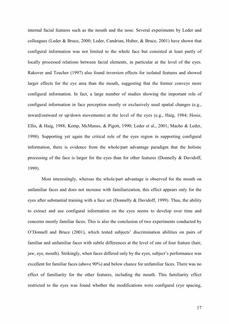
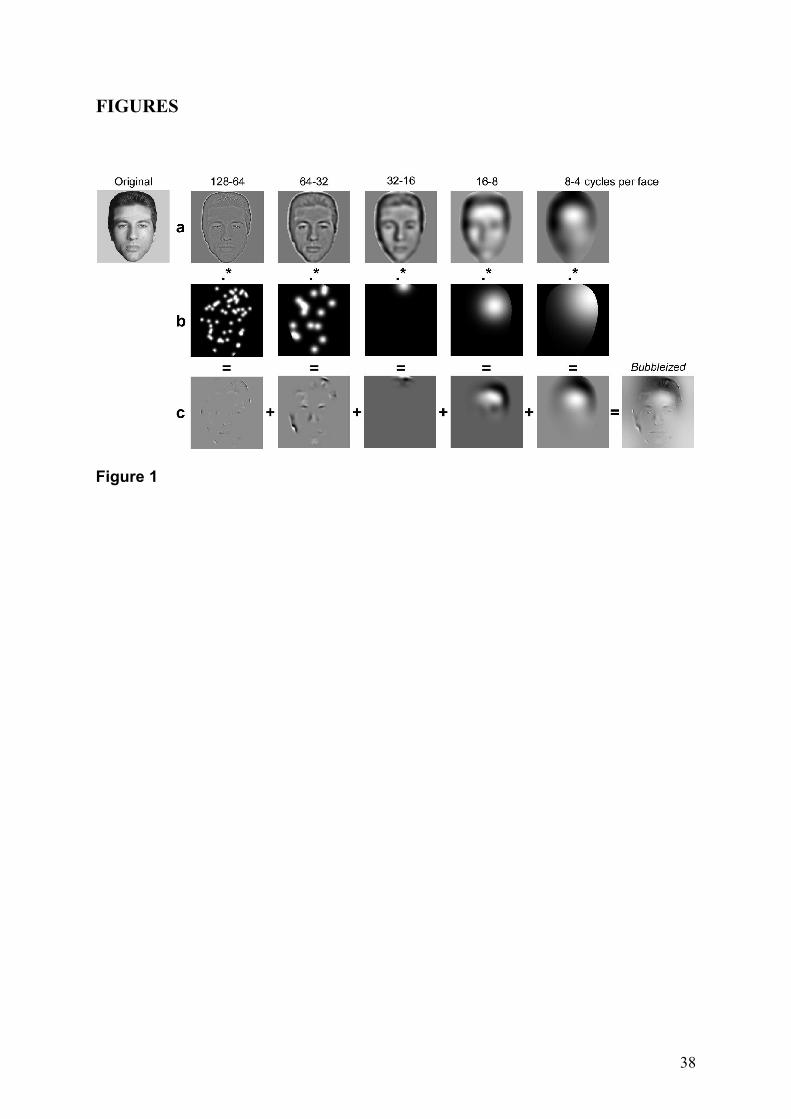
To specify PS’s use of facial information we used Bubbles (Gosselin and Schyns,
2001), a response classification technique, sampling the information in 3D space (2D image x
spatial frequencies). Bubbles samples an input space to present sparse versions of the faces as
stimuli (see Figure 1). PS and normal controls must categorize the sparse stimuli and Bubbles
keeps track of the samples of information that lead to correct and incorrect identification
responses. From this information, we can establish how each region of the input space is
selectively used in categorization tasks, and depict the selective use with an effective
stimulus.
In the present study, we applied this technique to PS and a group of control
participants, who identified a set of previously learned unfamiliar faces displaying two
possible expressions (neutral or happy) (see Methods - Schyns et al., 2002).
9
RESULTS
The proportion of the search space, revealed at each trial, was adjusted online to
maintain accuracy at 75% correct (see methods, and Gosselin & Schyns, 2001). The less this
revealed proportion, the better the sensitivity. For each subject session of trials, we averaged
among trials the proportion of information sampled (bubbles) required.
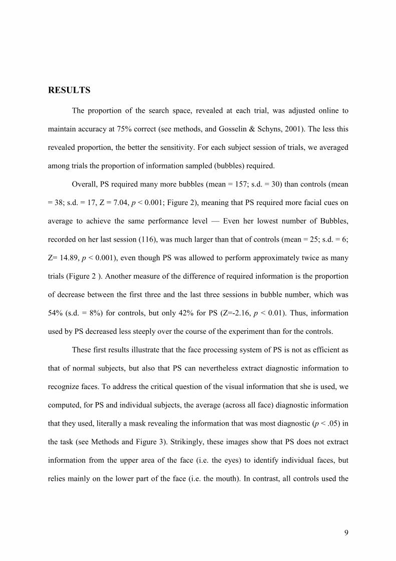
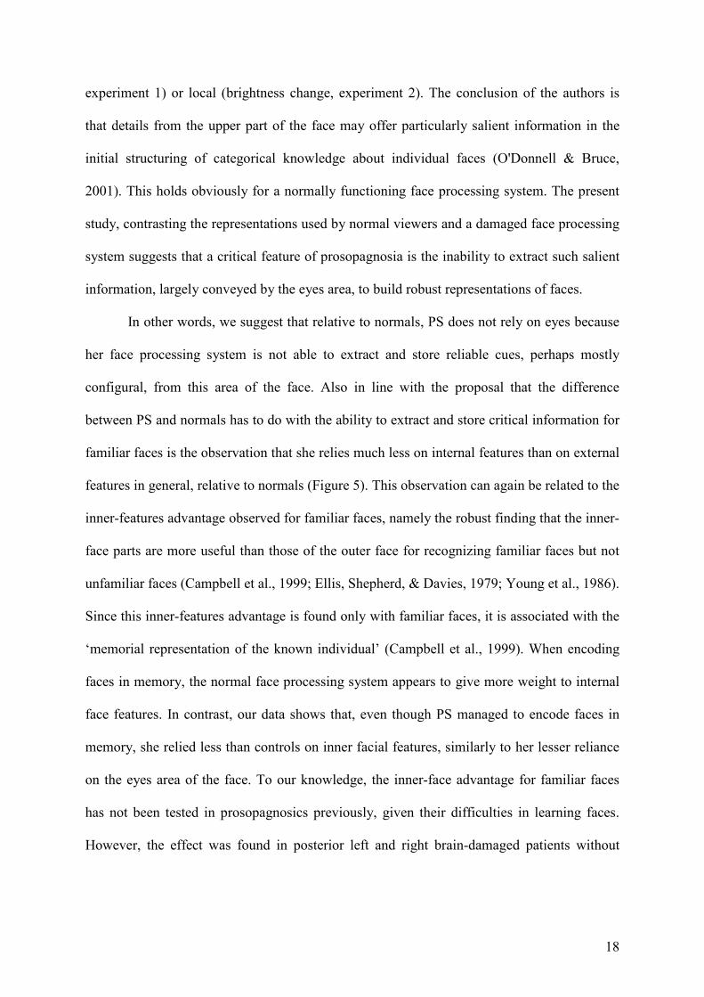
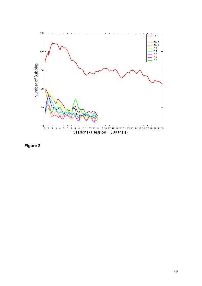
Overall, PS required many more bubbles (mean = 157; s.d. = 30) than controls (mean
= 38; s.d. = 17, Z = 7.04, p < 0.001; Figure 2), meaning that PS required more facial cues on
average to achieve the same performance level — Even her lowest number of Bubbles,
recorded on her last session (116), was much larger than that of controls (mean = 25; s.d. = 6;
Z= 14.89, p < 0.001), even though PS was allowed to perform approximately twice as many
trials (Figure 2 ). Another measure of the difference of required information is the proportion
of decrease between the first three and the last three sessions in bubble number, which was
54% (s.d. = 8%) for controls, but only 42% for PS (Z=-2.16, p < 0.01). Thus, information
used by PS decreased less steeply over the course of the experiment than for the controls.
These first results illustrate that the face processing system of PS is not as efficient as
that of normal subjects, but also that PS can nevertheless extract diagnostic information to
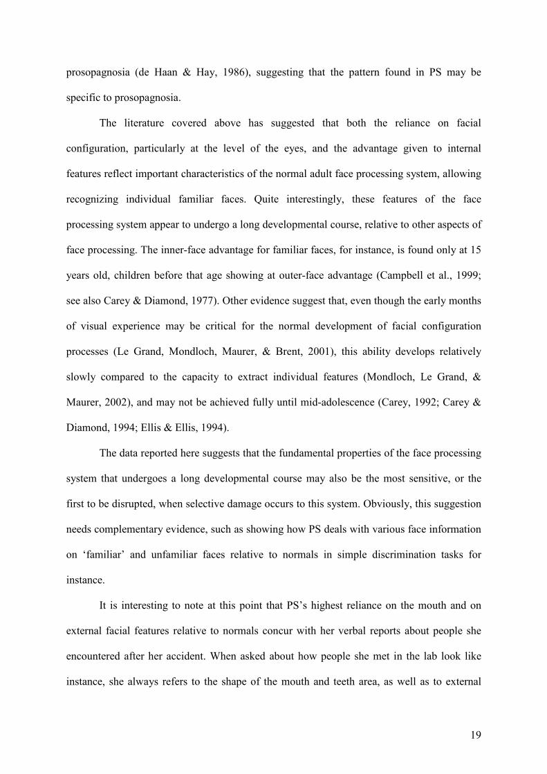
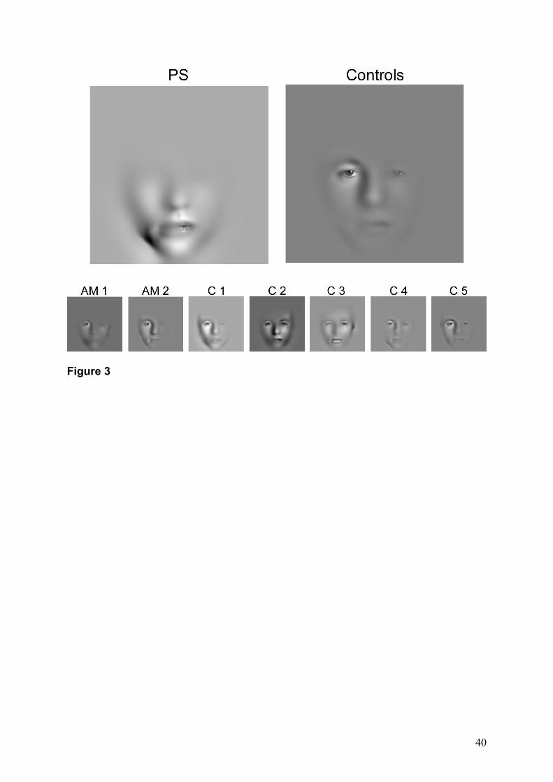
recognize faces. To address the critical question of the visual information that she is used, we
computed, for PS and individual subjects, the average (across all face) diagnostic information
that they used, literally a mask revealing the information that was most diagnostic (p < .05) in
the task (see Methods and Figure 3). Strikingly, these images show that PS does not extract
information from the upper area of the face (i.e. the eyes) to identify individual faces, but
relies mainly on the lower part of the face (i.e. the mouth). In contrast, all controls used the
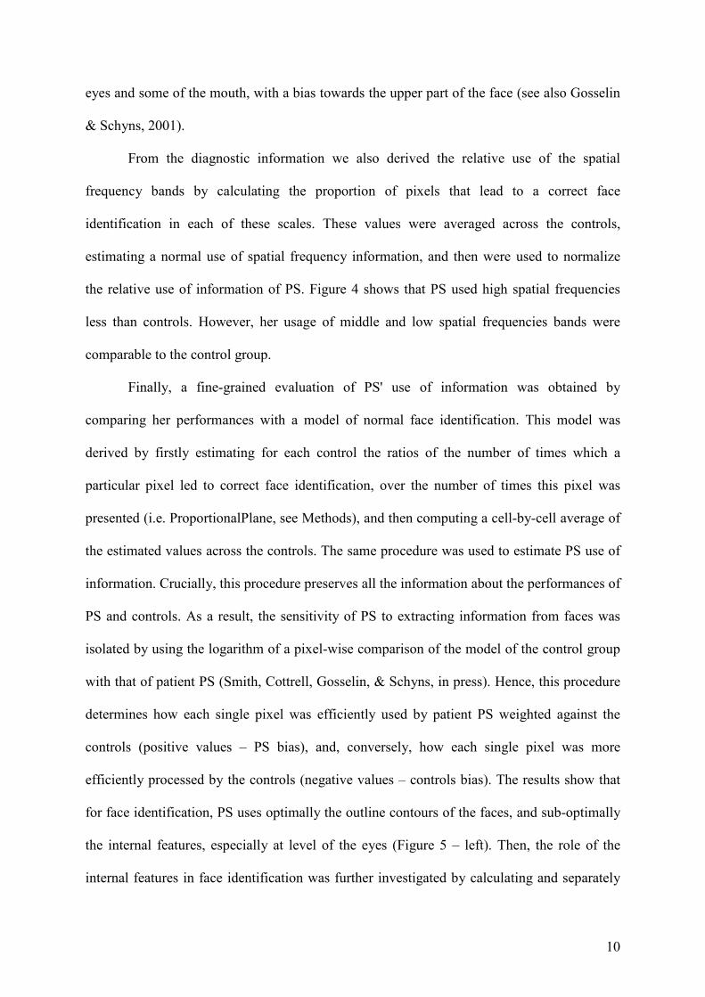
10
eyes and some of the mouth, with a bias towards the upper part of the face (see also Gosselin
& Schyns, 2001).
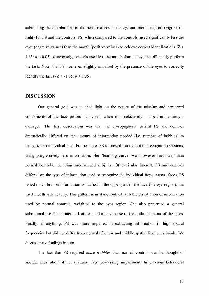
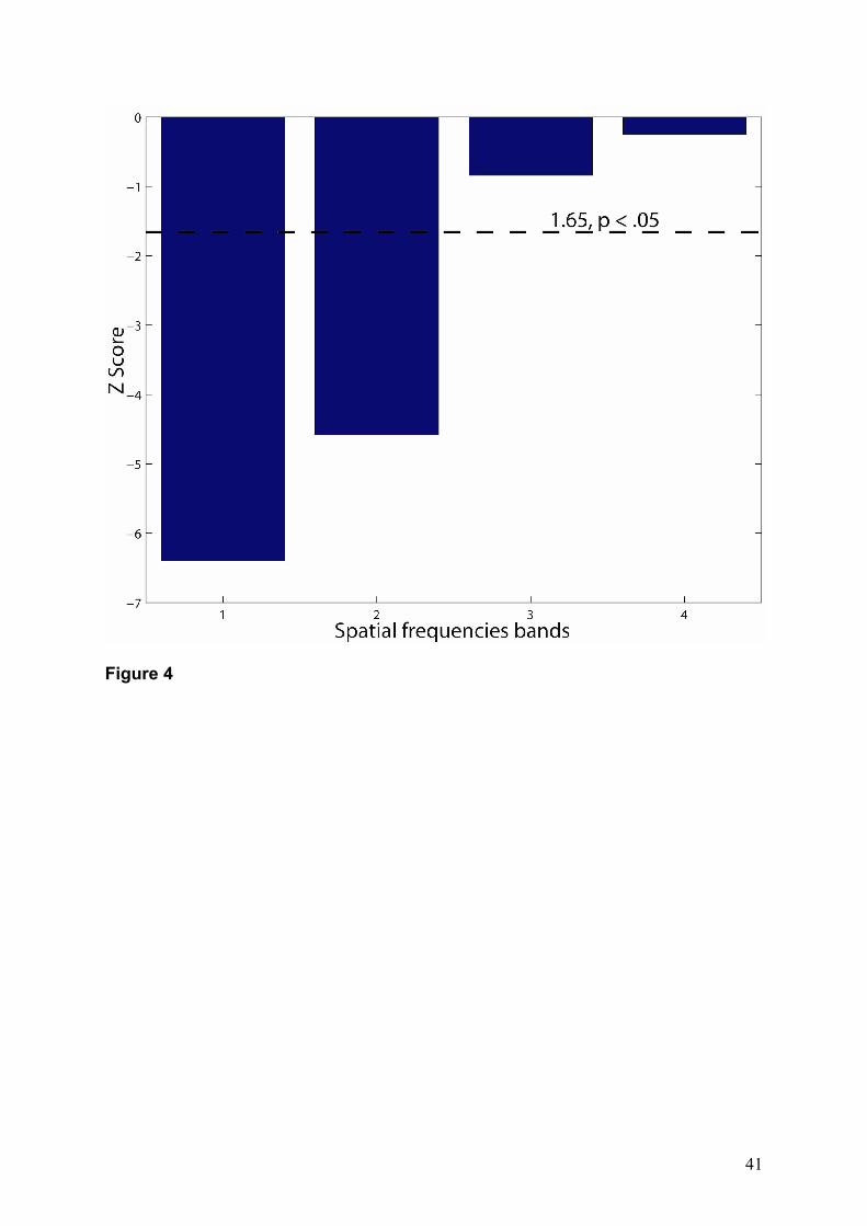
From the diagnostic information we also derived the relative use of the spatial
frequency bands by calculating the proportion of pixels that lead to a correct face
identification in each of these scales. These values were averaged across the controls,
estimating a normal use of spatial frequency information, and then were used to normalize
the relative use of information of PS. Figure 4 shows that PS used high spatial frequencies
less than controls. However, her usage of middle and low spatial frequencies bands were
comparable to the control group.
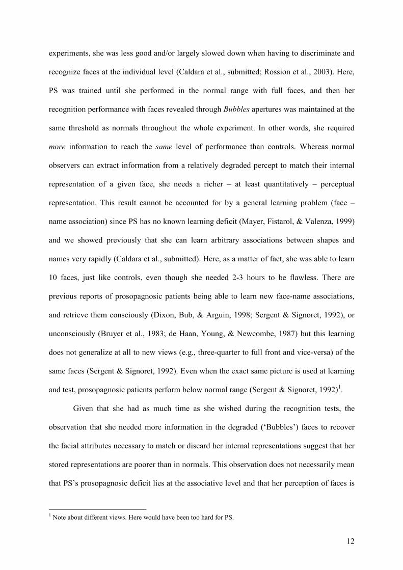
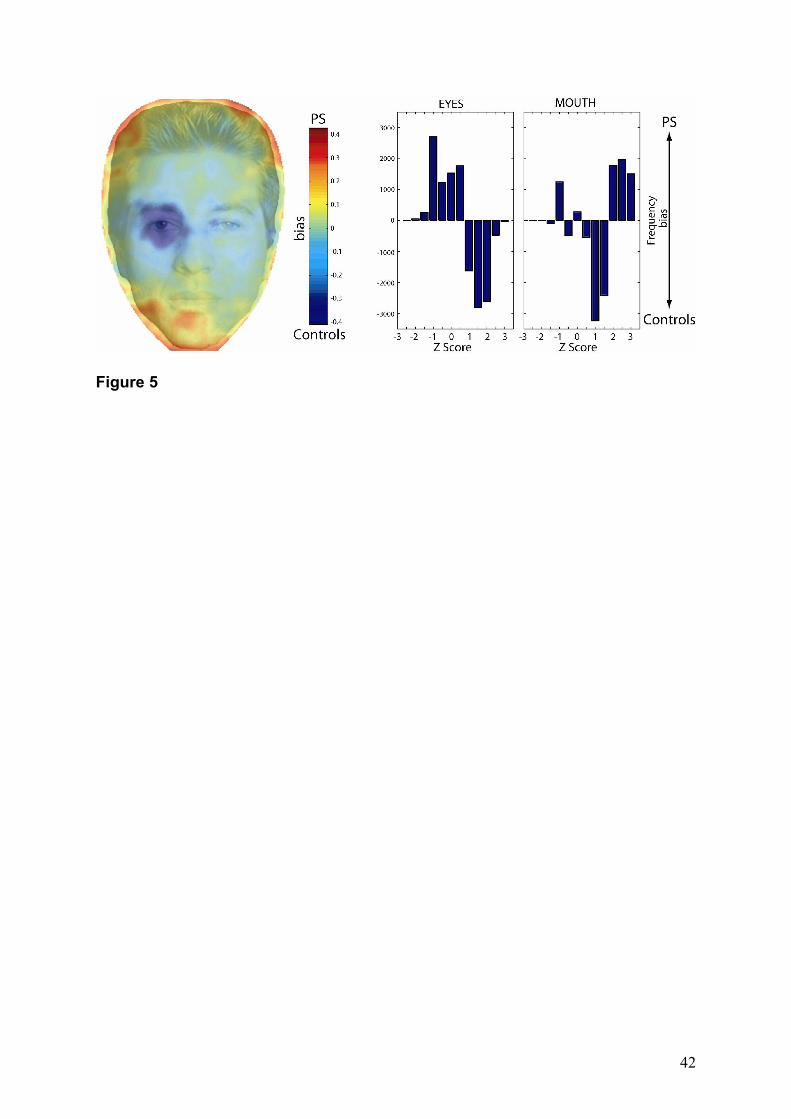
Finally, a fine-grained evaluation of PS' use of information was obtained by
comparing her performances with a model of normal face identification. This model was
derived by firstly estimating for each control the ratios of the number of times which a
particular pixel led to correct face identification, over the number of times this pixel was
presented (i.e. ProportionalPlane, see Methods), and then computing a cell-by-cell average of
the estimated values across the controls. The same procedure was used to estimate PS use of
information. Crucially, this procedure preserves all the information about the performances of
PS and controls. As a result, the sensitivity of PS to extracting information from faces was
isolated by using the logarithm of a pixel-wise comparison of the model of the control group
with that of patient PS (Smith, Cottrell, Gosselin, & Schyns, in press). Hence, this procedure
determines how each single pixel was efficiently used by patient PS weighted against the
controls (positive values – PS bias), and, conversely, how each single pixel was more
efficiently processed by the controls (negative values – controls bias). The results show that
for face identification, PS uses optimally the outline contours of the faces, and sub-optimally
the internal features, especially at level of the eyes (Figure 5 – left). Then, the role of the
internal features in face identification was further investigated by calculating and separately
11
subtracting the distributions of the performances in the eye and mouth regions (Figure 5 –
right) for PS and the controls. PS, when compared to the controls, used significantly less the
eyes (negative values) than the mouth (positive values) to achieve correct identifications (Z >
1.65; p < 0.05). Conversely, controls used less the mouth than the eyes to efficiently perform
the task. Note, that PS was even slightly impaired by the presence of the eyes to correctly
identify the faces (Z < -1.65; p < 0.05).
DISCUSSION
Our general goal was to shed light on the nature of the missing and preserved
components of the face processing system when it is selectively – albeit not entirely -
damaged. The first observation was that the prosopagnosic patient PS and controls
dramatically differed on the amount of information needed (i.e. number of bubbles) to
recognize an individual face. Furthermore, PS improved throughout the recognition sessions,
using progressively less information. Her ‘learning curve’ was however less steep than
normal controls, including age-matched subjects. Of particular interest, PS and controls
differed on the type of information used to recognize the individual faces: across faces, PS
relied much less on information contained in the upper part of the face (the eye region), but
used mouth area heavily. This pattern is in stark contrast with the distribution of information
used by normal controls, weighted to the eyes region. She also presented a general
suboptimal use of the internal features, and a bias to use of the outline contour of the faces.
Finally, if anything, PS was more impaired in extracting information in high spatial
frequencies but did not differ from normals for low and middle spatial frequency bands. We
discuss these findings in turn.
The fact that PS required more Bubbles than normal controls can be thought of
another illustration of her dramatic face processing impairment. In previous behavioral
12
experiments, she was less good and/or largely slowed down when having to discriminate and
recognize faces at the individual level (Caldara et al., submitted; Rossion et al., 2003). Here,
PS was trained until she performed in the normal range with full faces, and then her
recognition performance with faces revealed through Bubbles apertures was maintained at the
same threshold as normals throughout the whole experiment. In other words, she required
more information to reach the same level of performance than controls. Whereas normal
observers can extract information from a relatively degraded percept to match their internal
representation of a given face, she needs a richer – at least quantitatively – perceptual
representation. This result cannot be accounted for by a general learning problem (face –
name association) since PS has no known learning deficit (Mayer, Fistarol, & Valenza, 1999)
and we showed previously that she can learn arbitrary associations between shapes and
names very rapidly (Caldara et al., submitted). Here, as a matter of fact, she was able to learn
10 faces, just like controls, even though she needed 2-3 hours to be flawless. There are
previous reports of prosopagnosic patients being able to learn new face-name associations,
and retrieve them consciously (Dixon, Bub, & Arguin, 1998; Sergent & Signoret, 1992), or
unconsciously (Bruyer et al., 1983; de Haan, Young, & Newcombe, 1987) but this learning
does not generalize at all to new views (e.g., three-quarter to full front and vice-versa) of the
same faces (Sergent & Signoret, 1992). Even when the exact same picture is used at learning
and test, prosopagnosic patients perform below normal range (Sergent & Signoret, 1992)1.
Given that she had as much time as she wished during the recognition tests, the
observation that she needed more information in the degraded (‘Bubbles’) faces to recover
the facial attributes necessary to match or discard her internal representations suggest that her
stored representations are poorer than in normals. This observation does not necessarily mean
that PS’s prosopagnosic deficit lies at the associative level and that her perception of faces is
1 Note about different views. Here would have been too hard for PS.
13
normal (i.e. ‘a pure associative prosopagnosia’). In fact, her inability to match full face
pictures presented under identical or different viewpoints (Rossion et al., 2003; Caldara et al.,
submitted), point to a face deficit lying at the perceptual level. However, as pointed out
previously, disruption at the perceptual level of the face processing system may disable a set
of face processes, making it impossible to encode rich facial representations in memory
(Farah, 1990; Sergent & Signoret, 1992).
In what respects then, is PS’s internal representation of faces poorer than normals?
Behavioral and neurophysiological evidence suggest that faces are represented both in terms
of individual features (e.g., eyes, nose, mouth ...) and also as undissociated wholes (Perrett,
Hietanen, Oram, & Benson, 1992; Perrett, Rolls, & Caan, 1982; Tanaka & Farah, 1993).
Following PS’ brain damage, her representation of individual face features may be less
salient, or these features may be less well integrated in holistic facial representations, or both.
The observation that PS relied less on information contained in the high spatial frequencies
(HSFs) in the present experiment is not only in agreement with previous reports on non-facial
stimuli (Rizzo, Corbett, Thompson, & Damasio, 1986), but supports the hypothesis that PS’
representation of individual face features may be less salient, since HSFs mostly convey
detailed, featural information on faces (Morrison & Schyns, 2001; Sergent, 1986). However,
we have been collecting evidence elsewhere suggesting that PS presents a marked
impairment in building holistic facial representations, showing no inversion effect (Yin,
1969), no advantage in matching whole faces to wholes versus parts (Tanaka & Farah, 1993),
and no interference between face parts (Young, Hellawell, & Hay) (Rossion, Caldara,
Michel, & Mayer, in preparation). Although measured through different paradigms, this
inability to extract configural information on faces is typically observed in prosopagnosia
(Barton et al., 2002; Boutsen & Humphreys, 2002; Farah, Wilson, Drain, & Tanaka, 1998;
Sergent & Signoret, 1992). In short, it may well be that normal controls require less
14
information during the Bubbles experiment than PS because their internal representations are
more detailed at the level of single features and/or are better integrated, allowing to derive a
full representation from smaller fragments of the face.
How about the quality of PS’s internal representation of the faces? A dominant
feature is that it is weighted less on the upper part of the face (eyes/eyebrows) than on the
lower part (mouth area). The dominant reliance of PS on the mouth area was not true for all
faces that she was able to recognize, and there was also a certain amount of inter-individual
variability in normal controls. Yet, the general pattern was crystal clear: whereas the region
of the eyes dominated the picture for normal subjects, PS weighted strongly the area around
the mouth of the face. This finding suggests that PS’s internal representations of the faces
contain mostly information in the mouth area, whereas the normal face processing system
relies largely on the eyes (see also Gosselin & Schyns, 2001; Schyns et al., 2002).
The dominance of the eye region in normal face processing subjects is in agreement
with a large amount of observations. Primates in general, not only humans (Emery, 2000),
possess a great interest during social interactions in the eyes and the region around them, by
spending generally more time looking at the eye region of faces than the nose or mouth areas
(Keating & Keating, 1982; Kyes & Candland, 1987). A looking preference bias toward the
upper part of the visual field on facelike and nonfacelike patterns is present already in
newborns (Turati, Simion, Milani, & Umilta, 2002), as well as at the level of the face-
sensitive brain area in human adults (Caldara et al., submitted), and babies of less than a year
of age already follow other’s gaze (e.g., Butterworth, 1991). Neurophysiological evidence in
non-human primates also supports a dominant role of eyes in the representation of faces.
Single-cell recording studies in monkeys have shown that in the superior temporal sulcus
(STS) there are neurons specifically sensitive to the eyes (Perrett et al., 1985), which may be
involved in the recognition of the location where another individual is looking (Perrett et al.,
15
1992). In humans, neuroimaging studies show that the posterior part of the STS region is
implicated in gaze processing (Hoffman & Haxby, 2000; Puce, Allison, Bentin, Gore, &
McCarthy, 1998) (for a review, see Allison, Puce, & McCarthy, 2000). Electrophysiological
studies in humans corroborate the ‘special’ nature of the eyes in several ways. First, it has
been shown that isolated human eyes evoke particularly large and early visual responses
compared to whole face stimuli or other isolated facial features (e.g., Bentin, Allison, Puce,
Perez, & McCarthy, 1996; Taylor, Edmonds, McCarthy, & Allison, 2001). Second, the
occipito-temporal N170 (Bentin et al., 1996) evoked by isolated eyes is present earlier in
development than the same component elicited by whole face stimuli, suggesting a faster
maturation of the eye processing system compared to general face processes (Taylor et al.,
2001). Third, recent evidence using Bubbles in adults suggest that the eyes of a face evoke
the earliest and largest face-sensitive ERP responses (Schyns, Jentzsch, Johnson,
Schweinberger, & Gosselin, 2003).
Most importantly for the present findings, there is evidence supporting the view that
the eyes are dominant in the recognition of individual faces. Human adults can recognize and
remember faces with the eyes only (McKelvie, 1976) and experiments designed to measure
the relative importance of different facial features for individual face recognition have
consistently shown the dominance of the eye/eyebrow combination, followed by the mouth
and then the nose (Davies, Ellis, & Shepherd, 1977; Fraser, Craig, & Parker, 1990; Haig,
1985, 1986; Sergent, 1984; Tanaka & Farah, 1993; Walker Smith, 1978). In fact, recent
evidence even suggests that the eyebrows alone convey critical information to recognize
faces (Sadr, Jarudi, & Sinha, 2003; Sekuler et al., 2004; Vinette, Gosselin, & Schyns, 2004).
Given these considerations, there are several possible explanations regarding the
decreased reliance on the eyes area following prosopagnosia, as found in PS. First, following
her brain damage, PS may have lost the ability to extract other information than the cues
16
associated with identity on the eye region, and would have turned her interest to different
(e.g., the mouth) face cues when interacting with others. However, whereas she reports being
unable to recognize people, she does not complain about judging people’s expressions for
instance. Her judgment of expression is relatively preserved as compared to identity (Rossion
et al., 2003), and there is no evidence that she lost her ability to detect eye gaze direction,
although this should be tested in detail. Furthermore, PS’s lesions spare the region of the STS
entirely, anatomically and functionally (Sorger et al., 2004).
Another hypothesis that may account for PS’ decrease in the representation of the
eyes is also indirect. Given her inability to recognize faces, she may have developed some
strategies over time to be able to recognize people while avoiding the embarrassment caused
by staring. Indeed, it is well know that prosopagnosics report avoid looking people they
encounter in the eyes because they need quite some times and numerous cues to recognize
familiar persons and one cannot afford staring at people for long amounts of time (Gauthier et
al., 1999). According to these views, PS may be over-representing the mouth area for reasons
that have nothing to do with the structure of the face and the loss of her face individual
recognition abilities per se, but as a result of the development of new strategies following her
impairment.
Although these hypotheses cannot be completely dismissed, we favor the possibility
that her over-representation of the mouth area at the expense of the eyes has to do with the
interaction between the structure of the faces - what type of cues are conveyed by the eyes -
and the expertise of the face processing system with these cues. Several observations support
this hypothesis. When presented in isolation, the eyes area can be conceived as a visual
structure made up of several (pairs of) components (i.e. eyebrows, eyelid, eyelash, eyeball,
pupil and iris). As such, there is a lot of configural information (e.g., distance between the
eyes, between the eyebrows and the eyes …) in this structure, much more so than in other
17
internal facial features such as the mouth and the nose. Several experiments by Leder and
colleagues (Leder & Bruce, 2000; Leder, Candrian, Huber, & Bruce, 2001) have shown that
configural information was not limited to the whole face but consisted at least partly of
locally processed relations between facial elements, in particular at the level of the eyes.
Rakover and Teucher (1997) also found inversion effects for isolated features and showed
larger effects for the eye area than the mouth, suggesting that the former conveys more
configural information. In fact, a large number of studies showing the important role of
configural information in face perception mostly or exclusively used spatial changes (e.g.,
inward/outward or up/down movements) at the level of the eyes (e.g., Haig, 1984; Hosie,
Ellis, & Haig, 1988; Kemp, McManus, & Pigott, 1990; Leder et al., 2001; Macho & Leder,
1998). Supporting yet again the critical role of the eyes region in supporting configural
information, there is evidence from the whole/part advantage paradigm that the holistic
processing of the face is larger for the eyes than for other features (Donnelly & Davidoff,
1999).
Most interestingly, whereas the whole/part advantage is observed for the mouth on
unfamiliar faces and does not increase with familiarization, this effect appears only for the
eyes after substantial training with a face set (Donnelly & Davidoff, 1999). Thus, the ability
to extract and use configural information on the eyes seems to develop over time and
concerns mostly familiar faces. This is also the conclusion of two experiments conducted by
O’Donnell and Bruce (2001), which tested subjects’ discrimination abilities on pairs of
familiar and unfamiliar faces with subtle differences at the level of one of four feature (hair,
jaw, eye, mouth). Strikingly, when faces differed only by the eyes, subject’s performance was
excellent for familiar faces (above 90%) and below chance for unfamiliar faces. There was no
effect of familiarity for the other features, including the mouth. This familiarity effect
restricted to the eyes was found whether the modifications were configural (eye spacing,
18
experiment 1) or local (brightness change, experiment 2). The conclusion of the authors is
that details from the upper part of the face may offer particularly salient information in the
initial structuring of categorical knowledge about individual faces (O'Donnell & Bruce,
2001). This holds obviously for a normally functioning face processing system. The present
study, contrasting the representations used by normal viewers and a damaged face processing
system suggests that a critical feature of prosopagnosia is the inability to extract such salient
information, largely conveyed by the eyes area, to build robust representations of faces.
In other words, we suggest that relative to normals, PS does not rely on eyes because
her face processing system is not able to extract and store reliable cues, perhaps mostly
configural, from this area of the face. Also in line with the proposal that the difference
between PS and normals has to do with the ability to extract and store critical information for
familiar faces is the observation that she relies much less on internal features than on external
features in general, relative to normals (Figure 5). This observation can again be related to the
inner-features advantage observed for familiar faces, namely the robust finding that the inner-
face parts are more useful than those of the outer face for recognizing familiar faces but not
unfamiliar faces (Campbell et al., 1999; Ellis, Shepherd, & Davies, 1979; Young et al., 1986).
Since this inner-features advantage is found only with familiar faces, it is associated with the
‘memorial representation of the known individual’ (Campbell et al., 1999). When encoding
faces in memory, the normal face processing system appears to give more weight to internal
face features. In contrast, our data shows that, even though PS managed to encode faces in
memory, she relied less than controls on inner facial features, similarly to her lesser reliance
on the eyes area of the face. To our knowledge, the inner-face advantage for familiar faces
has not been tested in prosopagnosics previously, given their difficulties in learning faces.
However, the effect was found in posterior left and right brain-damaged patients without
19
prosopagnosia (de Haan & Hay, 1986), suggesting that the pattern found in PS may be
specific to prosopagnosia.
The literature covered above has suggested that both the reliance on facial
configuration, particularly at the level of the eyes, and the advantage given to internal
features reflect important characteristics of the normal adult face processing system, allowing
recognizing individual familiar faces. Quite interestingly, these features of the face
processing system appear to undergo a long developmental course, relative to other aspects of
face processing. The inner-face advantage for familiar faces, for instance, is found only at 15
years old, children before that age showing at outer-face advantage (Campbell et al., 1999;
see also Carey & Diamond, 1977). Other evidence suggest that, even though the early months
of visual experience may be critical for the normal development of facial configuration
processes (Le Grand, Mondloch, Maurer, & Brent, 2001), this ability develops relatively
slowly compared to the capacity to extract individual features (Mondloch, Le Grand, &
Maurer, 2002), and may not be achieved fully until mid-adolescence (Carey, 1992; Carey &
Diamond, 1994; Ellis & Ellis, 1994).
The data reported here suggests that the fundamental properties of the face processing
system that undergoes a long developmental course may also be the most sensitive, or the
first to be disrupted, when selective damage occurs to this system. Obviously, this suggestion
needs complementary evidence, such as showing how PS deals with various face information
on ‘familiar’ and unfamiliar faces relative to normals in simple discrimination tasks for
instance.
It is interesting to note at this point that PS’s highest reliance on the mouth and on
external facial features relative to normals concur with her verbal reports about people she
encountered after her accident. When asked about how people she met in the lab look like
instance, she always refers to the shape of the mouth and teeth area, as well as to external
20
contour information (hair, head size ...). Most importantly, it remains to be seen whether
other cases of prosopagnosia with at least relatively well-preserved non-face recognition
processes, show the lesser reliance on the eyes area and internal face features in general.
Recent investigations with simultaneous discrimination tasks on patient LR, a case of
acquired prosopagnosia restricted to faces following a lesion in the anterior part of the right
temporal lobe, support this view (Bukach, Bub, Gauthier, & Tarr, submitted).
CONCLUSIONS
Testing a single case of acquired prosopagnosia with Bubbles gives clues about how
faces can be represented by a selectively disrupted face processing system relative to
normals. The data collected show that PS’s internal representations of ‘familiar’ faces is less
robust and specific than in normals, requiring more information from the percept to be
activated and discriminated from other representations. Qualitatively, PS’s representations
appear to be weighted less toward the eyes and the internal features in general, which are
critical components of a normal adult face processing system having to build and store robust
representations of individual faces.
Finally, in the framework of high-level visual neuropsychological diseases, the
Bubbles technique might represent a promising instrument for identifying in the patients the
preserved visual representations and, as a consequence, exploiting this information for their
rehabilitation.
METHODS
Subjects
Case report
PS is a case of acquired prosopagnosia with normal object recognition who has been
reported in detail elsewhere (Rossion et al., 2003) and will be only briefly summarized here.
21
She is a 53 years old woman (born in 1950) who sustained a closed head injury in 1992,
causing lesions of the lateral part of the occipital and temporal lobes, bilaterally (see Figure 1
in Rossion et al., 2003). Despite the extent of the lesions in the visual system, PS has normal
low-level visual processing. PS is ranked as highly impaired on the Benton Face Matching
Test (Benton & van Allen, 1972), scoring 27/54 (percentile 1). She is also impaired on the
Short Recognition Memory Test for Faces, a set of the Camden Memory Tests (Warrington,
1984; Warrington, 1996), scoring 18/25 (percentile 3). Regarding the recognition of familiar
faces, when PS was confronted with the pictures of 60 famous people (all know by the
patient), she was able to classify 14 of them as familiar, and correctly classified all the
unfamiliar ones. Nevertheless, when she had to report the individual names of the faces
classified as familiar, as well as their associated semantic information, drastically, she was
correct for only 4 of them. Although much better than for individual discrimination, PS is not
as good as controls at gender and expression facial judgments but has normal performances
of age assessment on faces (Rossion et al., 2003). She is also able to draw correctly a
schematic face, and perfectly point out all the single features. Finally, PS has been tested
extensively with simultaneous and delayed face and non-face (cars and novel objects)
matching tasks in previous studies (Rossion et al., 2003). While she is consistently
dramatically impaired and slowed down for the face conditions, her performance with the
non-face objects are in the normal range.
Controls
5 young adults (mean age 27) and two age- and education level-matched controls (1
woman 54; 1 man 57) voluntarily participated in the experiment. All the subjects were
healthy, and had no neurological or psychiatric history.
Stimuli
22
Ten unfamiliar faces (5 females, 5 males) displaying two possible expressions (neutral
or happy) were used in the experiment. All faces had a normalized hairstyle, global
orientation and lighting (Schyns & Oliva, 1999). Bubbles (Gosselin & Schyns, 2001) was
applied to generate the stimuli (see Gosselin & Schyns, 2001 for details). Each face is first
decomposed in six non overlapping bands of spatial frequencies of 1 octave each. The cut-
offs of these bands were respectively 90, 45, 22.5, 11.25, 5.62, and 2.81 cycles per face, from
the finest to the coarsest scale. The coarsest band was a constant background and is not
illustrated in Figure 1. For each spatial frequency band, a number of randomly located
Gaussian apertures (called Bubbles) sampled facial information independently at each scale
to create a sparse stimulus – i.e. a subset of the original facial information. With sufficiently
many trials, this technique ensures that the entire face is sampled in the considered spatial
frequency bandwidths – i.e. Bubbles is an asymptomatically exhaustive non biased sampling.
Procedure
Prior to the Bubbles experiment, all participants received a printed version of all faces
and learned them at the individual level (e.g., their names). In a computer verification task
that assessed performances, participants were confronted with each face and instructed to
name them by pressing the corresponding computer keyboard key. Feedback displaying the
correct name informed subjects of their mistakes. This training procedure ended when
subjects performed a perfect identification of all the faces twice in a row. On average, for the
controls only ten minutes were sufficient to reach this level and start the experiment. The
same procedure was used with the patient PS. However, for her, a 3 hours training program
split in two days was necessary to achieve this level of face identification. In addition, PS
performed this verification procedure before starting each of the Bubbles sessions, and had to
succeed twice in a row.
23
The experiment ran on a Macintosh PowerBook G3 computer using a program written
with the Psychophysics (Brainard, 1997; Pelli, 1997) and Pyramid (Simoncelli, 1997)
Toolboxes for Matlab and consisted of sessions of 300 trials, presenting randomly 30 times
the same identity (15 neutral, 15 happy). All trials were taken into account for the analysis. In
each trial, a sparse face appeared on the screen, and the participants had to determine its
identity by pressing the corresponding computer keyboard key. The image remained on the
screen until the subject responded, and no feedback was provided. Note that PS and the
controls were not required to answer as fast as possible, and thus were free to finely examine
each stimulus. The number of bubbles was automatically adjusted in order to maintain
subjects' performance at 75% correct. PS performed a total of 9300 experimental trials
divided in 31 sessions of 300 trials, spanned over 16 weeks. Controls performed 4200 trials
divided in 14 sessions within three weeks.
Data analysis
We hypothesize that on any given trial, if the participant could correctly categorize
the sparse face on the basis of the information revealed by the bubbles, that information was
sufficient for that face identification. Across trials, we therefore kept track of the locations of
the bubbles leading to correct identification for each participant. To this end, for each scale
we added the masks with the bubbles leading to correct identifications in order to create a
CorrectPlane — henceforth, CorrectPlane(scale), where scale information is represented from
1 to 5, from fine to coarse (see Figure 1 for examples of masks). CorrectPlane(scale)
therefore encapsulates the locations, at each scale, where sampling of face information
(bubbles) led to correct identifications. We also added the masks with bubbles leading to both
correct and incorrect identifications to create TotalPlane(scale). So TotalPlane(scale)
represents, for each scale, the total sampling frequency of face information. From the
information in CorrectPlane(scale) and TotalPlane(scale), we determined, for each subject
24
separately and on a cell-by-cell basis, the ratio of the number of times a specific location led
to a successful face identification over the number of times this location was presented,
CorrectPlane(scale)/TotalPlane(scale). We refer to this ratio as ProportionPlane(scale).
Across subjects, the averaged ProportionPlane(scale) weighs the importance of the regions of
each scale for the identification task. If all regions were equally important,
ProportionPlane(scale) would be uniform across cells, and equal to the performance criterion
— here, .75. Consequently, regions significantly above (vs. below) the performance criterion
are more (vs. less) diagnostic. To determine this significance, we built a confidence interval
(p < .05) around the mean of the ProportionPlane(scale), for each proportion.
DiagnosticPlane(scale) was created by representing diagnostic (significant) proportions with
a filtering weight of 1 and nondiagnostic proportions with a filtering weight of 0. These
diagnostic weights were then used to filter the original stimulus to derive the effective
stimulus (see Figure 3), which depicts the selective use of information in the task. The
effective stimulus is simply obtained by multiplying the face information at each scale in
Figure 1 with the corresponding DiagnosticPlane(scale) convolved with bubbles of the size
used during the experiment. The importance of the information used at each spatial frequency
bands was compared between PS and the control group (see Figure 4), by normalizing patient
PS results with those of the control group, and selecting a confidence interval (p < .05)
around the mean.
The logarithm of a pixel-wise division of the ProportionPlane of patient PS with the
averaged ProportionPlane of the control group was computed to determine the
optimality/efficiency of information use by the patient compared to the control group (e.g.
Smith et al., in press). Negative and positive values are respectively related to a suboptimal
and an optimal processing of patient PS (Figure 5 - left). The 0 value represent a comparable
performance between patient PS and the control group. Finally, to reveal the quality of the
25
performances for the eyes and the mouth facial features, two separate masks covering these
face regions were applied to the ProportionPlane of PS and the controls, disclosing the
respective distributions for both face features and groups. Then, PS' distributions were
subtracted from the controls' distributions for each respective face region, revealing a
frequency bias for each facial feature in both populations (Figure 5 - right).
Acknowledgements
We are immensely grateful to PS for her patience and motivation during the
experiment. This work was supported by a grant ARC 01/06-267 (Communauté Française de
Belgique – Actions de Recherche Concertées) to Bruno Rossion. Roberto Caldara is
supported by a post-doctoral fellowship provided by the Swiss National Science Foundation.
26
REFERENCES
Allison, T., Puce, A., & McCarthy, G. (2000). Social perception from visual cues: Role of sts
region. Trends Cogn Sci, 4(7), 267-278.
Barton, J. J. (2003). Disorders of face perception and recognition. Neurol Clin, 21(2), 521-
548.
Barton, J. J., Cherkasova, M., & O'Connor, M. (2001). Covert recognition in acquired and
developmental prosopagnosia. Neurology, 57(7), 1161-1168.
Barton, J. J., Press, D. Z., Keenan, J. P., & O'Connor, M. (2002). Lesions of the fusiform face
area impair perception of facial configuration in prosopagnosia. Neurology, 58(1), 71-
78.
Bentin, S., Allison, T., Puce, A., Perez, E., & McCarthy, G. (1996). Electrophysiological
studies of face perception in humans. Journal of Cognitive Neuroscience, 8(6), 551-
565.
Benton, A., & van Allen, M. (1972). Prosopagnosia and facial discrimination. J Neurol Sci,
15, 167-172.
Bodamer, J. (1947). Die prosop-agnosie. Archive für Psychiatrie und Nervenkrankheiten,
179, 6-54.
Boutsen, L., & Humphreys, G. W. (2002). Face context interferes with local part processing
in a prosopagnosic patient. Neuropsychologia, 40(13), 2305-2313.
Bouvier, S. E., & Engel, S. A. (2004). Patterns of cortical damage in achromatopsia and
prosopagnosia. Journal of Vision, 4, Supplement, 205a.
Bruce, V., Burton, A. M., Hanna, E., Healey, P., & et al. (1993). Sex discrimination: How do
we tell the difference between male and female faces? Perception, 22(2), 131-152.

27
Bruce, V., Henderson, Z., Greenwood, K., Hancock, P. J. B., Burton, A. M., & Miller, P.
(1999). Verification of face identities from images captured on video. Journal of
Experimental Psychology:Applied, 5(4), 339-360.
Bruce, V., & Young, A. (1986). Understanding face recognition. British Journal of
Psychology, 77, 305-327.
Bruyer, R., Laterre, C., Seron, X., Feyereisen, P., Strypstein, E., Pierrard, E., et al. (1983). A
case of prosopagnosia with some preserved covert remembrance of familiar faces.
Brain Cogn, 2(3), 257-284.
Bukach, C. M., Bub, D. M., Gauthier, I., & Tarr, M. J. (submitted). Perceptual expertise
effects are not all or none: Local perceptual expertise for faces in a case of
prosopagnosia.
Burton, A. M., Wilson, S., Cowan, M., & Bruce, V. (1999). Face recognition in poor-quality
video: Evidence from security surveillance. Psychological Science, 10(3), 243-248.
Butterworth, G. (1991). The ontogeny and phylogeny of joint visual attention in human
infancy. In A. Whiten (Ed.), Natural theories of mind (pp. 223-232). Oxford:
Blackwell.
Caldara, R., Gauthier, I., Rossion, B., Schuller, A. M., Tarr, M. J., & Mayer, E. (submitted).
Prosopagnosia as an inability to develop expertise with visually homogeneous
categories: Evidence from a single-case study.
Caldara, R., Seghier, M., Rossion, B., Lazeyras, F., Michel, C., & Hauert, C.-A. (submitted).
A curvilinear vertical asymmetry tunes fusiform face area responses.
Calder, A. J., Burton, A. M., Miller, P., Young, A. W., & Akamatsu, S. (2001). A principal
component analysis of facial expressions. Vision Res, 41(9), 1179-1208.
28
Campbell, R., Coleman, M., Walker, J., Benson, P. J., Wallace, S., Michelotti, J., et al.
(1999). When does the inner-face advantage in familiar face recognition arise and
why? Visual Cognition, 6(2), 197-216.
Campbell, R., Landis, T., & Regard, M. (1986). Face recognition and lipreading. A
neurological dissociation. Brain, 109(Pt 3), 509-521.
Carey, S. (1992). Becoming a face expert. Philos Trans R Soc Lond B Biol Sci, 335(1273),
95-102; discussion 102-103.
Carey, S., & Diamond, R. (1977). From piecemeal to configurational representation of faces.
Science, 195(4275), 312-314.
Carey, S., & Diamond, R. (1994). Are faces perceived as configurations more by adults than
by children? In V. H. G. W. Bruce (Ed.), Object and face recognition. Special issue of
visual cognition (Vol. 1, pp. 253-274). Hove, England UK: Lawrence Erlbaum
Associates, Inc.
Damasio, A. R. (1985). Disorders of complex visual processing: Agnosias, achromatopsia,
balint's syndrome, and related difficulties in orientation and construction. In M.-M.
Mesulam (Ed.), Principles of behavioural neurology (pp. 259-288). Philadelphia: F.A.
Davis.
Damasio, A. R., Damasio, H., & Van Hoesen, G. W. (1982). Prosopagnosia: Anatomic basis
and behavioral mechanisms. Neurology, 32(4), 331-341.
Davies, G., Ellis, H., & Shepherd, J. (1977). Cue saliency in faces as assessed by the
"photofit" technique. Perception, 6(3), 263-269.
de Haan, E. H., & Hay, D. (1986). The matching of famous and unknown faces, given either
the internal or external features:A study onpatients with unilateral brain lesions. In H.
D. Ellis, F. Jeeves, F. Newcombe & A. W. Young (Eds.), Aspects of face processing
(pp. 302-309). Dordrecht: Martinus Nijhoff.
29
de Haan, E. H., Young, A., & Newcombe, F. (1987). Faces interfere with name classification
in a prosopagnosic patient. Cortex, 23(2), 309-316.
Diamond, R., & Carey, S. (1986). Why faces are and are not special: An effect of expertise. J
Exp Psychol Gen, 115(2), 107-117.
Dixon, M. J., Bub, D. N., & Arguin, M. (1998). Semantic and visual determinants of face
recognition in a prosopagnosic patient. Journal of Cognitive Neuroscience, 10(3),
362-376.
Donnelly, N., & Davidoff, J. (1999). The mental representations of faces and houses: Issues
concerning parts and wholes. Visual Cognition, 6(3-4), 319-343.
Ellis, D. M., & Ellis, H. D. (1994). Development of facial transformation processing skills in
children. Psychologie Francaise, 39(3), 287-300.
Ellis, H. D., & Florence, M. (1990). Bodamer's (1947) paper on prosopagnosia. Cognitive
Neuropsychology, 7(2), 81-105.
Ellis, H. D., Shepherd, J. W., & Davies, G. M. (1979). Identification of familiar and
unfamiliar faces from internal and external features: Some implications for theories of
face recognition. Perception, 8(4), 431-439.
Emery, N. J. (2000). The eyes have it: The neuroethology, function and evolution of social
gaze. Neurosci Biobehav Rev, 24, 581-604.
Farah, M., Levinson, K. L., & Klein, K. L. (1995). Face perception and within-category
discrimination in prosopagnosia. Neuropsychologia, 33(6), 661-674.
Farah, M. J. (1990). Visual agnosia.Cambridge: MIT Press.
Farah, M. J., McMullen, P. A., & Meyer, M. M. (1991). Can recognition of living things be
selectively impaired? Neuropsychologia, 29(2), 185-193.
Farah, M. J., Wilson, K. D., Drain, M., & Tanaka, J. N. (1998). What is "special" about face
perception? Psychol Rev, 105(3), 482-498.
30
Fraser, I. H., Craig, G. L., & Parker, D. M. (1990). Reaction time measures of feature
saliency in schematic faces. Perception, 19(5), 661-673.
Gauthier, I., Behrmann, M., & Tarr, M. J. (1999). Can face recognition really be dissociated
from object recognition? Journal of Cognitive Neuroscience, 11(4), 349-370.
Goldsmith, Z. G., & Liu, G. T. (2001). Facial recognition and prosopagnosia: Past and
present concepts. Neuro-Ophthalmology, 25, 177-192.
Gosselin, F., & Schyns, P. G. (2001). Bubbles: A technique to reveal the use of information
in recognition tasks. Vision Res, 41(17), 2261-2271.
Grüsser, O. J., & Landis, T. (1991). Visual agnosias and other disturbances of visual
perception and cognition. In Vision and visual dysfunction (Vol. 12). London:
Macmillan Press.
Haig, N. D. (1984). The effect of feature displacement on face recognition. Perception, 13(5),
505-512.
Haig, N. D. (1985). How faces differ--a new comparative technique. Perception, 14(5), 601-
615.
Haig, N. D. (1986). Exploring recognition with interchanged facial features. Perception,
15(3), 235-247.
Hill, H., Bruce, V., & Akamatsu, S. (1995). Perceiving the sex and race of faces: The role of
shape and colour. Proc R Soc Lond B Biol Sci, 261(1362), 367-373.
Hoffman, E. A., & Haxby, J. V. (2000). Distinct representations of eye gaze and identity in
the distributed human neural system for face perception. Nat Neurosci, 3(1), 80-84.
Hosie, J. A., Ellis, H. D., & Haig, N. D. (1988). The effect of feature displacement on the
perception of well-known faces. Perception, 17(4), 461-474.
Keating, C. F., & Keating, E. G. (1982). Visual scan patterns of rhesus monkeys viewing
faces. Perception, 11(2), 211-219.
31
Kemp, R., McManus, C., & Pigott, T. (1990). Sensitivity to the displacement of facial
features in negative and inverted images. Perception, 19(4), 531-543.
Kyes, R. C., & Candland, D. K. (1987). Baboon (papio hamadryas) visual preferences for
regions of the face. Journal of Comparative Psychology, 101(4), 345-348.
Laeng, B., & Caviness, V. S. (2001). Prosopagnosia as a deficit in encoding curved surface. J
Cogn Neurosci, 13(5), 556-576.
Landis, T., Regard, M., Bliestle, A., & Kleihues, P. (1988). Prosopagnosia and agnosia for
noncanonical views. An autopsied case. Brain, 111 (Pt 6), 1287-1297.
Le Grand, R., Mondloch, C. J., Maurer, D., & Brent, H. P. (2001). Neuroperceptionearly
visual experience and face processing. Nature, 410(6831), 890.
Leder, H., & Bruce, V. (2000). When inverted faces are recognized: The role of configural
information in face recognition. Q J Exp Psychol A, 53(2), 513-536.
Leder, H., Candrian, G., Huber, O., & Bruce, V. (2001). Configural features in the context of
upright and inverted faces. Perception, 30(1), 73-83.
Levine, D. N., & Calvanio, R. (1989). Prosopagnosia: A defect in visual configural
processing. Brain and Cognition, 10(2), 149-170.
Levine, D. N., Calvanio, R., & Wolf, E. (1980). Disorders of visual behavior following
bilateral posterior cerebral lesions. Psychol Res, 41(2-3), 217-234.
Macho, S., & Leder, H. (1998). Your eyes only? A test of interactive influence in the
processing of facial features. Journal of Experimental Psychology: Human Perception
& Performance, 24(5), 1486-1500.
Malone, D. R., Morris, H. H., Kay, M. C., & Levin, H. S. (1982). Prosopagnosia: A double
dissociation between the recognition of familiar and unfamiliar faces. Journal of
Neurology, Neurosurgery and Psychiatry, 45(9), 820-822.
32
Mayer, E., Fistarol, P., & Valenza, N. (1999). Prise en charge neuropsychologique d'une
patiente prosopagnosique. In P. Azouvi, D. Perrier & M. Van der Linden (Eds.), La
reeducation en neuropsychologie: Etudes de cas.Marseille: Solal.
McKelvie, S. J. (1976). The role of eyes and mouth in the memory of a face. American
Journal of Psychology, 89(2), 311-323.
Mondloch, C. J., Le Grand, R., & Maurer, D. (2002). Configural face processing develops
more slowly than featural face processing. Perception, 31(5), 553-566.
Morrison, D. J., & Schyns, P. G. (2001). Usage of spatial scales for the categorization of
faces, objects, and scenes. Psychon Bull Rev, 8(3), 454-469.
O'Donnell, C., & Bruce, V. (2001). Familiarisation with faces selectively enhances sensitivity
to changes made to the eyes. Perception, 30(6), 755-764.
Perrett, D. I., Hietanen, J. K., Oram, M. W., & Benson, P. J. (1992). Organization and
functions of cells responsive to faces in the temporal cortex. Philos Trans R Soc Lond
B Biol Sci, 335(1273), 23-30.
Perrett, D. I., Rolls, E. T., & Caan, W. (1982). Visual neurones responsive to faces in the
monkey temporal cortex. Exp Brain Res, 47(3), 329-342.
Perrett, D. I., Smith, P. A., Potter, D. D., Mistlin, A. J., Head, A. S., Milner, A. D., et al.
(1985). Visual cells in the temporal cortex sensitive to face view and gaze direction.
Proc R Soc Lond B Biol Sci, 223(1232), 293-317.
Puce, A., Allison, T., Bentin, S., Gore, J. C., & McCarthy, G. (1998). Temporal cortex
activation in humans viewing eye and mouth movements. The Journal of
Neuroscience, 18(6), 2188-2199.
Rakover, S. S., & Teucher, B. (1997). Facial inversion effects: Parts and whole relationship.
Perception & Psychophysics, 59(5), 752-761.
33
Rizzo, M., Corbett, J. J., Thompson, H. S., & Damasio, A. R. (1986). Spatial contrast
sensitivity in facial recognition. Neurology, 36(9), 1254-1256.
Rossion, B., Caldara, R., Michel, C., & Mayer, E. (in preparation). Holistic processing is
impaired in prosopagnosia: A case study.
Rossion, B., Caldara, R., Seghier, M., Schuller, A. M., Lazeyras, F., & Mayer, E. (2003). A
network of occipito-temporal face-sensitive areas besides the right middle fusiform
gyrus is necessary for normal face processing. Brain, 126(Pt 11), 2381-2395.
Sadr, J., Jarudi, I., & Sinha, P. (2003). The role of eyebrows in face recognition. Perception,
32(3), 285-293.
Schiltz, C., Sorger, B., Ahmed, F., Caldara, R., Mayer, E., Goebel, R., et al. (submitted).
Anomalous response to facial identity in the right middle fusiform gyrus underlies
impaired face identification in acquired prosopagnosia.
Schyns, P. G., Bonnar, L., & Gosselin, F. (2002). Show me the features! Understanding
recognition from the use of visual information. Psychol Sci, 13(5), 402-409.
Schyns, P. G., Jentzsch, I., Johnson, M., Schweinberger, S. R., & Gosselin, F. (2003). A
principled method for determining the functionality of brain responses. Neuroreport,
14(13), 1665-1669.
Schyns, P. G., & Oliva, A. (1999). Dr. Angry and mr. Smile: When categorization flexibly
modifies the perception of faces in rapid visual presentations. Cognition, 69(3), 243-
265.
Sekuler, A. B., Gaspar, C. M., Gold, J. M., & Bennett, P. J. (2004). Inversion leads to
quantitative, not qualitative, changes in face processing. Curr Biol, 14(5), 391-396.
Sergent, J. (1984). Configural processing of faces in the left and the right cerebral
hemispheres. Journal of Experimental Psychology Human Perception and
Performance, 10(4), 554-572.
34
Sergent, J. (1986). Microgenesis of face perception. In H. D. Ellis, F. Jeeves, F. Newcombe
& A. W. Young (Eds.), Aspects of face processing.Dordrecht: Martinus Nijhoff.
Sergent, J., & Signoret, J. L. (1992). Varieties of functional deficits in prosopagnosia. Cereb
Cortex, 2(5), 375-388.
Smith, M., Cottrell, G. W., Gosselin, F., & Schyns, P. G. (in press). Transmitting and
decoding facial expressions. Psychological Science.
Sorger, B., Schiltz, C., Caldara, R., Kriegeskorte, N., Mayer, E., Rossion, B., et al. (2004).
Functional neuroanatomy of the visual system in a prosopagnosic. Neuroimage, 22,
(Supplement 1), S40.
Tanaka, J. W. (2001). The entry point of face recognition: Evidence for face expertise. J Exp
Psychol Gen, 130(3), 534-543.
Tanaka, J. W., & Farah, M. J. (1993). Parts and wholes in face recognition. Quarterly Journal
of Experimental Psychology Human Experimental Psychology, 46a(2), 225-245.
Taylor, M. J., Edmonds, G. E., McCarthy, G., & Allison, T. (2001). Eyes first! Eye
processing develops before face processing in children. Neuroreport, 12(8), 1671-
1676.
Taylor, M. J., McCarthy, G., Saliba, E., & Degiovanni, E. (1999). Erp evidence of
developmental changes in processing of faces. Clin Neurophysiol, 110(5), 910-915.
Tranel, D., Damasio, A. R., & Damasio, H. (1988). Intact recognition of facial expression,
gender, and age in patients with impaired recognition of face identity. Neurology,
38(5), 690-696.
Turati, C., Simion, F., Milani, I., & Umilta, C. (2002). Newborns' preference for faces: What
is crucial? Dev Psychol, 38(6), 875-882.
35
Valentine, T. (1991). A unified account of the effects of distinctiveness, inversion, and race
in face recognition. Quarterly Journal of Experimental Psychology Human
Experimental Psychology, 43a(2), 161-204.
Vinette, C., Gosselin, F., & Schyns, P. G. (2004). Spatio-temporal dynamics of face
recognition in a flash: It�s in the eyes. Cognitive Science, 28, 289�301.
Walker Smith, G. J. (1978). The effects of delay and exposure duration in a face recognition
task. Perception and Psychophysics, 24(1), 63-70.
Warrington, E. (1984). Warrington recognition memory test.Los Angeles: Western
Psychological Services.
Warrington, E. K. (1996). The camden memory tests manual.Hove: Psychology Press.
Yin, R. K. (1969). Looking at upide-down faces. Journal of Experimental Psychology, 81(1),
141-145.
Young, A. W., Hellawell, D., & Hay, D. C. (1987). Configurational information in face
perception. Perception, 16(6), 747-759.
Young, A. W., McWeeny, K. H., Hay, D. C., & Ellis, A. W. (1986). Matching familiar and
unfamiliar faces on identity and expression. Psychological Research, 48(2), 63-68.
36
FIGURE LEGENDS
Figure 1. Application of Bubbles technique to the three-dimensional space composed of a
two-dimensional face and spatial scales on the third dimension. (a) The original image is
decomposed into five different scales. (b) The bubbles are randomly positioned at each scale
and covered approximately the same area across scales. (c) The bubbles were integrated with
the decomposed pictures and summed to result in a bubblized face.
Figure 2. Mean number of bubbles per session used by PS and 7 controls to correctly adapt to
the task (AM = Age-Matched; C = Controls, young adults). The performance for all the
participants was automatically kept to a 75% correct identification.
Figure 3. Top: Diagnostic information used by PS and the average for the Controls. Bottom:
Diagnostic information for each control subject (AM = Age-Matched; C = Control subject,
young adults). Note that PS is the only participant that did not use the eyes to adapt to the
task.
Figure 4. Patient PS' Z scores in the use of spatial frequencies bands. The dashed line
indicates a Z score of 1.65 (p < .05). PS used information in the high spatial frequencies
significantly less when compared to the control group. Note that no meaningful information
was found at the fifth frequency (coarsest scale) for PS or the controls.
Figure 5. Left: Optimality of the information used by PS compared to the control group. Dark
blue regions correspond to negative values, and illustrate a suboptimal use of information.
Light blue regions correspond to values close to 0, a comparable use of information. The red
and yellow areas correspond to positive values, reflecting the bias of PS in the task. Right:

37
Distribution of the performances in the Eyes and Mouth regions, after the subtraction of PS'
distribution from the controls. A positive value indicates a bias for PS, a negative value,
indicates a bias for the controls. Note, that PS was more efficient for the Mouth (Z > 1.65; p <
.05) than the Eyes region. The reverse pattern is observed for the controls.
38
FIGURES
Figure 1
39
Figure 2
40
Figure 3
41
Figure 4
42
Figure 5


![untitled 4 [mapageweb.umontreal.ca]mapageweb.umontreal.ca/reber/VanadiumCJASS.pdfTitle untitled 4 Author Christian Reber Created Date 6/7/2002 9:30:06 PM](https://img.pdfslide.us/doc/110x75/600ba85bc91dc438b367161f/untitled-4-title-untitled-4-author-christian-reber-created-date-672002-93006.jpg)