Embed Size (px)
Citation preview
Does Clearing of Axillary Lymph Nodes Contribute to Accurate Staging of Breast Carcinoma?
MONICA MORROW, MD, JAMES EVANS, MD, P. PETER ROSEN, MD, AND DAVID W. KINNE, MD
The major prognostic indicator in carcinoma of the breast is the presence of metastases in axillary lymph nodes. However, 25% of patients with negative axillary nodes by standard pathologic techniques are dead of metastatic breast carcinoma within 10 years. “Clearing” of the axillary fat has been shown to increase the yield of lymph nodes. Forty-two pathologic Stage I and I1 breast carcinoma specimens were cleared following routine pathologic examination to determine whether stage was changed by the clearing procedure. A total of 857 lymph nodes were recovered from 42 patients by routine techniques. Clearing increased the number of nodes found by 301, to 1114. In the 31 node-negative patients an additional 178 nodes were identified, increasing the mean number of nodes per patient from 20 to 26. The number of additional nodes found per specimen ranged from 0 to 19. None of the additional nodes identified contained metastases. In the node-positive patients, 79 additional nodes were found by clearing, including 33 with metastases. No change in stage resulted, although the mean number of nodes per patient was increased from 22 to 30. Although an occasional positive lymph node may be overlooked by manual dissection, the rarity of this event makes routine clearing of the axillary contents impractical for carcinoma of the breast except in a research setting. Whether this conclusion applies equally to other tumors and other lymph node groups requires further study.
Cancer 53:1329-1332, 1984.
HE IMPORTANCE of axillary lymph node examination T for metastatic spread from primary adenocarcinoma of the breast is indisputable. The significance of axillary nodal metastases as a prognostic factor has been noted by many investigators and needs no further emphasis.14 The fact that patients without axillary node metastases survive longer than those with such metastases is agreed on universally. However, approximately 25% of patients found to have negative lymph nodes by standard patho- logic examination die of metastatic breast carcinoma within 10 Efforts to identify those patients at risk have included sampling of node groups such as the interpectoral and internal mammary nodes which are not normally serial sectioning of lymph nodes to identify occult and “clearing” of the axillary fat to yield more nodes for examinat i~n .~*~*’’ It is well documented that clearing techniques increase the number of lymph nodes identified in a specimen when
compared with conventional manual dis~ection.~.’ I This study was designed to determine whether finding more lymph nodes changes the stage of node-negative breast carcinoma patients, and thus might contribute to more accurate determination of prognosis.
Materials and Methods
Forty-two patients were studied. Included were 27 con- secutive patients of one of the authors (D.W.K.) under- going radical or modified radical mastectomy. These women were all diagnosed as Stage I or I1 by routine pathologic techniques. An additional 15 node-negative patients were then identified at random among cases treated by other members of the Breast Service.
All specimens were processed initially by manual dis- section of the fresh axillary contents by pathologists who were unaware of this study. They selected lymph nodes and submitted the material for histologic examination for routine diagnostic purposes. Lymph nodes were then bisected and a single section was taken from each. -
Presented at the Annual Meeting ofThe Society ofsurgical Oncology, Denver, Colorado, May 1-4, 1983.
From the Breast Service, Department of Surgery and Department of Pathology. Memorial Sloan-Ketterinp. Cancer Center. I275 York Avenue.
The remaining axillary tissue was cleared after fixation in 10% neutral buffered formalin. All muscle was re- moved. the specimen was fragmented, daced in tissue - -
New Y&k, New York.
Center, 1275 York Avenue, New York, NY 10021.
cassettes, and left in 80% alcohol overnight. The cassettes were then passed through progressively increasing con- centrations of alcohol according to the following schedule:
Address for reprints: Breast Service, Memorial Sloan-Kettering Cancer
Accepted for publication August 18, 1983.
1329

1330 CANCER March 15 1984 VOl. 53
TABLE I . Lymph Nodes Identified by Clearing
Status of Total nodes axillary No. of by routine Total nodes Nodes found nodes patients examination. after clearing* by clearing
Negative 31 20 26 0-19 Positive I I 22 30 0-25
Total 42 20 21 0-25
* Mean
95% alcohol 95% alcohol absolute alcohol absolute alcohol
hour .5 hours hour
.5 hours
The cassettes were then placed in cedar oil overnight. All of these manipulations were carried out at room tem- perature. The tissue was then dissected over a light source. Nodes as small as 1 to 2 mm were identified, removed, and passed through xylene to be embedded in paraffin for histologic sectioning.
Results
A total of 857 lymph nodes were recovered from 42 patients by routine manual dissection, a mean of 20.4 nodes per specimen. This number was almost identical to the findings in a large series of mastectomy specimens recently reported from our hospital.” The number of nodes identified ranged from 7 to 41 per case.
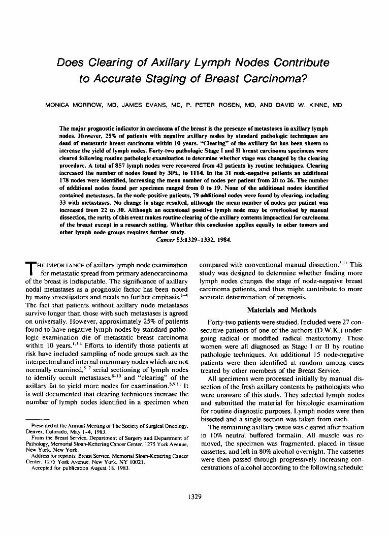
An additional 257 nodes were identified by clearing the specimen, averaging 6.1 per specimen and ranging from 0 to 25 per case. The total number of nodes identified was increased to I 1 14, a 30% increase in the number of nodes found (Table 1). In general, the lymph nodes iso- lated by clearing were a good deal smaller than those found by manual dissection (Fig. 1).
Nqutivr-Node Cast.s
The mean size of the primary tumor was 2.2 cm. A total of 6 I6 nodes were found by routine techniques in the 3 1 node-negative patients. Clearing increased the
FIG. I . Photograph of histologic sections of axillary lymph nodes. The five upper slides contain lymph nodes isolated manually. Small lymph nodes found in the cleared fat are on the lower six slides.

No. 6 LYMPH NODE CLEARING IN BREAST CARCINOMA - MOrrOW ef d. 1331
number of nodes found by 178 (mean, 25.6 nodes per patient). However, none of the additional nodes identified contained metastases. Consequently, stage of disease was not changed by the clearing procedure (Table 2).
Positive-Node Casm
The mean size of the primary tumor was 1.7 cm. Sev- enty-nine additional nodes were found in cleared tissue from the node-positive patients, including 33 with me- tastases (Table 2). The 33 positive nodes were found in three patients who had 17, 5, and 4 positive nodes, re- spectively, identified originally. No change in stage re- sulted, although the mean number of nodes per patient was increased from 2 I .9 to 29. I .
Discussion
Although most pathologists would agree that clearing the axillary fat results in a higher yield of lymph nodes in the specimen, there is not universal agreement on the nature and prognostic importance of the additional data collected. Durkin and Haagensen? reporting on 100 specimens examined by a clearing technique, stated that the percentage of nodes with metastatic disease, as well as the number of nodes identified, was increased. This comparison was made with a group of historical controls, and it compared the percentage of patients found to have nodal metastases in each group. From this data it was not possible to determine whether pathologic stage was actually changed by clearing. Four of 47 patients who had special study of the interpectoral nodes were found to have metastases at this site in the absence of axillary metastases. It is impossible to assess the contribution of clearing to staging these four patients since there was no control group examined by manual dissection of inter- pectoral nodes.
Hartveit and associates13 cleared 63 node-negative breast specimens, and found one node containing a mi- croscopic deposit of tumor which had not been identified by routine processing. They concluded that routine clear- ing of the specimens was not necessary.
Our study, using patients as their own controls, con- firms this. Although a mean of 6.1 additional nodes per patient was identified, stage of disease was not changed by the added information. Even in patients with gross nodal disease the majority of nodes detected by clearing techniques were negative. Manual techniques detected the majority of the positive nodes.
Fisher and Slack in a study of 2768 breast specimens from 43 institutions found that a mean of 17 nodes per specimen was identified by a variety of pathologic tech-
TABLE 2. Results of Clearing
No. of additional nodes by clearing for tumor
No. of positive
Node-negative patients I78 Node-positive patients 19
0 33
niques." Only one institution in this series used routine clearing of specimens. At Memorial Sloan-Kettering a mean of 20 nodes per specimen was identified, which showed little difference from these findings. This suggests that the lack of additional information provided by clear- ing is not secondary to a particularly diligent search for nodes by Memorial Sloan-Kettering pathologists. In ad- dition, the technique of clearing is rather time consuming and tedious, so it is unlikely to benefit the pathologist not willing to do a thorough manual dissection.
From these findings we conclude that clearing of axillary contents would not make a practical contribution to the pathologic examination of specimens obtained from pa- tients with breast carcinoma. Rarely, one may expect to find a positive lymph node overlooked when lymph nodes removed by manual dissection were free of metastases. Since this did not occur in any of the 3 I cases we studied, we expect this to be an infrequent event. Similarly, for patients found to have positive nodes by manual dissec- tion, discovery of additional affected nodes might be rel- evant to prognosis if the total positive nodes were increased from one to three to four or more. In view of the expense for reagents and manpower as well as the considerable time to carry out clearing and the delay in reporting, we believe that clearing is indicated only in a research setting with regard to breast carcinoma and the axillary lymph nodes. Additional study is necessary to determine whether this conclusion applies equally to other tumors, e.g., mel- anoma, or colon carcinoma, and other lymph node groups, inguinal or mediastinal.
Summary and Conclusions
Clearing the axillary fat from a mastectomy specimen yielded a higher number of lymph nodes from the axillary specimen than manual dissection alone.
Most of the additional nodes recovered did not contain metastases. No positive lymph nodes were found in 31 cases after the nodes were declared negative by manual dissection.
Clearing did not add useful prognostic information, and is not recommended as a practical procedure for the dissection of the axillary contents removed from patients with mammary carcinoma.

1332 CANCER March 15 1984 VOl. 53
Further study is required to determine if this conclusion is applicable to other tumor types and other lymph node groups.
REFERENCES
I . Rosen PP, Saigo PE, Braun DW Jr, Weathers E, Kinne DW. Prognosis in Stage I I (TINIMO) breast carcinoma. Ann Surg 1981: 194576.
2. Adair F, Berg J. Robbins G F ef a/. Long-term follow-up of breast cancer patients: The 30-year report. Cancer 1974; 33:l 145.
3. Urban J. Changing patterns of breast cancer. Cuncer 1976; 37: I I I- 117.
4. Valagussa P, Bonadonna G, Veronesi U. Patterns of relapse and survival following radical mastectomy. Cancer 1978: 4 I : I 170- I 178.
5 . Durkin K, Haagensen CD. An improved technique for the study of lymph nodes in surgical specimens. Ann Surg 1980: 191:419.
6. Donegan WL. The influence of untreated internal mammary me- tastases on the course of mammary cancer. Cuncer 1977: 39:533.
7. Veronesi U, Valagussa P. Inefficacy of internal mammary nodes dissection in breast cancer surgery. Cunccr 198 I : 47: 170-1 75.
8. Wilkinson EJ, Hause LL, Kuzma JF et a/. Occult axillary lymph node metastases in patients with invasive breast carcinoma. Lah Invesi 1981: 44:83A.
9. Pickren IW. Significance of occult metastases: A study of breast cancer. Cuncer I96 I ; 14: 1266.
10. Saphir 0, Amromin GD. Obscure axillary lymph node metastases in carcinoma of the breast. Cancer 1948: I :238-24 I .
I I . Fisher B, Slack N. Number of nodes examined in the prognosis of breast carcinoma. Surg Gynewd Ohsref I970 I3 I :79.
12. Rosen PP, Lesser ML, Kinne DW, Beattie EJ Jr. Discontinuous or “skip” metastases in breast carcinoma: Analysis of I228 axillary dissections. Ann SurR 1983: 197:276.
13. Hartveit F, Samsonsen G, Tangen M, Halversen JF. Routine histological investigation of the axillary nodes i n breast cancer. Clin Oncool 1982; 8: I2 I .