Embed Size (px)
DESCRIPTION
document
Citation preview
31
White Blood Nucleus Segmentation Using an
Automated Thresholding and Mathematical
Morphing [Anjali Gautam, H.S. Bhadauria, Annapurna Singh]
Abstract—The aim of this paper is to automate the process
of detection of leukocytes using image processing techniques such as automatic thresholding and mathematical morphing. We have used the segmentation to detect white blood nucleus. Now a day’s the automatic system is preferred as manual segmentation is very tedious, tardy and sometime prone to error, besides that the medical instruments which are used to detect white blood cells are very costly and may not be exist in all the hospitals and clinics. In automatic process, localization and segmentation of white blood cell are the most important stages. In this research we focus on white blood cell nucleus segmentation in which we firstly applied the mathematical techniques to identify the dark objects in an image like white blood cell and platelets then automatic segmentation on blood smears using the global Ostu thresholding technique which convert the segmented image to binary image and then mathematical morphing is applied on the binary image which removed the components which are not white blood cells and then the removal of border objects which are incomplete white blood cells results in final segmentation. The result obtained in this research shows a good accuracy rate as compared to other techniques for automatic segmentation.
Keywords—White Blood Cells, Mathematical Morphing,
Leukemia, Segmentation.
I. Introduction There are different kind of diseases in the body system and
many of the diseases are identified by the blood and the instruments which are used in the diagnosis of diseases are very costly so, in case of identification of white blood cells count that provide useful information to the doctors in diagnosis of different kind of diseases in the body system, so, automatic segmentation is preferred in the automatic localization and segmentation of white blood cells, as manually the segmentation is very tedious, tardy and sometimes to prone to errors. In normal human body there are three types of blood cells found namely red cells, white cells or leukocyte and blood platelets. Generally, red cells also
Anjali Gautam
Gobind Ballabh Pant Engineering College, Pauri Garhwal, India
Dr. H.S. Bhadauria
Gobind Ballabh Pant Engineering College, Pauri Garhwal ,India
Dr. Annnapurna Singh
Gobind Ballabh Pant Engineering College, Pauri Garhwal, India
called erythrocytes are simple and similar; the main function of them is to deliver oxygen to body tissues. While white blood cells which are also known as leukocytes protect the body against both the foreign material and the infectious diseases [2]. In human body there are five different and diverse types of leukocytes exist namely basophiles, neutrophils, eosinophils, lymphocytes and monocytes. They all are differing in their nucleus, size, texture affinity for different physiological colors and immune functions [1].
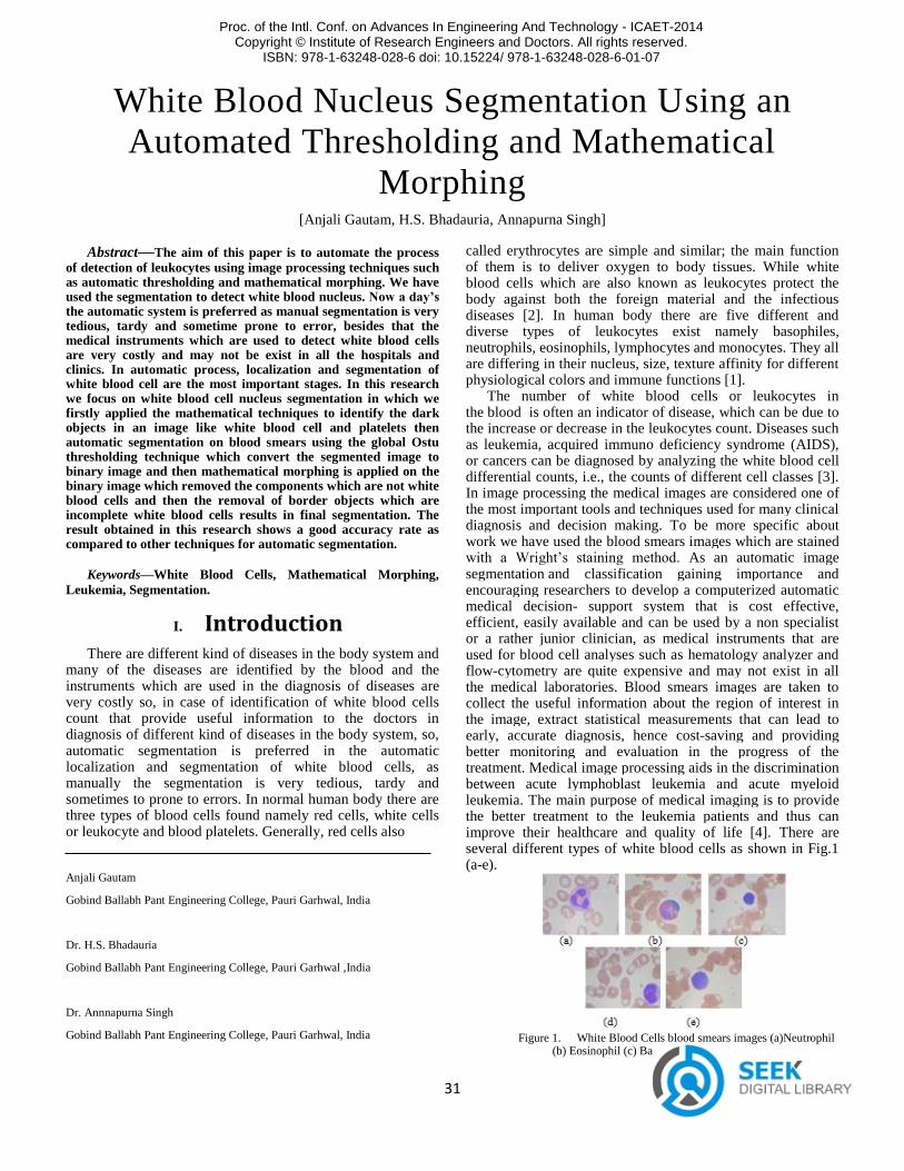
The number of white blood cells or leukocytes in the blood is often an indicator of disease, which can be due to the increase or decrease in the leukocytes count. Diseases such as leukemia, acquired immuno deficiency syndrome (AIDS), or cancers can be diagnosed by analyzing the white blood cell differential counts, i.e., the counts of different cell classes [3]. In image processing the medical images are considered one of the most important tools and techniques used for many clinical diagnosis and decision making. To be more specific about work we have used the blood smears images which are stained with a Wright’s staining method. As an automatic image segmentation and classification gaining importance and encouraging researchers to develop a computerized automatic medical decision- support system that is cost effective, efficient, easily available and can be used by a non specialist or a rather junior clinician, as medical instruments that are used for blood cell analyses such as hematology analyzer and flow-cytometry are quite expensive and may not exist in all the medical laboratories. Blood smears images are taken to collect the useful information about the region of interest in the image, extract statistical measurements that can lead to early, accurate diagnosis, hence cost-saving and providing better monitoring and evaluation in the progress of the treatment. Medical image processing aids in the discrimination between acute lymphoblast leukemia and acute myeloid leukemia. The main purpose of medical imaging is to provide the better treatment to the leukemia patients and thus can improve their healthcare and quality of life [4]. There are several different types of white blood cells as shown in Fig.1 (a-e).
Figure 1. White Blood Cells blood smears images (a)Neutrophil
(b) Eosinophil (c) Basophil (d) Monocyte (e) lymphocyte.
Proc. of the Intl. Conf. on Advances In Engineering And Technology - ICAET-2014 Copyright © Institute of Research Engineers and Doctors. All rights reserved.
ISBN: 978-1-63248-028-6 doi: 10.15224/ 978-1-63248-028-6-01-07
32
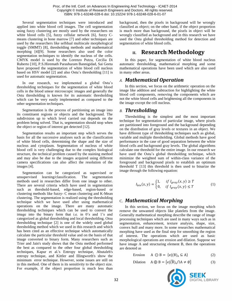
Several segmentation techniques were introduced and applied into white blood cell images. The cell segmentation using fuzzy clustering are mostly used by the researchers on white blood cells [5], fuzzy cellular network [6], fuzzy C-means clustering in bone marrow [7] and other techniques are used by the researchers like selfdual multiscale morphological toggle (SMMT) [8], thresholding methods and mathematical morphing [4][9]. Some researchers also used the color segmentation techniques to identify the nucleus of the cells. CMYK model is used by the Lorenzo Putzu, Cecilia Di Ruberto [10], P.S.Hiremath Parashuram Bannigidad, Sai Geeta have proposed the segmentation of white blood cell nucleus based on HSV model [2] and also Ostu’s thresholding [11] is used for automatic segmentation.
In our research, we implemented a global Ostu’s thresholding techniques for the segmentation of white blood cells in the blood smear microscopic images and generally the Ostu thresholding is known for its automatic thresholding which can be very easily implemented as compared to the other segmentation techniques.
Segmentation is the process of partitioning an image into its constituent regions or objects and the background. The subdivision up to which level carried out depends on the problem being solved. That is, segmentation should stop when the object or region of interest get detected [12].
Segmentation results an important step which serves the basis for all the successive analyses such as the classification of white blood cells, determining the shape and the size of nucleus and cytoplasm. Segmentation of nucleus of white blood cell is very challenging due to the complex biological structure, the technical problem caused by the staining method and may also be due to the images acquired using different camera specifications can also affect the resolution of the images [4].
Segmentation can be categorized as supervised or unsupervised learning/classification. The segmentation methods used in researches differ from one image to other. There are several criteria which have used in segmentation such as threshold-based, edge-based, region-based or clustering methods like fuzzy- C mean clustering and K-Mean clustering. The segmentation is realized using the thresholding technique which we have used after using mathematical operations on the image. There are many automatic thresholding techniques which can be used to convert the image into the binary form that i.e. in 0’s and 1’s and categorized as global thresholding and local thresholding. Otsu thresholding technique [2] is one of the widely used global thresholding method which we used in this research and which has been cited as an effective technique which automatically calculate the particular threshold value and on the basis of that image converted to binary form. Many researchers such as Trier and Jain's study shows that the Ostu method performed the best as compared to the other four global thresholding techniques, Kapur et al.'s Entropy technique, Abutaleb's entropy technique, and Kittler and Illingworth's show the minimum error technique. However, some issues are still on in this method. One of them is its sensitivity to the object size. For example, if the object proportion is much less than
background, then the pixels in background will be wrongly classified as object; on the other hand, if the object proportion is much more than background, the pixels in object will be wrongly classified as background and in this research we have used the Ostu global thresholding method for detection and segmentation of white blood cells.
II. Research Methodology In this paper, for segmentation of white blood nucleus
automatic thresholding, mathematical morphing and some mathematical operations have been used which are also used in many other areas.
A. Mathematical Operation In this section, we focus on the arithmetic operation on the
image like addition and subtraction for highlighting the white blood cell components, removing the components which are not the white blood cells and brightening all the components in the image except the cell nucleus.
B. Thresholding Thresholding is the simplest and the most important
technique for segmentation of particular image, where pixels are partitioned into foreground and the background depending on the distribution of gray levels or textures in an object. We have different type of thresholding techniques such as global, variable and multiple thresholding. Global methods have good performance in the case of good separation between the white blood cells and background gray levels. The global algorithms calculate one threshold for the entire image. In our research we have used the Ostu’s global thresholding technique, which minimize the weighted sum of within-class variance of the foreground and background pixels to establish an optimum threshold T [13] this threshold is then used to binarize the image through the following equation:
( ) { ( )
( ) (1)
C. Mathematical Morphing
In this section, we focus on the image morphing which remove the unwanted objects like platelets from the image. Generally mathematical morphing describe the range of image processing techniques which are used in many ways such as in segmentation, enhancement, texture analysis, shape, size, convex hull and many more. In some researches mathematical morphing have used as the final step for smoothing the region of interest. The operations which are used as basic morphological operations are erosion and dilation. Suppose we have image A and structuring element B, then the operations are denoted as
Erosion * ( ) + (2)
Dilation { ( ̂) } (3)
Proc. of the Intl. Conf. on Advances In Engineering And Technology - ICAET-2014 Copyright © Institute of Research Engineers and Doctors. All rights reserved.
ISBN: 978-1-63248-028-6 doi: 10.15224/ 978-1-63248-028-6-01-07
33
The opening and the closing operations are derived from
the erosion and the dilation of morphing. Opening used to
smoothes the contour of an object, breaks narrow isthmuses,
and eliminate thin protrusions. Closing is also used to smooth
contours but remove the small holes, fuses narrow breaks, long
thin gulfs, and fill the gaps in contour.
Opening ( ) (4)
Closing ( ) (5)
III. Proposed Method
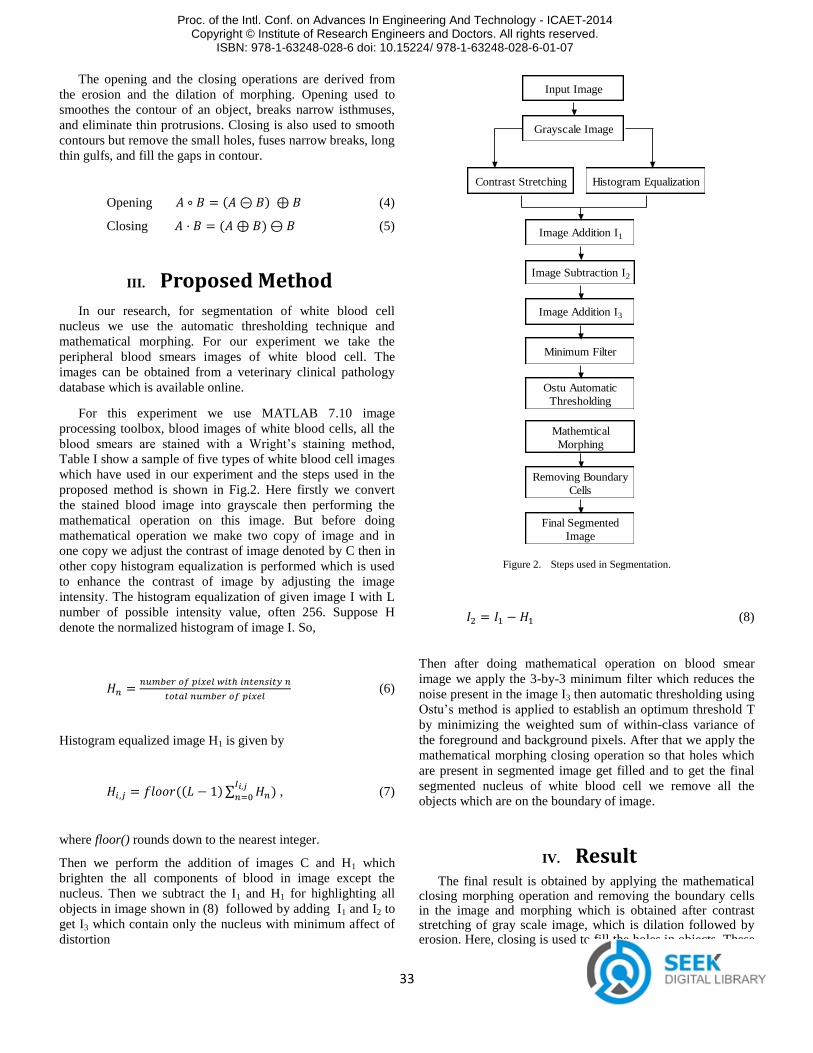
In our research, for segmentation of white blood cell
nucleus we use the automatic thresholding technique and
mathematical morphing. For our experiment we take the
peripheral blood smears images of white blood cell. The
images can be obtained from a veterinary clinical pathology
database which is available online.
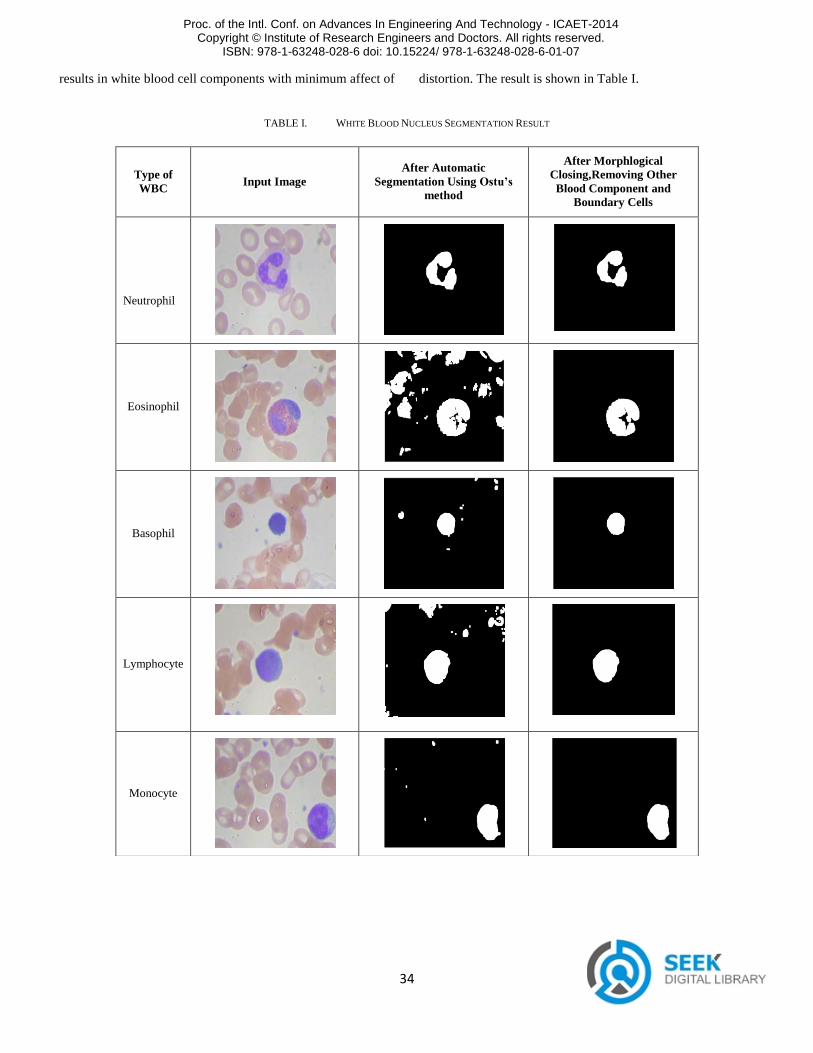
For this experiment we use MATLAB 7.10 image
processing toolbox, blood images of white blood cells, all the
blood smears are stained with a Wright’s staining method,
Table I show a sample of five types of white blood cell images
which have used in our experiment and the steps used in the
proposed method is shown in Fig.2. Here firstly we convert
the stained blood image into grayscale then performing the
mathematical operation on this image. But before doing
mathematical operation we make two copy of image and in
one copy we adjust the contrast of image denoted by C then in
other copy histogram equalization is performed which is used
to enhance the contrast of image by adjusting the image
intensity. The histogram equalization of given image I with L
number of possible intensity value, often 256. Suppose H
denote the normalized histogram of image I. So,
(6)
Histogram equalized image H1 is given by
(( )∑ ) , (7)
where floor() rounds down to the nearest integer.
Then we perform the addition of images C and H1 which
brighten the all components of blood in image except the
nucleus. Then we subtract the I1 and H1 for highlighting all
objects in image shown in (8) followed by adding I1 and I2 to
get I3 which contain only the nucleus with minimum affect of
distortion
Input Image
Grayscale Image
Contrast Stretching Histogram Equalization
Image Addition I1
Image Subtraction I2
Image Addition I3
Minimum Filter
Ostu Automatic
Thresholding
Mathemtical
Morphing
Removing Boundary
Cells
Final Segmented
Image
Figure 2. Steps used in Segmentation.
(8)
Then after doing mathematical operation on blood smear
image we apply the 3-by-3 minimum filter which reduces the
noise present in the image I3 then automatic thresholding using
Ostu’s method is applied to establish an optimum threshold T
by minimizing the weighted sum of within-class variance of
the foreground and background pixels. After that we apply the
mathematical morphing closing operation so that holes which
are present in segmented image get filled and to get the final
segmented nucleus of white blood cell we remove all the
objects which are on the boundary of image.
IV. Result The final result is obtained by applying the mathematical
closing morphing operation and removing the boundary cells in the image and morphing which is obtained after contrast stretching of gray scale image, which is dilation followed by erosion. Here, closing is used to fill the holes in objects. These
Proc. of the Intl. Conf. on Advances In Engineering And Technology - ICAET-2014 Copyright © Institute of Research Engineers and Doctors. All rights reserved.
ISBN: 978-1-63248-028-6 doi: 10.15224/ 978-1-63248-028-6-01-07
34
results in white blood cell components with minimum affect of distortion. The result is shown in Table I.
TABLE I. WHITE BLOOD NUCLEUS SEGMENTATION RESULT
Type of
WBC Input Image
After Automatic
Segmentation Using Ostu’s
method
After Morphlogical
Closing,Removing Other
Blood Component and
Boundary Cells
Neutrophil
Eosinophil
Basophil
Lymphocyte
Monocyte
Proc. of the Intl. Conf. on Advances In Engineering And Technology - ICAET-2014 Copyright © Institute of Research Engineers and Doctors. All rights reserved.
ISBN: 978-1-63248-028-6 doi: 10.15224/ 978-1-63248-028-6-01-07
35
V. Conclusion White blood cell identification and segmentation is the
ability to find the nucleus and removing all the other components of blood from the image. Until now most of the researcher for segmentation, manually crop the white blood cell image and then work on that image. Our study introduces the automatic white blood cell localization by using automatic thresholding for segmentation. In our research we firstly apply the Ostu global threshloding for segmentation and then mathematical morphing is applied which is followed by removing all the blood components which are on the image boundary, which results in the efficient segmentation of nuclei as compared to the other methods of automatic segmentation. This research introduces a method for white blood cell nucleus identification and segmentation as a first step towards a fully automatic system for diagnosis of different kind of diseases and classification using peripheral blood microscope image. White blood cell segmentation is the key procedure in the automatic leukemia diagnosis system.
References
[1]. Stanislav Mirčić and Nikola Jorgovanović, Automatic Classification of Leukocytes, Journal Of Automatic Control, University Of Belgrade, Vol 16, pp. 29-32, 2006 ©.
[2]. P.S.Hiremath, Parashuram Bannigidad and Sai Geeta, Automated Identification and Classification of White Blood Cells (Leukocytes) in Digital Microscopic Images, IJCA Special Issue on “Recent Trends in Image Processing and Pattern Recognition” RTIPPR, pp. 59-63, 2010.
[3]. Nipon Theera-Umpon and Sompong Dhompongsa, Morphological Granulometric Features of Nucleus in Automatic Bone Marrow White Blood Cell Classification, IEEE Transactions on Information Technology In Biomedicine, Vol. 11, No. 3, pp. 353-359, May 2007.
[4]. H.T Madhloom, S.A Kareem, H. Ariffin, A.A. Zaidan, H.O. Alanazi and B.B. Zaidan, An Automated White Blood Cell Nucleus Localization and Segmentation using Image Arithmetic and Automated Threshold, Journal of Applied Sciences 10(11), pp. 959-966, 2010©.
[5]. S. Chinwaraphat, A. Sanpanich, C. Pintavirooj, M. Sangworasil, and P. Tosranon, A Modified Fuzzy Clustering For White Blood Cell Segmentation, The 3rd International Symposium on Biomedical Engineering (ISBME), pp. 356-359, 2008.
[6]. Wang Shitong And Wang Min, A New Detection Algorithm (Nda) Based On Fuzzy Cellular Neural Networks For White Blood Cell Detection, IEEE Transactions on Information Technology in Biomedicine, Vol. 10, No. 1, pp. 5-10, January 2006.
[7]. Nipon Theera-Umpon, “Patch-Based White Blood Cell Nucleus Segmentation Using Fuzzy Clustering”, ECTI Transactions on Electrical Engg., Electronics, and Communications Vol.3, No.1, pp. 15-19, February 2005.
[8]. Leyza Baldo Dorini, Rodrigo Minetto, and Neucimar Jerˆonimo Leite
“Semiautomatic White Blood Cell Segmentation Based on Multiscale Analysis” IEEE Journal of Biomedical and Health Informatics, Vol. 17,
No. 1, pp. 250-256, January 2013.
[9]. Leyza Baldo Dorini Rodrigo Minetto Neucimar Jerˆonimo Leite, “White blood cell segmentation using morphological operators and scale-space
analysis”.
[10]. Lorenzo Putzu, Cecilia Di Ruberto “White Blood Cells Identification and Classification from Leukemic Blood Image” IWBBIO, pp.99-106,
2013.
[11]. R.C. Gonzales, R.E. Woods, Digitial Image Processing, pp. 712, 3.Auflage, ISBN 978-81-317-2695-2, Pearson Prentice Hall 2009.
[12]. Tapas Kanungo, David M. Mount, Nathan S. Netanyahu, Christine D.
Piatko, Ruth Silverman, and Angela Y. Wu, An Efficient k-Means Clustering Algorithm: Analysis and Implementation, IEEE Transactions
on Pattern Analysis and Machine Intelligence, Vol. 24, No. 7, pp. 881-
892, July 2002.
[13]. Ping-Sung Liao, Tse-Sheng Chen, and Pau-Choo Chung, A Fast Algorithm for Multilevel Thresholding, Journal of Information Science
and Engineering 17, pp. 713-727, 2001.
About Authors:
Anjali Gautam received her B. Tech
in Computer Science and
Engineering from Graphic Era
University, Uttarakhand India, in
2012. Currently she is pursing M.
Tech in Computer Science and
Engineering from G.B. Pant
Engineering College, Pauri Garhwal,
Uttarakhand, India. Her research
interest includes medical imaging,
mathematical morphing and image
processing.
Dr. H. S. Bhadauria received his B.
Tech in Computer Science &
Engineering, M. Tech. in Electronics
Engineering from Aligarh Muslim
University, Aligarh, and Ph.d. from
Indian Institute of Technology,
Roorkee, India in 1999, 2004 and
2013, respectively. He has published
some 40 research papers in
International and National Journals
and Conferences. His areas of
Interest are Digital Image Processing,
Digital Signal Processing and AdHoc
Network.
Dr. Annapurna Singh received M.
Tech. from Banasthali Vidyapeeth,
Banasthali, India in Dec 2003 and
PhD from Uttarakhand Technical
University, Dehradun in 2012 in
Computer Science Engineering. She
has published some 25 research
papers in International and National
Journals and Conferences. Her areas
of Interest are Digital Image
Processing, Computer Network and
AdHoc Network.
Proc. of the Intl. Conf. on Advances In Engineering And Technology - ICAET-2014 Copyright © Institute of Research Engineers and Doctors. All rights reserved.
ISBN: 978-1-63248-028-6 doi: 10.15224/ 978-1-63248-028-6-01-07