Embed Size (px)
DESCRIPTION
lalala...
Citation preview
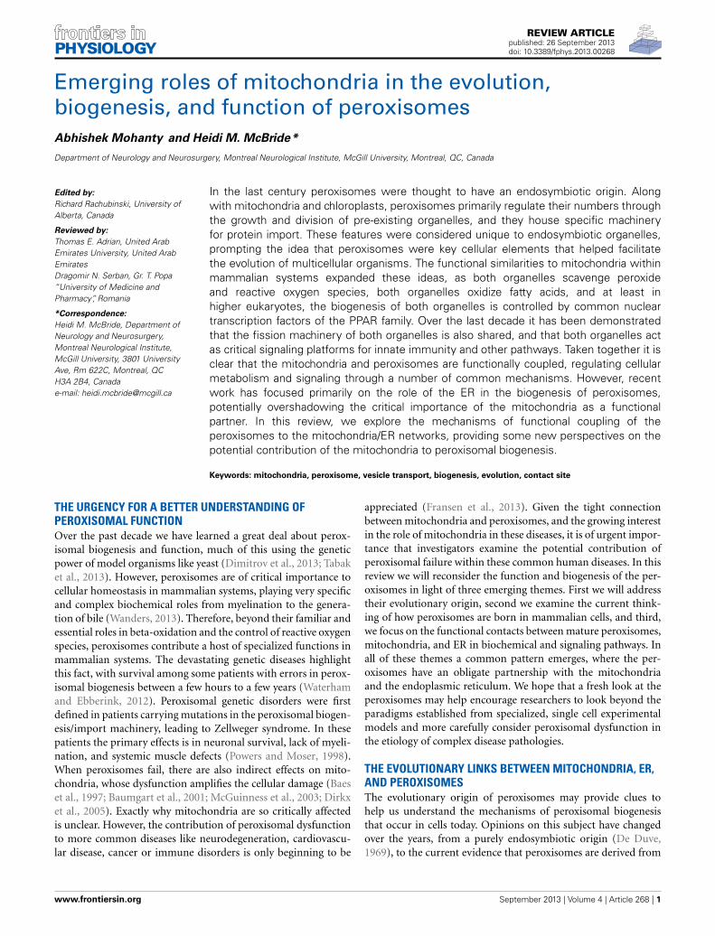
REVIEW ARTICLEpublished: 26 September 2013doi: 10.3389/fphys.2013.00268
Emerging roles of mitochondria in the evolution,biogenesis, and function of peroxisomesAbhishek Mohanty and Heidi M. McBride*
Department of Neurology and Neurosurgery, Montreal Neurological Institute, McGill University, Montreal, QC, Canada
Edited by:
Richard Rachubinski, University ofAlberta, Canada
Reviewed by:
Thomas E. Adrian, United ArabEmirates University, United ArabEmiratesDragomir N. Serban, Gr. T. Popa“University of Medicine andPharmacy”, Romania
*Correspondence:
Heidi M. McBride, Department ofNeurology and Neurosurgery,Montreal Neurological Institute,McGill University, 3801 UniversityAve, Rm 622C, Montreal, QCH3A 2B4, Canadae-mail: [email protected]
In the last century peroxisomes were thought to have an endosymbiotic origin. Alongwith mitochondria and chloroplasts, peroxisomes primarily regulate their numbers throughthe growth and division of pre-existing organelles, and they house specific machineryfor protein import. These features were considered unique to endosymbiotic organelles,prompting the idea that peroxisomes were key cellular elements that helped facilitatethe evolution of multicellular organisms. The functional similarities to mitochondria withinmammalian systems expanded these ideas, as both organelles scavenge peroxideand reactive oxygen species, both organelles oxidize fatty acids, and at least inhigher eukaryotes, the biogenesis of both organelles is controlled by common nucleartranscription factors of the PPAR family. Over the last decade it has been demonstratedthat the fission machinery of both organelles is also shared, and that both organelles actas critical signaling platforms for innate immunity and other pathways. Taken together it isclear that the mitochondria and peroxisomes are functionally coupled, regulating cellularmetabolism and signaling through a number of common mechanisms. However, recentwork has focused primarily on the role of the ER in the biogenesis of peroxisomes,potentially overshadowing the critical importance of the mitochondria as a functionalpartner. In this review, we explore the mechanisms of functional coupling of theperoxisomes to the mitochondria/ER networks, providing some new perspectives on thepotential contribution of the mitochondria to peroxisomal biogenesis.
Keywords: mitochondria, peroxisome, vesicle transport, biogenesis, evolution, contact site
THE URGENCY FOR A BETTER UNDERSTANDING OFPEROXISOMAL FUNCTIONOver the past decade we have learned a great deal about perox-isomal biogenesis and function, much of this using the geneticpower of model organisms like yeast (Dimitrov et al., 2013; Tabaket al., 2013). However, peroxisomes are of critical importance tocellular homeostasis in mammalian systems, playing very specificand complex biochemical roles from myelination to the genera-tion of bile (Wanders, 2013). Therefore, beyond their familiar andessential roles in beta-oxidation and the control of reactive oxygenspecies, peroxisomes contribute a host of specialized functions inmammalian systems. The devastating genetic diseases highlightthis fact, with survival among some patients with errors in perox-isomal biogenesis between a few hours to a few years (Waterhamand Ebberink, 2012). Peroxisomal genetic disorders were firstdefined in patients carrying mutations in the peroxisomal biogen-esis/import machinery, leading to Zellweger syndrome. In thesepatients the primary effects is in neuronal survival, lack of myeli-nation, and systemic muscle defects (Powers and Moser, 1998).When peroxisomes fail, there are also indirect effects on mito-chondria, whose dysfunction amplifies the cellular damage (Baeset al., 1997; Baumgart et al., 2001; McGuinness et al., 2003; Dirkxet al., 2005). Exactly why mitochondria are so critically affectedis unclear. However, the contribution of peroxisomal dysfunctionto more common diseases like neurodegeneration, cardiovascu-lar disease, cancer or immune disorders is only beginning to be
appreciated (Fransen et al., 2013). Given the tight connectionbetween mitochondria and peroxisomes, and the growing interestin the role of mitochondria in these diseases, it is of urgent impor-tance that investigators examine the potential contribution ofperoxisomal failure within these common human diseases. In thisreview we will reconsider the function and biogenesis of the per-oxisomes in light of three emerging themes. First we will addresstheir evolutionary origin, second we examine the current think-ing of how peroxisomes are born in mammalian cells, and third,we focus on the functional contacts between mature peroxisomes,mitochondria, and ER in biochemical and signaling pathways. Inall of these themes a common pattern emerges, where the per-oxisomes have an obligate partnership with the mitochondriaand the endoplasmic reticulum. We hope that a fresh look at theperoxisomes may help encourage researchers to look beyond theparadigms established from specialized, single cell experimentalmodels and more carefully consider peroxisomal dysfunction inthe etiology of complex disease pathologies.
THE EVOLUTIONARY LINKS BETWEEN MITOCHONDRIA, ER,AND PEROXISOMESThe evolutionary origin of peroxisomes may provide clues tohelp us understand the mechanisms of peroxisomal biogenesisthat occur in cells today. Opinions on this subject have changedover the years, from a purely endosymbiotic origin (De Duve,1969), to the current evidence that peroxisomes are derived from
www.frontiersin.org September 2013 | Volume 4 | Article 268 | 1
Mohanty and McBride Revisiting the links between the mitochondria and peroxisomes
the ER (Dimitrov et al., 2013). A bioinformatic analysis of thephylogeny of a number of peroxisomal proteins concluded thatperoxisomal proteins fall into two major categories, prokary-otic and eukaryotic (Gabaldon et al., 2006). Peroxisomal proteinsof eukaryotic origin (58% in yeast, 39% in rat) were primarilyinvolved in peroxisomal biogenesis. Peroxisomal proteins withbacterial or archeabacterial ancestry included about 13–18% ofthe peroxisomal proteins. A large proportion of proteins were dif-ficult to assign (∼25%), but these all had some homologies withprokaryotic proteins, although trees could not be constructedto distinguish bacterial or archeal origin. Of the assigned andunassigned proteins within the second category, all were func-tional enzymes. This suggests either that the perixosomes haveevolved as endosymbionts, or that these enzymes evolved frommitochondrial proteins sometime after the last common ances-tor. Since many of these enzymes remain dually targeted to bothorganelles, peroxisomal biologists generally suggest that theseenzymes were most likely retargeted to peroxisomes from mito-chondria (Gabaldon et al., 2006; Tabak et al., 2006). This wouldindicate that the peroxisome emerged as functionally special-ized mitochondria. Since we now know that mitochondria areable to sort specific proteins into vesicular carriers, we can beginto imagine how functionally distinct mitochondria may havetaken shape. These peroxisomal precursors would have housedenzymes responsible for breaking down a unique subclass of fattyacids, incorporated specialized enzymes regulating redox path-ways, and other biochemical pathways like plasmalogen synthe-sis. Eventually, peroxisomes would have adapted protein importmechanisms, and new signal sequences could direct precursorsdirectly to peroxisomes. Although the genetic expansion of theperoxisomal proteome provided a great deal of independencefrom the mitochondria, peroxisomes have retained the samemitochondrial machinery for their division, a central aspect ofperoxisomal biogenesis (Schrader et al., 2012).
If the functions of the peroxisome are largely variants of thosein mitochondria, why do they emerge from the endoplasmic retic-ulum? This should not be particularly surprising since the ER alsoprovides the mitochondria with the bulk of it’s lipid mass, andthe ER and mitochondria are functionally and physically cou-pled in many ways (de Brito and Scorrano, 2010; Rowland andVoeltz, 2012). Therefore, the relationship of the peroxisome tothe ER may also reflect an evolutionarily conserved variation onthe mechanisms of mitochondria/ER coupling. In the phylogenicanalysis of the peroxisomal proteins descended from a eukaryoticlineage, there was a clear relationship between the peroxisomalimport machinery and the components of the endoplasmic retic-ulum associated degradation, or ERAD pathway (Gabaldon et al.,2006; Schluter et al., 2006; Schliebs et al., 2010). For peroxiso-mal import, the receptor Pex5 binds to cytosolic precursors todeliver them to the peroxisome in a cycle that involves ubiquitina-tion and deubiquitination of Pex5 for the release of the substrate(for review see Schliebs et al., 2010). This appears to be analo-gous to the use of ubiquitin in the tagging and export of unfoldedproteins within the ER, which are ultimately delivered to the pro-teasome. Indeed 5 of the 6 conserved Pex genes show homologywith components of the ERAD machinery. Pex1 and Pex6 arehomologous to Cdc48 and p97 [which are themselves of bacterial
origin (Iyer et al., 2004)], whereby p97 is a AAA+ ATPase. Pex2and Pex10 are similar to the ubiquitin E3 ligase Hrd1 enzymethat tags unfolded ER proteins. Hrd1 has a binding partner Hrd3,which shows homology to Pex5, and Pex4 resembles a ubiquitinE2 ligase. Therefore, the authors concluded that the biogenesispathway of the peroxisomes evolved from the ER (Gabaldon et al.,2006; Schluter et al., 2006; Schliebs et al., 2010). The differenceis, of course that the peroxisome system would deliver ratherthan extract proteins. However, the only ubiquitinated cargo inperoxisomal import is actually the receptor Pex5, which is ubiqui-tinated in order to be extracted and recycled, following the releaseof the Pex5-bound import substrates in the peroxisome. In thisway, the Pex1/Pex6 complex is extracting Pex5, just as the ERADmachinery extracts ER proteins (Tabak et al., 2013).
Although it has been concluded that the similarly to theCdc48/p97 infers an ER origin of the peroxisomal importmachinery, it is important to note that this system has recentlybeen demonstrated to have a clear role at the mitochondrial mem-brane as well (Heo et al., 2010; Tanaka et al., 2010; Chan et al.,2011; Xu et al., 2011; Esaki and Ogura, 2012). In this case, targetedsubstrates are ubiquitinated by E3 ligases such as Parkin, a pro-tein that is mutated in familial cases of Parkinson’s disease. p97 isrequired for the retrotranslocation of these tagged proteins, whichare then targeted to the proteasome for degradation. Therefore,we would argue that this homology does not exclusively impli-cate the ER as the membrane of origin for the peroxisomal importmachinery, and equally supports a mitochondrial origin.
BEYOND EVOLUTION: CELL BIOLOGY OF PEROXISOMALBIOGENESIS TODAYA number of studies using yeast as a model organism haveunequivocally demonstrated that peroxisomes can be formed denovo from the endoplasmic reticulum (Dimitrov et al., 2013;Tabak et al., 2013). This information has effectively shelved thenotion that peroxisomes evolved as endosymbionts. Unlike mam-malian cells, yeast govern their peroxisomal numbers dependingon the carbon source, for example in the presence of oleic acid(Saccharomyces cerevisiae or Yarrow lipolytica) (Trotter, 2001) ormethanol (Hansenula polymorpha and Pichia pastoris) (Yurimotoet al., 2011). Since yeast mitochondria do not perform beta-oxidation, peroxisomes rapidly arise from the ER in order tocatabolize these fats, or to metabolize methanol. In this way, fungiare highly specialized organisms where peroxisomal function hasdiverged between evolutionary lineages. On the other hand, thelinkages to the mitochondria are much more obvious in multi-cellular organisms. For example, the transcriptional regulation ofmitochondria and peroxisomal biogenesis is not coupled in yeastas it is in mammals (Issemann and Green, 1990; Mandard et al.,2004; Scarpulla et al., 2012). In addition, the shared roles of per-oxisomes and mitochondria as signaling platforms (Dixit et al.,2010; Tait and Green, 2012) may not occur in yeast, and mostobviously, the metabolic functions of peroxisomes have divergedsignificantly throughout evolution (Islinger et al., 2010; Pieuchotand Jedd, 2012; Wanders, 2013). Therefore, fungal lineages mayhave lost some of the linkages between the mitochondria andperoxisomes, instead developing closer ties to the ER. We con-sider that there is likely a great deal of plasticity in the evolution
Frontiers in Physiology | Integrative Physiology September 2013 | Volume 4 | Article 268 | 2
Mohanty and McBride Revisiting the links between the mitochondria and peroxisomes
of peroxisomes, depending on the specific functional role theyplay across diverse species. Given this divergence, we suggest thatthere may not be unified theory for peroxisomal biogenesis acrossspecies, where, for example, significant differences are likely toexist between yeast and mammalian mechanisms.
The most compelling evidence to demonstrate the contribu-tion of the ER to peroxisomal biogenesis is the emergence ofPex-containing vesicles from the endoplasmic reticulum in yeastand mammals. A number of different experimental paradigmsand model systems have proven this point. First, fluorescentlytagged, membrane anchored Pex proteins, notably Pex3 andPex16, have been observed emerging from the ER in condi-tions where peroxisomes are either induced by growth conditionsor in pulse-chase type of rescue experiments (Titorenko andRachubinski, 1998; Hoepfner et al., 2005; Kragt et al., 2005;Tam et al., 2005; Kim et al., 2006; Motley and Hettema, 2007).Second, cell free budding assays from isolated ER have establishedsome of the machinery required to bud Pex-containing vesicles inyeast Saccharomyces cerevisiae (Lam et al., 2010). In this case, theauthors showed both Pex3p and Pex15p emerging within vesi-cles in a manner that depended on ATP and Pex19p, but notSar1, a GTPase essential for anterograde COPII budding events.The authors demonstrated a requirement for additional cytosolicfactors that are yet to be identified. Using a semi-permeable cellsystem in Pichia Pastoris, the authors also demonstrated a Pex19dependent, COPII-independent mechanism to generate Pex11-containing vesicles from the ER (Agrawal et al., 2011). Thesevesicles were generated even in the absence of Pex3, an essentialcomponent for peroxisome biogenesis. Similar data has shown anER origin for mammalian peroxisomes. Using human fibroblastslacking core proteins of the peroxisomal import machinery likePex16 or Pex3, the reintroduction of GFP-tagged Pex16 or Pex3can rescue the generation of new organelles from their ER local-ization (Kim et al., 2006; Toro et al., 2009; Yonekawa et al., 2011).This pathway was also shown to depend upon the ER budding fac-tor Sec16b (Yonekawa et al., 2011). Although many peroxisomalproteins target the mitochondria in the absence of peroxisomes(see next section for further discussion), these data clearly estab-lish the ER as a primary source of membrane in the generation ofnew peroxisomes.
How can you generate a mature peroxisome from an ERderived vesicle? Historically it has been assumed that the earlyperoxisomes would be import competent, and from there couldmature through the targeting and import of all the required func-tional enzymes. This maturation model did not require any fusionevents, instead all new peroxisomes would be formed from thegrowth and division of existing peroxisomes. However, now it isclear that small, vesicular carriers bud from the ER, which aretermed “pre-peroxisomes.” These vesicles must fuse with otherpre-peroxisomes, or with more mature peroxisomes, to generatea larger, functional organelle (Boukh-Viner et al., 2005; van derZand et al., 2012). Many studies have proven that mature per-oxisomes do not fuse, both in mammalian or yeast cells (Motleyand Hettema, 2007; Huybrechts et al., 2009; Bonekamp et al.,2012), raising the important question of specificity and regula-tion of fusion among/between pre-peroxisomes. Previous work inyeast Yarrow lipolytica demonstrated peroxisomal fusion in vitro,
and it was suggested then that fusion was limited to an “early”pool of peroxisomes that would then mature into fully func-tional organelles (Titorenko et al., 2000; Boukh-Viner et al.,2005). In Yarrow lipolytica, peroxisomal fusion was dependentupon the import factors Pex1 and Pex6, of the Cdc48/p97 family(Titorenko et al., 2000; Boukh-Viner et al., 2005). Although p97 isa AAA+ATPase that functions in the ERAD pathway, p97/Cdc48have also been shown to have an established role in ER andgolgi membrane fusion (Latterich et al., 1995; Hetzer et al., 2001;Uchiyama et al., 2006; Totsukawa et al., 2013).
Another important question is how the fusion of two pre-peroxisomes would lead to a functional peroxisome. Recent workhas answered this conundrum by revealing that the peroxiso-mal import machinery is sorted into two populations within theER. They observed two distinct populations of vesicles buddingfrom the ER, one containing the RING complex of the importmachinery, and the second carrying the docking complex (vander Zand et al., 2012). Heterotypic fusion between these twodistinct populations of pre-peroxisomes would then generate afunctional import machine, and by definition, a functional per-oxisome. This explains why peroxisomal import cannot occurinto the ER, since the machinery remains segregated. The mech-anisms for this segregation are not yet known. Consistent withprevious work, the authors could not observe any fusion eventsbetween pre-peroxisomes and mature peroxisomes, or betweenmature peroxisomes, indicating a highly selective mechanism forpre-peroxisomal fusion (van der Zand et al., 2012).
IS THERE A POTENTIAL ROLE FOR MITOCHONDRIALDERIVED VESICLES (MDVs) IN PEROXISOMAL BIOGENESIS?Since the emergence of peroxisomes from the ER is so clearlydemonstrated, is there any role for the mitochondria in the bio-genesis of peroxisomes beyond their evolutionary links? Certainlyour understanding of the flexibility of mitochondria has rapidlyincreased, and we appreciate how they fuse and divide in order todynamically position themselves both functionally and spatiallywithin cells. Our lab has also defined two distinct vesicular trans-port routes from the mitochondria (Neuspiel et al., 2008; Braschiet al., 2010; Soubannier et al., 2012a,b). Initially we described aroute between the mitochondria and the peroxisome (Neuspielet al., 2008), a pathway we also showed was dependent upon theretromer complex Vps35, Vps26 and Vps29 (Braschi et al., 2010).More recently we characterized another pathway between themitochondria and the late endosome/multivesicular body. Thislatter pathway selectively targets oxidized or damaged protein andlipid (Soubannier et al., 2012b), removing them from the mito-chondrial reticulum for degradation in the lysosome (Soubannieret al., 2012a). These two pathways open up new insights intohow the mitochondria may deliver their contents to other cellularorganelles.
What is the function of the vesicle transport route from themitochondria to peroxisomes? So far only one cargo was identi-fied in these vesicles, a membrane anchored protein called MAPL(mitochondrial anchored protein ligase, also called MUL1, GIDE,or HADES) (Neuspiel et al., 2008; Braschi et al., 2009). Thecarboxy-terminal of MAPL is exposed to the cytosol and con-tains a RING finger domain with strong SUMO E3 ligase activity.
www.frontiersin.org September 2013 | Volume 4 | Article 268 | 3
Mohanty and McBride Revisiting the links between the mitochondria and peroxisomes
Overexpression of MAPL drove massive mitochondrial fragmen-tation through the SUMOylation and activation of the mitochon-drial fission GTPase Drp1 (Neuspiel et al., 2008). Mitochondrialvesicles carrying MAPL fused with a subset of peroxisomes, onlyabout 10% of the total (Neuspiel et al., 2008; Braschi et al., 2009).These peroxisomes were able to import the transfected CFP-SKLmarker, indicating that they had functional import machinery.Although we have not yet determined the function of this vesic-ular transport route, we offer three potential functions here.First, MDVs may contribute to peroxisomal biogenesis, fusingwith the early, preperoxisomal population. Second, MDVs maycarry metabolites and target a functionally distinct subset of per-oxisomes, and third, MDVs may shuttle proteins that are notcompetent for peroxisomal import.
Given the evidence for a pre-peroxisomal population in cells,it is plausible that MAPL is targeted to this fusogenic popula-tion of peroxisomes and provides a mitochondrial component tothe maturing peroxisomes. In evolutionary terms, a phylogenicanalysis of MAPL indicates that it is of bacterial ancestry, withat least 5 prokaryotic domain structures (Andrade-Navarro et al.,2009). Therefore, this vesicle transport pathway may have played arole in the earliest segregation of specialized mitochondrial func-tion. This possibility has not been previously considered due tothe obvious assumption that the mitochondria were not com-petent to segregate cargo and bud vesicles. This assumption isfundamentally wrong. Our ongoing studies continue to charac-terize various classes of cargoes that are enriched in mitochondrialderived vesicles. For example, using an in vitro reconstitution sys-tem we demonstrated that the identity of the cargo within MDVsdestined for the lysosome depends greatly on the nature of theinsult (Soubannier et al., 2012b). We have a great deal of workahead to identify the mechanisms and regulation of mitochon-drial vesicle transport, but it is clearly a process that exists insteady-state conditions, suggesting a fundamental role for thesevesicles in cellular homeostasis.
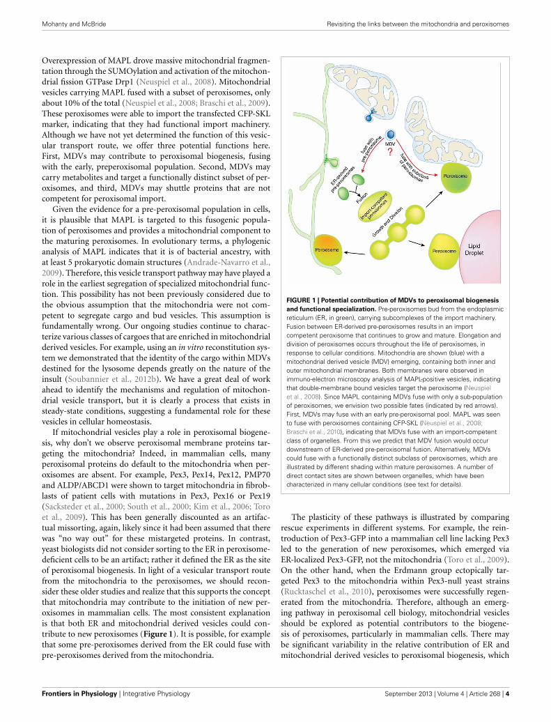
If mitochondrial vesicles play a role in peroxisomal biogene-sis, why don’t we observe peroxisomal membrane proteins tar-geting the mitochondria? Indeed, in mammalian cells, manyperoxisomal proteins do default to the mitochondria when per-oxisomes are absent. For example, Pex3, Pex14, Pex12, PMP70and ALDP/ABCD1 were shown to target mitochondria in fibrob-lasts of patient cells with mutations in Pex3, Pex16 or Pex19(Sacksteder et al., 2000; South et al., 2000; Kim et al., 2006; Toroet al., 2009). This has been generally discounted as an artifac-tual missorting, again, likely since it had been assumed that therewas “no way out” for these mistargeted proteins. In contrast,yeast biologists did not consider sorting to the ER in peroxisome-deficient cells to be an artifact; rather it defined the ER as the siteof peroxisomal biogenesis. In light of a vesicular transport routefrom the mitochondria to the peroxisomes, we should recon-sider these older studies and realize that this supports the conceptthat mitochondria may contribute to the initiation of new per-oxisomes in mammalian cells. The most consistent explanationis that both ER and mitochondrial derived vesicles could con-tribute to new peroxisomes (Figure 1). It is possible, for examplethat some pre-peroxisomes derived from the ER could fuse withpre-peroxisomes derived from the mitochondria.
FIGURE 1 | Potential contribution of MDVs to peroxisomal biogenesis
and functional specialization. Pre-peroxisomes bud from the endoplasmicreticulum (ER, in green), carrying subcomplexes of the import machinery.Fusion between ER-derived pre-peroxisomes results in an importcompetent peroxisome that continues to grow and mature. Elongation anddivision of peroxisomes occurs throughout the life of peroxisomes, inresponse to cellular conditions. Mitochondria are shown (blue) with amitochondrial derived vesicle (MDV) emerging, containing both inner andouter mitochondrial membranes. Both membranes were observed inimmuno-electron microscopy analysis of MAPL-positive vesicles, indicatingthat double-membrane bound vesicles target the peroxisome (Neuspielet al., 2008). Since MAPL containing MDVs fuse with only a sub-populationof peroxisomes, we envision two possible fates (indicated by red arrows).First, MDVs may fuse with an early pre-peroxisomal pool. MAPL was seento fuse with peroxisomes containing CFP-SKL (Neuspiel et al., 2008;Braschi et al., 2010), indicating that MDVs fuse with an import-competentclass of organelles. From this we predict that MDV fusion would occurdownstream of ER-derived pre-peroxisomal fusion. Alternatively, MDVscould fuse with a functionally distinct subclass of peroxisomes, which areillustrated by different shading within mature peroxisomes. A number ofdirect contact sites are shown between organelles, which have beencharacterized in many cellular conditions (see text for details).
The plasticity of these pathways is illustrated by comparingrescue experiments in different systems. For example, the rein-troduction of Pex3-GFP into a mammalian cell line lacking Pex3led to the generation of new peroxisomes, which emerged viaER-localized Pex3-GFP, not the mitochondria (Toro et al., 2009).On the other hand, when the Erdmann group ectopically tar-geted Pex3 to the mitochondria within Pex3-null yeast strains(Rucktaschel et al., 2010), peroxisomes were successfully regen-erated from the mitochondria. Therefore, although an emerg-ing pathway in peroxisomal cell biology, mitochondrial vesiclesshould be explored as potential contributors to the biogene-sis of peroxisomes, particularly in mammalian cells. There maybe significant variability in the relative contribution of ER andmitochondrial derived vesicles to peroxisomal biogenesis, which
Frontiers in Physiology | Integrative Physiology September 2013 | Volume 4 | Article 268 | 4
Mohanty and McBride Revisiting the links between the mitochondria and peroxisomes
would likely be linked to the functional diversity of the organellesin different tissues and organisms.
It is also possible that mitochondrial derived vesicles couldcarry cargo to functionally distinct peroxisomes, rather than play-ing a role in their biogenesis. There has been evidence thatperoxisomes can be functionally differentiated within single cells,having distinct densities, import competencies, and protein com-position (Schrader et al., 1994; Fahimi et al., 1996; Volkl et al.,1999; Islinger et al., 2012; Costa et al., 2013). However, it is notknown how this might be achieved. Given the functional cou-pling of the mitochondria and the peroxisomes—particularly inmammalian cells—a direct vesicular transport route may allowthe rapid shuttling of metabolites or proteins with very tight spa-tial and temporal regulation. A vesicular transport route couldselectively target “active” peroxisomes and deliver metabolitesor enzymes selectively to these organelles (Figure 1). Vesiclesmay also provide protection from potentially toxic, hydropho-bic, or more complex mitochondrial cargoes. These could includeheme, lipids like mitochondrial-generated PE or cardiolipin(Wriessnegger et al., 2007), or metabolic intermediates. It hasbeen largely concluded that catalase is imported into peroxi-somes in the heme-loaded form, indicating that heme would bean unlikely cargo for MDV transport. However, earlier work byLazarow and DeDuve used radiolabelled pulse-chase experimentsin rat liver elegantly demonstrated that heme loading of catalaseoccurred only after import (Lazarow and de Duve, 1973). Again,it cannot yet be excluded that some heme could be transportedinto peroxisomes from the mitochondria in vesicular carriers.
Finally, it is also possible that the mitochondrial proteinimport and folding machinery is more efficient than peroxisomes,leading some common enzymes to be shipped to peroxisomesonly after their rapid assembly in the mitochondria. MAPL, withit’s two transmembrane domains may fall into this category.Dually targeted proteins could potentially utilize both transportroutes, depending on the conditions.
There are a number of challenges remaining to identify themolecular machinery that regulates this vesicular transport route,to understand which types of cargoes are segregated into thevesicles, and how these vesicles select and fuse with a subset ofperoxisomes. Answers will likely come from large scale screen-ing efforts and the development of cell-free assay systems. Sucha screen of 4000 viable deletions in yeast provided compellingevidence for a direct link between the mitochondria and perox-isomes. This genome-wide scan of factors affecting peroxisomalbiogenesis in yeast identified only 4 ER proteins, but 41 mito-chondrial proteins whose loss affected peroxisomal numbers,shape, or function (Saleem et al., 2010). This can be compared tothe loss of 46 nuclear-targeted proteins that similarly affected theperoxisomes. Surprisingly, none of the 4 ER proteins are known tofunction in vesicle formation, instead were mapped to fatty acidsynthesis, farnysylation, lipid modifications and signal transduc-tion (Saleem et al., 2010). Clearly many ER proteins, includingmany of those required for ER transport are essential, and there-fore would not be seen in this screen of non-essential genes. Onthe other hand, the mitochondrial genes spanned multiple func-tions, from mitochondrial translation to respiration and mtDNAdistribution. The robust effects on peroxisomal function upon
the loss of so many mitochondrial genes should reinforce ourefforts to consider the dynamic interplay between peroxisomesand mitochondria in all organisms. Also notable in this screen wasthe fact that the loss of 9 vacuolar proteins also led to a reduc-tion in peroixomal numbers and content (Saleem et al., 2010).These proteins included fusion factors like the SNAREs Vam3 andNyv1, and the Rab GTPase, Ypt7. Future work will be required tounderstand the functions of these genes in peroxisomal behavior.
PEROXISOME AND MITOCHONDRIAL DYNAMICS: THEIMPLICATIONS OF A SHARED MECHANISMOne of the most striking parallels between the mitochondriaand peroxisomes is the conservation of the fission machinery indiverse organisms from yeast to plants. Deletion of the dynamin-related protein Drp1 [also called Dlp1 (dynamin-like protein) andDnm1 (in yeast)] led to elongated peroxisomes and mitochon-dria (Koch et al., 2003; Kuravi et al., 2006; Kobayashi et al., 2007;Motley et al., 2008). The recruitment of this cytosolic GTPaserequires its single-membrane spanning receptors Mff and Fis1,which are dually imported into both organelles (Koch et al., 2005;Delille and Schrader, 2008; Motley et al., 2008). Other mitochon-drial fission factors, including GDAP1 are also imported intoperoxisomes in a Pex19-dependent manner (Huber et al., 2013).Interestingly, MAPL activates mitochondrial fission through theSUMOylation of Drp1 (Braschi et al., 2009). As described above,MAPL is a cargo that is transported to the peroxisomes in mito-chondrial derived vesicles (MDVs) (Neuspiel et al., 2008; Braschiet al., 2010). Since MAPL was delivered only to a subpopulationof peroxisomes in HeLa cells, it suggests that MAPL-mediatedactivation of peroxisomal fission would be specific to either earlyperoxisomes, or functionally specialized organelles. Our lab con-tinues to work on this pathway to elucidate the impact of MAPLon peroxisomal function and dynamics. Peroxisomes employother factors, including the family of Pex11 proteins, which pro-mote peroxisomal elongation (Koch et al., 2004; Kobayashi et al.,2007), indicating that there are also organelle specific factorsregulating their division.
Having established that mitochondrial and peroxisomal fissionutilize common machinery, what does this mean for the cell? Onone hand, this fact further supports the idea that the mitochon-dria are a contributor to the ancestry of the peroxisome. But ofmore immediate relevance, it suggests that the mechanisms andsignaling pathways that activate the fission machinery of the twoorganelles are coupled. In other words, when mitochondria frag-ment, peroxisomes should also fragment. For example, is Drp1also stably recruited to peroxisomes during the apoptotic pro-gram, and would this contribute to the mechanisms of cell death(Frank et al., 2001; Wasiak et al., 2007)? In contrast, the inhibitionof Drp1 by PKA phosphorylation during autophagy could trig-ger peroxisomal elongation (Gomes et al., 2011; Rambold et al.,2011). Would this affect the breakdown of fatty acids during star-vation to promote gluconeogensis? Are longer peroxisomes alsofunctionally more efficient, or resistant to degradation by pex-ophagy? These are important questions that will hopefully beanswered soon.
Recent studies in mitochondrial fission have also highlighteda critical role for the endoplasmic reticulum in defining the site
www.frontiersin.org September 2013 | Volume 4 | Article 268 | 5
Mohanty and McBride Revisiting the links between the mitochondria and peroxisomes
of Drp1 scission sites (Friedman et al., 2011; Murley et al., 2013).Does Drp1 recruitment somehow mark a site to tether the per-oxisomes to the ER? Is the ER functionally required to marksites of peroxisomal division? These are questions that are likelythe topic of current investigation in many labs. There has beensome advance in our understanding of how peroxisomal divi-sion in yeast may be regulated by signaling pathways, through theperoxisome-specific fission factor Pex11. Yeast Pex11 was shownto be phosphorylated in both Pichia pastoris and Saccharomycescerevisiae in the presence of oleate (Knoblach and Rachubinski,2010; Joshi et al., 2012). In Saccharomyces the phosphorylationwas mediated by a cyclin-dependent kinase Pho85p, potentiallylinking peroxisomal fragmentation, and segregation during thecell cycle (Joshi et al., 2012). In Pichia, Pex11 phosphorylationfacilitated an interaction with Fis1 in steady state (Knoblach andRachubinski, 2010). This provides further evidence that peroxi-somal dynamics are tightly regulated through signaling cascadesin multiple organisms. Much more work remains to be doneto fully understand the functional implications of peroxisomallength, and whether/when fission may be controlled by signalingpathways.
Another emerging aspect of peroxisomal and mitochondrialdynamics is the contribution of organelle plasticity to qualitycontrol. It is clear that functionally aberrant, or damaged mito-chondria or peroxisomes must be removed in order to ensurethe survival of the cell. For peroxisomes, the primary mech-anism is through pexophagy, where the autophagic machineryengulfs “old” peroxisomes for degradation in the autophago-somes (Nordgren et al., 2013). This implies that there may beconserved mechanisms that target Drp1 selectively to fission siteswhere dysfunctional organelles will be removed. However, a rolefor Drp1 in pexophagy has not yet been established.
Peroxisomes also contain a number of proteases to degradeunfolded or misassembled complexes, including the LonP2enzyme which is paralagous to the mitochondrial LonP, anddegrades oxidized proteins in a similar manner (Kikuchi et al.,2004). Finally, retrotranslocation pathways called RADAR forReceptor Accumulation and Degradation in the Absence ofRecycling, functions in a ubiquitin-dependant manner similar tothe ERAD pathway of removal from the ER (Leon et al., 2006;Leon and Subramani, 2007). Whether errors in these pathwaysare a primary cause of disease is something that is becoming avery important area for future research.
A newly identified mechanism for mitochondrial quality con-trol is also the use of vesicular carriers that selectively removeoxidized proteins and lipids from otherwise intact organelles(Soubannier et al., 2012a,b). Interestingly, peroxisomes withinfungi like Neurospora crassa are known to segregate assembledcomplexes of Hex-1 protein oligomers that pinch off into special-ized organelles called woronin bodies. These structures target andphysically block the leakage of hyphal contents within broken fun-gal branches (Tenney et al., 2000; Tey et al., 2005). Mechanisticallythe Hex-1 oligomers have been shown to interact with peroxiso-mal protein import components within a subclass of peroxisomes,which stimulates import to fuel the generation of Hex-1 crys-tals (Liu et al., 2011). This process effectively “differentiated” thissubclass of peroxisomes to function in the generation of Hex-1
crystals rather than their other functions in redox control orbeta-oxidation. This segregation was effectively reconstituted ina yeast model ectopically expressing the Hex-1 protein. The gen-eration of the Hex-1 containing woronin bodies in this systemwas dependent on Dnm1/Vps1 for their division (Wurtz et al.,2008). In addition, peroxisomes in the yeast Hansenula polymor-pha were shown to segregate mutant catalase aggregates througha fission-dependent process, which were targeted to the autophago-some (Manivannan et al., 2013). This indicates that peroxisomesalso have a capacity to segregate cargo for their selective removal.Whether this processes involves the generation of small vesicles(∼100 nm with coat proteins, cargo enrichment mechanisms,etc.), or is done exclusively through the segregation and Drp1-dependent fission of larger, non-vesicular structures (or both)needs to be further explored. In any case, the segregation ofcargo is a specific process in cell biology that requires complexmechanisms and regulation.
A decade ago the field of mitochondrial dynamics was largelyconsidered phenomenological. However, time has proven the fun-damental importance of mitochondrial shape and position in theregulation of mitochondrial function (Nunnari and Suomalainen,2012). A similar future awaits the field of peroxisomal dynamics.At least in mammalian systems, the functional consequences ofprecise peroxisomal positioning and contacts within the cell, andthe question of regulated division and elongation during variouscellular transitions is primed for new discovery.
THE HABITS OF A MATURE PEROXISOMEFunctional peroxisomes have mechanisms for selective proteinturnover (Nordgren et al., 2013), but the organelle itself isthought to remain stable within the cell for 1–4 days (Price et al.,1962; Poole et al., 1969). During this time proteins are imported,and they perform a number of major functions including the betaoxidation of very long chain fatty acids, the breakdown of per-oxide, and the synthesis of specific compounds like bile acids,ether phospholipids like plasmalogen, etc (Wanders, 2013). Someof these functions, like the generation of plasmalogen, involvebiochemical pathways present in anaerobic bacteria, further sup-porting a prokaryotic lineage for peroxisomal enzymes (Goldfine,2010). However, the habits of peroxisomes in fungi, plants, andanimals can vary widely, where entire biochemical pathways havebeen lost and/or expanded across the species, from fungi to plantsand animals (Islinger et al., 2010). The generation of plasmalo-gens is one example, and the synthesis of bile is also specific toanimals. For each of these pathways, the substrates and productsof reactions performed within peroxisomes are acquired from,or targeted to, other cellular organelles. Historically, metabolitetransport was assumed to occur by free diffusion, without requir-ing any specific contact sites. However, emerging cell biologicalstudies continue to highlight the importance of direct organellecontacts between peroxisomes and the ER, mitochondria, andlipid droplets (Schrader et al., 2013). The task ahead is to deter-mine the molecular mechanisms and regulation of these contacts,and determine whether these contacts really play an essentialrole in the funneling of metabolites. If so, it is conceivable thatfunctionally distinct peroxisomes may favor contacts with justone partner organelle (i.e., mitochondria, ER, or lipid droplets),
Frontiers in Physiology | Integrative Physiology September 2013 | Volume 4 | Article 268 | 6
Mohanty and McBride Revisiting the links between the mitochondria and peroxisomes
leading to a type of peroxisomal differentiation within single cells(Figure 1). Given the technical limitations in visualizing peroix-isomal metabolism within living cells, we cannot yet distinguishthese possibilities. As cell biologists, the concept of free diffusionis very unappealing given the kinetic disadvantages comparedto regulated, targeted interorganelle transport and direct contact(Howe, 2005). Without entering into the biochemical details ofperoxisomal metabolism which are elegantly described elsewhere(Wanders, 2013), we describe three examples of the functionalcontacts that are currently under investigation in various cellmodels.
Plasmalogen is an ether phospholipid generated from enzymesin both the peroxisomes and the ER (Braverman and Moser,2012). Once it is synthesized, plasmalogen is localized with theER, but more significantly within golgi membranes, mitochon-dria and the nucleus. However, the bulk of plasmalogen is secretedfrom cells, and used in a variety of processes including the gen-eration of myelin (in brain), surfactant (in lung), and in thedevelopment of the lens in the eye (Gorgas et al., 2006). Manyof the severe phenotypes in patients with peroxisomal deficien-cies are due, in large part, to a loss in plasmalogen biosynthesis.The first three enzymes of this pathway are localized in the perox-isomes, and the last three enzymes reside in the ER. Plasmalogensynthesis begins with a fatty acid, which is likely stored in thelipid droplet. A peroxisomal surface enzyme called FAR1 convertsthe fatty acid into a fatty alcohol, which enters the peroxisome(Honsho et al., 2010). Given the close contact of the peroxisomeswith both the lipid droplets and ER, it has been suggested that“kiss and run” events help to facilitate the transfer of substratesand products between these organelles (Schrader et al., 2013).Indeed, lipids are transported from the ER into the mitochondriathrough well-established contact sites called MAM, for microso-mal associated microdomains (English and Voeltz, 2013). Similarsites appear to exist between the ER and mature peroxisomes(Raychaudhuri and Prinz, 2008), however, the molecular basis forthese contacts is unknown.
Peroxisomes are also essential in the production of bile acidsalts, which are secreted from the liver to emulsify dietary fatstravelling through the gut (Ferdinandusse et al., 2009; Lefebvreet al., 2009). The two fatty acids Di- and TrihydroxycholestanoicAcid (DHCA and THCA) are produced from cholesterol in theER, and are then transported to the peroxisomes, likely throughPMP70/ABCD3 (Morita and Imanaka, 2012). Following a fewrounds of beta-oxidation in peroxisomes, the resulting acetyl-CoA esters are converted to the taurine and glycine conjugatesfor export back into the cytosol. These bile acids are then releasedfrom the hepatocytes into the bile caniliculi, which can be storedin the gall bladder and secreted into the gut. Therefore, as in thesynthesis of plasmalogens, the generation of bile acid involves thetransport of metabolites between the ER, the peroxisomes, andthe plasma membrane. Whether or not the peroxisomes are incontact with the plasma membrane for direct flux of the bile saltsacross the membrane has not been explored.
The most conserved function for peroxisomes is the beta-oxidation of very long chain fatty acids. In fungi like yeast,peroxisomes are responsible for the beta-oxidation of allfatty acids, therefore there is no obvious requirement for
any mitochondrial/peroxisomal contacts in these organisms.However, yeast grown in the presence of oleic acid were shownto trigger significant direct contacts between peroxisomes, mito-chondria and the lipid droplet, hinting toward the direct trans-fer of fatty acids through these contact sites (Pu et al., 2011).In higher eukaryotes, peroxisomes catabolize very long chainfatty acids, and transport the medium chain products andacyl-CoA moieties into the mitochondria for further oxidation(Wanders, 2013). Therefore, beta-oxidation in mammals likelyinvolves direct contacts between the peroxisomes and mitochon-dria, although links to the lipid droplets are likely also implicated.Whether or not any lipids or substrates could be transported invesicular carriers between these organelles is also unknown.
In all of these instances, as well as numerous biochemical path-ways we haven’t described here, there is a constant need for theperoxisomes to be in direct contact with various intracellularorganelles. The primary partners are the ER and the mitochon-dria, although there is evidence for contacts with lipid dropletsas well. Future work will continue to explore the functionalimportance and molecular specificity of these contacts.
PEROXISOMES AND MITOCHONDRIA AS UNIQUESIGNALING PLATFORMSAs a final comment on the functional coupling between the per-oxisomes and the mitochondria, we end with their importantroles in intracellular signaling pathways. A well-established corefunction of both the mitochondria and peroxisomes is their abil-ity to scavenge damaging reactive oxygen species or peroxides(Starkov, 2008; Bonekamp et al., 2009). ROS scavenging is impor-tant to minimize cellular damage, but the contribution of ROSto signaling pathways is of equal importance (Tschopp, 2011;Murphy, 2012; Sena and Chandel, 2012). Recent studies utilizinga peroxisomal-targeted redox probe in both mammalian and yeastcells demonstrated significant variation in peroxisomal redoxstate depending on the environmental conditions (Ivashchenkoet al., 2011). Overall the peroxisomes and mitochondria exhib-ited much lower levels of oxidation than expected, given thefocus on these organelles as hot beds of reactive species. Thestrict control over the levels of ROS in these organelles reaf-firms their competence in neutralizing damage to protect the cell.As seen earlier in a number of peroxisomal mutant fibroblasts(Baumgart et al., 2001; Dirkx et al., 2005), disruption of per-oxisome redox status adversely affected the mitochondrial redoxstate, further highlighting the functional links between the two(Ivashchenko et al., 2011). In these experiments, individual per-oxisomes with very high oxidative status were eliminated throughpexophagy, consistent with the concept of selective autophagy(Nordgren et al., 2013). Whether the redox status of the mito-chondria and peroxisomes feeds back into changes within the ERremains unexplored.
Although the mitochondria and peroxisomes are able to min-imize the accumulation of reactive species, this does not excludea role for a highly localized and/or situation-specific use of oxi-dation mechanisms in signaling. Our own work investigating themolecular mechanisms that drive stress-induced mitochondrialfusion has shown that elevations in oxidized glutathione (GSSG)lead to the oligomerization and “priming” of the mitochondrial
www.frontiersin.org September 2013 | Volume 4 | Article 268 | 7
Mohanty and McBride Revisiting the links between the mitochondria and peroxisomes
fusion GTPases Mfn1 and Mfn2 (Shutt et al., 2012). This hasled us to consider a global role for increased local oxidation asa means to initiate protein modifications that may lead to theiractivation. A second example of this is the more established redoxsensor KEAP1, which normally targets the Nrf2 transcription fac-tor for ubiquitination by a Skp/Cul3 ubiquitin ligase complex(Itoh et al., 2010). Upon increasing levels of GSSG, new disul-fide bonds are formed within KEAP1, rendering it unable to bindNrf2, which is then targeted to the nucleus where it transcribesa host of stress response genes. KEAP1 has been localized tothe mitochondrial surface, through its interaction with the mito-chondrial outer membrane protein PGAM5 (Lo and Hannink,2008), suggesting that local redox transitions at the mitochondriacould effectively control Nrf2 transcriptional responses.
Perhaps the most surprising links between the mitochondriaand signaling pathways came a number of years ago with the iden-tification of the mitochondrial anti-viral signaling protein MAVS.MAVS was identified from 4 independent groups simultaneouslyas an essential protein for the viral-induced transcription of Nf-kB (Kawai et al., 2005; Meylan et al., 2005; Seth et al., 2005; Xuet al., 2005). Only one of the 4 groups examined this proteinby microscopy and realized that it is a mitochondrial membraneanchored protein (Seth et al., 2005). MAVS contains a carboxy-terminal transmembrane domain and a cytosolic CARD domain.Upon infection, the cytosolic double-stranded viral DNA forms acomplex with RIG-I, which binds the CARD domain of MAVS atthe mitochondrial surface. From there, a complex series of pro-tein interactions and oligomerization steps leads to the formationof extremely large, prion-like MAVS filaments (Hou et al., 2011;Berke et al., 2012). These filaments are even “contagious” as theycan seed the formation of MAVS filaments in ectopic situations.
MAVS has also been seen to signal even earlier from the per-oxisomes, again linking these two organelles as unique signalingplatforms (Dixit et al., 2010). So why do these things occur on themitochondrial or peroxisomal surface? Initially the mitochondriallocalization of MAVs suggested some role in delaying apoptosisuntil the infected cell could secrete cytokines to alert the neigh-boring cells. However, this has been challenging to prove, and thelocalization upon non-apoptotic peroxisomes suggests somethingdifferent. For example, one of the core observations during infec-tions is the spike of ROS that occurs, and has been shown to playa critical role in the host response (Soucy-Faulkner et al., 2010).There is evidence that mitochondria, and likely peroxisomes, con-tribute to these ROS spikes (Sena and Chandel, 2012). As ROSlevels increase on the surface of these organelles, it opens thepossibility that transient disulfide switching may mechanisticallyactivate the MAVS complexes (Xiong et al., 2011). The evolutionof conserved, redox-sensitive cysteine residues within MAVS orassociated proteins could help explain why these complexes tar-get the mitochondria and peroxisomes. More recently, MAVS wasshown to recruit the inflammasome to the mitochondrial surface,a process specific for certain classes of activators (Subramanianet al., 2013). So far the MAVs regulated complexes appear tobe specific to innate rather than adaptive immunity. A commontheme in immune activation is the requirement for ROS spikesupon infection (Tschopp, 2011). Therefore, we suspect that thereason the mitochondria and peroxisomes are commonly used as
signaling platforms is due to the high local concentrations of ROS(and subsequently oxidized glutathione) that can trigger con-formational changes through disulfide switching mechanisms.Future work will continue to explore these and other hypothesis.
CONCLUDING REMARKSIn this review we have highlighted a series of observations thatillustrate the very tight functional, spatial, and regulatory linksbetween the peroxisomes and the mitochondria. Evolutionaryanalysis coupled with the emergence of a vesicular transportroute between the mitochondria and peroxisomes propels usto consider a role for mitochondria in peroxisomal biogenesis.Since ER-derived pre-peroxisomes are fusogenic (Boukh-Vineret al., 2005; van der Zand et al., 2012), and the mitochon-drial cargo MAPL was seen to fuse with only a subpopulationof peroxisomes (Neuspiel et al., 2008; Braschi et al., 2010), wehypothesize that MDVs may contribute to early peroxisomal for-mation. Mature peroxisomes are also tightly integrated withincomplex biochemical cascades, funneling their substrates andproducts to the mitochondria, ER and sometimes lipid droplets(Schrader et al., 2013). Therefore, an alternative to a role in bio-genesis is that MDVs could also selectively deliver metabolites tofunctional subclasses of peroxisomes within a cell. An analysisof the extensive metabolite flux required to flow between theseorganelles helps to fuel our speculation about functional special-ization among peroxisomes. Clearly there is a great deal of workto do in order to distinguish these possibilities. Finally we pro-posed a general hypothesis where local oxidation may be used toactivate cellular signaling pathways, which may explain why themitochondria and peroxisomes work together as unique signalingplatforms.
The critical importance of peroxisomes in physiology is chron-ically underappreciated within the wider scientific community.Along with their established links to the ER, we hope that increas-ing awareness of the obligate coupling of the peroxisomes tothe mitochondria will encourage researchers to more carefullyconsider the contribution of peroxisomal dysfunction to diseaseprogression. For example, a great deal of attention is currentlybeing paid to the role of mitochondria in neurodegeneration,cancer and immunology, yet the impact of mitochondrial dys-function on peroxisomes is virtually unexplored in these diseasepathologies. There is a great deal of work to be done beforewe will fully understand the role of peroxisomal dysfunction inhuman disease. A first step will require a better characterizationof the molecular mechanisms that regulate the behavior and bio-chemistry of peroxisomes as a dynamic and tightly integratedorganelle.
ACKNOWLEDGMENTSResearch in the lab of Heidi M. McBride supporting thisreview was funded by the Heart and Stroke Foundation ofOntario (GIA#5769). Heidi M. McBride is also supported byresearch grants from the Canadian Institutes of Health Research(MOP#123398 and MOP#68833), and Juvenile Diabetes ResearchFoundation (#17-2010-519), along with an award from theCanada Research Chair Tier 1. Heidi M. McBride is currently aKillam Scholar.
Frontiers in Physiology | Integrative Physiology September 2013 | Volume 4 | Article 268 | 8
Mohanty and McBride Revisiting the links between the mitochondria and peroxisomes
REFERENCESAgrawal, G., Joshi, S., and Subramani,
S. (2011). Cell-free sorting of perox-isomal membrane proteins from theendoplasmic reticulum. Proc. Natl.Acad. Sci. U.S.A. 108, 9113–9118.doi: 10.1073/pnas.1018749108
Andrade-Navarro, M. A., Sanchez-Pulido, L., and McBride, H. M.(2009). Mitochondrial vesicles:an ancient process providingnew links to peroxisomes. Curr.Opin. Cell Biol. 21, 560–567. doi:10.1016/j.ceb.2009.04.005
Baes, M., Gressens, P., Baumgart, E.,Carmeliet, P., Casteels, M., Fransen,M., et al. (1997). A mouse model forZellweger syndrome. Nat. Genet. 17,49–57. doi: 10.1038/ng0997-49
Baumgart, E., Vanhorebeek, I.,Grabenbauer, M., Borgers, M.,Declercq, P. E., Fahimi, H. D., et al.(2001). Mitochondrial alterationscaused by defective peroxisomalbiogenesis in a mouse model forZellweger syndrome (PEX5 knock-out mouse). Am. J. Pathol. 159,1477–1494. doi: 10.1016/S0002-9440(10)62534-5
Berke, I. C., Yu, X., Modis, Y., andEgelman, E. H. (2012). MDA5assembles into a polar helical fila-ment on dsRNA. Proc. Natl. Acad.Sci. U.S.A. 109, 18437–18441. doi:10.1073/pnas.1212186109
Bonekamp, N. A., Sampaio, P.,de Abreu, F. V., Luers, G. H., andSchrader, M. (2012). Transient com-plex interactions of mammalianperoxisomes without exchangeof matrix or membrane markerproteins. Traffic 13, 960–978. doi:10.1111/j.1600-0854.2012.01356.x
Bonekamp, N. A., Volkl, A., Fahimi,H. D., and Schrader, M. (2009).Reactive oxygen species andperoxisomes: struggling for bal-ance. Biofactors 35, 346–355. doi:10.1002/biof.48
Boukh-Viner, T., Guo, T., Alexandrian,A., Cerracchio, A., Gregg, C.,Haile, S., et al. (2005). Dynamicergosterol- and ceramide-richdomains in the peroxisomal mem-brane serve as an organizingplatform for peroxisome fusion.J. Cell Biol. 168, 761–773. doi:10.1083/jcb.200409045
Braschi, E., Goyon, V., Zunino, R.,Mohanty, A., Xu, L., and McBride,H. M. (2010). Vps35 mediatesvesicle transport between themitochondria and peroxisomes.Curr. Biol. 20, 1310–1315. doi:10.1016/j.cub.2010.05.066
Braschi, E., Zunino, R., and McBride,H. M. (2009). MAPL is a newmitochondrial SUMO E3 ligase thatregulates mitochondrial fission.
EMBO Rep. 10, 748–754. doi:10.1038/embor.2009.86
Braverman, N. E., and Moser, A. B.(2012). Functions of plasmalogenlipids in health and disease. Biochim.Biophys. Acta 1822, 1442–1452. doi:10.1016/j.bbadis.2012.05.008
Chan, N. C., Salazar, A. M., Pham,A. H., Sweredoski, M. J., Kolawa,N. J., Graham, R. L., et al. (2011).Broad activation of the ubiquitin-proteasome system by Parkinis critical for mitophagy. Hum.Mol. Genet. 20, 1726–1737. doi:10.1093/hmg/ddr048
Costa, A., Drago, I., Zottini, M.,Pizzo, P., and Pozzan, T. (2013).Peroxisome ca(2+) homeosta-sis in animal and plant cells.Subcell. Biochem. 69, 111–133. doi:10.1007/978-94-007-6889-5_7
de Brito, O. M., and Scorrano, L.(2010). An intimate liaison: spa-tial organization of the endoplas-mic reticulum-mitochondria rela-tionship. EMBO J. 29, 2715–2723.doi: 10.1038/emboj.2010.177
De Duve, C. (1969). Evolution of theperoxisome. Ann. N.Y. Acad. Sci.168, 369–381. doi: 10.1111/j.1749-6632.1969.tb43124.x
Delille, H. K., and Schrader, M. (2008).Targeting of hFis1 to peroxisomesis mediated by Pex19p. J. Biol.Chem. 283, 31107–31115. doi:10.1074/jbc.M803332200
Dimitrov, L., Lam, S. K., andSchekman, R. (2013). The roleof the endoplasmic reticulum inperoxisome biogenesis. Cold SpringHarb. Perspect. Biol. 5:a013243. doi:10.1101/cshperspect.a013243
Dirkx, R., Vanhorebeek, I., Martens, K.,Schad, A., Grabenbauer, M., Fahimi,D., et al. (2005). Absence of peroxi-somes in mouse hepatocytes causesmitochondrial and ER abnormali-ties. Hepatology 41, 868–878. doi:10.1002/hep.20628
Dixit, E., Boulant, S., Zhang, Y., Lee,A. S., Odendall, C., Shum, B.,et al. (2010). Peroxisomes are sig-naling platforms for antiviral innateimmunity. Cell 141, 668–681. doi:10.1016/j.cell.2010.04.018
English, A. R., and Voeltz, G. K.(2013). Endoplasmic reticulumstructure and interconnectionswith other organelles. Cold SpringHarb. Perspect. Biol. 5:a013227. doi:10.1101/cshperspect.a013227
Esaki, M., and Ogura, T. (2012).Cdc48p/p97-mediated regulationof mitochondrial morphology isVms1p-independent. J. Struct. Biol.179, 112–120. doi: 10.1016/j.jsb.2012.04.017
Fahimi, H. D., Beier, K., Lindauer,M., Schad, A., Zhan, J., Pill, J.,
et al. (1996). Zonal heterogene-ity of peroxisome proliferationin rat liver. Ann. N.Y. Acad. Sci.804, 341–361. doi: 10.1111/j.1749-6632.1996.tb18627.x
Ferdinandusse, S., Denis, S., Faust,P. L., and Wanders, R. J. (2009).Bile acids: the role of peroxisomes.J. Lipid Res. 50, 2139–2147. doi:10.1194/jlr.R900009-JLR200
Frank, S., Gaume, B., Bergmann-Leitner, E. S., Leitner, W. W.,Robert, E. G., Catez, F., et al. (2001).The role of dynamin-related protein1, a mediator of mitochondrialfission, in apoptosis. Dev. Cell 1,515–525. doi: 10.1016/S1534-5807(01)00055-7
Fransen, M., Nordgren, M., Wang,B., Apanasets, O., and VanVeldhoven, P. P. (2013). Aging,age-related diseases and peroxi-somes. Subcell. Biochem. 69, 45–65.doi: 10.1007/978-94-007-6889-5_3
Friedman, J. R., Lackner, L. L., West,M., DiBenedetto, J. R., Nunnari,J., and Voeltz, G. K. (2011). ERtubules mark sites of mitochondrialdivision. Science 334, 358–362. doi:10.1126/science.1207385
Gabaldon, T., Snel, B., van Zimmeren,F., Hemrika, W., Tabak, H., andHuynen, M. A. (2006). Originand evolution of the peroxisomalproteome. Biol. Direct 1, 8. doi:10.1186/1745-6150-1-8
Goldfine, H. (2010). The appearance,disappearance and reappearanceof plasmalogens in evolution.Prog. Lipid Res. 49, 493–498. doi:10.1016/j.plipres.2010.07.003
Gomes, L. C., Di Benedetto, G.,and Scorrano, L. (2011). Duringautophagy mitochondria elongate,are spared from degradation andsustain cell viability. Nat. CellBiol. 13, 589–598. doi: 10.1038/ncb2220
Gorgas, K., Teigler, A., Komljenovic,D., and Just, W. W. (2006). Theether lipid-deficient mouse: track-ing down plasmalogen functions.Biochim. Biophys. Acta 1763,1511–1526. doi: 10.1016/j.bbamcr.2006.08.038
Heo, J. M., Livnat-Levanon, N., Taylor,E. B., Jones, K. T., Dephoure,N., Ring, J., et al. (2010). Astress-responsive system formitochondrial protein degra-dation. Mol. Cell 40, 465–480. doi:10.1016/j.molcel.2010.10.021
Hetzer, M., Meyer, H. H., Walther, T. C.,Bilbao-Cortes, D., Warren, G., andMattaj, I. W. (2001). Distinct AAA-ATPase p97 complexes function indiscrete steps of nuclear assembly.Nat. Cell Biol. 3, 1086–1091. doi:10.1038/ncb1201-1086
Hoepfner, D., Schildknegt, D.,Braakman, I., Philippsen, P., andTabak, H. F. (2005). Contribution ofthe endoplasmic reticulum to per-oxisome formation. Cell 122, 85–95.doi: 10.1016/j.cell.2005.04.025
Honsho, M., Asaoku, S., and Fujiki,Y. (2010). Posttranslational regu-lation of fatty acyl-CoA reductase1, Far1, controls ether glyc-erophospholipid synthesis. J. Biol.Chem. 285, 8537–8542. doi:10.1074/jbc.M109.083311
Hou, F., Sun, L., Zheng, H., Skaug,B., Jiang, Q. X., and Chen, Z.J. (2011). MAVS forms functionalprion-like aggregates to activate andpropagate antiviral innate immuneresponse. Cell 146, 448–461. doi:10.1016/j.cell.2011.06.041
Howe, C. L. (2005). Modeling the sig-naling endosome hypothesis: why adrive to the nucleus is better thana (random) walk. Theor. Biol. Med.Model. 2, 43. doi: 10.1186/1742-4682-2-43
Huber, N., Guimaraes, S., Schrader, M.,Suter, U., and Niemann, A. (2013).Charcot-Marie-Tooth disease-associated mutants of GDAP1dissociate its roles in peroxisomaland mitochondrial fission. EMBORep. 14, 545–552. doi: 10.1038/embor.2013.56
Huybrechts, S. J., Van Veldhoven,P. P., Brees, C., Mannaerts, G.P., Los, G. V., and Fransen, M.(2009). Peroxisome dynamics incultured mammalian cells. Traffic10, 1722–1733. doi: 10.1111/j.1600-0854.2009.00970.x
Islinger, M., Abdolzade-Bavil, A.,Liebler, S., Weber, G., and Volkl,A. (2012). Assessing heterogeneityof peroxisomes: isolation of twosubpopulations from rat liver.Methods Mol. Biol. 909, 83–96. doi:10.1007/978-1-61779-959-4_6
Islinger, M., Cardoso, M. J., andSchrader, M. (2010). Be different–the diversity of peroxisomes inthe animal kingdom. Biochim.Biophys. Acta 1803, 881–897. doi:10.1016/j.bbamcr.2010.03.013
Issemann, I., and Green, S. (1990).Activation of a member of thesteroid hormone receptor super-family by peroxisome prolifera-tors. Nature 347, 645–650. doi:10.1038/347645a0
Itoh, K., Mimura, J., and Yamamoto,M. (2010). Discovery of the neg-ative regulator of Nrf2, Keap1,a historical overview. Antioxid.Redox Signal. 13, 1665–1678. doi:10.1089/ars.2010.3222
Ivashchenko, O., Van Veldhoven, P.P., Brees, C., Ho, Y. S., Terlecky,S. R., and Fransen, M. (2011).
www.frontiersin.org September 2013 | Volume 4 | Article 268 | 9
Mohanty and McBride Revisiting the links between the mitochondria and peroxisomes
Intraperoxisomal redox balance inmammalian cells: oxidative stressand interorganellar cross-talk.Mol. Biol. Cell 22, 1440–1451. doi:10.1091/mbc.E10-11-0919
Iyer, L. M., Leipe, D. D., Koonin,E. V., and Aravind, L. (2004).Evolutionary history and higherorder classification of AAA+ATPases. J. Struct. Biol. 146, 11–31.doi: 10.1016/j.jsb.2003.10.010
Joshi, S., Agrawal, G., and Subramani,S. (2012). Phosphorylation-dependent Pex11p and Fis1pinteraction regulates peroxi-some division. Mol. Biol. Cell 23,1307–1315. doi: 10.1091/mbc.E11-09-0782
Kawai, T., Takahashi, K., Sato, S.,Coban, C., Kumar, H., Kato, H.,et al. (2005). IPS-1, an adap-tor triggering RIG-I- and Mda5-mediated type I interferon induc-tion. Nat. Immunol. 6, 981–988. doi:10.1038/ni1243
Kikuchi, M., Hatano, N., Yokota, S.,Shimozawa, N., Imanaka, T., andTaniguchi, H. (2004). Proteomicanalysis of rat liver peroxisome:presence of peroxisome-specificisozyme of Lon protease. J. Biol.Chem. 279, 421–428. doi: 10.1074/jbc.M305623200
Kim, P. K., Mullen, R. T., Schumann,U., and Lippincott-Schwartz, J.(2006). The origin and mainte-nance of mammalian peroxisomesinvolves a de novo PEX16-dependent pathway from theER. J. Cell Biol. 173, 521–532. doi:10.1083/jcb.200601036
Knoblach, B., and Rachubinski,R. A. (2010). Phosphorylation-dependent activation of peroxisomeproliferator protein PEX11 con-trols peroxisome abundance.J. Biol. Chem. 285, 6670–6680. doi:10.1074/jbc.M109.094805
Kobayashi, S., Tanaka, A., and Fujiki,Y. (2007). Fis1, DLP1, and Pex11pcoordinately regulate peroxisomemorphogenesis. Exp. Cell Res. 313,1675–1686. doi: 10.1016/j.yexcr.2007.02.028
Koch, A., Schneider, G., Luers, G.H., and Schrader, M. (2004).Peroxisome elongation and con-striction but not fission can occurindependently of dynamin-like pro-tein 1. J. Cell. Sci. 117, 3995–4006.doi: 10.1242/jcs.01268
Koch, A., Thiemann, M., Grabenbauer,M., Yoon, Y., McNiven, M. A.,and Schrader, M. (2003). Dynamin-like protein 1 is involved in per-oxisomal fission. J. Biol. Chem.278, 8597–8605. doi: 10.1074/jbc.M211761200
Koch, A., Yoon, Y., Bonekamp, N.A., McNiven, M. A., and Schrader,M. (2005). A role for Fis1 inboth mitochondrial and peroxiso-mal fission in mammalian cells.Mol. Biol. Cell 16, 5077–5086. doi:10.1091/mbc.E05-02-0159
Kragt, A., Voorn-Brouwer, T., van denBerg, M., and Distel, B. (2005).Endoplasmic reticulum-directedPex3p routes to peroxisomes andrestores peroxisome formationin a Saccharomyces cerevisiaepex3Delta strain. J. Biol. Chem.280, 34350–34357. doi: 10.1074/jbc.M505432200
Kuravi, K., Nagotu, S., Krikken,A. M., Sjollema, K., Deckers,M., Erdmann, R., et al. (2006).Dynamin-related proteins Vps1pand Dnm1p control peroxisomeabundance in Saccharomyces cere-visiae. J. Cell. Sci. 119, 3994–4001.doi: 10.1242/jcs.03166
Lam, S. K., Yoda, N., and Schekman,R. (2010). A vesicle carrier thatmediates peroxisome protein traf-fic from the endoplasmic reticu-lum. Proc. Natl. Acad. Sci. U.S.A.107, 21523–21528. doi: 10.1073/pnas.1013397107
Latterich, M., Frohlich, K. U., andSchekman, R. (1995). Membranefusion and the cell cycle: Cdc48pparticipates in the fusion of ERmembranes. Cell 82, 885–893. doi:10.1016/0092-8674(95)90268-6
Lazarow, P. B., and de Duve, C. (1973).The synthesis and turnover of ratliver peroxisomes. V. Intracellularpathway of catalase synthesis.J. Cell Biol. 59, 507–524. doi:10.1083/jcb.59.2.507
Lefebvre, P., Cariou, B., Lien, F.,Kuipers, F., and Staels, B. (2009).Role of bile acids and bile acidreceptors in metabolic regulation.Physiol. Rev. 89, 147–191. doi:10.1152/physrev.00010.2008
Leon, S., Goodman, J. M., andSubramani, S. (2006). Uniquenessof the mechanism of proteinimport into the peroxisome matrix:transport of folded, co-factor-bound and oligomeric proteinsby shuttling receptors. Biochim.Biophys. Acta 1763, 1552–1564. doi:10.1016/j.bbamcr.2006.08.037
Leon, S., and Subramani, S. (2007).A conserved cysteine residue ofPichia pastoris Pex20p is essentialfor its recycling from the per-oxisome to the cytosol. J. Biol.Chem. 282, 7424–7430. doi:10.1074/jbc.M611627200
Liu, F., Lu, Y., Pieuchot, L., Dhavale,T., and Jedd, G. (2011). Importoligomers induce positive feedback
to promote peroxisome differenti-ation and control organelle abun-dance. Dev. Cell 21, 457–468. doi:10.1016/j.devcel.2011.08.004
Lo, S. C., and Hannink, M. (2008).PGAM5 tethers a ternary complexcontaining Keap1 and Nrf2 tomitochondria. Exp. Cell Res. 314,1789–1803. doi: 10.1016/j.yexcr.2008.02.014
Mandard, S., Muller, M., and Kersten,S. (2004). Peroxisome proliferator-activated receptor alpha targetgenes. Cell. Mol. Life Sci. 61,393–416. doi: 10.1007/s00018-003-3216-3
Manivannan, S., de Boer, R., Veenhuis,M., and van der Klei, I. J. (2013).Lumenal peroxisomal proteinaggregates are removed by con-certed fission and autophagy events.Autophagy 9, 1044–1056. doi:10.4161/auto.24543
McGuinness, M. C., Lu, J. F., Zhang,H. P., Dong, G. X., Heinzer, A. K.,Watkins, P. A., et al. (2003). Role ofALDP (ABCD1) and mitochondriain X-linked adrenoleukodystrophy.Mol. Cell. Biol. 23, 744–753. doi:10.1128/MCB.23.2.744-753.2003
Meylan, E., Curran, J., Hofmann,K., Moradpour, D., Binder, M.,Bartenschlager, R., et al. (2005).Cardif is an adaptor protein inthe RIG-I antiviral pathway andis targeted by hepatitis C virus.Nature 437, 1167–1172. doi:10.1038/nature04193
Morita, M., and Imanaka, T. (2012).Peroxisomal ABC transporters:structure, function and role in dis-ease. Biochim. Biophys. Acta 1822,1387–1396. doi: 10.1016/j.bbadis.2012.02.009
Motley, A. M., and Hettema, E. H.(2007). Yeast peroxisomes mul-tiply by growth and division.J. Cell Biol. 178, 399–410. doi:10.1083/jcb.200702167
Motley, A. M., Ward, G. P., andHettema, E. H. (2008). Dnm1p-dependent peroxisome fissionrequires Caf4p, Mdv1p and Fis1p.J. Cell. Sci. 121, 1633–1640. doi:10.1242/jcs.026344
Murley, A., Lackner, L. L., Osman,C., West, M., Voeltz, G. K., Walter,P., et al. (2013). ER-associatedmitochondrial division links thedistribution of mitochondria andmitochondrial DNA in yeast. Elife2:e00422. doi: 10.7554/eLife.00422
Murphy, M. P. (2012). Modulatingmitochondrial intracellular locationas a redox signal. Sci. Signal. 5:pe39.doi: 10.1126/scisignal.2003386
Neuspiel, M., Schauss, A. C., Braschi,E., Zunino, R., Rippstein, P.,
Rachubinski, R. A., et al. (2008).Cargo-selected transport fromthe mitochondria to peroxisomesis mediated by vesicular carri-ers. Curr. Biol. 18, 102–108. doi:10.1016/j.cub.2007.12.038
Nordgren, M., Wang, B., Apanasets,O., and Fransen, M. (2013).Peroxisome degradation in mam-mals: mechanisms of action, recentadvances, and perspectives. Front.Physiol. 4:145. doi: 10.3389/fphys.2013.00145
Nunnari, J., and Suomalainen, A.(2012). Mitochondria: in sicknessand in health. Cell 148, 1145–1159.doi: 10.1016/j.cell.2012.02.035
Pieuchot, L., and Jedd, G. (2012).Peroxisome assembly and func-tional diversity in eukaryoticmicroorganisms. Annu. Rev.Microbiol. 66, 237–263. doi:10.1146/annurev-micro-092611-150126
Poole, B., Leighton, F., and De Duve,C. (1969). The synthesis andturnover of rat liver peroxisomes.II. Turnover of peroxisome pro-teins. J. Cell Biol. 41, 536–546. doi:10.1083/jcb.41.2.536
Powers, J. M., and Moser, H. W. (1998).Peroxisomal disorders: genotype,phenotype, major neuropathologiclesions, and pathogenesis. BrainPathol. 8, 101–120. doi: 10.1111/j.1750-3639.1998.tb00139.x
Price, V. E., Sterling, W. R., Tarantola,V. A., Hartley, R. W. Jr., andRechcigl, M. Jr. (1962). The kineticsof catalase synthesis and destruc-tion in vivo. J. Biol. Chem. 237,3468–3475.
Pu, J., Ha, C. W., Zhang, S., Jung,J. P., Huh, W. K., and Liu, P.(2011). Interactomic study oninteraction between lipid dropletsand mitochondria. Protein Cell2, 487–496. doi: 10.1007/s13238-011-1061-y
Rambold, A. S., Kostelecky, B., Elia, N.,and Lippincott-Schwartz, J. (2011).Tubular network formation protectsmitochondria from autophagoso-mal degradation during nutrientstarvation. Proc. Natl. Acad. Sci.U.S.A. 108, 10190–10195. doi:10.1073/pnas.1107402108
Raychaudhuri, S., and Prinz, W. A.(2008). Nonvesicular phospholipidtransfer between peroxisomesand the endoplasmic reticulum.Proc. Natl. Acad. Sci. U.S.A. 105,15785–15790. doi: 10.1073/pnas.0808321105
Rowland, A. A., and Voeltz, G. K.(2012). Endoplasmic reticulum-mitochondria contacts: functionof the junction. Nat. Rev. Mol.
Frontiers in Physiology | Integrative Physiology September 2013 | Volume 4 | Article 268 | 10
Mohanty and McBride Revisiting the links between the mitochondria and peroxisomes
Cell Biol. 13, 607–625. doi:10.1038/nrm3440
Rucktaschel, R., Halbach, A., Girzalsky,W., Rottensteiner, H., andErdmann, R. (2010). De novosynthesis of peroxisomes uponmitochondrial targeting of Pex3p.Eur. J. Cell Biol. 89, 947–954. doi:10.1016/j.ejcb.2010.06.012
Sacksteder, K. A., Jones, J. M., South,S. T., Li, X., Liu, Y., and Gould,S. J. (2000). PEX19 binds mul-tiple peroxisomal membrane pro-teins, is predominantly cytoplas-mic, and is required for peroxi-some membrane synthesis. J. CellBiol. 148, 931–944. doi: 10.1083/jcb.148.5.931
Saleem, R. A., Long-O’Donnell, R.,Dilworth, D. J., Armstrong, A.M., Jamakhandi, A. P., Wan, Y.,et al. (2010). Genome-wide analy-sis of effectors of peroxisome bio-genesis. PLoS ONE 5:e11953. doi:10.1371/journal.pone.0011953
Scarpulla, R. C., Vega, R. B., and Kelly,D. P. (2012). Transcriptional inte-gration of mitochondrial biogene-sis. Trends Endocrinol. Metab. 23,459–466. doi: 10.1016/j.tem.2012.06.006
Schliebs, W., Girzalsky, W., andErdmann, R. (2010). Peroxisomalprotein import and ERAD: vari-ations on a common theme. Nat.Rev. Mol. Cell Biol. 11, 885–890. doi:10.1038/nrm3008
Schluter, A., Fourcade, S., Ripp, R.,Mandel, J. L., Poch, O., and Pujol,A. (2006). The evolutionary originof peroxisomes: an ER-peroxisomeconnection. Mol. Biol. Evol. 23,838–845. doi: 10.1093/molbev/msj103
Schrader, M., Baumgart, E., Volkl,A., and Fahimi, H. D. (1994).Heterogeneity of peroxisomes inhuman hepatoblastoma cell lineHepG2. Evidence of distinct sub-populations. Eur. J. Cell Biol. 64,281–294.
Schrader, M., Bonekamp, N. A., andIslinger, M. (2012). Fission and pro-liferation of peroxisomes. Biochim.Biophys. Acta 1822, 1343–1357. doi:10.1016/j.bbadis.2011.12.014
Schrader, M., Grille, S., Fahimi,H. D., and Islinger, M. (2013).Peroxisome interactions andcross-talk with other subcellularcompartments in animal cells.Subcell. Biochem. 69, 1–22. doi:10.1007/978-94-007-6889-5_1
Sena, L. A., and Chandel, N. S.(2012). Physiological roles ofmitochondrial reactive oxygenspecies. Mol. Cell 48, 158–167. doi:10.1016/j.molcel.2012.09.025
Seth, R. B., Sun, L., Ea, C. K., andChen, Z. J. (2005). Identificationand characterization of MAVS, amitochondrial antiviral signalingprotein that activates NF-kappaBand IRF 3. Cell 122, 669–682. doi:10.1016/j.cell.2005.08.012
Shutt, T., Geoffrion, M., Milne, R.,and McBride, H. M. (2012). Theintracellular redox state is a coredeterminant of mitochondrialfusion. EMBO Rep. 13, 909–915.doi: 10.1038/embor.2012.128
Soubannier, V., McLelland, G. L.,Zunino, R., Braschi, E., Rippstein,P., Fon, E. A., et al. (2012a).A vesicular transport pathwayshuttles cargo from mitochon-dria to lysosomes. Curr. Biol.22, 135–141. doi: 10.1016/j.cub.2011.11.057
Soubannier, V., Rippstein, P.,Kaufman, B. A., Shoubridge, E.A., and McBride, H. M. (2012b).Reconstitution of mitochondriaderived vesicle formation demon-strates selective enrichment ofoxidized cargo. PLoS ONE 7:e52830.doi: 10.1371/journal.pone.0052830
Soucy-Faulkner, A., Mukawera,E., Fink, K., Martel, A., Jouan,L., Nzengue, Y., et al. (2010).Requirement of NOX2 and reac-tive oxygen species for efficientRIG-I-mediated antiviral responsethrough regulation of MAVS expres-sion. PLoS Pathog. 6:e1000930. doi:10.1371/journal.ppat.1000930
South, S. T., Sacksteder, K. A., Li,X., Liu, Y., and Gould, S. J.(2000). Inhibitors of COPI andCOPII do not block PEX3-mediated peroxisome synthesis.J. Cell Biol. 149, 1345–1360. doi:10.1083/jcb.149.7.1345
Starkov, A. A. (2008). The role ofmitochondria in reactive oxygenspecies metabolism and signaling.Ann. N.Y. Acad. Sci. 1147, 37–52.doi: 10.1196/annals.1427.015
Subramanian, N., Natarajan, K.,Clatworthy, M. R., Wang, Z.,and Germain, R. N. (2013). Theadaptor MAVS promotes NLRP3mitochondrial localization andinflammasome activation. Cell 153,348–361. doi: 10.1016/j.cell.2013.02.054
Tabak, H. F., Braakman, I., and vander Zand, A. (2013). Peroxisomeformation and maintenance aredependent on the endoplasmicreticulum. Annu. Rev. Biochem. 82,723–744. doi: 10.1146/annurev-biochem-081111-125123
Tabak, H. F., Hoepfner, D., Zand, A.,Geuze, H. J., Braakman, I., andHuynen, M. A. (2006). Formation
of peroxisomes: present and past.Biochim. Biophys. Acta 1763,1647–1654. doi: 10.1016/j.bbamcr.2006.08.045
Tait, S. W., and Green, D. R. (2012).Mitochondria and cell signalling.J. Cell. Sci. 125, 807–815. doi:10.1242/jcs.099234
Tam, Y. Y., Fagarasanu, A., Fagarasanu,M., and Rachubinski, R. A. (2005).Pex3p initiates the formation ofa preperoxisomal compartmentfrom a subdomain of the endoplas-mic reticulum in Saccharomycescerevisiae. J. Biol. Chem. 280,34933–34939. doi: 10.1074/jbc.M506208200
Tanaka, A., Cleland, M. M., Xu,S., Narendra, D. P., Suen, D. F.,Karbowski, M., et al. (2010).Proteasome and p97 mediatemitophagy and degradation ofmitofusins induced by Parkin.J. Cell Biol. 191, 1367–1380. doi:10.1083/jcb.201007013
Tenney, K., Hunt, I., Sweigard, J.,Pounder, J. I., McClain, C.,Bowman, E. J., et al. (2000).Hex-1, a gene unique to filamen-tous fungi, encodes the majorprotein of the Woronin body andfunctions as a plug for septal pores.Fungal Genet. Biol. 31, 205–217.doi: 10.1006/fgbi.2000.1230
Tey, W. K., North, A. J., Reyes, J.L., Lu, Y. F., and Jedd, G. (2005).Polarized gene expression deter-mines woronin body formationat the leading edge of the fun-gal colony. Mol. Biol. Cell 16,2651–2659. doi: 10.1091/mbc.E04-10-0937
Titorenko, V. I., Chan, H., andRachubinski, R. A. (2000). Fusionof small peroxisomal vesiclesin vitro reconstructs an early stepin the in vivo multistep peroxisomeassembly pathway of Yarrowialipolytica. J. Cell Biol. 148, 29–44.doi: 10.1083/jcb.148.1.29
Titorenko, V. I., and Rachubinski, R.A. (1998). Mutants of the yeastYarrowia lipolytica defective in pro-tein exit from the endoplasmic retic-ulum are also defective in peroxi-some biogenesis. Mol. Cell. Biol. 18,2789–2803.
Toro, A. A., Araya, C. A., Cordova, G.J., Arredondo, C. A., Cardenas, H.G., Moreno, R. E., et al. (2009).Pex3p-dependent peroxisomal bio-genesis initiates in the endoplas-mic reticulum of human fibroblasts.J. Cell. Biochem. 107, 1083–1096.doi: 10.1002/jcb.22210
Totsukawa, G., Matsuo, A., Kubota,A., Taguchi, Y., and Kondo, H.(2013). Mitotic phosphorylation
of VCIP135 blocks p97ATPase-mediated Golgi membranefusion. Biochem. Biophys. Res.Commun. 433, 237–242. doi:10.1016/j.bbrc.2013.02.090
Trotter, P. J. (2001). The genet-ics of fatty acid metabolismin Saccharomyces cerevisiae.Annu. Rev. Nutr. 21, 97–119. doi:10.1146/annurev.nutr.21.1.97
Tschopp, J. (2011). Mitochondria:sovereign of inflammation? Eur.J. Immunol. 41, 1196–1202. doi:10.1002/eji.201141436
Uchiyama, K., Totsukawa, G., Puhka,M., Kaneko, Y., Jokitalo, E., Dreveny,I., et al. (2006). p37 is a p97 adaptorrequired for Golgi and ER biogen-esis in interphase and at the end ofmitosis. Dev. Cell 11, 803–816. doi:10.1016/j.devcel.2006.10.016
van der Zand, A., Gent, J., Braakman,I., and Tabak, H. F. (2012).Biochemically distinct vesi-cles from the endoplasmicreticulum fuse to form peroxi-somes. Cell 149, 397–409. doi:10.1016/j.cell.2012.01.054
Volkl, A., Mohr, H., and Fahimi,H. D. (1999). Peroxisome sub-populations of the rat liver.Isolation by immune free flowelectrophoresis. J. Histochem.Cytochem. 47, 1111–1118. doi:10.1177/002215549904700902
Wanders, R. J. (2013). Peroxisomesin human health and disease:metabolic pathways, metabolitetransport, interplay with otherorganelles and signal transduction.Subcell. Biochem. 69, 23–44. doi:10.1007/978-94-007-6889-5_2
Wasiak, S., Zunino, R., and McBride,H. M. (2007). Bax/Bak promotesumoylation of DRP1 and itsstable association with mitochon-dria during apoptotic cell death.J. Cell Biol. 177, 439–450. doi:10.1083/jcb.200610042
Waterham, H. R., and Ebberink, M.S. (2012). Genetics and molec-ular basis of human peroxisomebiogenesis disorders. Biochim.Biophys. Acta 1822, 1430–1441. doi:10.1016/j.bbadis.2012.04.006
Wriessnegger, T., Gubitz, G., Leitner,E., Ingolic, E., Cregg, J., de laCruz, B. J., et al. (2007). Lipidcomposition of peroxisomes fromthe yeast Pichia pastoris grown ondifferent carbon sources. Biochim.Biophys. Acta 1771, 455–461. doi:10.1016/j.bbalip.2007.01.004
Wurtz, C., Schliebs, W., Erdmann,R., and Rottensteiner, H. (2008).Dynamin-like protein-dependentformation of Woronin bodies inSaccharomyces cerevisiae upon
www.frontiersin.org September 2013 | Volume 4 | Article 268 | 11
Mohanty and McBride Revisiting the links between the mitochondria and peroxisomes
heterologous expression of a singleprotein. FEBS J. 275, 2932–2941.doi: 10.1111/j.1742-4658.2008.06430.x
Xiong, Y., Uys, J. D., Tew, K. D.,and Townsend, D. M. (2011). S-glutathionylation: from molecularmechanisms to health outcomes.Antioxid. Redox Signal. 15, 233–270.doi: 10.1089/ars.2010.3540
Xu, L. G., Wang, Y. Y., Han, K. J., Li, L.Y., Zhai, Z., and Shu, H. B. (2005).VISA is an adapter protein requiredfor virus-triggered IFN-beta signal-ing. Mol. Cell 19, 727–740. doi:10.1016/j.molcel.2005.08.014
Xu, S., Peng, G., Wang, Y., Fang,S., and Karbowski, M. (2011).
The AAA-ATPase p97 is essen-tial for outer mitochondrialmembrane protein turnover.Mol. Biol. Cell 22, 291–300. doi:10.1091/mbc.E10-09-0748
Yonekawa, S., Furuno, A., Baba,T., Fujiki, Y., Ogasawara, Y.,Yamamoto, A., et al. (2011).Sec16B is involved in the endo-plasmic reticulum export of theperoxisomal membrane biogen-esis factor peroxin 16 (Pex16) inmammalian cells. Proc. Natl. Acad.Sci. U.S.A. 108, 12746–12751. doi:10.1073/pnas.1103283108
Yurimoto, H., Oku, M., and Sakai,Y. (2011). Yeast methylotrophy:metabolism, gene regulation and
peroxisome homeostasis. Int.J. Microbiol. 2011:101298. doi:10.1155/2011/101298
Conflict of Interest Statement: Theauthors declare that the researchwas conducted in the absence of anycommercial or financial relationshipsthat could be construed as a potentialconflict of interest.
Received: 02 August 2013; accepted: 10September 2013; published online: 26September 2013.Citation: Mohanty A and McBride HM(2013) Emerging roles of mitochondria inthe evolution, biogenesis, and function of
peroxisomes. Front. Physiol. 4:268. doi:10.3389/fphys.2013.00268This article was submitted to IntegrativePhysiology, a section of the journalFrontiers in Physiology.Copyright © 2013 Mohanty andMcBride. This is an open-access articledistributed under the terms of theCreative Commons Attribution License(CC BY). The use, distribution or repro-duction in other forums is permitted,provided the original author(s) or licen-sor are credited and that the originalpublication in this journal is cited, inaccordance with accepted academic prac-tice. No use, distribution or reproductionis permitted which does not comply withthese terms.
Frontiers in Physiology | Integrative Physiology September 2013 | Volume 4 | Article 268 | 12

Copyright of Frontiers in Physiology is the property of Frontiers Media S.A. and its contentmay not be copied or emailed to multiple sites or posted to a listserv without the copyrightholder's express written permission. However, users may print, download, or email articles forindividual use.


![Integrating the Healthcare Enterprise€¦ · Document Source Document ConsumerOn Entry [ITI Document Registry Document Repository Provide&Register Document Set – b [ITI-41] →](https://img.pdfslide.us/doc/110x75/5f08a1eb7e708231d422f7c5/integrating-the-healthcare-enterprise-document-source-document-consumeron-entry.jpg)