Embed Size (px)
Citation preview

Doctoral thesis: February2012 A. Troelsen, MD, PhD
- 1 -
Doctoral thesis:
Faculty of Health Sciences, Aarhus University, Denmark
Assessment of adult hip dysplasia and the outcome of surgical treatment
Anders Troelsen, MD, PhD

Doctoral thesis: February2012 A. Troelsen, MD, PhD
- 2 -

Doctoral thesis: February2012 A. Troelsen, MD, PhD
- 3 -
Preface
The studies that make up this doctoral thesis were performed during my research fellowship at the
Orthopaedic Research Unit, University Hospital of Aarhus, and during my ongoing employment at the
Department of Orthopedic Surgery, University Hospital of Hvidovre. My research fellowship was primarily
financed by a grant from the Danish Rheumatism Association.
I thank my co-authors: Lars Bolvig, Brian Elmengaard, John Gelineck, Steffen Jacobsen, Søren Kring, Steen
Ladelund, Inger Mechlenburg, Jens R. Nyengaard, Lone Rømer, and Kjeld Søballe for their important
theoretical and practical contributions to my studies. I am thankful for the always high standard of your
contributions. I thank Kasper K. Gosvig, Henrik Palm, and Stig Sonne-Holm who were my co-authors on a
scientific work first intended to be part of this thesis. I am especially indebted to Professor Kjeld Søballe for
his great support and encouragements through all aspects of my research. He has given me the great
opportunities that are the foundation of our fruitful scientific cooperation at present and in the future. I am
also very indebted to Steffen Jacobsen for his inspiration regarding my choice to follow the “dysplastic”
research path and for his willingness to assist me and let me be part of his research efforts.
I thank Tina Stenumgaard, Karen Fousing, Jeanette Slot, Britta R. Bundgaard, Jette Kirkegaard, and Margit
Jensen, University Hospital of Aarhus, for their efficient practical and logistic help; Alma B. Pedersen and
Frank Mehnert, University of Aarhus, for assistance in my inquiry to The Danish Hip Arthroplasty Register;
Theis Thillemann, University Hospital of Aarhus, for sharing statistical insights; Peter Gebuhr, University
Hospital of Hvidovre, for granting me leave of absence to write this thesis and for his general support of my
research; Michael Brix, University Hospital of Odense, and Henrik Husted, University Hospital of Hvidovre,
for sharing their interests in surgery and research with me.
I like my clinical work and research, but I love my family. This thesis is dedicated to my wife Signe and our
children Clara and Villads. Once again, thank you!
Hvidovre, September 2011
Anders Troelsen

Doctoral thesis: February2012 A. Troelsen, MD, PhD
- 4 -
Contents
Abstract 5
Abstract in Danish – Resumé på dansk 6
List of papers 7
Summary of papers 8
Abbreviations 10
1. Introduction 11
2. A short overview of hip dysplasia 12
2.1. Pathoanatomy and pathological biomechanics 12
2.2. Epidemiology and risk of osteoarthritis 13
2.3. The range of hip deformities 13
3. Diagnostic assessment of hip dysplasia 15
3.1. Errors in the diagnostic assessment of hip dysplasia 15
3.2. Weightbearing or supine assessment of hip dysplasia? 17
3.3. Acetabular retroversion in hip dysplasia 21
4. Assessment of acetabular labral tears in hip dysplasia 24
4.1. The role of the acetabular labrum in hip dysplasia 24
4.2. The role of MR arthrography, ultrasound, and clinical tests in acetabular labral tear diagnostics 24
4.3. Suggested strategy for diagnostic assessment of acetabular labral tears 29
5. Periacetabular osteotomy for surgical treatment of hip dysplasia in adults 30
5.1. Periacetabular osteotomy: outcome, problems, and perspectives 30
5.2. Predictors of outcome following periacetabular osteotomy: results and methodological limitations 35
5.3. The role of labral tears in the surgical treatment of hip dysplasia 38
6. Conclusions and perspectives 40
References 42

Doctoral thesis: February2012 A. Troelsen, MD, PhD
- 5 -
Abstract
Hip dysplasia and hip joint deformities in general are recognized as possible precursors of osteoarthritic development.
Early and correct identification of hip dysplasia is important in order to offer timely joint preserving treatment. In the
contemporary literature, several controversies exist, and some of these were the focus of this doctoral thesis.
Categorized into subjects, the major findings and their possible importance are listed below.
Diagnostic assessment of hip dysplasia
A multi-observer study quantified the variability of different methods for diagnostic assessment of hip dysplasia and
osteoarthritis and resulted in general recommendations regarding diagnostic assessment of hip dysplasia. Pelvic tilt
was shown to differ significantly between the supine and weight-bearing positions in patients with dysplastic hip
joints. This is a finding that adds controversy to the application of neutral pelvic positioning during assessment of hip
deformities because pelvic tilt affects the appearance of acetabular version. Weight-bearing assessment of acetabular
version showed the presence of retroversion in 33% of dysplastic hips. The establishment of retroversion as a rather
frequent entity in dysplastic hips is contradictory to the historical finding that hip dysplasia is characterized by
insufficient anterior and lateral coverage. In general, the findings have important implications for orthopedic surgeons
and radiologists dealing with diagnostic assessment of painful hips in young adults, and for surgeons planning and
performing joint-preserving periacetabular osteotomies.
Assessment of acetabular labral tears in hip dysplasia
The roles of ultrasound and clinical tests in acetabular labral tear diagnostics were established. After overcoming an
initial learning curve, ultrasound investigation was highly reliable in diagnosing labral tears, whereas only a positive
impingement or FABER test was reliable in identifying a labral tear. It seems that non-invasive and rapid ultrasound
examination performed by an experienced examiner can potentially alter the traditional diagnostic algorithm in which
magnetic resonance arthrography remains the gold standard.
Periacetabular osteotomy for surgical treatment of hip dysplasia in adults
Encouraging hip joint survival and clinical outcome were reported at medium-term follow-up after periacetabular
osteotomy. The small number of studies reporting the outcome beyond a 5-year follow-up is in contrast to the wide
application of the periacetabular osteotomy. The performed analysis of predictors of conversion to total hip
replacement following periacetabular osteotomy documented the importance of different biomechanical and
degenerative factors. Knowledge about factors predicting early conversion to total hip replacement has the potential
to refine patient selection and to improve treatment by periacetabular osteotomy. Cartilage thickness was
documented to be preserved up to 2½ years after periacetabular osteotomy. All but 1 hip joint had acetabular labral
tears, thus indicating that the presence of labral tears does not accelerate cartilage degeneration after periacetabular
osteotomy.

Doctoral thesis: February2012 A. Troelsen, MD, PhD
- 6 -
Abstract in Danish – Resumé på dansk
Det er almindeligt anerkendt, at hoftedysplasi og hoftedeformiteter generelt kan medføre udvikling af hofteslidgigt.
Ved tidlig identifikation af hoftedysplasi kan rettidig ledbevarende kirurgisk behandling tilbydes. I denne afhandling
behandles en række af de kontroversielle emner, der eksisterer I den nuværende litteratur. De vigtigste resultater og
perspektiverne af disse beskrives indenfor følgende emner:
Diagnostisk vurdering af hoftedysplasi
I et multiobservatør studie blev variabiliteten af forskellige metoder til diagnostik af hoftedysplasi og hofteslidgigt
undersøgt, og der blev givet rekommandationer for den diagnostiske vurdering af hoftedysplasi. Bækkenets
inklination blev vist at være signifikant forskellig mellem den liggende og stående stilling. Dette resultat gør brugen af
så kaldet neutral bækkeninklination i forbindelse med diagnostisk vurdering af hoftedeformiteter kontroversiel, idet
ændringer i bækkenets inklination vil ændre vurderingen af hofteskålenes orientering. Ved vurdering af hofteskålens
orientering med patienterne stående blev 33 % af dysplastiske hofteled fundet at have en retroverteret hofteskål. Den
hyppige forekomst af retroversion af hofteskålen er i modsætning til den historiske og almindelige opfattelse af
hoftedysplasi som en tilstand kendetegnet ved mangelfuldt anteriort og lateralt dække af lårbenshovedet. Generelt
har fundene betydning for ortopædkirurger og radiologer, der arbejder med diagnostisk vurdering af hoftesmerter hos
yngre voksne samt ved planlægning og udførelse af ledbevarende periacetabulær osteotomi.
Vurdering af hofteskålens ledlæbe i dysplastiske hofteled
Det blev undersøgt, hvilken plads ultralydsundersøgelse og udførelse af kliniske test har ved diagnostik af skader på
hofteskålens ledlæbe. Ultralydsundersøgelse viste sig pålidelig til diagnostik af skader på ledlæben, dog var metoden
forbundet med en initial læringskurve. Ved udførelse af kliniske tests var det kun en positiv ”impingement” eller
”FABER” test, der viste sig pålidelig. Ultralyd er en hurtig og non invasiv metode, som ved udførelse af en erfaren
radiolog, har potentiale for at ændre den traditionelle algoritme for udredning af skader på ledlæben. Traditionelt
betragtes magnetisk resonans kontrast-artrografi som diagnostisk guld standard.
Kirurgisk behandling af hoftedysplasi hos voksne med periacetabulær osteotomi.
Ved mellem lang opfølgning af patienter opereret med periacetabulær osteotomi blev der fundet gode kliniske
resultater og en god evne til at bevare dysplastiske hofteled. Periacetabulær osteotomi er en anerkendt og udbredt
ledbevarende kirurgisk behandling, men kun få studier rapporterer resultater med opfølgning længere end 5 år
postoperativt. En analyse af mulige prædiktorer for isættelse af total hoftealloplastik efter periacetabulær osteotomi
påviste betydningen af biomekaniske og degenerative faktorer for leddets bevarelse. Viden om disse prædiktorer kan
gøre det muligt at sikre en bedre selektion af patienter samt at forbedre det kirurgiske behandlingsresultat. I et studie
med 2½ års opfølgning efter periacetabulær osteotomi blev det vist, at bruskens tykkelse bevares. I alle hofteled
fraset et blev der fundet skade på ledlæben. Dette indikerer, at skade på ledlæben ikke accelererer degeneration af
brusken efter, at der er foretaget periacetabulær osteotomi.

Doctoral thesis: February2012 A. Troelsen, MD, PhD
- 7 -
List of papers
The doctoral thesis is based on the following papers, referred to in the text by Roman numerals (I-VII):
I. Troelsen A, Rømer L, Kring S, Elmengaard B, Søballe K. Assessment of hip dysplasia and osteoarthritis:
Variability of different methods. Acta Radiol 2010;51:187-193.
II. Troelsen A, Jacobsen S, Rømer L, Søballe K. Weightbearing anteroposterior pelvic radiographs are
recommended in DDH assessment. Clin Orthop Relat Res 2008;466:813-819.
III. Troelsen A, Rømer L, Jacobsen S, Ladelund S, Søballe K. Cranial acetabular retroversion is common in
DDH in the weightbearing position. Acta Orthop 2010;81(4):x-x.
IV. Troelsen A, Jacobsen S, Bolvig L, Gelineck J, Rømer L, Søballe K. Ultrasound versus MR arthrography
in acetabular labral tear diagnostics. Acta Radiol 2007;48:1004-1010.
V. Troelsen A, Mechlenburg I, Gelineck J, Bolvig L, Jacobsen S, Søballe K. What is the role of clinical tests
and ultrasound in acetabular labral tear diagnostics? Acta Orthop 2009;80:314-318.
VI. Troelsen A, Elmengaard B, Søballe K. Medium-term outcome of periacetabular osteotomy and
predictors of conversion to total hip replacement. J Bone Joint Surg Am 2009;91:2169-2179.
VII. Mechlenburg I, Nyengaard JR, Gelineck J, Søballe K, Troelsen A. Cartilage thickness in the hip joint
measured by MRI and stereology before and after periacetabular osteotomy. Clin Orthop Relat Res
2010;468:1884-1890.

Doctoral thesis: February2012 A. Troelsen, MD, PhD
- 8 -
Summary of papers
I. Assessment of hip dysplasia and osteoarthritis: variability of different methods
To investigate the intra- and interobserver variability of different methods for the diagnostic assessment of
hip dysplasia and osteoarthritis, 4 observers each did 2 assessments by vision and 2 assessments by angle
construction in 50 hip joints. All measures were compared to those made on computed tomography scans.
Angle construction showed less variability and improved the observers’ ability to diagnose hip dysplasia. In
general, assessment of osteoarthritis showed poor agreement with findings on computed tomography
scan. The results of the study suggest that angles should always be drawn in connection with assessment of
hip dysplasia, and that osteoarthritis should be assessed by measuring the joint space width or by
classifying the Tönnis grade as either 0-1 or 2-3.
II. Weightbearing anteroposterior pelvic radiographs are recommended in DDH assessment
In sets of supine and weightbearing anteroposterior pelvic radiographs in 31 patients with hip dysplasia,
measurements were made of pelvic and acetabular orientation, joint space width, femoral head
translation, and common dysplastic radiographic indices. For both genders, a significant change in pelvic tilt
occurred when repositioning the pelvis. Also, the appearance of the acetabular version changed after
repositioning the pelvis, whereas the remaining parameters were similar in both views. It was
recommended that weightbearing anteroposterior pelvic radiographs be obtained to assess hip dysplasia
given the differences in pelvic tilt and acetabular version. The weightbearing position secures the best
coherence between symptoms, functional appearance, and hip deformities.
III. Cranial acetabular retroversion is common in DDH in the weightbearing position
In weightbearing anteroposterior pelvic radiographs of 95 dysplastic hip joints, measurements were made
of the acetabular height and the distance from the acetabular roof to the point of crossing of the
acetabular rims, if present. Acetabular retroversion was found in 31 of 95 dysplastic hip joints. In 28 of 31
hip joints with retroversion, crossover of the acetabular rims was positioned within the cranial 30% sector.
The degree of pelvic tilt differed between retroverted and anteverted dysplastic hip joints. If assessed on
pelvic radiographs obtained with the patient supine and included only if the degree of pelvic tilt met
standardized criteria, the prevalence of acetabular retroversion may be underestimated.
IV. Ultrasound versus MR arthrography in acetabular labral tear diagnostics: a prospective study in 20
dysplastic hips
In a prospective study of 20 dysplastic hips, the ability of noninvasive ultrasound examination to diagnose
acetabular labral tears was assessed and compared to that of MR arthrography. The ability to diagnose
acetabular labral tears by ultrasound examination was calculated: sensitivity 44%, specificity 75%, positive
predictive value 88%, and negative predictive value 25%. It was concluded that the accuracy of ultrasound
examination was not yet good enough, and that further development and experience were needed,
especially regarding the interpretation of ultrasound examinations.
Doctoral thesis: February2012 A. Troelsen, MD, PhD
- 9 -
V. What is the role of clinical tests and ultrasound in acetabular labral tear diagnostics?
In 18 patients who previously had periacetabular osteotomies due to symptomatic hip dysplasia, the ability
of clinical tests and ultrasound to diagnose acetabular labral tears was compared to MR arthrography. The
impingement test, FABER test, resisted straight leg raise test, ultrasound examination, and MR
arthrography were performed in all hips. Ultrasound had a sensitivity of 94% and a positive predictive value
of 94%. The impingement test showed the best diagnostic ability of the clinical tests, and it was concluded
that it is helpful in identifying acetabular labral tears. The use of ultrasound could reliably diagnose most
tears of the acetabular labrum. If the ultrasound examination is negative and the patient has continuing
specific symptoms, MR arthrography is indicated.
VI. Medium-term outcome of periacetabular osteotomy and predictors of conversion to total hip
replacement
Medium-term outcome was assessed in 116 periacetabular osteotomies 5.2 to 9.2 years after surgery. At
follow-up, patients were interviewed, had clinical and radiographic examinations, and were asked to fill in
WOMAC and SF-36 questionnaires. An analysis of radiographic and patient-related factors predicting
conversion to total hip replacement was performed. Kaplan-Meier survival analysis showed a hip joint
survival rate of 81.6%. The results showed that the outcome of periacetabular osteotomy was good at
medium-term follow-up. Seven factors predicting conversion to total hip replacement were identified. To
further improve the outcome, focus should be on the potentially negative influence of parameters, such as
the preoperative grade of osteoarthritis, the presence of an os acetabuli, and severe acetabular dysplasia.
VII. Cartilage thickness in the hip joint measured by MRI and stereology before and after periacetabular
osteotomy
Twenty-two dysplastic hip joints were followed up prospectively with magnetic resonance imaging before
periacetabular osteotomy and again 1 year and 2½ years after the surgery. We determined whether there
were any changes in the thickness of the cartilage in the hip after periacetabular osteotomy. Further, 18 of
the 26 hip joints underwent magnetic resonance arthrography to determine how many had a labral tear
and whether labral tears were associated with thinning of the cartilage after periacetabular osteotomy. A
labral tear was found in 17 of 18 hip joints. We found that cartilage thickness was unchanged 2½ years after
periacetabular osteotomy, although most dysplastic hip joints had labral tears.

Doctoral thesis: February2012 A. Troelsen, MD, PhD
- 10 -
Abbreviations
AI acetabular index
AP anteroposterior
CE center-edge
CT computed tomography
FABER flexion, abduction, external rotation
FAI femoroacetabular impingement
JSW joint space width
LCPD Legg-Calvé-Perthes disease
MRA magnetic resonance arthrography
PAO periacetabular osteotomy
SF-36 short form-36
THR total hip replacement
WOMAC Western Ontario and McMaster Universities

Doctoral thesis: February2012 A. Troelsen, MD, PhD
- 11 -
1. Introduction
In 1939, Wiberg outlined a new paradigm for hip dysplasia research (1). He called attention to hip dysplasia
as a possible precursor of premature osteoarthritic development. Among the most important contributors
to this paradigm in the half century that followed were Stulberg and Harris, Cooperman et al., Hasegawa et
al., and Murphy et al., who all investigated the suggestions of Wiberg on the association between hip
dysplasia and osteoarthritis (2-6).
The understanding that structural hip deformity may cause osteoarthritis was also the focus of other
important research of the 1960s. Murray, followed by Solomon and Harris, suggested that structural hip
deformities of the proximal femur (i.e. pistol grip deformities) were associated with osteoarthritic
development (7-12). The understanding that proximal femoral and acetabular deformities give rise to
disturbed biomechanics and early degeneration has been addressed by Ganz and colleagues by introducing
the concept of femoroacetabular impingement (FAI) (13-14).
Contemporary research into hip dysplasia was founded by the introduction of the periacetabular
osteotomy (PAO) for the treatment of symptomatic hip dysplasia in adults by Ganz and colleagues in 1988
(15). The systematic understanding of the role of the acetabular labrum and of the pathological
biomechanics characterizing the dysplastic hip joint was initiated by a description of the “acetabular rim
syndrome” by Klaue et al. a few years later (35). Research efforts have been progressive, but more than 20
years later controversies and unanswered questions still exist.
The aims of the studies presented in this doctoral thesis were inspired by these controversies, unanswered
questions, and questions not yet asked in the contemporary research on hip dysplasia. Diagnostic
assessment and treatment of structural hip deformity make a thorough radiographic understanding an
important tool. In this thesis focus has been on the contemporary controversies of reliability of
radiographic assessment, optimal positioning of the patient for pelvic radiography and assessment, and
acetabular retroversion in hip dysplasia and the issues related to the dependence of these factors on pelvic
positioning (I-III). In accordance with the present intense interest in labral pathology of, the aim of part of
the research has been to ascertain the possible role of ultrasound and clinical examination in acetabular
labral tear diagnostics (IV-V). Based on a 20-year follow-up of periacetabular osteotomies, it seems that the
procedure can successfully preserve selected dysplastic hip joints (73). However, few data about the
medium- and long-term efficacy of PAO have been forthcoming, and knowledge about predictors of
outcome following this major surgical procedure remains sparse. These subjects and the role of acetabular
labral tears for the outcome of treatment of hip dysplasia have been investigated in this doctoral thesis (VI-
VII).

Doctoral thesis: February2012 A. Troelsen, MD, PhD
- 12 -
2. A short overview of hip dysplasia
2.1 Pathoanatomy and pathological biomechanics
Hip dysplasia is characterized by a steep and shallow acetabulum and insufficient acetabular coverage of
the femoral head (Figure 2.1). Because of the decreased area of acetabular and femoral head contact, load
forces on the joint increase. Acetabular structural change is often transmitted distally during development,
and excessive femoral neck anteversion and varying degrees of coxa valga can result (30-34,38-40). To
further complicate the 3-dimensional pathoanatomy of hip dysplasia, it has been documented that varying
degrees of acetabular retroversion coexist in as many as 40% of the hips (41-42).
For decades it has been speculated that hip joint deformity could cause osteoarthritis (2-12). During the
past decade, knowledge of the pathologically altered biomechanics caused by hip deformities and which
lead to osteoarthritis of the hip joint has grown exponentially. The biomechanical concept of FAI has been
introduced, and it is now commonly accepted that repeated collisions between the acetabular rim and the
femoral head or femoral head-neck junction with time cause tearing of the labrum and subsequent joint
deterioration (13,14,17-28). Hip dysplasia is one of the hip deformities found to be associated with FAI and
osteoarthritic development. General joint instability and a shearing kind of impingement may cause
repeated, chronic overload of the acetabular rim, with possible tearing of the labrum and subsequent
destruction of adjacent cartilage. In classical cases, the lack of coverage has an anterolateral location, and
this is the most frequent location of labral tears in dysplastic hips (35-37).
Figure 2.1.
A section of an anteroposterior pelvic
radiograph showing the right hip. Hip dysplasia
is present, and the center-edge angle of Wiberg
is ≤20°.

Doctoral thesis: February2012 A. Troelsen, MD, PhD
- 13 -
2.2 Epidemiology and risk of osteoarthritis
Few population-based prevalence estimates of hip joint deformities including hip dysplasia exist (45-47,52).
Gosvig et al. and Jacobsen et al. report population-based prevalence estimates of hip dysplasia by studying
the Copenhagen Osteoarthritis Substudy with 4151 included individuals (47,52). Standardized
weightbearing anteroposterior (AP) pelvic radiographs were obtained, and using a center-edge (CE) angle
of ≤ 20° as radiographic cut-off Gosvig et al. reported hip dysplasia in 4.3 % of males and 3.6 % of females
(52). These prevalences of hip dysplasia are grossly similar to that previously reported in both sexes (45-46).
The evidence that hip dysplasia can cause osteoarthritic development through pathologically altered
biomechanics resulting in labral tearing is primarily derived from clinical observations in small, highly
selected cohorts (16,34-37,56-58). In a study of 96 symptomatic dysplastic hip joints, Jessel et al. identified
the presence of an acetabular labral tear as an independent predictor of substantial osteoarthritis (62). A
few population-based risk estimates of osteoarthritic development in dysplastic hip joints exist. Reijman et
al. (60) and Lane et al. (61) performed prospective population-based cohort studies. Lane et al. (61) claimed
increased risk (OR: 3.3) of incident hip osteoarthritis in elderly (all > 65 years) white women with mild
dysplasia (CE-angle < 30°). In the study hip dysplasia was defined as a CE angle <30°, which is not coherent
with the commonly accepted cut-off values of 20° or 25° used in clinical and epidemiological studies, and
only 3 subjects in the study had a CE angle < 25°.Reijman et al. (60) found an increased risk (OR: 2.4) of
incident hip osteoarthritis in male and female subjects ≥ 55 years old with hip dysplasia (CE-angle < 25°).
Jacobsen et al. (47,59) have previously identified hip dysplasia as a significant risk factor for osteoarthritic
development in the cross-sectional population-based study setting of the Copenhagen Osteoarthritis
Substudy. In the same setting Gosvig el al. (52) established risk estimates of hip dysplasia and other major
hip deformities and adjusted for the risk induced by other hip deformities, which had not been done in
previous studies (47,59-61). Acetabular dysplasia showed a risk ratio of 1.6, but it fell short of being a
significant risk factor given the predefined level of significance (p=0.053).
Based on the results of clinical and epidemiological studies it is commonly acknowledged that hip dysplasia
is associated with an increased risk of joint overload, shearing impingement, labral tearing and
development of osteoarthritis. The structural deformity of hip dysplasia is considered the major contributor
to the increased risk of osteoarthritis. However, it should be acknowledged that in the context of hip
dysplasia a greater understanding is needed of risk factors (intrinsic and extrinsic) that can help explain why
some individuals live a life span without osteoarthritic development and others develop early
osteoarthritis.
2.3 The range of hip deformities
When evaluating patients with hip or groin pain the radiographic assessment of structural hip deformity is
very important. An assessment should address all structural deformities. The range of hip deformities
found to be associated with FAI and osteoarthritic development include hip dysplasia, a deep acetabular
socket, acetabular retroversion, and pistol grip deformity of the proximal femur (14,22,29,52).
A deep acetabular socket is seen in coxa profunda and protrusio acetabuli. There is global overcoverage of
the femoral head, and collisions take place between the femoral neck and the acetabular rim. The
mechanism is named “pincer” type FAI, in which direct damage to the labrum occurs in the anterior part of

Doctoral thesis: February2012 A. Troelsen, MD, PhD
- 14 -
the joint. Secondary contrecoup-like chondral damage in the posteroinferior part of the acetabulum can be
seen due to leverage of the head into the acetabulum. Focal overcoverage, as seen in the acetabular
retroversion, can also cause collisions between the femoral neck and the acetabular rim at the site of
overcoverage. An aspherical shape of the femoral head with a prominence extending beyond the
anterolateral femoral head-neck junction is characteristic of the deformity underlying “cam” type FAI. It is
named a pistol grip deformity because of its appearance on anteroposterior (AP) radiographs. The head-
neck offset is reduced, and the relative prominence of the head-neck junction is jammed into the
acetabulum. Distinct from the damage pattern in pincer FAI is that cam FAI initially produces a progressive
chondral delamination starting at the junction between the labrum and cartilage (13,14,17-28,43,44).
Only few population-based prevalence estimates of hip joint deformities exist: An overall prevalence of
pistol grip deformity of 8% has been reported in an investigation of 2655 human skeletons (48). Population-
based prevalence estimates by Gosvig et al. show that pistol grip deformity is predominantly a male
condition (19.6% of males vs. 5.2% of females) with a male to female ratio of approximately 4:1 (52). A
deep acetabular socket was found to be a common hip joint deformity in both sexes, with prevalences of
15.2% in males and 19.4% in females (52).
The evidence that these hip joint deformities can cause osteoarthritic development through FAI
mechanisms is primarily derived from clinical observations in small, highly selected cohorts (13,14,18-
28,53,55-58,64-66). However, in the population-based setting a deep acetabular socket (risk ratio: 2.4), and
pistol grip deformity (risk ratio: 2.2) has been identified as significant risk factors for the development of
osteoarthritis (52).

Doctoral thesis: February2012 A. Troelsen, MD, PhD
- 15 -
3. Diagnostic assessment of hip dysplasia
3.1. Errors in the diagnostic assessment of hip dysplasia
Patients with symptomatic hip dysplasia may benefit from joint preserving surgery (67). Therefore,
identification of these young adult patients is an important task. Delay of diagnosis may result in
progression of joint degeneration into an advanced stage necessitating hip replacement surgery.
Assessment of hip dysplasia includes the patient history, clinical examination, and radiographic evaluation.
The AP pelvic radiograph is the traditional cornerstone of initial conventional radiographic assessment of
any hip deformity (I-III). For clinical use it should be supplemented with a cross-table or frog-leg lateral view
of each hip. On the AP pelvic radiograph, the most commonly used radiographic indices for assessment of
hip dysplasia are the CE angle of Wiberg and the acetabular index angle of Tönnis (1,68). However, several
radiographic indices can be used to describe the degree of hip dysplasia (2,4,69-71). It is widely accepted
that a CE angle <25° is diagnostic of hip dysplasia in symptomatic patients in a clinical setting. A CE angle of
>20° <25° is often referred to as borderline dysplasia. The cut-off value of ≤20°corresponds approximately
to the lower limit of 2 standard deviations from the mean value in the population (52). Thus, this cut-off is
used in the epidemiological setting of population-based surveys (47,52,59). It is also commonly accepted
that an AI angle >10° is pathological. Initial assessment of osteoarthritis is important because hips with
advanced stages of osteoarthritis are not candidates for joint-preserving surgery (72-74, VI). In the
literature pertaining to the treatment of hip dysplasia, osteoarthritis has classically been assessed by means
of the Tönnis classification (0-3) or less frequently by measuring the minimum joint space width (JSW) (72-
74, VI). Computed tomography (CT) scans represent a high diagnostic standard in the assessment of hip
dysplasia and aid the surgeon during preoperative planning.
When assessing conventional AP pelvic radiographs for the presence of hip dysplasia and osteoarthritis, it is
important to realize that assessment of the commonly used parameters has inherent intra- and inter-
observer variability (75-79, II). This has implications for the intra- and intraobserver interpretation of the
diagnosis of hip dysplasia and the degree of hip dysplasia and osteoarthritis. Because of these variations,
opinions on the indication for joint-preserving surgery may vary.
Troelsen et al. (I) conducted a blinded, 4-observer study with each observer performing 2 assessments by
vision and 2 assessments by angle construction in 50 hip joints. All measures were compared to those made
on CT scan. The intra- and inter-observer variability of angle assessment was less when angles were
constructed compared with assessment by vision. Intra- observer variability was confined within
approximately ± 10° for assessment by vision and within approximately ± 5° to ± 7° for assessment by angle
construction. Inter-observer assessments showed slightly higher variability, and a similar difference
between assessments by vision and by angle construction. The observers’ ability to diagnose hip dysplasia
were in general improved when angles were constructed compared with assessment by vision. Assessment
of osteoarthritis in general showed poor agreement with findings on CT scan, with assessment of a JSW
<2mm and a dichotomized grading of the Tönnis classification in grades 0-1 and 2-3 showing the best
agreement with findings on CT scan (Table 3.1). Previous studies have reported measures of intraobserver
variability of the CE and AI angle of approximately ± 5° (75,79, II), which is comparable to the findings by
Troelsen et al.(I). Other studies that investigated the intra- and interobserver variability for angle measures
used in assessment of hip dysplasia reported the results as intraclass coefficients, which does not convey
Doctoral thesis: February2012 A. Troelsen, MD, PhD
- 16 -
information on the actual magnitude of the variability (77,78). A Bland-Altman approach should be the
means of presenting these data (105,106). The study by Troelsen et al. (I) is the first to report the variability
of angle assessment by vision, and to compare the findings on conventional radiographs to that on CT
scans. Troelsen et al. (I) confirmed that assessment of hip dysplasia and osteoarthritis is very reliable on CT
scans, and previous studies have found similar satisfactory levels of intraobserver variability (33,34).
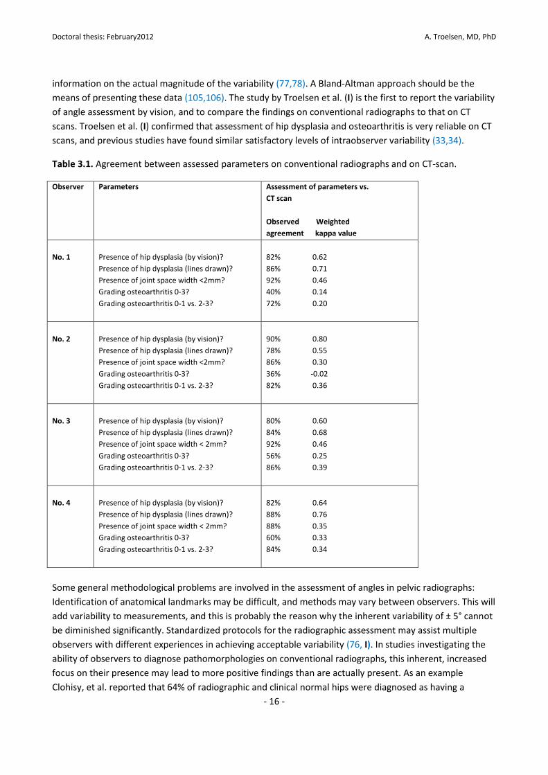
Table 3.1. Agreement between assessed parameters on conventional radiographs and on CT-scan.
Observer
Parameters Assessment of parameters vs.
CT scan
Observed Weighted
agreement kappa value
No. 1
Presence of hip dysplasia (by vision)?
Presence of hip dysplasia (lines drawn)?
Presence of joint space width <2mm?
Grading osteoarthritis 0-3?
Grading osteoarthritis 0-1 vs. 2-3?
82% 0.62
86% 0.71
92% 0.46
40% 0.14
72% 0.20
No. 2
Presence of hip dysplasia (by vision)?
Presence of hip dysplasia (lines drawn)?
Presence of joint space width <2mm?
Grading osteoarthritis 0-3?
Grading osteoarthritis 0-1 vs. 2-3?
90% 0.80
78% 0.55
86% 0.30
36% -0.02
82% 0.36
No. 3
Presence of hip dysplasia (by vision)?
Presence of hip dysplasia (lines drawn)?
Presence of joint space width < 2mm?
Grading osteoarthritis 0-3?
Grading osteoarthritis 0-1 vs. 2-3?
80% 0.60
84% 0.68
92% 0.46
56% 0.25
86% 0.39
No. 4
Presence of hip dysplasia (by vision)?
Presence of hip dysplasia (lines drawn)?
Presence of joint space width < 2mm?
Grading osteoarthritis 0-3?
Grading osteoarthritis 0-1 vs. 2-3?
82% 0.64
88% 0.76
88% 0.35
60% 0.33
84% 0.34
Some general methodological problems are involved in the assessment of angles in pelvic radiographs:
Identification of anatomical landmarks may be difficult, and methods may vary between observers. This will
add variability to measurements, and this is probably the reason why the inherent variability of ± 5° cannot
be diminished significantly. Standardized protocols for the radiographic assessment may assist multiple
observers with different experiences in achieving acceptable variability (76, I). In studies investigating the
ability of observers to diagnose pathomorphologies on conventional radiographs, this inherent, increased
focus on their presence may lead to more positive findings than are actually present. As an example
Clohisy, et al. reported that 64% of radiographic and clinical normal hips were diagnosed as having a

Doctoral thesis: February2012 A. Troelsen, MD, PhD
- 17 -
pathomorphological finding in a multi-observer study (76). Finally, the radiographic diagnostic assessment
of hip pathomorphologies should always be evaluated in a clinical context with knowledge of the patient’s
medical history and the findings on clinical examination. This likely has the potential to improve the ability
to correctly diagnose hip deformities, but it has not been done in radiographic reliability studies (76, I).
In conclusion, any observer, regardless of his or her level of experience, should refrain from assessment of
radiographic angles using only vision. This will result in an unacceptably high variability of angle measures
and a poorer ability to correctly diagnose the presence of hip dysplasia (I). One should be aware of the
inherent variability of approximately ± 5° for angle measures used in the diagnostic assessment of hip
dysplasia (75,79, I, II), and as a consequence consider reevaluation by CT scan in symptomatic patients in
whom a CE angle of 20° to 30° has been measured on conventional pelvic radiographs (I). Angle
measurements of hip dysplasia performed on a CT scan by a senior consultant radiologist represent a high
radiographic standard with diminishment of variability between measurements to approximately ± 3° and
excellent agreement between assessments of the presence of hip dysplasia (I). Measurement of the JSW or
a dichotomized assessment of the Tönnis grade should be preferred for the most reliable assessment of
osteoarthritis (I).
3.2. Weightbearing or supine assessment of hip dysplasia?
Not only intra- and interobserver variability of radiographic assessment can affect the interpretation of
radiographs. The position of the pelvis during recording has also been hypothesized to affect the
interpretation of AP radiographic indices (II). Previous studies have shown that AP radiographic indices of
hip dysplasia are not affected beyond inherent measuring errors unless the pelvis is excessively rotated or
tilted (79-81). Siebenrock et al. reported an easily understood relationship between the degree of pelvic tilt
and the appearance of acetabular version on AP pelvic radiographs (82). Based on the finding of such a
relationship, it has been recommended that pelvises be neutrally positioned in the supine position for
radiographic assessment of acetabular deformities (82). Accordingly, well-defined limits for neutral pelvic
positioning have been published and are now applied in studies pertaining to AP radiographic assessment
of the acetabular version (82-86). Clearly, the application of neutral pelvic positioning is only meaningful if
pelvic tilt is not affected significantly by differing patient positioning within the physiological range of
motion (i.e. repositioning from supine to weightbearing position). However, in the literature, there has
been controversy regarding the effect of change of position from supine to weightbearing on pelvic tilt (87-
93). Traditionally, AP pelvic radiographs have been recorded with patients in the supine position.
Troelsen et al. assessed AP radiographic indices of hip dysplasia, pelvic tilt, and acetabular version in 31 sets
of supine and weightbearing pelvic radiographs (II). Small mean reductions in the CE angle (1.3° – 1.6°) and
small mean increases in the AI angle (1.6° – 2.3°) were observed when repositioning from the supine to the
weightbearing position. However, the changes in angle measures were contained within the inherent
intraobserver variability (III). Fuchs-Winkelmann et al. (94) assessed 61 sets of supine and weightbearing
radiographs, and reported a marginally bigger, significant reduction in the CE angle (3.6°) when
repositioning than did Troelsen et al. Intra- and interobserver variability measures were, however,
expressed by Pearson’s correlation coefficients, leaving no possibility to assess the actual magnitude of the
variability. Fuchs-Winkelmann et al. observed a significant mean reduction in JSW of 0.49 mm after
repositioning (94). Troelsen et al. observed a significant reduction in male left hips of 0.67 mm on

Doctoral thesis: February2012 A. Troelsen, MD, PhD
- 18 -
repositioning (II). Still, the measure in both studies is confined within the observed intra-observer variability
of the JSW (II). Overall and in females, Troelsen et al. did not observe significant changes on repositioning,
and this is supported by findings in other studies (95,96, II). Troelsen et al. did not find any relevant femoral
head translation after repositioning from the supine to the weightbearing position (II).
Several studies have evaluated the effect of repositioning (i.e. from supine to weightbearing) on changes in
pelvic tilt (Table 3.2). Using varying methods for assessment of change in pelvic tilt, some studies report no
significant changes (87,90,92) and others a significant extension (backward rotation or reclination) of the
pelvis of approximately 4° to 8° (88,89,91,93). To support the notion that pelvic mobility is rarely excessive
during repositioning, Nishihara et al. and Babisch et al. found that pelvic tilt did not exceed a change of 10°
in 90% and 83% of their cases, respectively (87,90,93). Using an indirect measure of change in pelvic tilt,
Troelsen et al. reported a significant change in pelvic tilt of 13° to 14° in females and 6° to 7° in males after
repositioning (II). A moderately strong correlation between the distance from the symphysis to the
sacrococcygeal joint (used by Troelsen et al. (II)) and the degree of pelvic tilt has been reported (97) (Figure
3.1). Overall, the studies agree that an extension of the pelvis takes place on repositioning (87-93, II).
Comparison of studies is not possible because a wide variety of methods are used and study populations
show both intra- and interstudy heterogeneity (Table 3.2). It seems, however, that study populations with
normal subjects or hip dysplasia are associated with the report of significant changes of pelvic tilt during
repositioning (88,89,91,93, II). At least in patients with hip dysplasia, the often generalized instability and
coverage deficiency could contribute to increased pelvic mobilization during repositioning.
It is clear that results are diverging, but despite controversy, there are studies to support the hypothesis
that pelvises are significantly extended when repositioned from supine to weightbearing (88,89,91,93, II).
In the light of this, the application of standardized, so-called neutral pelvic positioning (i.e. assuming no
difference in pelvic tilt between supine and weightbearing positions) is controversial (82). This is further
amplified by Troelsen et al. who reported that only 32% of patients in the weightbearing position were
confined within the limits of neutral pelvic positioning suggested by Siebenrock et al. (82, II). Also the
position of the patient was found to affect the appearance of acetabular version because 11 patients
showed signs of retroversion in the supine position versus 4 patients in the weightbearing position (II). This
is explained by the extension of the pelvis in connection with repositioning.
In summary, pelvic radiographs for assessment of hip deformities are usually recorded with the patient
supine, and neutral pelvic positioning has been advocated (82). However, based on the present literature, a
significant pelvic extension may take place after repositioning from the supine to the weightbearing
position (88,89,91,93, II). Further, pain originating from prearthritic structural deformities is often
attenuated or only present during function. It is thought that weightbearing radiographs secure the best
coherence between symptoms, functional appearance, and hip deformities. Finally, AP radiographic indices
of hip dysplasia, femoral head translation, and the JSW show only minor differences between the supine
and weightbearing positions (94, II). Troelsen et al. recommend weightbearing AP pelvic radiographs for
assessment of hip deformities (II).

Doctoral thesis: February2012 A. Troelsen, MD, PhD
- 19 -
Figure 3.1.
A section of an anteroposterior pelvic radiograph showing both hip joints. The line with arrowheads marks
the distance from the symphysis to the sacrococcygeal joint.

Doctoral thesis: February2012 A. Troelsen, MD, PhD
- 20 -
Table 3.2. Studies evaluating the effect of repositioning on changes in pelvic tilt
Study No. of patients
(females/males)
Description of
patients
Method of
assessment
Results
(diff. supine to
weightbearing)
Conclusion
Anda et al.
1990 (87)
40
(27/13)
Healthy young
adults
Pelvic
inclinometer
Females:
Extension: 2.3°
Males:
Extension: 0.4°
No significant
changea
Konishi et al.
1993 (88)
54
(27/27)
Normal subjects
15-79 years
Lateral x-rays
correlated with
AP x-rays
Females:
Extension: 5°
Males:
Extension 5°
Significant
change
(p<.0001)
Eddine et al.
2001 (89)
24
(9/15)
Healthy subjects
24-41 years
Lateral x-rays:
supine and
standing
Females and
males:
Extension app.
6°-8°
Significant
change
(p=.0001)
Nishihara et al.
2003 (90)
101
(71/30)
Degeneration: 91
Loose THR: 10
23-81 years
Image matching
between CT and
AP x-ray
Females and
males:
Extension: 2°
No significant
change
Lembeck et al.
2005 (91)
30
(13/17)
Healthy subjects
20-43 years
Pelvic
inclinometer
Females and
males:
Extension: 4°
Significant
change
(p=0.02)a
Mayr et al.
2005 (92)
120
(60/60)
Adults volunteers
21-91 years
30 of 120 were
young and healthy
Pelvic landmarks
digitized
percutaneously
Young and healthy;
females and males:
Extension: 1.0°
No significant
changea
Babisch et al.
2008 (93)
30
(24/6)
Hip dysplasia: 17
Osteoarthritis:13
Standing lateral
x-ray and supine
CT scan
Females and
males:
Extension: 5.4°
Significant
change
(p<.001)
Troelsen et al.
2008 (II)
31
(24/7)
Unilateral (5) or
bilateral (26) hip
dysplasia
16-56 years
Supine and
standing AP x-
rays
Female:
Extension: 13°-14°b
Male:
Extension: 6°-7°b
Significant
change
(p<.0001 and
p=0.0042)
a: p values are calculated from the original data.
b: Values of the mean differences in distances from the symphysis to the sacrococcygeal joint are converted according
to Tannast et al. (97).

Doctoral thesis: February2012 A. Troelsen, MD, PhD
- 21 -
3.3. Acetabular retroversion in hip dysplasia
Acetabular retroversion has been recognized as a possible precursor of osteoarthritic development and as a
source of hip pain (43,44,86,98). The entity has been found to be associated with the presence of labral
tears, and it is incorporated into the biomechanical concept of FAI as a focal overcoverage causing pincer
impingement (14,56,57). The first assessment of acetabular retroversion is made on an AP pelvic
radiograph by identification of a crossing of the anterior and posterior acetabular rims. Reynolds et al.
described this so called “crossover” sign approximately a decade ago (98) (Figure 3.2). Jamali et al. found
the crossover sign to be highly valid in assessment of acetabular retroversion on AP pelvic radiographs (99).
Siebenrock et al. reported that acetabular retroversion gets more pronounced with increasing pelvic flexion
(inclination or forward rotation) (82).
Historically, hip dysplasia has been described as a condition associated with lateral and anterior acetabular
deficiency and acetabular anteversion (30,31,33,34,37,100). It is therefore somewhat surprising that during
the last 5 to 10 years, especially the last 2 years, acetabular retroversion has been reported to coexist with
hip dysplasia in a considerable minority of dysplastic hip joints (41,42,83-86,101,102, III). The reported
prevalences of acetabular retroversion in dysplastic hips ranges from 15% to 42% (Table 3.3). The crossing
of acetabular rims is most frequently seen in the cranial third of the dysplastic acetabulum (86,101, III). The
degree of hip dysplasia, quantified by the CE angle, does not seem to differ between retroverted acetabuli
and normally oriented acetabuli (42,84,86, III).
As already outlined (see section 3.2), it is controversial whether hip deformities should be assessed in AP
pelvic radiographs with the pelvis neutrally positioned or in the weightbearing position. The appearance of
acetabular version and its extent depend on the degree of pelvic tilt, and thus the prevalence estimates of
acetabular retroversion will depend on the radiographic method applied (82, II, III). Most studies reporting
the prevalence of acetabular retroversion in dysplastic hips include only radiographs if they meet certain
standardized criteria with respect to pelvic tilt (Table 3.3). In light of the believed difference in pelvic tilt
after repositioning and the coherence between deformities and functional appearance in the weight-
bearing position, Troelsen et al. assessed the prevalence of retroversion in weightbearing AP pelvic
radiographs (III). Troelsen et al. found acetabular retroversion in 33% of dysplastic hips, which is higher
than the estimates of approximately 15% to 20% reported in the majority of studies (83-86,101, III). This
difference is explained by the exclusion of pelvises with excessive flexion (inclination or forward rotation).
That is, those pelvises are exclude that by nature are excessively flexed and therefore are prone to have a
more pronounced appearance of retroversion. In general, studies report a satisfying or good intra- and
inter- observer variability in the assessment of the crossover sign (99,103,104, III). Assessment of the
acetabular rims and the crossover sign demands good quality radiographs. As an alternative, the ischial
spine sign has been introduced as a valid indicator of acetabular retroversion (103).
The clinical importance of acetabular retroversion in hip dysplasia and its implications for performance of a
redirective PAO are not yet fully understood. Recently, hip dysplasia with acetabular retroversion was
found to be associated with an earlier onset of pain (86). Also, recent studies have stressed the importance
of recognizing acetabular retroversion during preoperative planning and performance of redirective PAO
(84,85,102). Failure to do so will result in continued or even aggravated retroversion with a decreased

Doctoral thesis: February2012 A. Troelsen, MD, PhD
- 22 -
range of motion and a postoperative FAI with continued joint deterioration. Acetabular retroversion has
been reported to persist in 10% to 62% of dysplastic hips following redirective procedures. (84,85,102).
In summary, the identification of acetabular retroversion in dysplastic hips potentially has important clinical
and surgical implications that need further investigation. Retroverted acetabuli are surprisingly frequent in
dysplastic hips (15%-42%) (41,42,83-86,101,102, III). Because the appearance of acetabular retroversion
depends on the pelvic tilt (82, II, III), the position of the patient during radiographic recording is a
potentially important factor that needs further exploration.
Figure 3.2.
A section of an anteroposterior pelvic
radiograph showing the left hip. The dashed line
marks the anterior acetabular rim and the solid
line the posterior acetabular rim. A “crossover”
sign is present as the dashed and solid lines
intersect.

Doctoral thesis: February2012 A. Troelsen, MD, PhD
- 23 -
Table 3.3. Studies reporting the prevalence of acetabular retroversion in dysplastic hips
Study No. of hips (patients)
(females/males)
Age
Dysplastic hips
included
Radiographs included Prevalence of
retroversion
Li and Ganz
2003 (101)
232 (199)
(136/63)
30yrs (12-61)
CE angle ≤25° Coccyx to symphysis distance was
0-2 cm
17%
Mast et al.
2004 (41)
235 (153)
(125/28)
NR
CE angle <20° Coccyx to symphysis distance was ≤
2 cm
37%
Ezoe et al.
2006
(83)
74 (64)
(56/8)
36 yrs (14-54)
CE angle <20° Sacrococcygeal joint to symphysis:
distance 25-40 mm in males;
40-55 mm in females
18%
Peters et al.
2006
(102)
83 (73)
(55/18)
28 yrs (15-47)
Operated hips with
CE angle: -20° to
34°
NRa
28%
Kiyama et al.
2009 (84)
180 (155)
(NR/NR)
NR
Operated hips with
CE angle <16°
Sacrococcygeal joint to symphysis:
distance 25-40 mm in males;
40-55 mm in females
18%
Nehme et al.
2009 (42)
195 (174)
(137/37)
30 yrs (15-56)
CE angle <20° Coccyx to symphysis: distance 0-2
cm
42%
Xie et al.
2010 (85)
106 (88)
(NR/NR)
38 yrs (15-52)
Operated hips with
CE angle: mean
app. 9°; SD app. ±8°
Sacrococcygeal joint to symphysis:
distance 25-40 mm in males;
40-55 mm in females
15%
Fujii et al.
2010 (86)
96 (59)
(52/7)
40 yrs (15-60)
CE angle <20° Sacrococcygeal joint to symphysis:
distance 25-40 mm in males;
40-55 mm in females
18%
Troelsen et al.
2010 (III)
95 (54)
(44/10)
36 yrs (14-57)
CE angle <25° Weightbearing radiographs
(defined protocol)
33%
a = NR: Not reported.

Doctoral thesis: February2012 A. Troelsen, MD, PhD
- 24 -
4. Assessment of acetabular labral tears in hip dysplasia
4.1 The role of the acetabular labrum in hip dysplasia.
During the last decade, the understanding of the relationship between hip joint deformities and
osteoarthritic development has increased significantly. Tearing of the acetabular labrum or the adjacent
cartilage is recognized as the key to joint deterioration in all cases of biomechanically induced osteoarthritis
(14). The description of acetabular labral tears associated with hip dysplasia is not new, but the actual
biomechanical properties of the acetabular labrum and its role in initiation of joint degeneration have now
been documented (16,35-37,107-110). The labrum is hypothesized to have a load-sharing role, at least in
hip dysplasia, and to act as a seal optimizing the properties of hip joint lubrication (108,111). Furthermore,
the labrum has a stabilizing function that protects against critical biomechanical alterations in the hip joint
(112).
The biomechanical changes induced by the osseous deformities in hip dysplasia, together with the
instability of the joint, are thought to make the acetabular labrum susceptible to overload and tearing.
There is theoretical and clinical evidence that a “shearing” kind of impingement, with repeated micro
trauma to the labrum, subsequent degeneration, and finally a tear or detachment of the labrum in the
chondrolabral transition zone, underlies the biomechanical concept (16,35-37,113,114). Acetabular labral
tears in hip dysplasia are most frequently found in the anterior region of the acetabulum, which may be
explained by the demanding biomechanics, with increased joint load and a weaker mechanical structure of
the labrum particularly in this region (109,110,115,116, IV,V). Another characteristic feature of the labrum
is its often hypertrophic state in dysplastic hips (35,115).
Tearing of the acetabular labrum is a frequent finding in symptomatic dysplastic hips. In 170 hips with
dysplasia, McCarthy and Lee found a labral tear on hip arthroscopy in 72% (113). Studies utilizing magnetic
resonance arthrography (MRA) to evaluate the labrum in symptomatic dysplastic hips found labral tearing
in approximately 80% (115, IV). The findings suggest that joint overload and labral tearing play an
important role in the development symptoms in patients with hip dysplasia. Classical symptoms of hip
dysplasia are sharp groin pain and clicking or locking of the hip, all of which correspond well with a labral
tear and continuous joint overload.
4.2 The role of MR arthrography, ultrasound, and clinical tests in acetabular labral tear diagnostics
The identification of an acetabular labral tear as a cause of pain and a precursor of hip joint degeneration
has focused attention on reliable diagnostic assessment. MRA has been established as the radiographic
gold standard method for the diagnostic assessment of acetabular labral tears (Figure 4.1). Recent studies
have reported a good ability of MRA to diagnose labral tears (117-119). Toomayan et al. performed MRA in
30 hips and found a sensitivity of 92% and specificity of 100% when MRA findings were compared with
those obtained during hip arthroscopy (117). Chan et al. reported a sensitivity of 100% and an accuracy of
94% (in 18 hips undergoing subsequent hip arthroscopy) (118). Freedman et al. reported that 22 (96%) of
23 labral tears diagnosed on hip arthroscopy had been found on MRA images (119). Ziegert et al. found a
detection rate of labral tears of 97.2% on MRA in 144 hips with proven tears at arthroscopy (120). Czerny et
al. published the first report of its kind in 1996 (n=22 hip MRAs) and found a sensitivity of 90% and an
accuracy of 91% for MRA compared with arthroscopic findings (121). In a later study, Czerny et al. showed

Doctoral thesis: February2012 A. Troelsen, MD, PhD
- 25 -
that MRA can be used to correctly stage labral tears (122); however, the staging seems of less prognostic
value (119). The intrareader reliability of MRA readings has been reported to be excellent (119,121, IV, V).
In contrast to these encouraging results, Keeney et al. (n=104 hips) and Leunig et al. (n=23 hips) reported
sensitivities and specificities of approximately 40% to 70% for MRA in labral tear detection (123,124). It
should be acknowledged that the studies reporting the diagnostic ability of MRA often suffer
methodological problems, such as, a retrospective design, with lack of a clear prospective protocol for
image readings; bias induced by lack of blinding of radiologists to the arthroscopic findings; selection-
induced bias because all hips may have been included in the retrospective study due to the finding of a
labral tear on hip arthroscopy; and interobserver variation because several radiologists or arthroscopic
surgeons had assessed the presence of the labral tears.
In the literature MRA, has been established as the radiographic gold standard in labral tear diagnostics.
However, the method is time-consuming and uncomfortable for the patients. Ultrasound is widely used in
musculoskeletal diagnostic radiology, and it has been hypothesized that it may have the ability to diagnose
acetabular labral tears reliably (IV, V) (Figure 4.2). Few studies have investigated the ability of ultrasound
examination. Mitchell et al. reported the results of 8 ultrasound examinations in hips that had arthroscopic
assessment of joint pathology: in 1 of 8 examinations ultrasound diagnosed the pathology present. Given
the methodological flaws of this study, conclusions cannot be drawn, and the authors make no mention of
ultrasound in their suggested diagnostic approach to hip pain (125). Sofka et al. reported a subjective
improvement in visualization of labral pathology by ultrasound during intra-articular steroid injections in 21
hip joints. Magnetic resonance imaging (MRI) without contrast was performed in 14 of the 21 hips, and on
review, anterior labral tears were found in 13 hips on both MRI and ultrasound examination. The authors
did not quantify the diagnostic ability of ultrasound. The study might represent a show of the potential
success of ultrasound to diagnose labral pathology, but any conclusions are made invalid by the
retrospective design that meant review of only cases with a positive finding of labral pathology during
ultrasound examination (126). A prospective comparison of ultrasound with MRA in labral tear diagnostics
was performed by Troelsen et al. (IV). Examinations were performed in 20 consecutive dysplastic hip joints
presenting with pain. The prospective protocol included predefined criteria for description of labral tears
and blinding of the MRA radiologist and the ultrasound radiologist to the findings of the other examiner.
The corresponding findings on ultrasound and MRA are presented in Table 4.1. The resulting sensitivity was
44% and the specificity was 75%. In a subsequent study by Troelsen et al. (V), the authors examined the
ability of ultrasound to detect labral tears, applying a protocol for the performance of examinations similar
to the one used in the previous study by Troelsen et al. (IV). The hip joints of 18 patients who previously
had had periacetabular osteotomies were examined. The findings on MRA and ultrasound are presented in
Table 4.2. Thus, the sensitivity of ultrasound in labral tear diagnostics was 94%. The studies (IV, V) were
strengthened by the prospective protocol used for the performance of the examinations, but limited by the
relatively small sizes of the study cohorts, and by the fact that the radiographic findings were not verified
by hip arthroscopy. However, MRA is well established as the radiographic gold standard in acetabular labral
tear diagnostics, with an excellent correlation to arthroscopic findings in recent studies (117-119). The
intra- and interobserver variability of ultrasound in labral tear diagnostics remains uninvestigated.

Doctoral thesis: February2012 A. Troelsen, MD, PhD
- 26 -
Figure 4.1.
Figure 4.2.
Coronal view of hip MR arthrography visualizing
an acetabular labral tear (arrow). Contrast
medium is seen running through the base of the
labrum.
Ultrasound examination visualizes an acetabular
labral tear. There is a hypoechoic cleft running
through the base of the labrum (thick arrow),
and a cystic formation is visible just superior to
the labrum (thin arrows). The crosses mark the
limits of the labrum.
Doctoral thesis: February2012 A. Troelsen, MD, PhD
- 27 -
A thorough patient history and a clinical examination should be able to raise suspicion of a labral tear as the
cause of hip pain. But how reliable are commonly used clinical tests in the assessment of acetabular labral
tears? Evidence in this field is limited. Narvani et al. (51) conducted a study examining 18 hips by an
“internal rotation, flexion, axial compression” test and using MRA as diagnostic reference. The sensitivity
was 75% and the specificity was 43%. In a study by Martin et al. (127) 6 orthopedic surgeons, specializing in
hip pain, performed clinical examinations in 8 patients. The clinical examinations were performed as
preferred by each specialist. Based on the clinical examination, the orthopedic surgeons agreed 63% of the
time with the finding of a labral tear on the following hip arthroscopy. Troelsen et al. (V) investigated the
ability of the impingement test, the FABER test, and the resisted straight leg raise test to diagnose labral
tears. The clinical findings in 18 hips were compared to MRA findings of labral tears, and the diagnostic
ability of the tests was calculated (Table 4.3). Of the clinical tests, the impingement test showed the best
diagnostic ability, with a sensitivity of 59% and a specificity of 100%. Martin and Sekiya investigated the
interrater reliability of the impingement test and the FABER test and found a moderate agreement
between observers (kappa: 0.58) for the impingement test and a substantial agreement between observers
(kappa: 0.63) for the FABER test (128). The few studies investigating the diagnostic ability of clinical tests
are in general limited by small study populations (51,127, V). Furthermore, the study by Troelsen et al. (V)
is limited by the frequent presence of a labral tear in the selected study population. The diagnostic ability of
the impingement test in patients with a normal labrum is thus difficult to assess. The prospective protocol
of examinations and blinding of both the clinical, ultrasound, and MRA examiners to each other’s findings is
a methodological strength, and one should bear in mind that patients presenting in an outpatient clinic
dealing specifically with hip problems are highly selected.
In conclusion, MRA has been established as the diagnostic gold standard in acetabular labral tear
diagnostics. The results of studies on the diagnostic ability of MRA have been conflicting. In the most
recently published studies, however, MRA has been reported to have excellent diagnostic properties (117-
119). Ultrasound is a new and promising tool in labral tear diagnostics. The improvement in the diagnostic
ability of ultrasound demonstrated by a comparison of the results of the two studies by Troelsen et al. (IV,
V) suggests that a learning curve is associated with the use of ultrasound in labral tear diagnostics. Even in
the hands of an experienced ultrasound examiner, as in the studies by Troelsen et al., issues of creating
optimal visualization and interpretation of findings represented methodological difficulties that had to be
overcome during the first study (IV). Clinical examination to detect labral tears is the “every-day tool” of
the orthopedic hip surgeon, and even the most widely used tests (impingement and FABER tests) are not
very reliable in labral tear diagnostics (51,127, V). This issue emphasizes the need for reliable radiographic
assessment. The knowledge base regarding the role of ultrasound and clinical tests in acetabular labral tear
diagnostics is limited, and the role of their use in unselected cohorts remains uninvestigated.

Doctoral thesis: February2012 A. Troelsen, MD, PhD
- 28 -
Table 4.1. Labral tear diagnostics: findings on ultrasound examination and resulting reliability measures
(Troelsen et al. (IV))
Ultrasound: Labral tear
Ultrasound: No labral tear
MRA: Labral tear
7
9
16
MRA: No labral tear
1
3
4
8
12
20
Sensitivity (true positives)
Specificity (true negatives)
Positive predictive value
Negative predictive value
7/16 = 44%
3/4 = 75%
7/8 = 88%
3/12 = 25%
Table 4.2. Labral tear diagnostics: findings on ultrasound examination and resulting reliability measures
(Troelsen et al. (V))
Ultrasound: Labral tear
Ultrasound: No labral tear
MRA: Labral tear
16
1
17
MRA: No labral tear
1
0
1
17
1
18
Sensitivity (true positives)
Specificity (true negatives)
Positive predictive value
Negative predictive value
16/17 = 94%
0/1 = Not reported
16/17 = 94%
0/1 = Not reported
Table 4.3. The diagnostic ability of the impingement test and the FABER test in labral tear diagnostics
(Troelsen et al. (V)). The resisted straight leg raise test was positive in 1 of 18 cases and thus results were
not analyzed further.
Impingement test
FABER test
Sensitivity (true positives)
Specificity (true negatives)
Positive predictive value
Negative predictive value
10/17 = 59%
1/1 = 100%
10/10 = 100%
1/8 = 13%
7/17 = 41%
1/1 = 100%
7/7 = 100%
1/11 = 9%
Doctoral thesis: February2012 A. Troelsen, MD, PhD
- 29 -
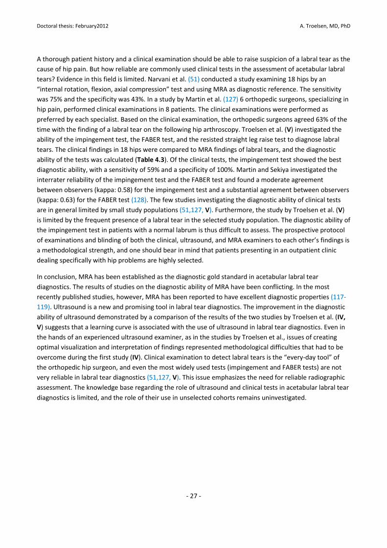
4.3 Suggested strategy for diagnostic assessment of acetabular labral tears
Patients presenting with hip-related pain, especially sharp groin pain, and in some a history of clicking or
locking of the hip joint should be suspected to have a tear of the acetabular labrum (Figure 4.3) (129,130).
A weightbearing AP pelvic radiograph and a lateral view of the hips are required to diagnose coexisting hip
deformities and/or osteoarthritis (129,130, II, III).
To further assess the suspicion of a labral tear in these selected patients, an impingement test and perhaps
the FABER test should be carried out, because the use of these tests is supported by the literature
(51,127,128, V). Both tests have been reported to have a positive predictive value of 100%, meaning that
on reproduction of sharp groin pain, the patient is very likely to have a labral tear (V). Previous studies have
reported ≥95% of impingement tests to be positive in patients with surgically verified labral tears
(19,49,50). On the other hand, not all labral tears are diagnosed by the impingement or FABER tests
(sensitivities of 59% to 75% and 41%, respectively), and a negative outcome of the tests is unreliable (51,
V). In this case, further radiographic assessment is warranted.
If an experienced ultrasound examiner with interest in development of this tool in labral tear diagnostics is
available, ultrasound examination can be performed to assess a potential tear of the labrum (IV, V). In the
hands of an examiner who has overcome the learning curve regarding the interpretation of the
examinations, the method is sensitive (94%) in diagnosing acetabular labral tears (V). A finding of labral
tearing on ultrasound makes it very likely that the patient actually has a tear (positive predictive value: 94%
(V)). The present literature is inconclusive regarding the reliability of not finding a labral tear and
ultrasound therefore should be considered unreliable in this situation. (IV, V). Further radiographic
assessment is then warranted.
MRA is the established gold standard in radiographic assessment of labral tears. The main problem related
to clinical tests and ultrasound is the lack of reliability if findings are negative (51, IV,V). Thus, MRA should
be performed in patients if groin pain is not produced by the impingement or FABER test and a labral tear
cannot be visualized on ultrasound examination, and the patient continues to have specific hip-related pain
(V). In the most recent experience with MRA, the diagnostic ability has been reported to be excellent, with
sensitivity, specificity, and accuracy measures in the range of 92% to 100% (117-119). However, failure to
diagnose a labral tear cannot be ruled out, and on continued suspicion, hip arthroscopy should be
performed.
Figure 4.3. Suggested strategy for diagnostic assessment of acetabular labral tears (V)

Doctoral thesis: February2012 A. Troelsen, MD, PhD
- 30 -
5. Periacetabular osteotomy for surgical treatment of hip dysplasia in adults
5.1 Periacetabular osteotomy: outcome, problems, and perspectives.
Since its introduction more than 20 years ago, PAO has been adopted as the preferred contemporary joint
preserving surgical treatment for symptomatic hip dysplasia in adults (15,72-74,102,131-147). The clinical
aims are to relieve hip joint pain, improve function and health related quality of life, and to prevent
osteoarthritic development necessitating conversion to THR. The surgical aim is a 3-dimensional
reorientation of the acetabulum that will optimize femoral head coverage, decrease hip joint load forces,
and relieve the overload of the acetabular labrum and adjacent cartilage and soft tissues (15,72,148-150)
(Figure 5.1).
Whereas numerous studies describe the short-term outcome following PAO (132-144), only a few studies
report the outcome at medium- and long-term follow-up (i.e. more than a minimum follow-up of 5 years)
(72-74,131, VI). This lack of studies reporting the outcome at medium- and long-term follow-up is deeply
contrasted by the wide acceptance and worldwide application of this major surgical procedure. In the
studies investigating the medium- and long-term outcome following PAO, the main endpoint indicating
failure is conversion to THR (72-74,131, VI).
Troelsen et al. reported the medium-term clinical and radiographic outcome in 116 periacetabular
osteotomies 5.2 to 9.2 years postoperatively. Seventeen hips were converted to THR, and the Kaplan-Meier
hip joint survival rate with conversion to THR as endpoint was 90.5% (95% CI: 83.5-94.6) at 5 years, and
81.6% (95% CI: 69.7-89.3) at 9.2 years [VI]. Other authors reporting the medium- or long-term hip joint
survivorship show rates comparable to these numbers (72-74) (Table 5.1). Further, as outlined in Table 5.1,
the study groups are grossly comparable. Short-term hip joint survival rates (i.e. less than a minimum
follow-up of 5 years) are most frequently reported to be >90% (132-144).
Table 5.1. Studies reporting the medium- or long-term outcome after periacetabular osteotomy.
Author
(year)
No. hips
(% females)
Hips with
DDH
Follow-up
Mean (range)
No.
surgeons
Preoperative CE
angles (range)
Hips with
Tönnis 0-1
Hip joint
survivorship
Kralj et al.
2005 (72)
26 (85%)
26 12 yrs (7-15) 3 Mean 15°
(7-26)
81% 85% (mean 12y)
Steppacher et al.a
2008 (73)
75 (77%) 75 20.4 yrs (19-23) 1 Mean 6°
(-24-25)
76% KMb 87.6%(10y)
KMb60.5% (20y)
Matheney et al.
2009 (74)
135 (88%) 135 9 yrs (NRc)
(SD:±2.2)
1 Median 0° to 3°
(NRc)
82% KMb84% (10y)
Troelsen et al.
2009 (VI)
116 (78%) 102
(14 LCPDd)
6.8 yrs (5.2-9.2) 1 Median 11°
(-29-30)
90% KMb81.6%(9.2y)
a = Steppacher et al. reports the outcome of the same cohort as in the study by Siebenrock et al. 1999 (131).
b = KM is the Kaplan-Meier survivorship rate. Only Kralj et al. (72) did not report a Kaplan-Meier estimate.
c = NR: Not reported.
d = LCPD: Legg-Calvé-Perthes disease.
The extensive follow-up of PAO patients by Troelsen et al. comprised an interview, a clinical examination, a
radiographic examination (weightbearing anterior-posterior pelvic radiograph), and Short Form (SF)-36
(151) and Western Ontario and McMaster Universities Osteoarthritis Index (WOMAC) (152) questionnaires

Doctoral thesis: February2012 A. Troelsen, MD, PhD
- 31 -
(VI). The results of the follow-up evaluation are presented in Table 5.2. Key features of the interviews and
clinical examinations were as follows: Median pain scores on the visual analog scale were 0 at rest and 1
after 15 minutes of normal walking. The groin was the most frequent location of hip-related pain or
discomfort. Clicking or locking of the hip joint was seen in 25% of hip joints, and a positive impingement
test was found in 18%. Hip joint osteoarthritis and possible coexisting intraarticular problems may explain
these findings. Steppacher et al. noted the impingement test was positive in 24% of hip joints at 10-year
evaluation of patients undergoing PAO surgery (73). Troelsen et al. reported hip range of motion with a
median hip flexion of 100° similar to the 10-year follow-up result found by Steppacher et al. (73, VI).
Removal of screws was performed in 18% of the operated hips at the time of medium-term follow-up,
making it a matter of importance during the initial preoperative patient information session (VI). Troelsen
et al. shed new socioeconomic insights on the subject of PAO surgery because only 19% had to change jobs
after surgery due to hip problems (VI).
The Harris Hip Score or the Merle d’Aubigné & Postel Score have been the preferred hip-specific functional
scores in previous studies reporting the outcome of PAO surgery (72,73,102,132,133,135-137). It is
doubtful whether these scores provide a satisfying assessment in joint preserving PAO surgery in young
adults. Troelsen et al. assessed the outcome at medium-term follow-up after PAO surgery using the
contemporary SF-36 and WOMAC scores (Table 5.2) (VI). Comparison to the few other studies using the SF-
36 and WOMAC scores is compromised by different durations of follow-up and differing strategies of data
transformation (72,144,145). Comparison of the SF-36 scores reported at medium-term follow-up by
Troelsen et al. with normative data for Danish citizens of a relevant age group showed, not surprisingly,
that the physical component score was lower in the hip patients and that the mental component scores
were comparable. (151, VI). Thus, it seems that mental health is not negatively influenced by the
deterioration in physical health in patients after PAO surgery at medium-term follow-up. In addition, the
median total WOMAC score of 84.4 found at medium-term follow-up is satisfactory. Of studies reporting
the outcome of PAO surgery at medium- to long-term follow-up, the studies by Kralj et al. (72) (using the
WOMAC score) and Steppacher et al. (73) (using the Merle d’Anbigné & Postel Score) report both pre- and
postoperative scores, whereas the studies by Matheney et al. (74) (using the WOMAC pain subscale) and
Troelsen et al. (VI) are limited by reporting only the postoperative follow-up scores. Finally, a hip-specific
patient-related outcome measure specially constructed for young patients undergoing joint preserving
surgery, such as the PAO, remains to be developed.
Even though widely accepted, the use of conversion to THR as an end point is in some ways problematic. At
the time of follow-up, more patients may have developed end-stage hip joint osteoarthritis or functionally
compromising pain. Those hip joints may also qualify for a THR in the near future, and thus the true failure
rate may be underestimated. There is no commonly accepted definition of which secondary end points to
consider. Matheney et al. considered a score of ≥10 (of 20) on the WOMAC pain subscale at the time of
follow-up to be a clinical failure. Consequently, an additional 12% (16 of 135) of the study group qualified as
clinical failures (74). Kralj et al. reported that 8 (31%) of 26 hips had end-stage osteoarthritis (Tönnis grade
3) or a deterioration of the WOMAC total score of >20 on medium to long-term follow-up after PAO. This
group was separated from the groups of patients with an apparently satisfying clinical outcome and
patients who had had a THR (72). Steppacher et al. report outcomes in the same cohort as in the study by
Siebenrock et al. (131) and found that end-stage osteoarthritis (Tönnis grade 3) had developed in 5 (7%) of
68 hips. Two hips were graded as having a poor result according to the Merle d’Anbigné & Postel Score at

Doctoral thesis: February2012 A. Troelsen, MD, PhD
- 32 -
last follow-up, but whether the hips also had end-stage osteoarthritis is unclear (73). Troelsen et al. found
that end-stage osteoarthritis (Tönnis grade 3) had developed in 6 (6%) of 93 hips (VI). Patients lost to
follow-up constitute another problem related to the use of conversion to THR as an end point. How many
of these patients have had their PAO converted to a THR at another institution? In the study by Matheney
et al. 23 (15%) of the initial 158 eligible dysplastic hips were lost to follow-up, with no trace of the hips
beyond the first year after surgery (74). Kralj et al. lost track of 20 (36%) of 55 consecutive patients during
their follow-up. They also excluded 9 of 35 respondents, of which 2 had a THR performed (72). Steppacher
et al. lost track of 5 (7%) of 75 hips. However, 3 of these were seen at follow-up 10 to 12 years
postoperatively and showed good to excellent clinical results and no conversions to THR (73). In the study
by Troelsen et al. 12 (10%) of 116 hip joints were lost to follow-up (VI). However, this study bears the
advantage that inquiry to the Danish Hip Arthroplasty Register (63,153) was made regarding conversion to
THR in all 116 hip joints, thus optimizing the completeness of the follow-up. None of the hips lost to follow
up had had a THR.
Patients lost to follow-up, unless accounted for by inquiry to a national joint arthroplasty register, add
uncertainty to the reported hip joint survival rates, and evaluating a clinical and/or radiographic end point
other than conversion to THR may add significantly to the failure rate. Interstudy differences concerning
these issues can easily explain even significant differences in the reported survivorship rates. The lack of
consensus regarding the report of outcome is problematic, and to assure a uniform, transparent and
sufficient quality of studies reporting the outcome following PAO surgery, the following criteria are
suggested: 1) Conversion to THR should be reported. 2) Development of end-stage osteoarthritis and/or a
score of ≥10 (of 20) on the WOMAC pain subscale should be reported. 3) Inquiry to national arthroplasty
registers, if possible, regarding otherwise undetected conversion to THR should be made. Otherwise, no
more than 10% to 15% should be lost to follow-up. 4) Follow-up should be performed using contemporary
WOMAC and/or SF-36 questionnaires.
The methodological weaknesses of the studies reporting medium- or long-term outcomes following PAO
surgery can generally be attributed to their retrospective designs (72-74,131, VI). Further, they rely on
conventional radiography to diagnose the presence and degree of osteoarthritis, and therefore focal or
generalized cartilage deterioration may go undetected. Future studies reporting the outcome of PAO
surgery should follow a strictly prospective protocol, using contemporary scoring systems, and magnetic
resonance imaging to clarify the intraarticular constitution of the hip joint. Finally, with some cynicism, one
might argue that the surgeons and researchers in the field of PAO orthopedics have forgotten to perform a
randomized study that would determine whether patients obtain sufficient benefits by undergoing a PAO
compared to, for example, conservative treatment. On the other hand, randomizing young patients with
clear-cut hip dysplasia, no osteoarthritis, and invalidating pain to conservative treatment or THR would
seem unethical when studies have shown that PAO surgery preserves hip joints for up to 2 decades and
yields good clinical results in selected patients (72-74,131, VI).

Doctoral thesis: February2012 A. Troelsen, MD, PhD
- 33 -
Figure 5.1.
A postoperative anteroposterior pelvic radiograph showing both hip joints. The right hip has been treated
with a redirective joint preserving periacetabular osteotomy. A center-edge angle of Wiberg of 35° has
been achieved. Hip dysplasia is noted in the left hip.

Doctoral thesis: February2012 A. Troelsen, MD, PhD
- 34 -
Table 5.2. The medium-term outcome following periacetabular osteotomy (VI).
PARAMETER
Interview and examination (n=77 procedures)
Primary localization of hip pain or discomfort
Number (%), localization
VAS pain score; median (interquartile range) (range)
At rest
After 15 minutes of normal walking
Clicking or locking of the hip joint
Number (%)
Pain or discomfort related to spine or lower extremities
Number (%), condition
Subsequent hardware removal
Number (%)
Change of job since surgery due to hip problems
Number (%)
Dysesthesia of the lateral femoral cutaneous nerve
Number (%)
Positive Impingement test
Number (%)
Range of motion; median (interquartile range) (range)
Flexion
Extension
Abduction
Adduction
Internal rotation
External rotation
26 (34%) groin; 7 (9%) trochanter
3 ( 4%) buttock; 2 (3%) femur
0 (0-1) (0-7)
1 (0-3) (0-10)
19 (25%)
21 (27%) spine problems; 4 (5%) knee problems
14 (18%)
15 (19%)
37 (48%)
14 (18%)
100˚ (95-120) (80-140)
15˚ (5-20) (0-30)
45˚ (40-50) (20-60)
20˚ (20-30) (0-50)
15˚ (10-30) (0-45)
30˚ (20-40) (5-60)
Questionnaires (n=87 procedures)
SF-36 Physical Component Score (0-100)
Median (interquartile range) (range)
SF-36 Mental Component Score (0-100)
Median (interquartile range) (range)
WOMAC total score (0-100)
Median (interquartile range) (range)
48.31 (39.34-54.65) (15.50-59.12)
57.95 (51.39-61.07) (18.14-68.54)
84.44 (70.20-95.83) (38.05-100.00)
Radiographic (weightbearing pelvic, n=76 procedures)
Presence of cross over sign
Number (%)
Minimal joint space width
Mean (95% confidence interval) (range)
Tönnis grade of osteoarthritis
Number (%); grade
20 (26%)
3.8 mm (3.5-4.1) (0.0-6.4)
24 (32%) grade 0
38 (50%) grade 1
8 (10%) grade 2
6 ( 8%) grade 3

Doctoral thesis: February2012 A. Troelsen, MD, PhD
- 35 -
5.2 Predictors of outcome following periacetabular osteotomy: results and methodological limitations.
Increased knowledge regarding patient selection criteria and further focus on aspects of acetabular
reorientation are likely to facilitate future improvements in the outcome of PAO surgery. Using mainly
descriptive and comparative statistical approaches, previous studies have suggested that advanced
preoperative osteoarthritis (Tönnis grades 2 to 3) is an important predictor of hip joint failure and
conversion to THR (72,131-133,154). Few studies have produced actual risk estimates. Steppacher et al.
found a statistically significant hazard ratio of 3.39 per Tönnis grade higher (73). Millis et al. reported a
statistically significant hazard ratio of 2.19 for hips with a preoperative Tönnis grade of 2 (142). Troelsen et
al. found a statistically significant hazard ratio of 5.54 if preoperative Tönnis osteoarthritis grades 2 to 3
were present (VI). In addition to the studies using conventional radiography to diagnose the Tönnis grade
of osteoarthritis, Cunningham et al. found the dGEMRIC (delayed gadolinium-enhanced magnetic
resonance imaging of cartilage) index, used as an early measure of osteoarthritis, to be the most important
predictor of failure of PAO surgery (143). A cost-effectiveness study supports the above findings in clinical
studies because primary THR was shown to be more cost-effective than PAO in Tönnis grade 3
osteoarthritis, and in Tönnis grade 2 osteoarthritis; however, PAO did became more cost-effective if
patients survived more than approximately 18 years (155). Very few hips with moderate or advanced
osteoarthritis can be expected to last more than 18 years (73).
In the analysis of predictors of conversion to THR following PAO surgery, Troelsen et al. extensively
analyzed demographic and radiographic (conventional and computed tomographic (CT)) pre- and
postoperative parameters (1,35,68,156,157, II, VI). Using the Cox proportional hazards model and adjusting
for the preoperative grade of osteoarthritis, the following 7 statistically significant predictors (1 to 5
assessed on conventional radiographs, 6 to 7 on preoperative CT-scans) were identified: 1) a preoperative
CE angle of <0°, 2) a postoperative width of the acetabular sclerotic zone of <2.5 cm, 3) an x coordinate of
femoral head translation of ≥2.0 cm (larger values of x means relative lateralization of the femoral head
proportional to the acetabulum), 4) a y coordinate of femoral head translation of ≥10.8 cm (larger values of
y means relative cranialization of the femoral head proportional to the acetabulum), 5) preoperative
presence of an os acetabuli, 6) a coronal CE-angle on the CT scan of <5°, and 7)an acetabular anteversion
angle of <10°. The crude and adjusted hazard ratios as well as the level of significance are presented in
Table 5.3 for all predictors with a crude (unadjusted) hazard ratio differing statistically significantly from 1.0
(VI). The predictors identified can all be explained in a biomechanical paradigm: Both a low preoperative CE
angle and reduced acetabular anteversion represent factors that may cause preoperative overload and
preosteoarthritic lesions to the labrum and adjacent cartilage. Further, in cases with such diversions from
normal anatomy, it is difficult to achieve proper acetabular reorientation. Preoperative presence of os
acetabuli is evidence of advanced damage to the acetabular labrum and rim caused by overload and
possible shearing impingement (35,37), and explains why these hips will eventually fail due to already
extensive deterioration at the time of surgery. Increasing postoperative x and y coordinates and a narrow
postoperative width of the acetabular sclerotic zone are thought to represent hip joints that will continue
to overload the acetabular rim after acetabular reorientation, resulting in joint deterioration (VI).
Steppacher et al. found in their study increasing age to be a significant risk factor for conversion to THR
following PAO (hazard ratio: 1.08) (73). In the study by Troelsen et al., age at surgery ≥45 years was not
found to be a significant risk factor after adjustment for the preoperative Tönnis grade of osteoarthritis

Doctoral thesis: February2012 A. Troelsen, MD, PhD
- 36 -
(VI). This implies that age itself is of less importance compared to the progression and stage of cartilage
deterioration. This argument is supported by a retrospective study of 70 patients (87 hips) reviewed 2 to 13
years after PAO (minimum age at surgery 40 years) that showed a significantly increased risk of conversion
to THR if Tönnis grade 2 was present preoperatively compared with Tönnis grades 0-1 (142). Somewhat
surprising, Matheney et al. (74) in their multivariate logistic regression analysis identified age >35 years to
be an independent predictor of failure following PAO surgery. However, in the same analysis, advanced
Tönnis grades of preoperative osteoarthritis were not found to be a significant independent predictor.
One of the surgical aims of PAO is to achieve a CE angle of 30° to 40°. In the analysis of predictors of
conversion to THR, Troelsen et al. found that a postoperative CE angle outside this interval was a significant
risk factor when assessed in relation to the unadjusted (crude) hazard ratio, but after adjusting for the
preoperative Tönnis grade of osteoarthritis (adjusted hazard ratio), the p value was adjusted to the 0.05
level (hazard ratio: 4.37) (VI). At least, the finding indicates the importance of proper reorientation. This is
supported by Steppacher et al. who report that insufficient acetabular coverage, as measured by the
extrusion index, is a significant risk factor (hazard ratio: 1.11) for conversion to THR (73).
In the task to identify the predictors of conversion to THR following PAO surgery, the sound methodological
approach is to apply the Cox proportional hazards model. It analyzes the time-dependent association
between possible predictors and the time to conversion to THR, thus taking into account the differences in
“time at risk” for the operated hips in the study cohort. This method was applied in the studies by Troelsen
et al. (VI) and Steppacher et al. (73). Both studies found, like numerous other studies, that an advanced
stage of osteoarthritis (Tönnis grades 2-3) is a factor negatively influencing the outcome of PAO (72,131-
133,142,154). Therefore Troelsen et al. appropriately adjusted for its presence in their analysis of
predictors, whereas Steppacher et al. did not, and the actual effect, independent of concomitant
osteoarthritis, of the predictors they identified is not clear (73, VI). Given the sample sizes of 116 hips and
75 hips in the studies by Troelsen et al. and Steppacher et al, respectively, both studies are limited in their
analysis of predictors of conversion to THR by being underpowered (73, VI). The studies are therefore
exploratory in nature, and the findings need to be reproduced in larger studies. On the other hand, the
studies are quite sizeable when appraised in the paradigm of clinical studies evaluating PAO surgery, and
they will offer guidance for future, larger studies.
In conclusion, PAO surgery should primarily be performed in hip joints with no or only slight signs of
osteoarthritis (Tönnis grades 0-1). Performing a PAO in hip joints with advanced osteoarthritis should be
restricted to special indications, such as young age of the patient. In addition, when selecting patients for
PAO surgery, special focus should be on factors negatively influencing the biomechanical environment both
pre-and postoperatively.
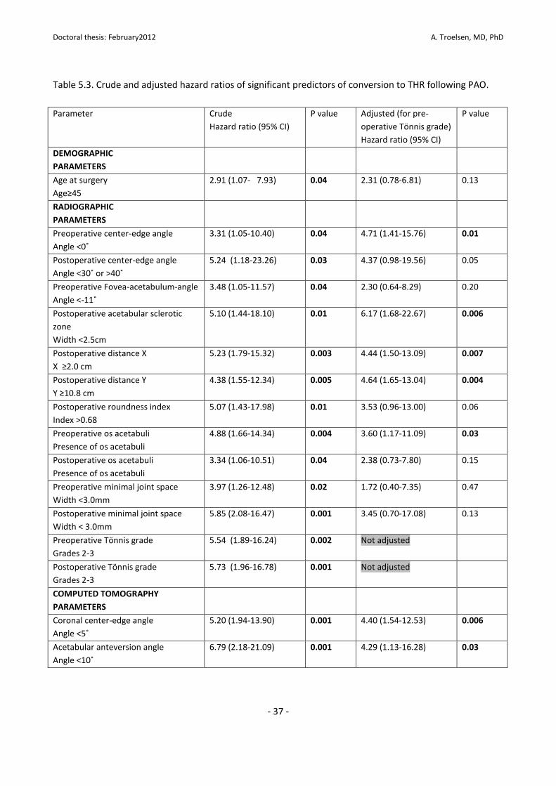
Doctoral thesis: February2012 A. Troelsen, MD, PhD
- 37 -
Table 5.3. Crude and adjusted hazard ratios of significant predictors of conversion to THR following PAO.
Parameter Crude
Hazard ratio (95% CI)
P value Adjusted (for pre-
operative Tönnis grade)
Hazard ratio (95% CI)
P value
DEMOGRAPHIC
PARAMETERS
Age at surgery
Age≥45
2.91 (1.07- 7.93) 0.04 2.31 (0.78-6.81) 0.13
RADIOGRAPHIC
PARAMETERS
Preoperative center-edge angle
Angle <0˚
3.31 (1.05-10.40) 0.04 4.71 (1.41-15.76) 0.01
Postoperative center-edge angle
Angle <30˚ or >40˚
5.24 (1.18-23.26)
0.03 4.37 (0.98-19.56) 0.05
Preoperative Fovea-acetabulum-angle
Angle <-11˚
3.48 (1.05-11.57) 0.04 2.30 (0.64-8.29) 0.20
Postoperative acetabular sclerotic
zone
Width <2.5cm
5.10 (1.44-18.10) 0.01 6.17 (1.68-22.67) 0.006
Postoperative distance X
X ≥2.0 cm
5.23 (1.79-15.32) 0.003 4.44 (1.50-13.09) 0.007
Postoperative distance Y
Y ≥10.8 cm
4.38 (1.55-12.34) 0.005 4.64 (1.65-13.04) 0.004
Postoperative roundness index
Index >0.68
5.07 (1.43-17.98) 0.01 3.53 (0.96-13.00) 0.06
Preoperative os acetabuli
Presence of os acetabuli
4.88 (1.66-14.34) 0.004 3.60 (1.17-11.09) 0.03
Postoperative os acetabuli
Presence of os acetabuli
3.34 (1.06-10.51) 0.04 2.38 (0.73-7.80) 0.15
Preoperative minimal joint space
Width <3.0mm
3.97 (1.26-12.48) 0.02 1.72 (0.40-7.35) 0.47
Postoperative minimal joint space
Width < 3.0mm
5.85 (2.08-16.47) 0.001 3.45 (0.70-17.08) 0.13
Preoperative Tönnis grade
Grades 2-3
5.54 (1.89-16.24)
0.002 Not adjusted
Postoperative Tönnis grade
Grades 2-3
5.73 (1.96-16.78) 0.001 Not adjusted
COMPUTED TOMOGRAPHY
PARAMETERS
Coronal center-edge angle
Angle <5˚
5.20 (1.94-13.90) 0.001 4.40 (1.54-12.53) 0.006
Acetabular anteversion angle
Angle <10˚
6.79 (2.18-21.09) 0.001 4.29 (1.13-16.28) 0.03

Doctoral thesis: February2012 A. Troelsen, MD, PhD
- 38 -
5.3 The role of labral tears in the surgical treatment of hip dysplasia.
The role of acetabular labral tears in the management of hip dysplasia remains a discussed and
controversial issue in PAO surgery. It is generally accepted that deformities of the hip, including hip
dysplasia, can cause FAI with repeated trauma to the acetabular labrum, subsequent labral tearing, and
cartilage degeneration (13,14,17-28,35-37,107-110,113,114). Studies have identified the presence of labral
tearing in up to 80% of symptomatic dysplastic hip joints, indicating that labral tearing is an important
factor in the development of pain and the risk of joint deterioration in hip dysplasia (115, IV).
The performance of arthrotomy to address labral tears during PAO surgery is inconsistently described in the
literature. A positive preoperative impingement test has been suggested by Steppacher et al. as a
significant predictor of failure following PAO surgery (73). They hypothesized that the presence of a
preoperative labral tear is the reason for the worsened prognosis and further suggested that performing
PAO with additional arthrotomy and intraarticular intervention may improve outcome (73). However,
drawing conclusions based on outcome of the impingement test is, as previously outlined, doubtful (V).
Peters et al. reported the outcome of PAO in 83 hips, with a mean follow-up of 46 months, and in 11 hips
with known labral tears, he reported an average Harris Hip score of 90 and no signs progressive
osteoarthritis at the most recent follow-up (102). Further, Matheney et al. were not able to find that the
presence of a labral tear predicted failure of the PAO in their multivariate regression analysis (74). The hip
joint survival rates reported in studies in which the joint and labrum were left untouched are encouraging
(140,141, VI). Arthroscopic treatment of labral tearing without addressing the hip dysplasia by means of a
PAO is controversial. Treatment may cause an adverse outcome (accelerated osteoarthritis), and results
beyond short-term follow-up are unknown (158).
Mechlenburg et al. (VII) measured cartilage thickness in the hip joint in 26 patients by using magnetic
resonance imaging and application of a stereologic method (159) before and up to 2½ years after PAO
surgery. Traction was applied to the leg undergoing magnetic resonance imaging to separate acetabular
from femoral cartilage. In addition, 18 patients underwent MRA of the operated hip to diagnose acetabular
labral tears. It was found that preoperative acetabular and femoral cartilage thicknesses were similar to
measurements 2½ years after surgery. At measurements 1 year postoperatively, the acetabular cartilage
was significantly, however marginal, thicker than at 2½ years postoperatively (1.47 mm vs. 1.35 mm).
Seventeen of 18 patients undergoing MRA had an acetabular labral tear.
Some methodological limitations should be acknowledged (VII). The leg traction approach used during
magnetic resonance imaging may have led to an underestimation of mean cartilage thickness due to
difficulty in separating cartilages in the thickest central parts of the joint. Due to the randomization of
measurements used in this stereological method, small local areas with thinning over time may go
undetected, with only a marginal effect on mean cartilage thickness. This may be important because joint
deterioration may initially be characterized by local cartilage damage adjacent to a labral tear. There was
no mention of pre- and postoperative functional outcome measures to document the clinical effect of the
PAOs performed or of the correlation of outcomes with the findings of cartilage thickness and labral tears.
However, the report of pre- and postoperative visual analog scale pain scores documented the major relief
of hip pain. Magnetic resonance arthrographies were not performed preoperatively and therefore it cannot
Doctoral thesis: February2012 A. Troelsen, MD, PhD
- 39 -
be documented whether labral tears were present preoperatively. However, tears of the labrum have been
documented in up to 80% of dysplastic hip joints (115, IV).
If one acknowledges that small local areas with cartilage damage may go undetected, it appears that
cartilage thickness is preserved up to 2½ years following PAO surgery. None of the hips in the study had an
arthrotomy and labral intervention during PAO surgery, and knowing that 17 of 18 arthrographies detected
a labral tear, it seems that during short-term follow-up the presence of a labral tear does not accelerate
cartilage destruction.
In conclusion, no studies of sufficient methodological value are yet available to definitively clarify whether
arthrotomy and labral intervention should be performed or not. According to existing studies, both
approaches can be chosen. The redistribution and decrease in load forces together with the resulting relief
of overload on the acetabular rim thought to be caused by the PAO (15,54,72,148-150) may explain why
pain is relieved in defiance of the presence of a labral tear and why cartilage destruction is seemingly
prevented. Future prospective studies with thorough preoperative magnetic resonance-based diagnostics
of labral tearing and comparison of clinical and radiographic outcome are warranted.

Doctoral thesis: February2012 A. Troelsen, MD, PhD
- 40 -
6. Conclusions and perspectives
Awareness of the limitations and controversies of diagnostic assessment of hip joint deformities and
osteoarthritis are important because correct diagnosis has great implications for candidates for joint
preserving surgery. An extensive quantification of the variability of different methods for the assessment of
hip dysplasia and osteoarthritis was carried out (I). The suggestions made regarding assessment of hip
dysplasia have implications for all orthopedic surgeons and radiologists dealing with painful hips in young
adults. Evidence was given that in patients with hip dysplasia, pelvic tilt may differ between the supine and
weightbearing positions (II). Supported by other studies (88,89,91,93), this finding questions the use of
standardized neutral pelvic positioning during assessment of hip joint deformities because the AP
radiographic appearance of acetabular version is affected by the degree of pelvic tilt. In an evaluation of
acetabular version in dysplastic hip joints in weightbearing AP pelvic radiographs, acetabular retroversion
was seen in 33% of hips (III). Awareness of the possible importance of patient positioning and of the
frequent finding of acetabular retroversion in dysplastic hip joints is particularly important during
assessment of hip dysplasia and the planning and performance of a joint preserving PAO. Larger scale
studies recording both lateral and AP pelvic radiographs of both normally structured hips and hips with
deformities are needed to shed further light on the importance of patient positioning.
Tearing of the acetabular labrum has been identified as a key feature in the initiation of early osteoarthritic
development in hips with structural deformities (13,14,17-28,35-37,107-110,113,114). The role of
ultrasound and various clinical tests to diagnose acetabular labral tears was established (IV, V). After
overcoming an initial learning curve, ultrasound was showed to be highly reliable in diagnosing labral tears,
whereas only a positive finding with the impingement or FABER tests was reliable in identifying a labral
tear. Very little was previously known about the ability of these modalities to diagnose labral tears. It seems
that noninvasive and rapid ultrasound examination performed by the experienced examiner can potentially
alter the traditional diagnostic algorithm in which MRA remains the gold standard. However, further
investigations are needed to explore the full potential of ultrasound. As experienced by orthopedic
surgeons from around the world, clinical examination remains unreliable in diagnosing labral tears, even in
the hands of hip specialists (127). Because this situation may never improve, supplementary radiographic
examination is needed.
In line with the few other studies (72-74,131) reporting the medium- and long-term follow-up results after
PAO for the treatment of hip dysplasia, encouraging hip joint survival and clinical outcome were reported
(VI). The still small number of reports on the outcome beyond 5-year follow-up is contrasted by the wide
application of the PAO. Surgeons and patients are awaiting further consolidation of the so far encouraging
joint-preserving abilities of PAO, and various aspects, especially those perceived by the patient to be
important, are undergoing investigation or remain uninvestigated. The aim should be the performance of
truly prospective follow-up studies. The performed analysis of predictors of conversion to THR after PAO
documented the importance of various biomechanical and degenerative factors (VI). Knowledge about
factors predicting early conversion to THR has the potential to refine patient selection and to improve the
potentials of PAO. The previous documentation of such factors, beyond the negative influence of
preexisting hip joint osteoarthritis, was very limited. Cartilage thickness was documented to be preserved
up to 2½ years after PAO (VII). All but one hip joint had acetabular labral tears, thus indicating that the
presence of labral tears does not accelerate cartilage degeneration after PAO. However, the issue of labral

Doctoral thesis: February2012 A. Troelsen, MD, PhD
- 41 -
intervention during PAO remains highly controversial. Because final conclusions cannot be drawn from the
present insufficient knowledge base, a prospective, MRA-based, follow-up study assessing outcome of PAO
is being conducted.

Doctoral thesis: February2012 A. Troelsen, MD, PhD
- 42 -
References
1. Wiberg G. Studies on dysplastic acetabula and congenital subluxation of the hip. With special reference to the
complication of osteoarthritis. Acta Chir Scand 1939;83(suppl 58):5-135.
2. Stulberg, S. D. Acetabular dysplasia and development of osteoarthritis of hip. Harris, W. H. The hip. Proceedings of
the second open scientific meeting of the hip society. 82-93. 1974. St.Louis, C.V.Mosby.
3. Stulberg, S. D. Unrecognized childhood hip disease: a major cause of idiopathic osteoarthritis of the hip. Cordell, LD,
Harris, WH, Ramsey, PL, and MacEwen, GD. Proceedings of the third open scientific meeting of the hip society , 212-
228. 1975. St.Louis, C.V.Mosby.
4. Cooperman DR, Wallensten R, Stulberg SD. Acetabular dysplasia in the adult. Clin Orthop Relat Res 1983;175:79-85.
5. Hasegawa Y, Iwata H, Mizuno M, Genda E, Sato S, Miura T: The natural course of osteoarthritis of the hip due to
subluxation or acetabular dysplasia. Arch Orthop Trauma Surg 1992;111:187-191.
6. Murphy SB, Ganz R, Muller ME: The prognosis in untreated dysplasia of the hip. A study of radiographic factors that
predict the outcome. J Bone Joint Surg Am 1995;77:985-989.
7. Murray RO. The aetiology of primary osteoarthritis of the hip. Br J Radiol 1965;38:810-24.
8. Solomon L. Patterns of osteoarthritis of the hip. J Bone Joint Surg Br 1976;58:176-83.
9. Solomon L, Schnitzler CM, Browett JP. Osteoarthritis of the hip: the patient behind the disease. Ann Rheum Dis
1982;41:118-25.
10. Solomon L. Geographical and anatomical patterns of osteoarthritis. Br J Rheumatol 1984;23:177-80.
11. Harris WH. Primary osteoarthritis of the hip: a vanishing diagnosis. J Rheumatol 1983;Suppl 9:64.
12. Harris WH. Etiology of osteoarthritis of the hip. Clin Orthop Relat Res 1986;(213):20-33.
13. Ganz R, Parvizi J, Beck M, Leunig M, Nötzli H, Siebenrock KA. Femoroacetabular impingement: a cause for
osteoarthritis of the hip. Clin Orthop Relat Res 2003;(417):112-20.
14. Ganz R, Leunig M, Leunig-Ganz K, Harris WH. The etiology of osteoarthritis of the hip: an integrated mechanical
concept. Clin Orthop Relat Res 2008;466:264-72.
15. Ganz R, Klaue K, Vinh TS, Mast JW. A new periacetabular osteotomy for the treatment of hip dysplasias. Technique
and preliminary results. Clin Orthop Relat Res 1988;232:26-36.
16. Pitto RP, Klaue K, Ganz R, Ceppatelli S. Acetabular rim pathology secondary to congenital hip dysplasia in the adult.
A radiographic study. Chir Organi Mov. 1995;80:361-368.
17. Ganz R, Gill TJ, Gautier E, Ganz K, Krügel N, Berlemann U. Surgical dislocation of the adult hip a technique with full
access to the femoral head and acetabulum without the risk of avascular necrosis. J Bone Joint Surg Br 2001;83:1119-
24.
18. Ito K, Minka MA 2nd, Leunig M, Werlen S, Ganz R. Femoroacetabular impingement and the cam-effect. A MRI-
based quantitative anatomical study of the femoral head-neck offset. J Bone Joint Surg Br 2001;83:171-6.

Doctoral thesis: February2012 A. Troelsen, MD, PhD
- 43 -
19. Ito K, Leunig M, Ganz R. Histopathologic features of the acetabular labrum in femoroacetabular impingement. Clin
Orthop Relat Res 2004;(429):262-71.
20. Kassarjian A, Yoon LS, Belzile E, Connolly SA, Millis MB, Palmer WE. Triad of MR arthrographic findings in patients
with cam-type femoroacetabular impingement. Radiology 2005;236:588-92.
21. Jäger M, Wild A, Westhoff B, Krauspe R. Femoroacetabular impingement caused by a femoral osseous head-neck
bump deformity: clinical, radiological, and experimental results. J Orthop Sci 2004;9:256-63.
22. Beck M, Kalhor M, Leunig M, Ganz R. Hip morphology influences the pattern of damage to the acetabular cartilage:
femoroacetabular impingement as a cause of early osteoarthritis of the hip. J Bone Joint Surg Br 2005;87:1012-8.
23. Siebenrock KA, Wahab KH, Werlen S, Kalhor M, Leunig M, Ganz R. Abnormal extension of the femoral head
epiphysis as a cause of cam impingement. Clin Orthop Relat Res 2004;(418):54-60.
24. Wagner S, Hofstetter W, Chiquet M, Mainil-Varlet P, Stauffer E, Ganz R, Siebenrock KA. Early osteoarthritic
changes of human femoral head cartilage subsequent to femoro-acetabular impingement. Osteoarthritis Cartilage
2003;11:508-18.
25. Tannast M, Goricki D, Beck M, Murphy SB, Siebenrock KA. Hip damage occurs at the zone of femoroacetabular
impingement. Clin Orthop Relat Res 2008;466:273-80.
26. Leunig M, Beck M, Kalhor M, Kim YJ, Werlen S, Ganz R. Fibrocystic changes at anterosuperior femoral neck:
prevalence in hips with femoroacetabular impingement. Radiology 2005;236:237-46.
27. Leunig M, Beck M, Woo A, Dora C, Kerboull M, Ganz R. Acetabular rim degeneration: a constant finding in the aged
hip. Clin Orthop Relat Res 2003;(413):201-7.
28. Pfirrmann CW, Mengiardi B, Dora C, Kalberer F, Zanetti M, Hodler J. Cam and pincer femoroacetabular
impingement: characteristic MR arthrographic findings in 50 patients. Radiology 2006;240:778-85.
29. Harris WH. The correlation between minor or unrecognized developmental deformities and the development of
osteoarthritis of the hip. Instr Course Lect 2009;58:257-9.
30. Nakamura S, Yorikawa J, Otsuka K, Takeshita K, Harasawa A, Matsushita T. Evaluation of acetabular dysplasia using
a top view of the hip on three-dimensional CT. J Orthop Sci. 2000;5:533-539.
31. Anda S, Terjesen T, Kvistad KA, Svenningsen S. Acetabular angles and femoral anteversion in dysplastic hips in
adults: CT investigation. J Comput Assist Tomogr. 1991;15:115-120.
32. Noble P, Kamaric E, Sugano N, Matsubara M, Harada Y, Ohzono K, Paravic V. Three-dimensional shape of the
dysplastic femur: Implications for THR. Clin Orthop Relat Res. 2003;417:27-40.
33. Jacobsen S, Rømer L, Søballe K. The other hip in unilateral hip dysplasia. Clin Orthop Relat Res. 2006;446:239-246.
34. Jacobsen S, Rømer L, Søballe K. Degeneration in dysplastic hips. A computer tomography study. Skeletal Radiol.
2005;7:1-7.
35. Klaue K, Durnin CW, Ganz R. The acetabular rim syndrome. A clinical presentation of dysplasia of the hip. J Bone
Joint Surg Br. 1991;73:423-429.

Doctoral thesis: February2012 A. Troelsen, MD, PhD
- 44 -
36. Byrd JW, Jones KS. Hip arthroscopy in the presence of dysplasia. Arthroscopy 2003;19:1055-60.
37. Jacobsen S. Adult hip dysplasia and osteoarthritis. Studies in radiology and clinical epidemiology. Acta Orthop.
2006;77(Suppl 324):1-37.
38. Mavcic B, Pompe B, Antolic V, Daniel M, Iglic A, Kralj-Iglic V. Mathematical estimation of stress distribution in
normal and dysplastic human hips. J Orthop Res. 2002;20:1025-30.
39. Russell ME, Shivanna KH, Grosland NM, Pedersen DR. Cartilage contact pressure elevations in dysplastic hips: a
chronic overload model. J Orthop Surg. 2006;1:6.
40. Mavcic B, Iglic A, Kralj-Iglic V, Brand RA, Vengust R. Cumulative hip contact stress predicts osteoarthritis in DDH.
Clin Orthop Relat Res 2008;466:884-91.
41. Mast JW, Brunner RL, Zebrack J. Recognizing acetabular version in the radiographic presentation of hip dysplasia.
Clin Orthop Relat Res 2004;418:48-53.
42. Nehme A, Trousdale R, Tannous Z, Maalouf G, Puget J, Telmont N. Developmental dysplasia of the hip: is
acetabular retroversion a crucial factor? Orthop Traumatol Surg Res 2009;95:511-9.
43. Giori NJ, Trousdale RT. Acetabular retroversion is associated with osteoarthritis of the hip. Clin Orthop Relat Res
2003;417:263-9.
44. Kim WY, Hutchinson CE, Andrew JG, Allen PD. The relationship between acetabular retroversion and osteoarthritis
of the hip. J Bone Joint Surg Br 2006;88:727-9.
45. Croft P, Cooper C, Wickham C, Coggon D. Osteoarthritis of the hip and acetabular dysplasia. Ann Rheum Dis.
1991;50:308-310.
46. Smith RW, Egger P, Coggon D, Cawley MI, Cooper C. Osteoarthritis of the hip joint and acetabular dysplasia in
women. Ann Rheum Dis. 1995;54:179-181.
47. Jacobsen S, Sonne-Holm S. Hip dysplasia: a significant risk factor for the development of hip osteoarthritis. A cross-
sectional survey. Rheumatology (Oxford). 2005;44:211-218.
48. Goodman DA, Feighan JE, Smith AD, Latimer B, Buly RL, Cooperman DR. Subclinical slipped capital femoral
epiphysis. Relationship to osteoarthrosis of the hip. J Bone Joint Surg Am 1997;79:1489-1497.
49. Burnett RS, Della Rocca GJ, Prather H, Curry M, Maloney WJ, Clohisy JC. Clinical presentation of patients with tears
of the acetabular labrum. J Bone Joint Surg Am 2006;88:1448-57.
50. Fitzgerald RH. Acetabular labrum tears. Diagnosis and treatment. Clin Orthop Relat Res 1995;311:60-68.
51. Narvani AA, Tsiridis E, Kendall S, Chaudhuri R, Thomas P. A preliminary report on prevalence of acetabular labrum
tears in sports patients with groin pain. Knee Surg Sports Traumatol Arthrosc 2003;11:403-8.
52. Gosvig K, Jacobsen S, Sonne-Holm S, Palm H, Troelsen A. Prevalence of malformations of the hip joint and their
relationship to sex, groin Pain and the risk of osteoarthritis: a population-based survey. J Bone Joint Surg Am
2010;92:1162-1169.

Doctoral thesis: February2012 A. Troelsen, MD, PhD
- 45 -
53. Gosvig KK, Jacobsen S, Palm H, Sonne-Holm S, Magnusson E. A new radiological index for assessing asphericity of
the femoral head in cam impingement. J Bone Joint Surg Br 2007;89:1309-1316.
54. Maeyama A, Naito M, Moriyama S, Yoshimura I. Periacetabular osteotomy reduces the dynamic instability of
dysplastic hips. J Bone Joint Surg Br 2009;91:1438-42.
55. Nötzli HP, Wyss TF, Stoecklin CH, Schmid MR, Treiber K, Hodler J. The contour of the femoral head-neck junction as
a predictor for the risk of anterior impingement. J Bone Joint Surg Br 2002;84:556-60.
56. Wenger DE, Kendell KR, Miner MR, Trousdale RT. Acetabular labral tears rarely occur in the absence of bony
abnormalities. Clin Orthop Relat Res 2004;426:145-150.
57. Peelle MW, Della Rocca GJ, Maloney WJ, Curry MC, Clohisy JC. Acetabular and femoral radiographic abnormalities
associated with labral tears. Clin Orthop Relat Res 2005;441:327-33.
58. Guevara CJ, Pietrobon R, Carothers JT, Olson SA, Vail TP. Comprehensive morphologic evaluation of the hip in
patients with symptomatic labral tear. Clin Orthop Relat Res 2006;453:277-85.
59. Jacobsen S, Sonne-Holm S, Soballe K, Gebuhr P, Lund B. Hip dysplasia and osteoarthrosis: a survey of 4151 subjects
from the Osteoarthrosis Substudy of the Copenhagen City Heart Study. Acta Orthop 2005;76:149-158.
60. Reijman M, Hazes JM, Pols HA, Koes BW, Bierma-Zeinstra SM. Acetabular dysplasia predicts incident osteoarthritis
of the hip: the Rotterdam study. Arthritis Rheum 2005;52:787-93.
61. Lane NE, Lin P, Christiansen L, Gore LR, Williams EN, Hochberg MC, Nevitt MC. Association of mild acetabular
dysplasia with an increased risk of incident hip osteoarthritis in elderly white women: the study of osteoporotic
fractures. Arthritis Rheum 2000;43:400-4.
62. Jessel RH, Zurakowski D, Zilkens C, Burstein D, Gray ML, Kim YJ. Radiographic and patient factors associated with
pre-radiographic osteoarthritis in hip dysplasia. J Bone Joint Surg Am 2009;91:1120-9.
63. The Danish Hip Replacement Register. The Danish Hip Replacement Register. Department of Orthopedic surgery,
University Hospital of Aarhus, Denmark. 2004;1-49.
64. Ecker TM, Tannast M, Puls M, Siebenrock KA, Murphy SB. Pathomorphologic alterations predict presence or
absence of hip osteoarthrosis. Clin Orthop Relat Res 2007;465:46-52.
65. Barros HJ, Camanho GL, Bernabé AC, Rodrigues MB, Leme LE. Femoral head-neck junction deformity is related to
osteoarthritis of the hip. Clin Orthop Relat Res 2010;468:1920-5.
66. Tanzer M, Noiseux N. Osseous abnormalities and early osteoarthritis: the role of hip impingement. Clin Orthop
Relat Res 2004;429:170-7.
67. Clohisy JC, Schutz AL, St John L, Schoenecker PL, Wright RW. Periacetabular osteotomy: a systematic literature
review. Clin Orthop Relat Res 2009;467:2041-52.
68. Tönnis D. Congenital Dysplasia and Dislocation of the Hip in Children and Adults. Berlin, Heidelberg, New York:
Springer; 1987.
69. Sharp I. Acetabular dysplasia. J Bone Joint Surg Br. 1961;43:268-272.
Doctoral thesis: February2012 A. Troelsen, MD, PhD
- 46 -
70. Heyman CH, Herndon CH. Legg-Perthes Disease. A method for the measurement of the roentgenographic result. J
Bone Joint Surg Am. 1950;32:767-778.
71. Goodman SB. Comparison of radiographic parameters for analysis of normal and dysplastic hips in the adult.
Contemp Orthop 1990;20:505-511.
72. Kralj M, Mavcic B, Antolic V, Iglic A, Kralj-Iglic V. The Bernese periacetabular osteotomy: clinical, radiographic and
mechanical 7-15-year follow-up of 26 hips. Acta Orthop 2005;76:833-840.
73. Steppacher SD, Tannast M, Ganz R, Siebenrock KA. Mean 20-year follow-up of Bernese periacetabular osteotomy.
Clin Orthop Relat Res 2008;466:1633-1644.
74. Matheney T, Kim YJ, Zurakowski D, Matero C, Millis M. Intermediate to long-term results following the Bernese
periacetabular osteotomy and predictors of clinical outcome. J Bone Joint Surg Am 2009;91:2113-23.
75. Troelsen A, Elmengaard B, Rømer L, Søballe K. Reliable Angle Assessment During Periacetabular Osteotomy with a
Novel Device. Clin Orthop Relat Res 2008;466:1169-1176.
76. Clohisy JC, Carlisle JC, Trousdale R, Kim YJ, Beaule PE, Morgan P, Steger-May K, Schoenecker PL, Millis M.
Radiographic evaluation of the hip has limited reliability. Clin Orthop Relat Res 2009;467:666-75.
77. Nelitz M, Guenther KP, Gunkel S, Puhl W. Reliability of radiological measurements in the assessment of hip
dysplasia in adults. Br J Radiol 1999;72:331-4.
78. Omeroglu H, Biçimoglu A, Aguş H, Tümer Y. Measurement of center-edge angle in developmental dysplasia of the
hip: a comparison of two methods in patients under 20 years of age. Skeletal Radiol 2002;31:25-9.
79. Portinaro NM, Murray DW, Bhullar TP, Benson MK. Errors in measurement of acetabular index. J Pediatr Orthop
1995;15:780-4.
80. Ball F, Kommenda K. Sources of error in the roentgen evaluation of the hip in infancy. Ann Radiol 1968;11:298-303.
81. Jacobsen S, Sonne-Holm S, Lund B, Søballe K, Kiær T, Rovsing H, Monrad H. Pelvic orientation and assessment of
hip dysplasia in adults. Acta Orthop 2004;75:721-729.
82. Siebenrock K, Kalbermatten D, Ganz R. Effect of pelvic tilt on acetabular retroversion: a study of pelves from
cadavers. Clin Orthop Relat Res 2003;407:241-8.
83. Ezoe M, Naito M, Inoue T. The prevalence of acetabular retroversion among various disorders of the hip. J Bone
Joint Surg Am 2006;88:372-9.
84. Kiyama T, Naito M, Shiramizu K, Shinoda T. Postoperative acetabular retroversion causes posterior osteoarthritis
of the hip. Int Orthop 2009;33:625-31.
85. Xie J, Naito M, Maeyama A. Evaluation of acetabular versions after a curved periacetabular osteotomy for
dysplastic hips. Int Orthop 2010;34:473-7.
86. Fujii M, Nakashima Y, Yamamoto T, Mawatari T, Motomura G, Matsushita A, Matsuda S, Jingushi S, Iwamoto Y.
Acetabular retroversion in developmental dysplasia of the hip. J Bone Joint Surg Am 2010;92:895-903.
87. Anda S, Svenningsen S, Grøntvedt T, Benum P. Pelvic inclination and spatial orientation of the acetabulum. A
radiographic, computed tomographic and clinical investigation. Acta Radiol 1990;31:389-94.

Doctoral thesis: February2012 A. Troelsen, MD, PhD
- 47 -
88. Konishi N, Mieno N, Mieno T. Determination of acetabular coverage of the femoral head with use of a single
anteroposterior radiograph. A new computerized technique. J Bone Joint Surg Am 1993;75:1318-1333.
89. Eddine TA, Migaud H, Chantelot C, Cotten A, Fontaine C, Duquennoy A. Variations of pelvic anteversion in the lying
and standing positions: analysis of 24 control subjects and implications for CT measurement of position of a prosthetic
cup. Surg Radiol Anat 2001;23:105-10.
90. Nishihara S, Sugano N, Nishii T, Ohzono K, Yoshikawa H. Measurements of pelvic flexion angle using three-
dimensional computed tomography. Clin Orthop Relat Res 2003;411:140-151.
91. Lembeck B, Mueller O, Reize P, Wuelker N. Pelvic tilt makes acetabular cup navigation inaccurate. Acta Orthop
2005;76:517-23.
92. Mayr E, Kessler O, Prassl A, Rachbauer F, Krismer M, Nogler M. The frontal pelvic plane provides a valid reference
system for implantation of the acetabular cup: spatial orientation of the pelvis in different positions. Acta Orthop
2005;76:848-53.
93. Babisch JW, Layher F, Amiot LP. The rationale for tilt-adjusted acetabular cup navigation. J Bone Joint Surg Am
2008;90:357-65.
94. Fuchs-Winkelmann S, Peterlein CD, Tibesku CO, Weinstein SL. Comparison of pelvic radiographs in weightbearing
and supine positions. Clin Orthop Relat Res 2008;466:809-12.
95. Altman RD, Bloch DA, Dougados M, Hochberg M, Lohmander S, Pavelka K, Spector T, Vignon E. Measurement of
structural progression in osteoarthritis of the hip: the Barcelona consensus group. Osteoarthritis Cartilage
2004;12:515-524.
96. Conrozier T, Lequesne MG, Tron AM, Mathieu P, Berdah L, Vignon E. The effects of position on the radiographic
joint space in osteoarthritis of the hip. Osteoarthritis Cartilage 1997;5:17-22.
97. Tannast M, Murphy S, Langlotz F, Anderson S, Siebenrock K. Estimation of pelvic tilt on anteroposterior x-rays—a
comparison of six parameters. Skeletal Radiol 2006;35:149-55.
98. Reynolds D, Lucas J, Klaue K. Retroversion of the acetabulum. A cause of hip pain. J Bone Joint Surg Br
1999;81:281-288.
99. Jamali AA, Mladenov K, Meyer DC, Martinez A, Beck M, Ganz R, Leunig M. Anterior pelvic radiographs to assess
acetabular retroversion: high validity of the ‘cross-over-sign.’ J Orthop Res 2007;25:758-765.
100. Anda S, Terjesen T, Kvistad KA. Computed tomography measurements of the acetabulum in adult dysplastic hips:
which level is appropriate? Skeletal Radiol 1991;20:267-71.
101. Li PL, Ganz R. Morphologic features of congenital acetabular dysplasia: one in six is retroverted. Clin Orthop Relat
Res 2003;416:245-53.
102. Peters CL, Erickson JA, Hines JL. Early results of the Bernese periacetabular osteotomy: the learning curve at an
academic medical center. J Bone Joint Surg Am 2006;88:1920-1926.
103. Kalberer F, Sierra RJ, Madan SS, Ganz R, Leunig M. Ischial spine projection into the pelvis : a new sign for
acetabular retroversion. Clin Orthop Relat Res 2008;466:677-83.

Doctoral thesis: February2012 A. Troelsen, MD, PhD
- 48 -
104. Kappe T, Kocak T, Neuerburg C, Lippacher S, Bieger R, Reichel H. Reliability of radiographic signs for acetabular
retroversion. Int Orthop 2010 May 10. [Epub ahead of print]
105. Bland JM, Altman DG. Statistical methods for assessing agreement between two methods of clinical
measurement. Lancet 1986;1:307-310.
106. Bland JM, Altman DG. Applying the right statistics: analyses of measurement studies. Ultrasound Obstet Gynecol
2003;22:85-93.
107. Ferguson SJ, Bryant JT, Ganz R, Ito K. The influence of the acetabular labrum on hip joint cartilage consolidation: a
poroelastic finite element model. J Biomech 2000;33:953-60.
108. Ferguson SJ, Bryant JT, Ganz R, Ito K. An in vitro investigation of acetabular labral seal in hip joint mechanics. J
Biomech 2003;36:171-8.
109. McCarthy JC, Noble PC, Schuck MR, Wright J, Lee J. The role of labral lesions to development of early
degenerative hip disease. Clin Orthop Relat Res 2001;393:25-37.
110. McCarthy JC, Noble PC, Schuck MR, Wright J, Lee J. The watershed labral lesion: its relationship to early arthritis
of the hip. J Arthroplasty 2001;16 Suppl.1:81-7.
111. Dorrell JH, Catterall A. The torn acetabular labrum. J Bone Joint Surg Br 1986;68:400-3.
112. Crawford MJ, Dy CJ, Alexander JW, Thompson M, Schroder SJ, Vega CE, Patel RV, Miller AR, McCarthy JC, Lowe
WR, Noble PC. The 2007 Frank Stinchfield Award. The biomechanics of the hip labrum and the stability of the hip. Clin
Orthop Relat Res 2007;465:16-22.
113. McCarthy JC, Lee JA: Acetabular dysplasia: a paradigm of arthroscopic examination of chondral injuries. Clin
Orthop Relat Res 2002;405:122-128.
114. Maeyama A, Naito M, Moriyama S, Yoshimura I. Evaluation of dynamic instability of the dysplastic hip with use of
triaxial accelerometry. J Bone Joint Surg Am 2008;90:85-92.
115. Leunig M, Podeszwa D, Beck M, Werlen S, Ganz R. Magnetic resonance arthrography of labral disorders in hips
with dysplasia and impingement. Clin Orthop Relat Res 2004;418:74-80.
116. Cashin M, Uhthoff H, O'Neill M, Beaulé PE. Embryology of the acetabular labral-chondral complex. J Bone Joint
Surg Br 2008;90:1019-24.
117. Toomayan GA, Holman WR, Major NM, Kozlowicz SM, Vail TP. Sensitivity of MR arthrography in the evaluation of
acetabular labral tears. Am J Roentgenol 2006;186:449-53.
118. Chan YS, Lien LC, Hsu HL, Wan YL, Lee MS, Hsu KY et al. Evaluating hip labral tears using magnetic resonance
arthrography: a prospective study comparing hip arthroscopy and magnetic resonance arthrography diagnosis.
Arthroscopy 2005;21:1250.
119. Freedman BA, Potter BK, Dinauer PA, Giuliani JR, Kuklo TR, Murphy KP. Prognostic value of magnetic resonance
arthrography for Czerny stage II and III acetabular labral tears. Arthroscopy 2006;22:742-7.
120. Ziegert AJ, Blankenbaker DG, De Smet AA, Keene JS, Shinki K, Fine JP. Comparison of standard hip MR
arthrographic imaging planes and sequences for detection of arthroscopically proven labral tear. AJR Am J Roentgenol
2009;192:1397-400.

Doctoral thesis: February2012 A. Troelsen, MD, PhD
- 49 -
121. Czerny C, Hofmann S, Neuhold A, Tschauner C, Engel A, Recht MP et al. Lesions of the acetabular labrum:
accuracy of MR imaging and MR arthrography in detection and staging. Radiology 1996;200:225-30.
122. Czerny C, Hofmann S, Urban M, Tschauner C, Neuhold A, Pretterklieber M et al. MR arthrography of the adult
acetabular capsular-labral complex: correlation with surgery and anatomy. Am J Roentgenol 1999;173:345-9.
123. Keeney JA, Peelle MW, Jackson J, Rubin D, Maloney WJ, Clohisy JC. Magnetic resonance arthrography versus
arthroscopy in the evaluation of articular hip pathology. Clin Orthop Relat Res 2004;429:163-9.
124. Leunig M, Werlen S, Ungersbock A, Ito K, Ganz R. Evaluation of the acetabular labrum by MR arthrography. J
Bone Joint Surg Br 1997;79:230-4.
125. Mitchell B, McCrory P, Brukner P, O'Donnell J, Colson E, Howells R. Hip joint pathology: clinical presentation and
correlation between magnetic resonance arthrography, ultrasound, and arthroscopic findings in 25 consecutive cases.
Clin J Sport Med 2003;13:152-6.
126. Sofka CM, Adler RS, Danon MA. Sonography of the acetabular labrum: visualization of labral injuries during intra-
articular injections. J Ultrasound Med 2006;25:1321-26.
127. Martin RL, Kelly BT, Leunig M, Martin HD, Mohtadi NG, Philippon MJ, Sekiya JK, Safran MR. Reliability of clinical
diagnosis in intraarticular hip diseases. Knee Surg Sports Traumatol Arthrosc 2010;18:685-90.
128. Martin RL, Sekiya JK. The interrater reliability of 4 clinical tests used to assess individuals with musculoskeletal hip
pain. J Orthop Sports Phys Ther 2008;38:71-7.
129. Clohisy JC, Beaulé PE, O'Malley A, Safran MR, Schoenecker P. AOA symposium. Hip disease in the young adult:
current concepts of etiology and surgical treatment. J Bone Joint Surg Am 2008;90:2267-81.
130. Troelsen A, Rømer L, Søballe K. Hip dysplasia: clinical assessment, radiologic evaluation and reference. Ugeskr
Laeger 2007;169:394-6.
131. Siebenrock KA, Scholl E, Lottenbach M, Ganz R. Bernese periacetabular osteotomy. Clin Orthop Relat Res
1999;363:9-20.
132. Trousdale RT, Ekkernkamp A, Ganz R, Wallrichs SL. Periacetabular and intertrochanteric osteotomy for the
treatment of osteoarthrosis in dysplastic hips. J Bone Joint Surg Am 1995;77:73-85.
133. Trumble SJ, Mayo KA, Mast JW. The periacetabular osteotomy. Minimum 2 year followup in more than 100 hips.
Clin Orthop Relat Res 1999;363:54-63.
134. Murphy SB, Millis MB. Periacetabular osteotomy without abductor dissection using direct anterior exposure. Clin
Orthop Relat Res 1999;364:92-98.
135. Clohisy J, Barrett S, Gordon J, Delgado E, Schoenecker P. Periacetabular osteotomy for the treatment of severe
acetabular dysplasia. J Bone Joint Surg Am 2005;87:254-259.
136. Crockarell J Jr, Trousdale RT, Cabanela ME et al. Early experience and results with the periacetabular osteotomy.
The Mayo Clinic experience. Clin Orthop Relat Res 1999;363:45-53.
137. Matta JM, Stover MD, Siebenrock K. Periacetabular osteotomy through the Smith-Petersen approach. Clin Orthop
Relat Res 1999;363:21-32.

Doctoral thesis: February2012 A. Troelsen, MD, PhD
- 50 -
138. Pogliacomi F, Stark A, Wallensten R. Periacetabular osteotomy. Good pain relief in symptomatic hip dysplasia, 32
patients followed for 4 years. Acta Orthop Scand 2005;76:67-74.
139. Garras DN, Crowder TT, Olson SA. Medium-term results of the Bernese periacetabular osteotomy in the
treatment of symptomatic developmental dysplasia of the hip. J Bone Joint Surg Br 2007;89:721-724.
140. Troelsen A, Elmengaard B, Søballe K. A new minimally invasive transsartorial approach for periacetabular
osteotomy. J Bone Joint Surg Am 2008;90:493-498.
141. Troelsen A, Elmengaard B, Søballe K. Comparison of the minimally invasive and ilioinguinal approaches for
periacetabular osteotomy: 263 single-surgeon procedures in well-defined study groups. Acta Orthop 2008;79:777-84.
142. Millis MB, Kain M, Sierra R, Trousdale R, Taunton MJ, Kim YJ, Rosenfeld SB, Kamath G, Schoenecker P, Clohisy JC.
Periacetabular osteotomy for acetabular dysplasia in patients older than 40 years: a preliminary study. Clin Orthop
Relat Res 2009;467:2228-34.
143. Cunningham T, Jessel R, Zurakowski D et al. Delayed gadolinium-enhanced magnetic resonance imaging of
cartilage to predict early failure of Bernese periacetabular osteotomy for hip dysplasia. J Bone Joint Surg Am
2006;88:1540-8.
144. Biedermann R, Donnan L, Gabriel A, Wachter R, Krismer M, Behensky H. Complications and patient satisfaction
after periacetabular osteotomy. Int Orthop. 2007 [Epub ahead of print]. DOI 10.1007/s00264-007-0372-3
145. van Bergayk AB, Garbuz DS. Quality of life and sports-specific outcomes after Bernese periacetabular osteotomy.
J Bone Joint Surg Br 2002;84:339-43.
146. Troelsen A. Surgical advances in periacetabular osteotomy for treatment of hip dysplasia in adults. Acta Orthop
Suppl 2009;80(332):1-33.
147. Søballe K. Pelvic osteotomy for acetabular dysplasia. Acta Orthop Scand 2003;74:117-118.
148. Mechlenburg I, Nyengaard J, Rømer L, Søballe K. Changes in load-bearing area after Ganz periacetabular
osteotomy evaluated by multislice CT scanning and stereology. Acta Orthop Scand 2004;75:147-153.
149. Hipp JA, Sugano N, Millis NB, Murphy SB. Planning acetabular redirection osteotomies based on joint contact
pressures. Clin Orthop Relat Res 1999;364:134-143.
150. Trousdale RT, Cabanela ME. Lessons learned after more than 250 periacetabular osteotomies. Acta Orthop Scand
2003;74:119-126.
151. Bjørner JB, Damsgaard MT, Watt T, Bech P, Rasmussen NK, Kristensen TS, Modvig J, Thunedborg K. Danish
manual for SF-36. Copenhagen: Lif; 1997.
152. Bellamy N. WOMAC osteoarthritis index. User guide VIII. WOMAC® is a registered trademark in Canada of
Nicholas Bellamy.
153. Pedersen A, Johnsen S, Overgaard S, Søballe K, Sørensen HT, Lucht U. Registration in the Danish hip arthroplasty
registry: completeness of total hip arthroplasties and positive predictive value of registered diagnosis and
postoperative complications. Acta Orthop Scand 2004;75:434-441.
154. Murphy S, Deshmukh R. Periacetabular osteotomy: preoperative radiographic predictors of outcome. Clin Orthop
Relat Res 2002;405:168-174.

Doctoral thesis: February2012 A. Troelsen, MD, PhD
- 51 -
155. Sharifi E, Sharifi H, Morshed S et al. Cost-effectiveness analysis of periacetabular osteotomy. J Bone Joint Surg Am
2008;90:1447-1456.
156. Nötzli HP, Müller SM, Ganz R. The relationship between fovea capitis femoris and weight bearing area in the
normal and dysplastic hip in adults: a radiologic study. Z Orthop 2001;139:502-506.
157. Okano K, Enomoto H, Osaki M, Shindo H. Outcome of rotational acetabular osteotomy for early hip osteoarthritis
secondary to dysplasia related to femoral head shape. 49 hips followed for 10-17 years. Acta Orthopaedica
2008;79:12-17.
158. Parvizi J, Bican O, Bender B, Mortazavi SM, Purtill JJ, Erickson J, Peters C. Arthroscopy for labral tears in patients
with developmental dysplasia of the hip: a cautionary note. J Arthroplasty 2009;24(6 Suppl):110-3.
159. Mechlenburg I, Nyengaard JR, Gelineck J, Soballe K. Cartilage thickness in the hip joint measured by MRI and
stereology--a methodological study. Osteoarthritis Cartilage 2007;15:366-71.


![DOCTORAL THESIS [TESIS DOCTORAL] - UAM](https://img.pdfslide.us/doc/110x75/62162d5b17f9b23eb24b8a87/doctoral-thesis-tesis-doctoral-uam.jpg)