Embed Size (px)
Citation preview
IMMUNOPROPHYLAXIS OF TICK INFESTATION IN
BOVINE
Zakir Ali
2005-VA-146
A Thesis Submitted in the Partial Fulfillment of the Requirement for
the Degree
OF
DOCTOR OF PHILOSOPHY
IN
PARASITOLOGY
UNIVERSITY OF VETERINARY AND ANIMAL SCIENCES,
LAHORE
2010
CONTENTS
DEDICATION (i)
ACKNOWLEDGMENT (ii)
LIST OF TABLES (iv)
LIST OF FIGURES (v)
LIST OF ABBREVIATIONS (vi)
CHAPTER# NAME OF CHAPTER Page#
1. INTRODUCTION 01
2. REVIEW OF LITERATURE 05
1. Tick Infestation 05
1.1 Tick classification, structure and bionomics 05 1.2 Prevalence study 06 1.2.1 Ambient environmental conditions for ticks 06 1.2.2 Prevalence study in Pakistan 07 1.2.3 Prevalence study in other countries 08
1.3 Host susceptibility to tick infestation 09
1.4 Ticks as potential vectors of pathogens 10 1.4.1 Pathogens vector by ticks 10 1.4.2 Role of salivary gland in disease transmission 10 1.4.3 Detection of pathogens in ticks 11
1.5 Tick Infestation Based Economic Losses 13
2. Control of ticks 15 2.1 Chemotherapy 15 2.2 Immunotherapy 17
2.2.1 Antigen 17 2.2.2 Development of immunity 18
2.2.3 Commercial vaccines 19
2.2.4 Development of new vaccines 20
2. Growing intestinal cell as monolayer 21 3. Summary 21
3. MATERIALS AND METHODS 23 1. Collection and Morphological study of tick 23
2. Preparation of intestinal, salivary gland and whole homogenate tick vaccines and their inoculation into experimental animals 24
3. Monitoring sero-conversion of vaccinated rabbits and calves 30
3.1 AGPT 30
3.2 CFT 33
4. Monitoring capacity of Hyalomma whole homogenate tick vaccine in controlling tick infestation in crossbred animals by ELISA 39
5. Detection of T.annulata by PCR 43
6. Growing of intestinal epithelial cells of hard tick as monolayer 46
4. RESULTS 49
1. Tick prevalence in cattle and buffaloes 49
2. Preparation of different whole homogenate oil based vaccines 51
3. Monitoring sero-conversion of vaccinated rabbits and
calves 52
4. Monitoring efficacy of Hyalomma whole homogenate vaccine in Crossbred animals 55
5. Detection of T. annulata in hard ticks by PCR 58
6. Growing of intestinal epithelial cells of hard tick as
monolayer 61
5. DISCUSSION 62
1. Prevalence study 62
2. Detection of T. annulata in tick species by PCR 68
3. Tick susceptibility due to differences in breed, sex and age
of host 69
4. Effects on tick reproductive efficiency by immunization 70
5. Vaccination studies 74 5.1 Different tick organ tissues as candidate antigen 74 5.2 Cross-reactivity study 76 5.3 Dose of antigen 79
6. Growth of tick intestinal cell as monolayer 81
6. SUMMARY 82
7. LITERATURE CITED 84
8. APPENDICES 108

[i]

[ii]
I thank Almighty Allah for bestowing upon me the exhilarating opportunity, sense of
direction and courage to accomplish this work.
I owe a deep sense of obligation to Prof. Dr. Azhar Maqbool for his supervision, keen
interest, constant encouragement, understanding and guidance during the course of study.
I am thankful to Dr. Kamran Ashraf, Chairman, Department of Parasitology for his
participatory role in this writing.
I gratefully acknowledge invaluable help rendered by Prof. Dr. Khushi Muhammad,
Chairman, Department of Microbiology, during my research work.
I am also thankful to Prof. Dr. Muhammad Sarwar Khan for his constructive and valuable
guidance during the write up of this dissertation.
I am highly indebted to the following persons for their support and help during the
conduct of my research work.
Dr. Imran Altaf, Lecturer, WTO Quality Control Laboratory, UVAS, Lahore.
Dr. Imran Rashid, Assistant Professor, Department of Parasitology, UVAS,
Lahore.
[iii]
Dr. Anwar Hussain, Director PVTV, L & DD, Punjab.
Dr. Rana Abdul Rashid, Farm Superintendant, LES, Shergarh, Distt. Okara.
Dr. Munir, Veterinary Officer, Buffalo Research Institute, Pattoki.
Dr. Amjad, Veterinary Officer, Distt. Vehari.
Funds for this project were provided by Higher Education Commission, Pakistan under
the scheme of Indigenous Ph. D. fellowship (Batch 111) which is a great contribution in
promoting research and science in Pakistan. International Research Supportive Initiative
Program (IRSIP) was availed at Kansas State University, USA during this scheme which
is also funded by HEC.
I would like to thank tremendously to Dr. Roman Ganta who was mentor and advisor
during this externship training and helped a lot in learning molecular techniques and in
preparing this thesis.
DR. ZAKIR ALI
JUNE, 2010
[iv]
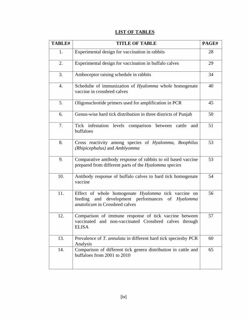
LIST OF TABLES
TABLE# TITLE OF TABLE PAGE#
1. Experimental design for vaccination in rabbits 28
2. Experimental design for vaccination in buffalo calves
29
3. Amboceptor raising schedule in rabbits 34
4. Schedulte of immunization of Hyalomma whole homogenate vaccine in crossbred calves
40
5. Oligonucleotide primers used for amplification in PCR
45
6. Genus-wise hard tick distribution in three districts of Punjab
50
7. Tick infestation levels comparison between cattle and buffaloes
51
8. Cross reactivity among species of Hyalomma, Boophilus (Rhipicephalus) and Amblyomma
53
9. Comparative antibody response of rabbits to oil based vaccine prepared from different parts of the Hyalomma species
53
10. Antibody response of buffalo calves to hard tick homogenate vaccine
54
11. Effect of whole homogenate Hyalomma tick vaccine on feeding and development performances of Hyalomma anatolicum in Crossbred calves
56
12. Comparison of immune response of tick vaccine between vaccinated and non-vaccinated Crossbred calves through ELISA
57
13. Prevalence of T. annulata in different hard tick speciesby PCR Analysis
60
14. Comparison of different tick genera distribution in cattle and buffaloes from 2001 to 2010
65
[v]
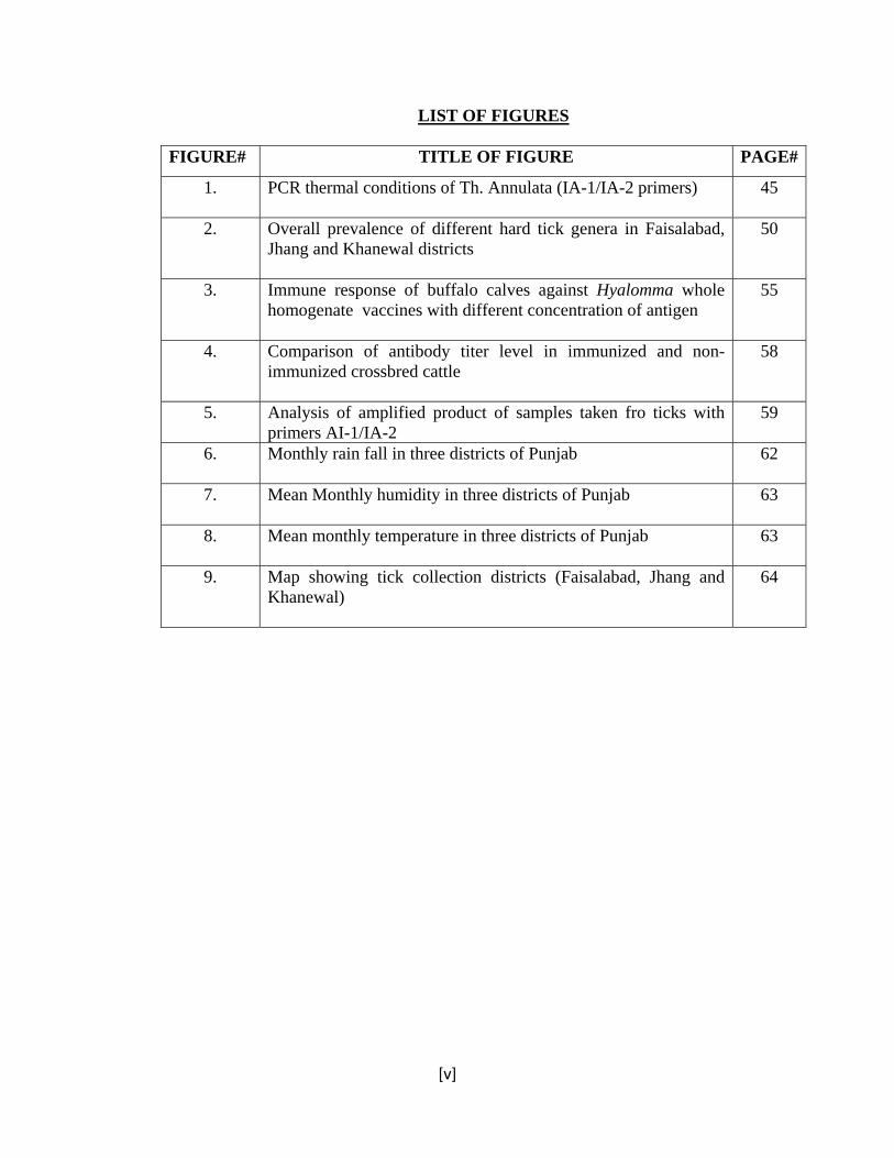
LIST OF FIGURES
FIGURE# TITLE OF FIGURE PAGE#
1. PCR thermal conditions of Th. Annulata (IA-1/IA-2 primers)
45
2. Overall prevalence of different hard tick genera in Faisalabad, Jhang and Khanewal districts
50
3. Immune response of buffalo calves against Hyalomma whole homogenate vaccines with different concentration of antigen
55
4. Comparison of antibody titer level in immunized and non-immunized crossbred cattle
58
5. Analysis of amplified product of samples taken fro ticks with primers AI-1/IA-2
59
6. Monthly rain fall in three districts of Punjab
62
7. Mean Monthly humidity in three districts of Punjab
63
8. Mean monthly temperature in three districts of Punjab
63
9. Map showing tick collection districts (Faisalabad, Jhang and Khanewal)
64
[vi]
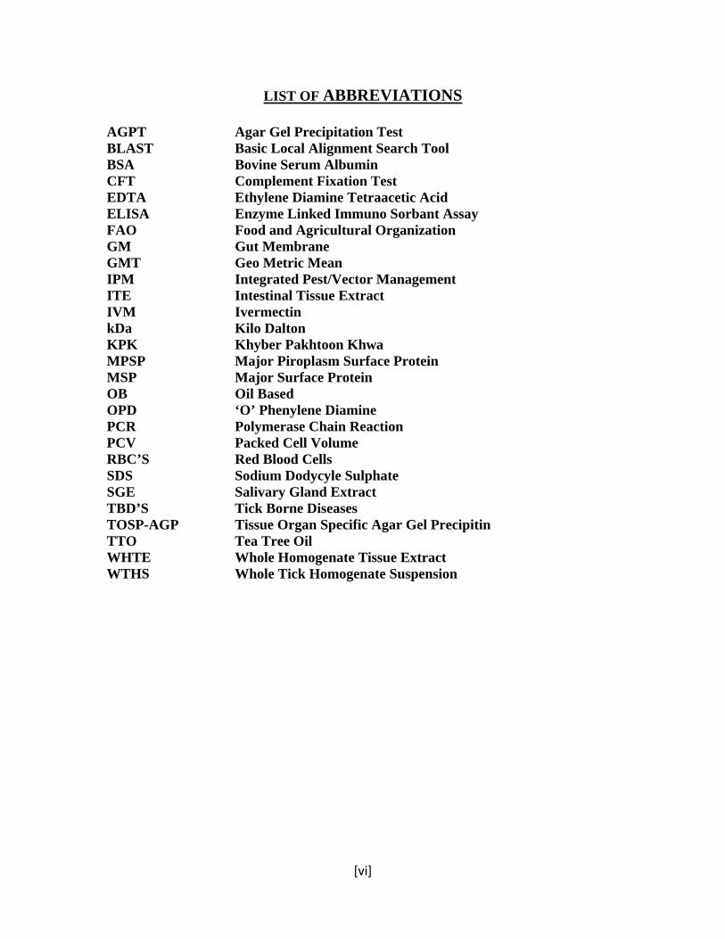
LIST OF ABBREVIATIONS
AGPT Agar Gel Precipitation Test BLAST Basic Local Alignment Search Tool BSA Bovine Serum Albumin CFT Complement Fixation Test EDTA Ethylene Diamine Tetraacetic Acid ELISA Enzyme Linked Immuno Sorbant Assay FAO Food and Agricultural Organization GM Gut Membrane GMT Geo Metric Mean IPM Integrated Pest/Vector Management ITE Intestinal Tissue Extract IVM Ivermectin kDa Kilo Dalton KPK Khyber Pakhtoon Khwa MPSP Major Piroplasm Surface Protein MSP Major Surface Protein OB Oil Based OPD ‘O’ Phenylene Diamine PCR Polymerase Chain Reaction PCV Packed Cell Volume RBC’S Red Blood Cells SDS Sodium Dodycyle Sulphate SGE Salivary Gland Extract TBD’S Tick Borne Diseases TOSP-AGP Tissue Organ Specific Agar Gel Precipitin TTO Tea Tree Oil WHTE Whole Homogenate Tissue Extract WTHS Whole Tick Homogenate Suspension

[vii]
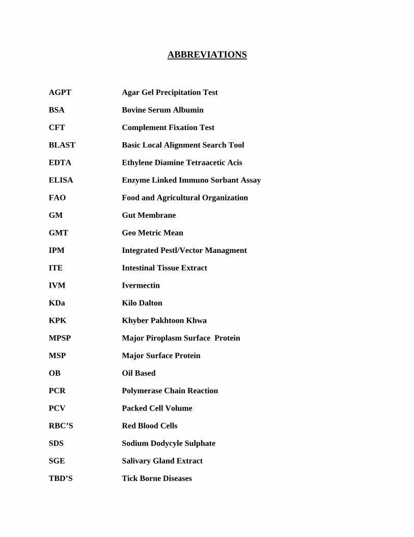
ABBREVIATIONS
AGPT Agar Gel Precipitation Test
BSA Bovine Serum Albumin
CFT Complement Fixation Test
BLAST Basic Local Alignment Search Tool
EDTA Ethylene Diamine Tetraacetic Acis
ELISA Enzyme Linked Immuno Sorbant Assay
FAO Food and Agricultural Organization
GM Gut Membrane
GMT Geo Metric Mean
IPM Integrated Pestl/Vector Managment
ITE Intestinal Tissue Extract
IVM Ivermectin
KDa Kilo Dalton
KPK Khyber Pakhtoon Khwa
MPSP Major Piroplasm Surface Protein
MSP Major Surface Protein
OB Oil Based
PCR Polymerase Chain Reaction
PCV Packed Cell Volume
RBC’S Red Blood Cells
SDS Sodium Dodycyle Sulphate
SGE Salivary Gland Extract
TBD’S Tick Borne Diseases

TOSP-AGP Tissue Organ Specific Agar Gel Precipitin
TTO Tea Tree Oil
WHTE Whole Homogenate Tissue Extract
WTHS Whole Tick Homogenate Suspension
�����������������������������������������������������������������������������������������������������������������������������������������������������������������������������������������������
[1]
CHPTER 1
INTRODUCTION
Tick infestation in livestock is a common problem in countries having hot and humid
environment. Exotic cattle breeds are more vulnerable to tick infestation as compared to local
stocks due to non-adaptive immune factors, grooming activity, skin color and thickness, area of
skin available for infestation or length of fur (Mattioli, 1998 and Meltzer, 1996). Tick infestation
not only reduces appetite and body condition of the host but also reduces the hide quality up to
20-30% in leather industry (Ghosh et al., 2007). Even ticks with smaller mouthparts like
Rhipicephalus (Boophilus) may cause similar losses when present in large number. Ticks with
long and massive mouthparts such as Amblyomma and Hyalomma may induce abscesses,
lameness and loss of teats in their hosts. The saliva of certain ticks like Dermacenter can cause
toxicosis and paralysis as their salivary secretions contain toxins (Jongejan and Uilenberg, 2004).
Tick infestation also causes severe anemia, loss of production, weakness and immuno-
suppression in cattle and buffaloes (Gwakisa et al., 2001). Moreover, it can cause transmission of
viral, bacterial and protozoan pathogens causing diseases like hemorrhagic fever, ehrlichiosis,
anaplasmosis, theileriosis and babesiosis in meat and dairy animals. It has been studied that
about 80% of the world cattle population is infested with ticks (Bowman et al., 1996) and Food
and Agricultural Organization (FAO)of the United Nation estimated the global cost of hard tick
infestation to be US $ 7.0 billion annually (Harrow et al., 1991). Proposed new agents producing
�����������������������������������������������������������������������������������������������������������������������������������������������������������������������������������������������
[2]
clinical signs are increasingly reported from ticks mainly by sequencing Poly Chain Reaction
products and doing comparison of these sequences by BLAST in GeneBank, (Telfor III and
Goethert, 2004).
Ticks belong to the order Acarina and class Arachnida. Two families of ticks exist, the hard ticks
(Ixodidae) with 670 species and soft ticks (Argicidae) about 150 species (Urquhart et al., 1996).
In Pakistan various species of ticks of the genera Rhipecephalus, Boophilus, Hyalomma,
Amblyomma and Hemaphysalis have been reported to infest the dairy and meat animals. Breed,
age, sex, environmental conditions, nutritional and lactation status of an animal, however, are the
factors that mainly determine the density of tick infestation (Sajid et al., 2008). The prevalence
of ticks has been found higher in May through August which is the hottest duration of the
summer in Pakistan. The most favorable sites for tick attachment are udder, ear, neck, inside of
thighs and vulvolar regions (Yakhachali et al., 2004)
Injectables, burning of pastures, selection of breeds with a higher natural resistance to tick
infestation and applications of acaricides are different methods for tick control typically used in
Pakistan. Application of acaricides is the most common way to control tick infestation which is
applied in the form of a spray, shower or spot on. This method is not sustainable due to
expensiveness, residual effect and development of resistance against many tick species (Makala
et al., 2003). The resistance to ticks is primarily an acquired rather than innate (Wagland, 1978).
Moreover acaricides cause environmental pollution and health hazards to persons applying
acaricides.
�����������������������������������������������������������������������������������������������������������������������������������������������������������������������������������������������
[3]
An important component of integrated pest/vector management (IPM) is immunoprophylaxis
(Ghoshe et al., 2007). Different types of vaccines have been developed throughout the world to
control tick infestation in cattle and buffaloes. They include whole tick lysate vaccine, tick
cement protein vaccine, and recombinant tick vaccine (Bishop et al., 2002). Progress in
developing suitable anti tick vaccines is slow primarily due to difficulty in identifying suitable
sources of antigens. Concealed as well as exposed antigens have been used to develop vaccine
against different tick species. Attachment of ticks to a vaccinated host results into mitigation of
engorgement weight and reproductive performance due to antibody binding with gut wall which
causes its lysis (Nuttal et al., 2004). Complete and well organized immunization regime may
promote success in controlling tick burden to dairy and meat animals.
Pertinent literature regarding immunoprophylaxis of tick infestation is scanty. This study had
therefore been designated with the following objectives.
1. Characterization of hard ticks collected from the area of study by observing its
morphological features under microscope.
2. Development and evaluation of a vaccine prepared from field isolates of hard ticks.
3. Hard ticks collected from infected animals may be contaminated with blood borne
parasites that might ultimately contaminate the tick vaccine. In this study, efforts
therefore were made to identify the presence of Theileria annulata using a PCR
technique in homogenized material of ticks. This parasite was chosen for the analysis
�����������������������������������������������������������������������������������������������������������������������������������������������������������������������������������������������
[4]
because it is the major parasite found in ticks and also has a significant impact on cross- breed
animals in Pakistan.
4. Efforts were also made to grow intestinal epithelial cells of hard ticks as a monolayer for
subsequent production of a vaccine.
������ ��������� ���
[5]
CHAPTER 2
REVIEW OF LITERATURE
1. Tick Infestation
1.1 Tick classification, structure and bionomics
Most of vertebrates living on earth are susceptible to tick infestation; their
warmth and odor are attractive to ticks (Harwood and James, 1979). Suborder Ixodidea and
subclass aracri (Arachnida) contain two tick families Ixodidae and Argasidae (Urquhart, 1996;
Soulsby, 2006 and Porto Neto et al., 2011). Ixodidae can be differentiated from Argasidae by
having a hard, chintinous shield or scutum. Scutum covers only the anterior part behind the
capitulum in immature and female Ixodes, while in mature male it covers the dorsum fully. All
tick species pass through four stages (egg, larva, nymph and adult) from six weeks to three years.
The larva hatched from egg is six-legged and remain in this condition until moults into nymph.
The nymph has four pair of legs and transforms into sexually matured adult. (Harwood and
James, 1979; Urquhart, 1996 and Soulsby, 2006). Female ticks can become greatly distended and
when fully engorged are bean shaped; they are 200 times greater in weight as compared to
unengorged (Harwood and James, 1979 and Urquhart, 1996).
Ixodes tick species that remain on the host during the two molting periods are known as one-host
ticks. In two host species, the molt to the nymphal stage occurs on the host but engorged nymph
������ ��������� ���
[6]
leaves the host, molts in the environment and then finds a new host. In the three host tick life
cycle, both the larvae and nymph leave the host to molt, attaching to host again after each molt
(Urquhart, 1996 ; Minjauw and McLeod, 2003and Zajac et al., 2006), while ticks of Argasidae
are free living (Jongejan and Uilenberg, 2004). ). Ixodes ticks lay eggs in batches, each batch
contains upto 18000 eggs and female dies after hatching (Harwood and James, 1979 and
Soulsby, 2006). In transtadial transmisstion, tick can transmit disease pathogens from one molt to
another while in transovarion transmission, adult female can transmit pathogens to larva through
infecation of ovaries (Bowman et al., 2003). There are almost 900 species of ticks that are
endemic to most continents (Barker and Murrell, 2004) and important are Hyalomma anatolicum
anatolicum, Boophilus (Rhipecephalus) microplus and Amblyomma americanum (Porto Neto et
al., 2011 and Atif et al., 2012). The highest mean pre-oviposition period was during spring while
it was the lowest in autumn; the mean oviposition period was also the highest in spring. The
incubation period of the ova of Hyalomma species varied in different seasons, no oviposition was
recorded at the temperature 100oC and 85% humidity. The maximum number of eggs was laid at
34oC and the lowest egg production occurs at 15oC. The maximum number of eggs hatches at
32oC and 85% humidity. The variation in relative humidity has no appreciable effect on rate of
development of ticks while the number of eggs laid increase with the rise in temperature
(Durrani and Shakoori, 2009).
1.2 Prevalence study
1.2.1 Ambient environmental conditions for ticks
The ability of Ixodes ticks to survive off the host is astounding and much of this time is spent in
an active state resembling to diapauses. Phenomenon of diapause is mostly found in temperate
������ ��������� ���
[7]
countries, it may also occur in tropical countries. The prevalence to ticks is more in areas with
higher humidity and main stress factor for their life is desiccation during off host period
(Marquardt et al., 2005). The highest tick infestation was recorded when mean temperature was
27oC and relative humidity as 85% in Rawalpind and Islamabad areas of Pakistan during month
of June-July (Rehman et al., 2004).
1.2.2 Prevalence study in Pakistan
Tick infestation study was conducted in cattle, buffaloes, goats and sheep in Rawalpindi and
Islamabad regions. It showed prevalence of five species namely, Haemaphysalis (H) sulcata, H.
anatolicum, H. anatolicum anatolicum, H. marginatum and Haemaphysalis (R) erinacei at 74%,
14%, 12%, 1% and at1%, respectively. They reported the prevalence of Haemaphysalis (H)
sulcata and H. anatolicum highly significant (Rehman et al., 2004).In Peshawar district of
Khyber Pakhtoon Khwa (KPK) province the hard tick prevalence was conducted in randomly
selected livestock farms. It was found that overall 13% of the observed farm animals were tick
infested showing the highest infestation in cattle (20%) followed by sheep (13%), goats (12%),
buffaloes (11%) and donkeys (six %). The highly prevalent ticks were Boophilus species (46%)
followed by Hyalomma species (31%), Rhipicephalus species (18%) and Amblyomma species
(five %). It was found that tick infestation was higher in late summer and lower in winter (Manan
et al. 2007). Similar study was conducted in Layyah and Muzaffargarh districts in population of
cattle, buffaloes, sheep, and goats. The cattle at 75% were found the highest followed by goats at
52% and buffaloes at 40%, respectively. No case of tick infestation was found in camels and
sheep. Hyalomma anatolicum was the species which was the most abundant followed by
������ ��������� ���
[8]
Rhipicephalus sanguineus (Sajid et al., 2008). A survey to study the prevalence of Hyalomma
species was conducted in Rawalpindi, Multan and Lahore districts of Punjab province in
Pakistan. In cattle genera wise study showed the highest prevalence (12%) of Hyalomma species
and the lowest prevalence (four %) of Rhipicephalus species. The results showed the highest
prevalence (67%) of ticks in district Lahore. A survey was conducted to study the prevalence of
Hyalomma species in Rawalpindi, Multan and Lahore districts of Punjab province in Pakistan
(Durrani and Shakoori, 2009). In Sargodha, Khushab and Rawalpindi districts of Punjab
province, overall prevalence of cattle tick infestation was 54.76%. The prevalence of Hyalomma
anatolicum anatolicum, Rhipicephalus (boophilus) microplus, Rhipicephalus (boophilus)
annulatus and Haemaphysalis species were reported in this study (Atif et al., 2012).
1.2.3 Prevalence study in other countries
In Oshnavich suburb, West Azerbaijan Ixodid tick distributions per animal were 5, 3-4, 4-5, 2-3
and 1-2 in cattle, calves, buffaloes, female buffaloes and sheep, respectively. The prevalence of
ticks was 44%, 41% and 47% in cattle, buffaloes and sheep, respectively. Two genera of ixodid
ticks were found in cattle, Hyalomma at (64%) and Rhipicephalus at (four %). Six species were
found in cattle, Hyalomma anatolicum excavatum (four %), H. anatolicum anatolicum (five %),
H. asiaticum asiaticum (16%), H. marginatum (four %), H. dromedarii (13%) and Rhipicephalus
bursa (four %). Hyalomma species (63%) and Rhipicephalus species (four %) were also present
in buffaloes. Five genera of ixodid ticks are found in sheep, Hyalomma species (two %), R.
species (23%), Haemaphysalis species (two %), Dermacentor species (27%) and Boophilus
species(two %) (Yakhachali et al., 2004). In Mazandran provine, Iran nine species were
������ ��������� ���
[9]
identified: Rhipicephalus (Boophilus) microplus (51.3%), Rhipicephalus bursa (16.8%),
Haemaphysalis punctata (6.3%), Ixodes ricinus (68%), Hyalomma anatolicum (12.5%),
Hyalomma anatolicum exacavatum (5.2%), Hyalomma asiaticum (0.6%), Hyalomma detrium
(0.2%) and Dermacenter species (0.1%). Boophilus (Rhipicephalus) annulata, Rhipicephalus
bursa and Hyalomma species were dominant in the study area (Razmi et al., 2007). In different
upazila of Chittagong District from a total number of 380 cattle were examined, of which 138
(36.31%) cattle were found infested. Three species of ticks were identified namely Boophilus
microplus, Rhipicephalus sanguineus and Haemaphysalis bispinosa. . Tick infestation was more
prevalent in local (43.82%) cattle than the cross-bred (24.13%) cattle (Kabir et al., 2011).
1.3 Host susceptibility to tick infestation
Bos indicus are naturally more resistant to tick infestation as compared to Bos Taurus. Less than
one % of ticks feed successfully on Bos indicus (local breed), while response of Sahiwal breed
against control measures was significant. In case of Bos taurus breeds more than 50% may feed
to repletion. Tick infestation effects differ considerable due to variation in susceptibility and
resistance to the host, which are also influenced by grooming and grazing behavior, the
difference in quality and quantity of grazing and the timing and abundance of tick burden (Latif
and Pegram, 1992). Taurine crosses with zebu were more vulnerable to ticks as compared to pure
zebu cattle under similar field conditions. This tick resistant in Zebu cattle may be due to
presence of significantly higher serums complement level in their blood as compared to
crossbred cattle (Wambura et al., 1998). It was documented the highest tick incidence in young
������ ��������� ���
[10]
calves at 48%, following growing stock at41%, heifer at 40% and adult cattle at 35% (Das,
1994).
1.4 Ticks as potential vectors of pathogens
1.4.1 Pathogens vector by ticks
The ecology and physiology of ticks have made them second most important vectors after
mosquitoes. Ticks transmit a large variety of intercellular bacteria in the Rickettsia group like
Rickettsia, Ehrlichia and Anaplasma. Similarly several piroplasm protozoa like T. annulata,
T.parva and Babesiosis bigemmina are also transmitted specifically by ticks (Marquardat et al.,
2005). Tropical theileriosis is disease of cattle and caused by Theileria annulata; the disease is
transmitted mainly by H. detritum and Hyalomma excavatum (Fesharki, 1988). Transmission of
Anaplasma marginale occurs by ticks, both male tick and host become persistently infected with
A.marginale and serve as reservoir of infection. As erythrocytes are major site of infection, A.
marginale undergoes developmental cycle in the tick. The cycle begins in gut cells and
transmission occurs to susceptible host through salivary glands (Kocan et al., 2010).
1.4.2 Role of salivary gland in disease transmission
Ticks like other haematophagus arthropods must cope with coagulation, platelet aggregation and
pain or itch responses for obtaining blood meal successfully (Ribeiero, 1995a). Salivary glands
are necessary for the biological success of ticks during off-feed period and on-host period. Ticks
return back to host about 70% of the fluid and ion components of blood meal during the
phenomenon of salivation. Moreover, they also indicated that the salivary glands are sites of
������ ��������� ���
[11]
pathogen development and ticks transmit different pathogens through saliva. The multi-
functionary activities of salivary glands for tick survival and vector potential portray the gland as
target for intervention (Bowman and Sauer, 2004). Theileria sporoblast was detected in salivary
glands of Hylomma detrium detrium. Ninteen percent (24/127) of Hylomma detrium detrium
were infected with theileria species. Among these infected ticks more than 50% had five or more
sporoblasts in their salivary glands (Flach and Ouheli, 1992).
1.4.3 Detection of pathogens in ticks
PCR analysis was performed to detect of Theileria annulata in Hyalomma ticks using primers
derived from a gene which code for 30kDa major merozoite surface antigen. It was found that
infection rate in the adult ticks is high at 62% which was decreased to zero for one day in tick
groups which were treated with Butalex TM and again increases to 30% two days later. Two
hundred two adult ticks were collected from low theileria parasitemic zebu cattle from
Mauritania and are tested for Theileria annulata by the PCR. In H. dromedarii 73% and 57%
infection rates were detected from Gorgol and Trarza regions, respectively. While in Gorgol
region seven percent Hyalomma marginatum rufipes are positive. These findings confirmed that
PCR analysis was a useful tool for determining the infections in ticks and that H. dromedarii was
the main vector of T. annulata in the area of study (D’Oliviera et al., 1997). Twenty-eight field
isolated theileria parasite DNAs were obtained from dairy and beef cattle in distinct geographical
areas of Thailand and characterized them by using polymerase chain reaction amplification with
six sets of oligonucleotide primers. Three sets of them are modified from two genes of
immunodominant major piroplasm surface protein (MPSP) coding for 32kDa (p32) of T. sergenti
������ ��������� ���
[12]
and 33/34kDa (p33/34) of T. buffeli, and MPSP of Theileria species (Thai-isolate). The other
three sets of primers were basically generated from three alleles of MPSP which were specific
for Japanese strains of theilerial organisms. The results indicated that 14 out of 28 isolates were
amplified by the Thai-specific primers whereas six isolates were amplified by the p32 specific
primers and the other five isolates were amplified by the p32 and Thai-specific primers. In
addition, by using the allele-specific PCR, 14 out of 28 isolates contained C-type MPSP whereas
three isolates contained B1 type parasites. Interestingly, 20 out of 28 isolates could be amplified
by the Thai-specific primer. The majority of theileria parasites distributed in Thailand contained
Thai type parasites, whereas C-type parasites showed the mixed population with B1 and Thai
type parasites. No I-type parasite is detected (Sarataphan et al., 1999). T. parva infections in
field-collected ticks and bovine samples from Tanzania were detected by PCR. They found that
infection prevalence was high at 75% in unfed adult Rhipicephalus appendiculatus, but low at
three percent in unfed adult R. appendiculatus. Tick infection prevalence rate was similar to that
in previous studies in which T. parva was detected in salivary glands of field collected ticks by
staining technique (Ogden et al., 2003). The potential of R. appendiculatus to transmit T. parva
during different time periods was studied by using PCR analysis. In this experiment infected
adult ticks were allowed to engorge to naive mice for graded lengths of time. They observed that
infected ticks started to transmit T. parva after 72 hours of their attachment to mice and
transmission of T. parva from ticks to mouse increased as duration of tick attachment increases.
On day four, five, six and seven post-tick attachment, the transmission of the parasites was 78%,
100%, 85% and 100%, respectively (Konnai et al.,2007).
������ ��������� ���
[13]
1.5 Tick Infestation Based Economic Losses
Over 250 million cattle population of world is under the threat of tropical theileriosis which is an
important tick transmitted protozoan disease (Susana et al., 1989). Acaricidal application for the
control of ticks and tick-borne is costly and still leave treated animals susceptible to tick-borne
diseases (Meltzer et al., 1993). The maturity age in cows at first calving was 3.9 years in tick free
herds while in tick infested herds it was 4.2 years. Similarly, the weight gain to adult tick free
cow was 221kg in contrast to 215kg in tick infested adult cow. Mortality in cows in tick infested
herds was five % while in tick free it was four %. Calving interval was 511days in tick free cows
while in the tick infested cows it was 591 days (Pegram et al., 1996). It was estimated that one
fully engorged tick was responsible for the loss of 8.9ml in milk production and 1.0g of
bodyweight per day during the trial period. In tick free cows, the dry matter intake was 0.83kg
greater than that of infested cows. There was no significant difference in milk composition,
packed cell volume (PCV) and total plasma protein (TPP) between control and experimental
cows (Jonsson et al., 1998). Eighty % of cattle population of world was at risk of tick
infestation and estimate of productivity losses due to ticks and tick borne diseases was about US
$ 7000 million (McCoskar, 2004). Estimated economic losses of tropical theileriosis in an
endemic region of the North of Tunisia was about $9388.20; a major proportion of these
economic losses (52%) were due to asymptomatic infection. The disease condition with clinical
signs of anaemia led to highest losses in weight gain, whereas disease cases with typical signs
led to economic losses of 24%; death was the most important element in this case (Gharbi et al.,
2006). Public health and economic importance of ticks had been established due to their
������ ��������� ���
[14]
capability to transmit diseases to people and animals. In this way ticks cause great economic
losses to livestock by adversely affecting livestock hosts in different ways. Anemia is direct
effect of ticks as they act as potential vector for blood-protozoa and helminthes parasites. In
heavily tick infested animals, there is a reduction in live weight and anemia among domestic
animals, while their bites also reduce the quality of hides in leather industry. Major world-wide
economic losses caused by ticks are due to their ability to transmit different viral, bacterial and
protozoan pathogens (Iqbal et al., 2006). Tick-borne diseases (TBDs) especially anaplasmosis,
babesiosis, cowdriosis and theileriosis, constrain cattle production and improvement in Tanzania,
leading to considerable economic losses due to production losses, treatment and control costs.
The total annual national loss due to TBDs was estimated to be 364 million US $, including an
estimated mortality of 1.3 million cattle. Theileriosis accounted for 68% of total loss, while
anaplasmosis and babesiosis each accounted for 13% and cowdriosis accounted for six percent of
the total loss. Cost associated with mortality, chemotherapy and acaricides application accounted
for 49%, 2%, and 14% of the total estimated annual TBDs losses, respectively. Treatment cost,
milk loss and weight loss accounted for one %, six % and nine % (Kivaria, 2006). Overall cost
was as $4,096,000 per annum in 1998 to the dairy industry of Queensland due to cattle ticks and
tick borne diseases. About 49% of this cost was incurred on control measures and 51% was due
to losses in production (Jonsson et al. 2008).
������ ��������� ���
[15]
2 Control of ticks
2 .1 Chemotherapy
Effects of four chemicals namely, coumaphos, diazinon, permethrin and ivermectin were
evaluated on the basis of reduction in tick count on the body of host. It was proved that
ivermectin was the best in efficacy at 83 to 86%, followed by diazinon at 67 to 70%, permethrin
at 66 to 62% and coumaphos at 53 to 54%. These trials were performed in sheep and goats which
were kept in experimental and control group (Khan et al., 1998). The biocontrol agents play
significant role towards limiting tick population. These agents include many bacteria, fungi,
spiders, ants, beetles, rodents, birds and other living things. Fungi of the genera Beauveria and
Metarhizium are the agents having good potential for controlling ticks (Samish and Rehacek,
1999). Ticks have numerous natural enemies, but only a few species have been evaluated as tick
bio-control agents. The most promising entomopathogenic fungi appears to be Metarhizium
anisopole and Beauveria bassiana strains which are already commercially available for the
control of some pests (Samish et al., 2004). Effect of essential oil of Melaleucaalternifolia (tea
tree oil, TTO) at different doses and for different exposure times on nymphs of tick Ixodes
ricinus was studied. A dose of eight micro liters TTO was found to be lethal for more than 70%
of ticks when inhaled and this effect was increased when the dose was enhanced to 10μl (>80%).
The findings of the experiments showed that TTO had acaricidal properties and could be
potentially useful in controlling the ticks (Iori et al., 2005). Efficacy of ivermectin and
moxidectin by administering a single dose of each endectocide subcutaneously was determined.
Ivermectin and moxidectin were administered at the dose rate of 200μg per kg live body weight
to control all parasitic developmental stages of Boophilus microplus in cattle. The reproductive
������ ��������� ���
[16]
capability of the surviving females was reduced by 99%, for each of endectocide. Treated
females produced egg masses weighing six-times less than egg masses produced by female ticks
in untreated group (Ronald et al., 2005). Bioacaricidal activity of a recombinant baculovirus was
tested which expressed a chitinase gene. The virus expresses a chitinase enzyme (AcMNPV-
CHT1) from Haemaphysalis longicornis and found to have a novel acaricidal effects against
ticks. Synergistically, recombinant virus was used with chitinase as a mixture. This mixture was
found to kill ticks faster as compared to pure chitinase and recombinant virus independently.
Moreover, it was also found that a mixture of recombinant virus and flumethrin could decrease
the dose of the chemical acaricide to half (Assenga et al., 2005). Hyalomma anatolicum
anatolicum infestations in bovines was treated with ivermectin and cypermethrin and compared
their efficacy of two drugs. After examining ticks, it was indicated that ivermectin and
cypermethrin compounds were effective in vitro condition against H. a. anatolicum. On the other
hands, cypermethrin pour-on showed a higher in vivo efficacy as compared to IVM after 15 days
of treatment. During study it was observed that most of the farmers are using acaricides
wrongfully and indiscriminately along with bad husbandry practices on their farms (Sajid et al.,
2009). The use of chemicals to control ticks usually results in induction of drug resistance,
environment deterioration and increases cost treatment in addition to the necessity for
laborious and repeated applications (de la Fuente et al., 1998). Field reports of development of
ivermectin resistance in ticks (Martin et al., 2001) also necessitate work on other biological tick
control method.
������ ��������� ���
[17]
2.2 Immunotherapy
2.2.1 Antigens
Immune response of membrane antigens extracted from the mid-gut (GM) of the Boophilus
microplus was monitored in sheep and cattle by enzyme-linked immunosorbent assay (ELISA).
Dose of one milligram of GM induces an antibody response in sheep comparable to that induced
by five milligram GM. It was indicated single or divided doses of 5mg GM induced the same
antibody levels in sheep during 12 weeks trial. Cattle vaccinated with either 500mg GM in two
doses or with 50 or 500mg GM in three doses had detectable and significant antibody responses
and protected as 89%, 80% and 95%, respectively against challenge tick infestation. In another
experiment, cattle vaccinated with 2.95mg GM divided into 12 doses over six months had
antibody levels that protected challenge infestation successfully at 96% (Jackson and Opdebeeck
1989). An experiment was performed to study cross protection of antigen Bm86, isolated from B.
microplus against other Boophilus species. In many parts of the world, B. microplus existed with
other Boophilus species, mainly B. annulatus and B. decoloratus. To test cross reactivity of
Bm86 antigen vaccine against other Boophilus species, Gavac immunized cattle were challenged
with B. annulatus larvae artificially. The results of the experiment proved high efficacy of
Gavac in the control of B. annulatus infestations (Fragoso et al., 1998). Possible interactions of
the Bm86 with other vaccine antigens was studied. In an experiment a potent stimulatory effect
of the recombinant Bm86 is demonstrated when Bm86 was administered with recombinant
Hepatitis B surface antigen in mice and with inactivated infectious Bovine Rhinothraquetis virus
in cattle. The results led to conclusion that Bm86 antigen was a good candidate for combining
vaccines for cattle because of its immunogen and adjuvant role (Garcia et al., 1998). Tick saliva
������ ��������� ���
[18]
of Ixodes recinus blocked hemolysis of sheep red blood cells by following alternative pathway of
complement. Acquired immune response of BALB/c mice is also regulated by tick salivary
pharmacopeia. Some constituents of salivary glands down regulated response of draining lymph
node T cells and sensitized splenic T cells in vitro; the response of naive splenic T cells to Con
A stimulation in vitro was also down regulated by tick saliva (Mejri et al., 2001). Glycoproteins
of 34 kDa from the larvae of H. anatolicum anatolicum and 29 kDa were isolated from the larvae
of B. microplus. In animals the supporting efficacy of the antigens with incomplete Freund’s
adjuvant gave protection against 56% of larvae and 52% of adults of H. a. anatolicum,
respectively, whereas the efficacy was 40% against adults of B. microplus. Seventy percent and
64% efficacy was calculated against larvae and adults of H. a. anatolicum, respectively, when
antigens were used in combinations. In case of adults of B. microplus this efficacy was 63%.
Above 30 weeks protection period was indicated against H. a. anatolicum and B. microplus
infestations when immunogens in combination with incomplete Freund’s adjuvant were used
(Ghosh et al., 2005).
2.2.2 Development of immunity
In animals acquired immunity against tick infestation can be produced by two ways; repeated
tick infestation and active immunization with crude or purified or recombinant antigens
(Mulenga et al., 2000). Tick resistance may lead to ten times reduction in engorgement weight of
adult ticks and a significant decline in engorgement weight of the nymphs. Rhipicephalus
zambeziensis resistant rabbits had no protection against Amblyomma hebraeum. Feeding of tick
on resistant rabbits resulted into severe gut damage which was associated with the binding of
host IgG to mid-gut cells and complement mediated lysis of such complexes. Abnormally fed
������ ��������� ���
[19]
ticks were characterized by light to pale color and normally fed ticks are dark grey in color.
Abnormally light color appearance of ticks was possible due to inability of ticks to access the
deep blood vessel owing to immunological reaction at host skin. In this way ticks sucked
extracellular fluid which is deficient of RBC’s making ticks lighter in color (Fivaz et al., 1991;
Saran et al., 1996). Protective effect of tick gut extract antigen was studied during a period of
two years. Two groups of male Holstein-Friesian calves were made randomly, five animals in
each group. The test group was inoculated three times with the adjuvant antigen. Control group
was administered with Freund's adjuvant. Cattle were challenged with fifty pairs of homologous
adult tick. Feeding and fertility of H.anatolicum anatolicum were recorded in immunized
animals and compared with the unimmunized animals of control group. A significant reduction
in percentage of engorgement, engorged weight, feeding index, percentage of oviposition, egg
mass and fertility index are observed in the ticks fed on the test calves (Tabar et al., 2004). In
cattle humoral immune response was monitored to salivary gland, ovary and larval extracts by
ELISA. Antibodies levels were monitored weekly for a period of nine weeks post infestation. An
increase of the antibody level was observed after one week post infestation and reaches in a peak
at 9th week and decreased thereafter (Nikpay et al., 2008).
2.2.3 Commercial vaccines
The development process of effective vaccines against tick is slow, despite its introduction as
TickGARD and GAVAC to the market in 1994. Exposed antigen and concealed antigens are two
sources which can be used as source of antigen in commercial vaccine development. A third
group of antigens having properties of both exposed and concealed antigens, shows
transmission-blocking and protective activity against a tick-borne pathogen (Nuttal et al., 2006).
������ ��������� ���
[20]
Friesian cattle were immunized with two inoculations of Tick-GARD vaccine and challenged 30
and 90 days later with Boophilus annulatus larvae, derived from 1.2g of eggs. No nymphs or
adult ticks were found on the immunized cattle during four weeks after challenge. Repeated
infestations (two to four) with larvae on three other calves during a period of 160 and 390 days
after the immunization did not result in development of nymphal and adult stages. (Pipano et al.,
2003).
2.2.4 Development of new vaccines
Vaccines are feasible tick control method that offers a cost-effective and environmentally
friendly alternative to chemical control. Identification of tick-protective antigens remain one of
the hurdles in vaccine development; both exposed and concealed are proven to be good
candidates for vaccine development. Development of vaccines against multiple tick species is
possible using highly conserved tick-protective antigens or by antigens showing immune cross-
reaction to different tick species (de la Fuente and Kocan 2006). DNA vaccination of Merino
crossbred sheep against B. microplus by usingBm86 full length gene was monitored. The
antibody titer of the immunised animals was low but marginal decrease in mean engorgement
weight of femaleticks was observed (De Rose et al., 1999). We can manipulate tick
endosymbionts and use them to control ticks by using chemotherapeutic, immunological and
microbiological approaches. (Node et al., 1997; Benson et al., 2004). Immunological approach
may be used to hit the endosmybionts of ticks instead of hitting vector (Shanmugam et al.,
1976).
������ ��������� ���
[21]
3. Growing intestinal cell as monolayer
IDE8 is of Virginia isolate of A. marginale continuous cell line and is derived from embryonic
source of Ixodes scapularis. They evaluated the potential of this organism for the use as an
antigen for serologic tests and vaccines. The range of structural conservation of the major surface
proteins (MSPs) between the cell culture-derived organism and the bovine erythrocytic stage was
determined. Presently, A. marginale found in bovine erythrocytic is the source of A. marginale
antigen (Barbet et al.,1999). IDE8 cell line was also used to propagate Cowdria ruminantium
continuously for over 500 days by using the Gardel isolate from bovine endothelial cells as an
inoculum. Infection of the tick cells was confirmed by PCR analysis, karyotyping, electron
microscopy, and reinfection of bovine cells (Bell-Sekyi et al., 2000). Lyme disease spirochete
Borrelia burgdorferi was propogated to test ability of cell lines to phagocytose the agent. Cell
lines used were ISE6 and IDE12 from Ixodes scapularis and DAE15 from Dermacentor
andersoni Stiles. This study also showed that infection due to endosymbionts did not intervene
significantly with the phagocytic activity of immunocompetent tick cells (Mattila et al., 2007).
4. Summary
Tick infestation is historical issue of livestock and other animal species in hot humid countries
particularly in Asia (Pakistan, India, Bangladesh, Iran, Afghanistan etc.) and Africa. Ticks as
vector can transmit a more pathogens as compared to any other arthropod. Ten % of currently
known tick species act as vectors of pathogens in people and domestic animals. In Pakistan
exotic cattle breed are more susceptible to ticks as compared to local e.i. Sahiwal and Dhanee
������ ��������� ���
[22]
breeds. There are almost 900 species of ticks that are endemic to most continents and while
feeding they can vector different bacterial, viral and bacterial diseases among the hosts. In
order to devise control measures for tick infestation, it is necessary to conduct tick prevalence in
the area. The use of acaricides was the commonest method for controlling ticks and ivermectin
and trichlorfan are acaricides which were being normally used by the farmers. Uncontrolled and
irrational use of acaricides is creating drug residue effects and selection of tick resistance against
these chemicals. To mitigate the economical losses due to tick infestation it is necessary to work
on the immunotherapy of ticks. Exposed (salivary gland) and concealed (midgut) antigens can be
used for the preparation of tick vaccine. Different attempts have been made to immunize animals
against the ticks by using crude extract. Tick GUARD and Gawac are two commercial vaccines
available in the market for the control of ticks. Tick intestinal cell can be grown in cell culture as
monolayer and further be harvested as source of antigen in vaccine preparation.
���������������������������������������������������������������������������������������������������������������������������������������������� ������������ �� ���
[23]
CHAPTER 3
MATERIALS AND METHODS
1. Collection and Morphological study of ticks
1.1 Study Area
This study was conducted at 30 randomly selected livestock farms of districts Faisalabad,
Jhang and Khanewal in the Punjab province, Pakistan. The area is canal irrigated and
heavily populated with livestock. Total livestock population in these three districts is
estimated to be 1.75 million cattle (local, cross bred and exotic breeds), 2.88 million
buffaloes, 0.59 million sheep and 1.98 million goats (Ahmad et al., 2000). Cattle on these
farms are mostly cross breed and exotic and are kept in cemented and bricked farm
buildings.
1.2 Collection of Ticks and tick infestation level study
The ticks were collected in morning and evening in the months of July and August, 2007
from 710 cattle and 320 buffaloes. The tick collection and tick infestation level study was
conducted with the help of field workers of Nestle Pakistan Ltd. Tick infestation rate and
tick infestation level were estimated in all these bovine, 10 farms from each of three
districts was selected randomly for these studies. Tick infestation level study was
performed by categorizing cattle and buffalo population into three levels i.e. low,
moderate and high infestation levels. Animals having 1-25 ticks were designated as low
infestation level, while animals having 26-50 and above 50 ticks were characterized as
moderate and high infestation levels, respectively. With the help of small forceps ticks
were collected systemically as per Muhammad et al. (2008) starting from head towards
���������������������������������������������������������������������������������������������������������������������������������������������� ������������ �� ���
[24]
tail direction and placed in a Petri dish. Care was taken to avoid decapitation and
shedding of legs. The tick samples were dispatched to Parasitology laboratory in clean
and dry properly labeled plastic bottles. The mouths of these plastic bottles were covered
by cheese cloth for proper aeration.
1.3 Tick collection for the T. annulata detection
Twenty morphologically speciated ticks were taken from each of five species (Hyalomma
anatolidum, H. dromedari, H.marginatum, Rhipicephalus microplus and Amblyomma
variegatum) and were used for detection of T. Annulata by PCR. Tick collection for this
experiment was made in sterile glass bottles from cross bred cattle.
1.4 Identification of Ticks
In the laboratory, the ticks were kept in 70% ethyl alcohol for the purpose of
preservation. The collected ticks were characterized microscopically on the basis of
morphology with the help of dichotomous key described by Hoogstral (1956).
1.5 Separation of female hard ticks from the male After genera identification of collected ticks, female adult ticks of each genus were
separated by observing small area of scutum on the anterior dorsum of each tick
(Urquhart et al., 1996).
2. Preparation of intestinal, salivary gland and whole homogenate tick vaccines and their inoculation into experimental animals
2.1 Source of ticks
Tick colony was established on a cross bred calf maintained in an experimental
laboratory animal house, Department of Parasitology, University of Veterinary and
���������������������������������������������������������������������������������������������������������������������������������������������� ������������ �� ���
[25]
Animal Sciences, Lahore, with 30 ±3°C temperature and 75±5% relative humidity (Khan
et al., 1982). Partially engorged Hyalomma female ticks were collected from the calf and
were used to make whole homogenate and organ based vaccines.
2.2 Salivary gland antigen isolation
Salivary glands were removed by the method of Walker et al. (1979). Partially engorged
live female ticks were embedded on wax and salivary gland removed and stored at -40oC
until required. These glands were thawed and homogenized in phosphate buffer saline
(PBS) as shown in Appendix-1, pH 7.2, using a sterile glass homogenizer. The resultant
material was sonicated in 10-15 bursts of 30sec each with simultaneous cooling.
Homogenate was then centrifuged 2000 revolution per minute (rpm) at 4oC for 20min.
The supernatant was used as Salivary Gland Extract (SGE) and stored at -40oC until
required.
2.3 Intestinal antigen isolation
Mid-gut was removed from semi engorged ticks as described by Das et al. (2000) during
dissection and homogenized in 0.1M PBS, 1mM disodium Ethylene DTA, 0.02%
merthiolate, pH 7.2, containing 1mM phenylmethane sulphonyl flourid (PMSF), in a
glass homogenizer. The homogenate was sonicated four times for 30sec with interception
of 10sec and then centrifuged at 14,000g for 10min at 4oC. Supernatant was collected and
pooled at -40oC as intestinal tissue extract (ITE).
2.4 Whole homogenate antigen isolation
For whole homogenate vaccine ticks were cleaned up with 70% alcohol. Cleaned ticks
were washed three times with normal saline to remove the debris, ground in tissue
���������������������������������������������������������������������������������������������������������������������������������������������� ������������ �� ���
[26]
grinder and filtered through muslin cloth to get the Whole Homogenate Tick Extract
(WHTE). The WHTE was centrifuged at 6000g for 30min and supernatant was stored at -
40oC.
2.5 Protein Estimation of isolated antigens
The protein contentso f each SGE, ITE and WHTE was estimated by the method of
Lowry et al. (1951) using protein estimation kit (Human, Germany: 157004).
Principle: Cupric ions react with protein in alkaline to form a purple complex. The
absorbance of this complex is proportional to the protein concentration in the sample.
Reagent Composition:
Sodium hydroxide 200mM/L
Potasium sodium tartrate 32mM/L
Copper sulphate 12mM/L
Potassium oxide 30mM/L
Standard Solution:
Protein 8g/dL
Sodium azide 0.095%
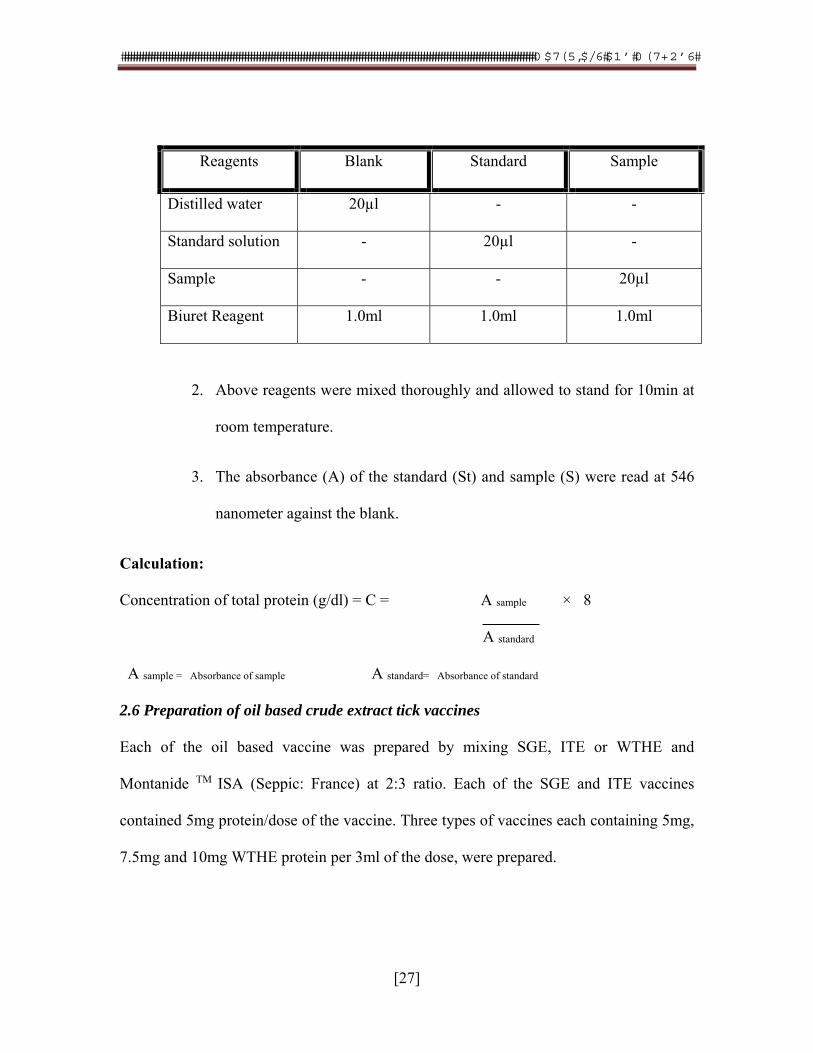
Procedure:
1. Protein estimation was performed on spectrophotometer. The different
materials are pipetted in cuvettes having one centimeter optical path by
following pipetting scheme.
���������������������������������������������������������������������������������������������������������������������������������������������� ������������ �� ���
[27]
Reagents Blank Standard Sample
Distilled water 20µl - -
Standard solution - 20µl -
Sample - - 20µl
Biuret Reagent 1.0ml 1.0ml 1.0ml
2. Above reagents were mixed thoroughly and allowed to stand for 10min at
room temperature.
3. The absorbance (A) of the standard (St) and sample (S) were read at 546
nanometer against the blank.
Calculation:
Concentration of total protein (g/dl) = C = A sample × 8
A standard
A sample = Absorbance of sample A standard= Absorbance of standard
2.6 Preparation of oil based crude extract tick vaccines
Each of the oil based vaccine was prepared by mixing SGE, ITE or WTHE and
Montanide TM ISA (Seppic: France) at 2:3 ratio. Each of the SGE and ITE vaccines
contained 5mg protein/dose of the vaccine. Three types of vaccines each containing 5mg,
7.5mg and 10mg WTHE protein per 3ml of the dose, were prepared.
���������������������������������������������������������������������������������������������������������������������������������������������� ������������ �� ���
[28]
2.6.1 Safety test
The vaccines were injected at dose rate of 0.5ml to each of three rabbits for the
confirmation of its safety.
2.6.2 Sterility
The vaccine was streaked on three different culture media (Thioglycerlate media,
MacConkey agar and Tryptose blood agar). The media was observed for any growth after
keeping them at incubation at 37oC for 48 hours.
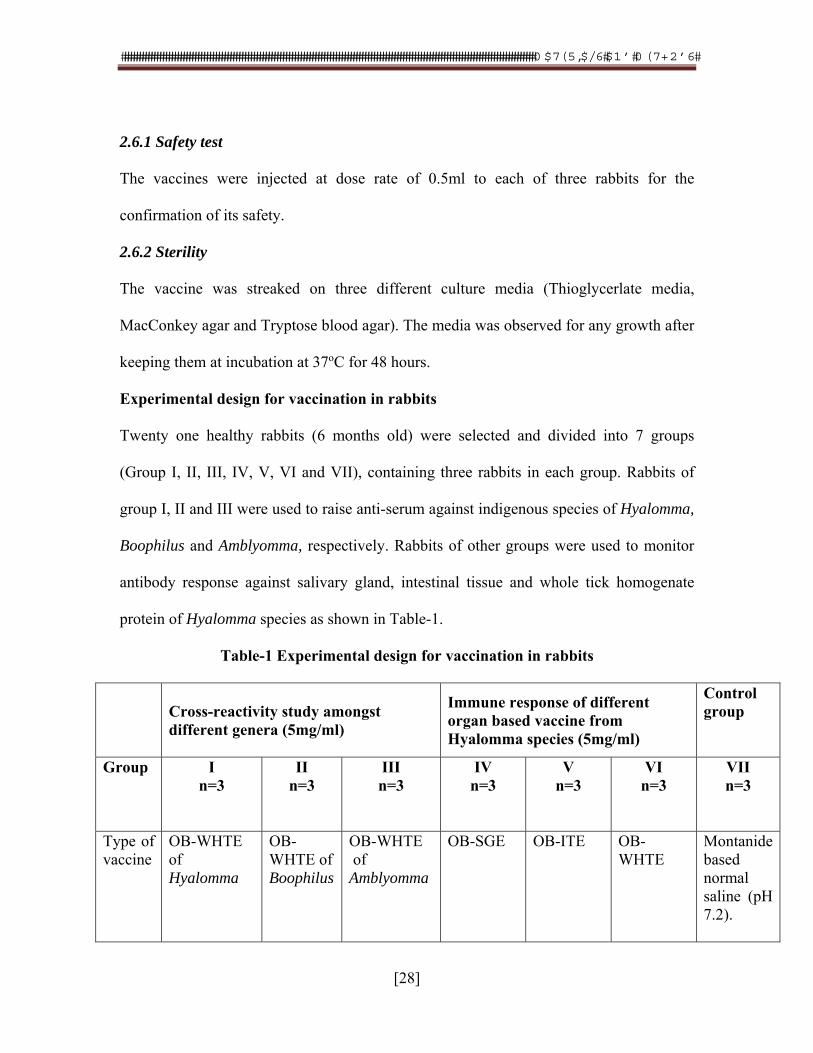
Experimental design for vaccination in rabbits
Twenty one healthy rabbits (6 months old) were selected and divided into 7 groups
(Group I, II, III, IV, V, VI and VII), containing three rabbits in each group. Rabbits of
group I, II and III were used to raise anti-serum against indigenous species of Hyalomma,
Boophilus and Amblyomma, respectively. Rabbits of other groups were used to monitor
antibody response against salivary gland, intestinal tissue and whole tick homogenate
protein of Hyalomma species as shown in Table-1.
Table-1 Experimental design for vaccination in rabbits
Cross-reactivity study amongst different genera (5mg/ml)
Immune response of different organ based vaccine from Hyalomma species (5mg/ml)
Control group
Group I n=3
II n=3
III n=3
IV n=3
V n=3
VI n=3
VII n=3
Type of vaccine
OB-WHTE of Hyalomma
OB-WHTE of Boophilus
OB-WHTE of Amblyomma
OB-SGE
OB-ITE
OB-WHTE
Montanide based normal saline (pH 7.2).
���������������������������������������������������������������������������������������������������������������������������������������������� ������������ �� ���
[29]
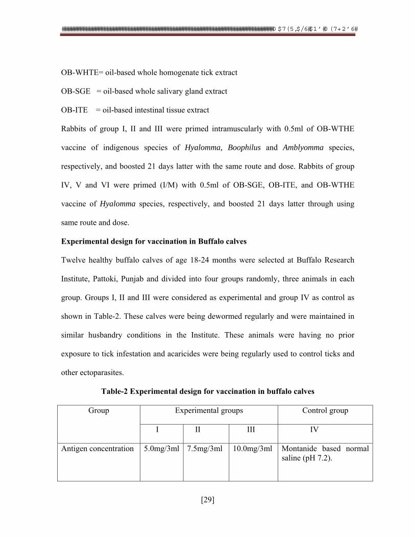
OB-WHTE= oil-based whole homogenate tick extract
OB-SGE = oil-based whole salivary gland extract
OB-ITE = oil-based intestinal tissue extract
Rabbits of group I, II and III were primed intramuscularly with 0.5ml of OB-WTHE
vaccine of indigenous species of Hyalomma, Boophilus and Amblyomma species,
respectively, and boosted 21 days latter with the same route and dose. Rabbits of group
IV, V and VI were primed (I/M) with 0.5ml of OB-SGE, OB-ITE, and OB-WTHE
vaccine of Hyalomma species, respectively, and boosted 21 days latter through using
same route and dose.
Experimental design for vaccination in Buffalo calves
Twelve healthy buffalo calves of age 18-24 months were selected at Buffalo Research
Institute, Pattoki, Punjab and divided into four groups randomly, three animals in each
group. Groups I, II and III were considered as experimental and group IV as control as
shown in Table-2. These calves were being dewormed regularly and were maintained in
similar husbandry conditions in the Institute. These animals were having no prior
exposure to tick infestation and acaricides were being regularly used to control ticks and
other ectoparasites.
Table-2 Experimental design for vaccination in buffalo calves
Group Experimental groups Control group
I II III IV
Antigen concentration 5.0mg/3ml 7.5mg/3ml 10.0mg/3ml Montanide based normal saline (pH 7.2).
���������������������������������������������������������������������������������������������������������������������������������������������� ������������ �� ���
[30]
Each buffalo calf of all experimental groups was primed (3ml/dose) using intramuscular
route and boosted with the same dose and route 28 days post priming. Amount of tick
protein/dose was 5mg, 7.5mg and 10mg for each calf of group I, II and III, respectively.
The control animals were inoculated with equal volume of the Montanide based normal
saline (pH 7.2).
Blood sample of each rabbit of all groups and each buffalo calf was taken on 0, 15, 30
and 45 day post-priming. The serum was separated from each sample and transferred to
each of the labeled vials and stored at -200C till required for antibody monitoring.
3. Monitoring sero-conversion of vaccinated rabbits and calves
Serum of each rabbit of each group was subjected to Agar Gel Precipitin Test ((Akhtar,
1995) to monitor the antibody response against each part (SGE, ITE and WTHE) of
Hyalomma species and to monitor the cross reactivity among three genera of the hard
ticks. The antibody titer in buffalo calves against tick extract was determined using
Complement Fixation Test (CFT; Hudson and Hay, 1976). The anti-crude extract
complement fixing antibody titer of each serum was determined.
3.1 Agar Gel Precipitation Test
Specific reactions in AGPT were indicated by precipitin lines between the tick antigen
and the test serum. The results were compared with the identification of the reaction
between the same antigen and that of the standard positive serum raised against tick
antigen.
���������������������������������������������������������������������������������������������������������������������������������������������� ������������ �� ���
[31]
3.1.1 Optimization of AGPT
Optimization and standardization of the test was performed by using three different
compositions of purified Noble agar. For this purpose 0.9gm, 1.0gm and 1.1 gm purified
Noble agar was used in same weights (8gm) of sodium chloride and sodium azide
(0.01gm) and same volume (100ml) of distilled water. It was found that gel was solidified
optimally in that Petri dish in which poured molten agar with 0.9 gm purified Noble agar
was used.
3.1.2 Preparation of agar gel plate
According to standardization results, 0.9% was prepared according to following
composition.
Nobel Agar (Oxoid: LP0028) 0.9gms
Sodium chloride 8.0gms
Sodium azide 0.01gms
Distilled water 100ml
All above three ingredients were weighed and mixed in 100ml dH2O water and then
heated to boil in microwave oven until a uniform suspension was obtained. The uniform
suspension of the agar was then poured in to Petri dishes kept at room temperature and
leveled surface and allowed to solidify. Approximately 70ml of gel in 100ml Petri dishes
and 35ml in 60mm Petri dishes were poured. The thickness of gel was 4mm and
solidified gel was transferred to a refrigerator till further used.
���������������������������������������������������������������������������������������������������������������������������������������������� ������������ �� ���
[32]
3.1.3 Punching of wells
Well guiding template of required well diameter got prepared from local market and was
used for punching the wells in the agar gel. The diameter of the holes and the distance
between the wells was 5mm, respectively. The lid of the plate was removed and the
template was placed on agar gel plate. Care was taken that the template should not touch
the surface of the agar gel plate. A metal gel borer was used to punch the wells through
template in the agar gel. The punched gel in the wells was removed with great care using
vacuum pump fitted with sterilized Pasteur pipette. The bottom of all wells was sealed by
adding 20µl molten agar. This was done to minimize the leakage of antigen or antiserum
between gel and glass plate.
3.1.4 Charging of wells
The known antigen (50µl/well) was added in three of peripheral wells and normal saline
(50µl/ well) was added in 4th well, while unknown antiserum (50µl/well) was added in the
central well. The plate was incubated for 72h in humidity chamber. The positive samples
after thawing, were two fold diluted with PBS in microtiter plate with the help of pipette
starting from the 1st well up to 10th of each dilution. All the two fold dilutions of positive
samples were added to wells (50µl/well) to agar gel plate to monitor the antibody titer
level. Appearance of the precipitin lines between the tick antigen and the serum sample
within 72h was recorded.
���������������������������������������������������������������������������������������������������������������������������������������������� ������������ �� ���
[33]
3.2 CFT
In present study, CFT was carried out for the evaluation of tick vaccine as described by
(Hudson and Hay, 1976), using Hyalomma whole tick homogenate suspension (WTHS)
as antigen which was prepared in previous experiment and was stored at -40oC for future
use. In this test, test sera were raised in rabbits and experimental calves kept at Buffalo
Research Institute, Pattokee. Oil based vaccines with 5mg, 7.5mg and 10.0mg per does
were injected to these animals. Five percent washed sheep RBCs, rabbit anti-sheep
erythrocytes antiserum containing sub-agglutinating level and Veronal buffer (Appendix-
2) as diluents/stabilizer were used to conduct the test.
3.2.1 Amboceptor (rabbit anti-sheep erythrocytes antiserum)
Amboceptor (antibodies against sheep erythrocytes) were raised in rabbits by following
the technique as described by Merchant and Packer (1983).
(a) Washing of sheep red RBC’S
I. A blood volume of 2ml was collected aseptically from the jugular vein of a sheep
kept at Animal House of Department of Microbiology, University of Veterinary and
Animal Sciences, Lahore. An anticoagulant 5mg sodium citrate was added in 5ml sterile
syringe before the collection of blood. Afterwards, the syringe containing blood was
slightly agitated to mix the anticoagulant with the blood.
II. Washing of Red Blood Cells (RBC’s) was performed with help of PBS. For this
purpose blood was shifted to centrifuge tube and PBS was added to make total volume
10ml. The tube was centrifuged at 2000rpm for two min. The supernatant was discarded
and PBS equal to packed cell volume (PCV) was added to the centrifuge tube carefully.
���������������������������������������������������������������������������������������������������������������������������������������������� ������������ �� ���
[34]
III. The packed RBC’s were re-suspended in the saline by slight inverted motion of
centrifuge tube and centrifuged at the same speed and time. The supernatant was again
discarded and same quantity of PBS was again added. The process of RBC’s washing
was repeated thrice. Final volume of PCV 1.5ml was obtained and in this way 3.5ml PBS
was added to get 10% suspension of the sheep erythrocytes. This suspension percentage
of sheep RBC’s was used in the rabbits to raise antiserum against it.
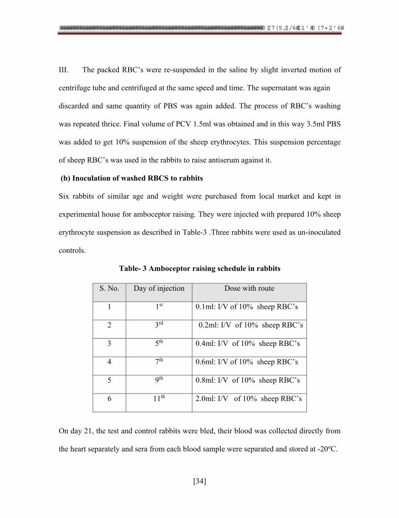
(b) Inoculation of washed RBCS to rabbits
Six rabbits of similar age and weight were purchased from local market and kept in
experimental house for amboceptor raising. They were injected with prepared 10% sheep
erythrocyte suspension as described in Table-3 .Three rabbits were used as un-inoculated
controls.
Table- 3 Amboceptor raising schedule in rabbits
On day 21, the test and control rabbits were bled, their blood was collected directly from
the heart separately and sera from each blood sample were separated and stored at -20oC.
S. No. Day of injection Dose with route
1 1st 0.1ml: I/V of 10% sheep RBC’s
2 3rd 0.2ml: I/V of 10% sheep RBC’s
3 5th 0.4ml: I/V of 10% sheep RBC’s
4 7th 0.6ml: I/V of 10% sheep RBC’s
5 9th 0.8ml: I/V of 10% sheep RBC’s
6 11th 2.0ml: I/V of 10% sheep RBC’s
���������������������������������������������������������������������������������������������������������������������������������������������� ������������ �� ���
[35]
(c) Calculation of sub-agglutination titer
The serum samples were processed for titration of the amboceptor and determination of
its sub-agglutination titer. A U-shaped, 96 well immuno-plates were used for the titration
of the amboceptor.
1. Veronal buffer (50µl: pH 7.2, Appendix- 2) was added in each wells of three rows
of the plate using multi-channeled micropipette.
2. Amboceptor (50µl) was added in all wells of first row except well No.12 and its
two-fold dilution was made up to well No.11 discarding 50µl from well No. 11.
3. Washed sheep RBS’s (50µl: five percent suspension) were added from well 1 to
well 12 and the plate was incubated at the room temperature for 30 min.
4. Both the test and the control rabbit sera were monitored for the amboceptor
titeration.
The general plan for determination of agglutination level of the amboceptor is presented in
Appendix -3.
The highest dilution of the rabbit serum showing agglutination was considered as 1 HA
unit titer. Sub-agglutination level of amboceptor i.e., the 50% dilution of 1 HA unit titer
of the amboceptor was used for sensitization of 10% washed sheep RBS’s.
(d) Sensitization of sheep RBC’s
1. The sub-agglutination level of the amboceptor (10ml) was taken and treated at
56oC for half an hour in water bath to inactivate the rabbit complement and then added to
10ml of the 10% washed sheep RBC’s suspension.
2. This mixture was incubated at 37oC for 20min for sensitization of sheep RBC’s.
���������������������������������������������������������������������������������������������������������������������������������������������� ������������ �� ���
[36]
3. The mixture was centrifuged at 2000rpm for two min and the supernatant was
discarded and the pellet of the RBC’s was re-suspended.
4. The centrifugation process was repeated thrice by re-suspending the sensitized
sheep RBC’s in the saline.
5. Finally 0.2% suspension of the sheep RBCs was made (0.2ml of the sensitized R
BC’s was added in 99.8ml of the saline) to be used for titration of the complement.
(f) Complement
Complement was used as indicator in hemolytic system in CFT and serum from guinea
pig was used as complement source as guinea pig serum contains a greater concentration
of all complements than that of other animals (Merchant and Packer, 1983). Four guinea
pigs were kept at the Experimental house of Department of Parasitology, University of
Veterinary and Animal Sciences, Lahore.
Before bleeding, guinea pigs were kept under air conditioned environment for overnight.
Each guinea pig was sacrificed and bled in large beaker. Serum was collected after
coagulation of blood and its aliquots of two micro liters were made in Eppendorf tubes.
This serum was used as source of complement.
3.2.2 Calculation of hemolytic units of guinea pig serum
Four hemolytic units (4 HL) of guinea pig serum were calculated and U-shaped 96-well
micro titration plate was used for titration of complement. Following protocol was used:
1. Using multi-channeled micropipette, 50µl of PBS was added from well No.1 to
well no.12 in all rows.
���������������������������������������������������������������������������������������������������������������������������������������������� ������������ �� ���
[37]
2. A 50µl amount of the guinea pig serum was added in the first well and two-fold
dilution up to well No.10 was made of first row.
3. The 50µl the sensitized sheep RBC’s (0.2%) were added from well 1 to well no.
12.
4. Well number 12 was kept as negative control. Plate was incubated at 4oC for
overnight.
The plane for calculation of 4-Hemolytic unit is presented in Appendix-4
Positive= Lysis= Clear suspension of the sensitized sheep RBC’s
Negative= No lysis/button formation=sedimentation of the sensitized sheep RBC’s
Procedure of CFT
The CFT was performed as recommendation of Merchant and Packer (1983).
Brief steps are as follows:
1. An amount of 50µl PBS solution was added from well number 1 to well number
12 of the immunotitration plate.
2. The 50µl of the immune serum was added in well no. 1 and its two-fold dilutions
were made up to well number 9.
3. The 50µl of tick antigen (whole tick extract) was added from well number 1 to
well number 10.
4. Then 50µl of the complement containing 4 HI units was added from well number
1 to well number 11 and the plate was incubated at 37oC for ten min.
5. Finally, 50µl of the sensitized sheep RBCs (0.2%) was added from well number 1
to well number 12
���������������������������������������������������������������������������������������������������������������������������������������������� ������������ �� ���
[38]
In this way each of the immune serum sample (test serum) and control serum samples
was titrated against tick antigen.
Statistical Analysis:
The geometric mean titer (GMT) of antibody data of AGPT and CFT was calculated
by the technique described by Villages (1998).
In case of AGPT it is highest dilution of the serum showing Agar Gel Precipitation
lines against specific antigen, while, it the highest dilution of no hemolysis in
quantitative assay in Complement Fixation Test. \
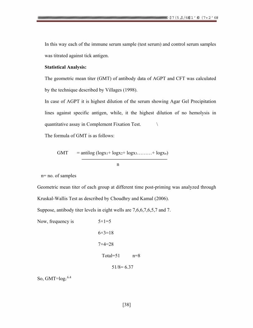
The formula of GMT is as follows:
GMT = antilog (logx1+ logx2+ logx3………+ logxn) n n= no. of samples Geometric mean titer of each group at different time post-priming was analyzed through
Kruskal-Wallis Test as described by Choudhry and Kamal (2006).
Suppose, antibody titer levels in eight wells are 7,6,6,7,6,5,7 and 7.
Now, frequency is 5×1=5
6×3=18
7×4=28
Total=51 n=8
51/8= 6.37
So, GMT=log2 6.4
���������������������������������������������������������������������������������������������������������������������������������������������� ������������ �� ���
[39]
The general plan for calculation of anti-tick immunogen- complement fixing antibody units is shown in Appendix-5.
4. Monitoring capacity of Hyalomma whole homogenate tick vaccine in controlling tick infestation in crossbred animals by ELISA
Twelve crossbred male calves of same age (6-9) and weight were selected from Aslam
Gujjar Livestock Farm, Lahore. These animals were thoroughly examined for tick
infestation and found free of ticks. These calves have no previous exposure to tick
infestation because acaricides were regularly used in these animals. The animals were
divided randomly into two groups, six animals in each group. Group I was assigned as
experimental control and group II was assigned as control.
4.1 Vaccination of crossbred calves with Hyalomma whole homogenate tick vaccine
Crossbred calves of group I (calf no. AGF-1, AGF-2, AGJ-4, AGF-10, AGF-19, and
AGF-20) were kept as control. Crossbred calves of group I1 (calf no. AGF-9, AGF-12,
AGF-14, AGF-22 AGF-23 and AGF-24) were vaccinated with oil-based Hyalomma
whole homogenate tick vaccine with 7.5mg/dose antigen concentration.
���������������������������������������������������������������������������������������������������������������������������������������������� ������������ �� ���
[40]
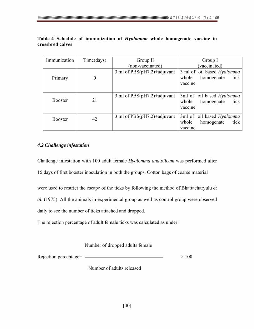
Table-4 Schedule of immunization of Hyalomma whole homogenate vaccine in crossbred calves
4.2 Challenge infestation
Challenge infestation with 100 adult female Hyalomma anatolicum was performed after
15 days of first booster inoculation in both the groups. Cotton bags of coarse material
were used to restrict the escape of the ticks by following the method of Bhattacharyulu et
al. (1975). All the animals in experimental group as well as control group were observed
daily to see the number of ticks attached and dropped.
The rejection percentage of adult female ticks was calculated as under:
Number of dropped adults female
Rejection percentage= × 100
Number of adults released
Immunization Time(days) Group II (non-vaccinated)
Group I (vaccinated)
Primary 0 3 ml of PBS(pH7.2)+adjuvant 3 ml of oil based Hyalomma
whole homogenate tick vaccine
Booster 21 3 ml of PBS(pH7.2)+adjuvant 3ml of oil based Hyalomma
whole homogenate tick vaccine
Booster 42 3 ml of PBS(pH7.2)+adjuvant 3ml of oil based Hyalomma
whole homogenate tick vaccine
���������������������������������������������������������������������������������������������������������������������������������������������� ������������ �� ���
[41]
Dropped ticks were kept at 29oC and 85% RH (Andreotti et al., 2002) for three weeks of
incubation period and weight of laid egg was measured. The dropped ticks were keenly
observed and classified into abnormally (white or pale yellow in color) and normally fed
(dark grey color) ticks according to their appearance. Differences in rejection percentage,
engorgement period, engorgement weight, egg mass and reproductive index in
immunized and control group were calculated statistically by Student-t test to find the
efficacy of the vaccine in experimental group and control group.
Blood samples from cross-bred calves were collected on day 0, 15, 30, 45, 60, 75, 90,
105 and 120 and serum samples were stored at -20oC until used. Antibody response in
controlled and vaccinated group was determined by Indirect Enzyme Linked
Immunosorbant Assay (ELISA).
4.3 Indirect ELISA
The ELISA was performed for detection of antibodies against antigen prepared from
whole tick homogenate (Ismael et al., 2003). For this purpose horse radish peroxidase
conjugated antibovine IgG (Sigma- Aldrich: Cat # A 8917) in rabbits as conjugate and
‘O’ phenylene diamine (OPD) with hydrogen peroxidase in citrate phosphate buffer as
substrate were used. Incubation period for antigen and conjugate concentration were
optimized by Checker board titration method and following testing procedure was
performed:
4.3.1 Preparation of coating buffer
50 mM Carbonate buffer (pH 9.6) was prepared for coating the antigen to ELISA plate.
(Appendix-6)
���������������������������������������������������������������������������������������������������������������������������������������������� ������������ �� ���
[42]
4.3.2 Preparation of washing buffer
The washing buffer (0.05% Tween 20) was prepared by adding 0.5ml Tween20 in
1000ml Phosphate buffered saline (Appendix-7).
4.3.3 Standardization of solid Phase
Three types of 96 well microtiter plates (U bottom, Flat bottom and flat bottom EIA)
were used for the coating of antigen.
4.3.4 Preparation of substrate
Substrate was prepared by dissolving 30mg OPD tablet in 30µl of 30 % H2O2 v/v in 75
ml citrate buffer (Appendix-8).
4.3.5 Coating of plate with tick antigen
Plates were coated with tick antigen prepared in previous experiment. Different
concentrations of this antigen ranging from 25-200ug/µl were prepared in coating buffer.
4.3.6 Dilution of conjugate
The conjugated Antibovine IgG in rabbit (A 8917:Sigma Aldrich) was reconstituted and
its 1:2000 dilution was made in solvent (BSA 4%+PBS 1X). Aliquots of the solution
were prepared and stored at 4oC till further used.
4.3.7 Dilution of serum samples
The serum samples were diluted 1:100 with diluent (BSA 4%+PBS 1X).
Protocol for Indirect ELISA:
1. Plates were coated with 100µl of diluted antigen/well and kept at 4oC for
overnight. The plates were covered with adhesive plastic sheet to prevent
evaporation of antigen. For each dilution of antigen two rows of plates were used.
���������������������������������������������������������������������������������������������������������������������������������������������� ������������ �� ���
[43]
2. Next day the plates were washed three times with washing buffer (1X PBS
+Tween 0.05%) 300µl per well
3. Blocking (2% bovine serum albumin) solution was added in volume of 150µl/
well and then plates were incubated for one hour at 37oC.
4. The plate were emptied and washed with PBS Tween 20 washing buffer.
5. Two fold serial dilution of serum samples were made in PBS. From each dilution
100µl of serum/ well was dispensed from well 1-10 in each row. Plates were incubated
for one hour at 37oC.
6. Plates were washed three times with washing buffer.
7. Conjugate was added in all wells (100µl/well) and plates were incubated at 37oC
for two hours.
8. Substrate (‘O’ phenylenediamine with Hydrogen peroxide in citrate buffer) was
added to all wells (100µl/ well) and was incubated at 37oC for 30 minutes.
9. After 15min the reaction was stopped by adding 100µl of NaOH (0.2M).
10. Observations were recorded on ELISA reader at 405nm wavelength.
5. Detection of T.annulata by PCR
5.1 Processing of samples for DNA extraction
DNA was extracted from ticks by Phenol-Chloroform method as described by D’Oliveira
et al., 1997. In brief, ticks were removed from ethanol (70%) and dissected into two
halves with the help of sterile blade. New blade was used for each tick dissection. Both
halves of the dissected ticks were boiled in 1.5ml microcentrifuge tube in 200µl PBS for
���������������������������������������������������������������������������������������������������������������������������������������������� ������������ �� ���
[44]
10min. One percent SDS was added to 150µl boiled tick sample which was removed
thereafter with phenol: choloroform (1:1) and phenol: isoamyl alcohol (24:1),
respectively. Two micro liters of extracted DNA was used for PCR reaction. DNA
quantity was measured at 260 nm. The ratios of 260/280 were ranging from 1.80 to 1.95
for purified DNA samples which showed that preparations were free of any major
protein. After quantification, DNA samples thus extracted were stored in -40oC till used
for PCR analysis.
5.2 Preparation of final reaction volume and PCR analysis
The mixture for PCR was prepared in volume of 50µl containing 200uM of dNTPS,
1.5mM of MgCl2, 10mM of Tric HCl, 50mM of KCl, 200pM of each of reverse and
forward primer, 0.1% of Tritone and 2.5IU units of Taq DNA polymerase. The
temperature cycles used for PCR analysis are: initial heating for five minutes at 94oC,
following by 35 cycles of 94oC for 30sec, 55oC for 30sec and 72oC for one minute. Final
extension step was programed at 72oC for seven minutes (Sirigireddy and Ganta, 2005).
Two controls, one negative and one positive were included in each test. The reaction
mixtures were performed in automatic conventional DNA thermocylcler (Esco
Technologies, Inc.: USA).
5.3 Primers
Primers (IA-1 and IA-2: First base, Singapor) were used for the amplification of the gene
coding for surface protein of the merozoite (Table-5).
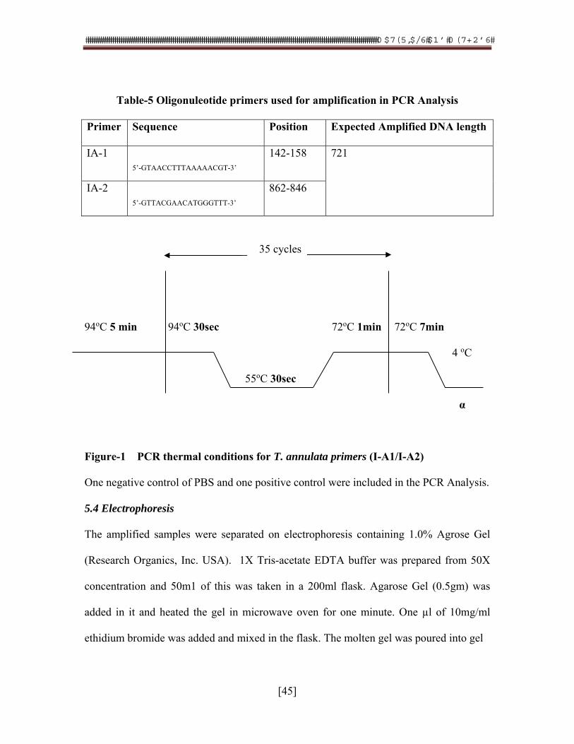
���������������������������������������������������������������������������������������������������������������������������������������������� ������������ �� ���
[45]
Table-5 Oligonuleotide primers used for amplification in PCR Analysis
Primer Sequence Position Expected Amplified DNA length
IA-1
5’-GTAACCTTTAAAAACGT-3’ 142-158 721
IA-2
5’-GTTACGAACATGGGTTT-3’ 862-846
35 cycles
94oC 5 min 94oC 30sec 72oC 1min 72oC 7min
4 oC
55oC 30sec
α
Figure-1 PCR thermal conditions for T. annulata primers (I-A1/I-A2)
One negative control of PBS and one positive control were included in the PCR Analysis.
5.4 Electrophoresis
The amplified samples were separated on electrophoresis containing 1.0% Agrose Gel
(Research Organics, Inc. USA). 1X Tris-acetate EDTA buffer was prepared from 50X
concentration and 50m1 of this was taken in a 200ml flask. Agarose Gel (0.5gm) was
added in it and heated the gel in microwave oven for one minute. One µl of 10mg/ml
ethidium bromide was added and mixed in the flask. The molten gel was poured into gel
���������������������������������������������������������������������������������������������������������������������������������������������� ������������ �� ���
[46]
plate (with inserted comb) and allowed to solidify for 20min at room temperature. Two
hundred and fifty milli liters prepared 1X Tris-acetate EDTA buffer as dissolving
medium was poured in the gel tank and five micro liters of ethidium bromide was also
added in it.
Bromopehonl blue (Fermentas: Europe) was added as loading dye in the PCR product.
For this purpose, 2µl of 6x Bromophenol blue was added in 5µl of PCR product and
mixed thrice by micropipette. One Kb plus DNA ladder and λ hindIII were run along
with PCR product for the detection of any non specific reaction and to compare the size
of band. The bands were monitored under UV light and after proper cropping of the
images the data was saved for record keeping.
6. Growing of intestinal epithelial cells of hard tick as monolayer
6.1 Preparation of culture medium
Cell culture medium of required concentration (13.99mg/L) was prepared using 1X L-15
(MP: Biomedicals, LCC: USA) as insect medium to grow tick intestinal cell (Bel-sekyi et
al., 2000). The prepared combined medium was kept in incubator for 48h at 37oC and
checked for any growth.
6.2 Surface sterlization of Hyalomma ticks
Ten semi-engorged adult female Hyalomma ticks were collected from tick colony and
sterilized by dipping and intermittent shaking in 0.5% benzalkonium chloride solution.
The ticks were then washed with 70% alcohol and rinsed in three changes of sterilized
distilled water.
���������������������������������������������������������������������������������������������������������������������������������������������� ������������ �� ���
[47]
6.3 Isolation of intestinal cell
After surface sterilization ticks were dissected aseptically and intestines were removed
and placed in culture medium placed in small Petri dish. Suspension medium was filtered
through muslin cloth. For breaking the gut into cells filtrate was mixed thereafter on hot
plate stirrer by keeping heat knob off for 15min. These broken cells were shifted to
Karrel flask (25ml) in safety cabin.
6.4 Examination of tick intestinal cell viability
A solution with following composition was prepared for the examination of tick intestinal
viability under compound microscope.
1. Cell suspension (from Karrel flask) 0.2ml
2. Basal salt solution 0.4ml
3. Typin blue (4%) 0.4ml
A drop from above solution was poured on hemocytometer slide and examined under
inverted microscope for viability. Afterwards cell culture flask was kept in incubator with
5% CO2 at 37oC and media was changed regularly after 3-4 days.
���������������������������������������������������������������������������������������������������������������������������������������������� ������������ �� ���
[48]
6.5 Examining confluence rate of tick intestinal cells For the examining the confluence rate, T-25 flask was scratched and with the help of
pipette and 200µl volume of cell suspension was shifted to micro centrifuge tube. The
tube was centrifuged at 400rpm for 2min and one drop from bottom was taken on glass
slide. After air drying the slide, it was stained by using Hema staining kit for which
following reagents with given duration of staining were used:
1. Hema-3 (fixative solution) 20sec
2. Hema-3 Solution 1 7sec
3. Hema-3 Solution 2 4sec
After staining the slide, it was rinsed with tap water and allowed to air dry at room
temperature. Examination of confluence percentage was examined under oil immersion
lens under light microscope.
������������������������������������������������������������������������������������������������������������������������������������������������������������������������������������������
[49]
CHAPTER 4
RESULTS
1. Tick prevalence in cattle and buffaloes
This study was conducted in the months of July and August, 2007. Temperature and
humidity during study period were 30-350C and 60-70%, respectively (Appendix-9). In
all three districts Hyalomma species were found the highest in prevalence at 61% as
compared to other species (Table-6, Figure-5). Faisalabad district was the highest for
Hyalomma species being 43% followed by Jhang at 28% and Khanewal at 29%.
Rhipicephalus (Boophilus) species were the second most prevalent species with an
average prevalence of 28%, Faisalabad being the highest at 66% followed by Jhang at
26% and Khanewal at 7%. Amblyomma species were the third major species detected
(average=eight percent). This tick species was the highest in Jhang at 86% compared to
Faisalabad at eight percent and Khanewal at six percent. Haemaphysalis species were the
lowest at three percent in prevalence in three districts; Jhang being the highest at 58%
followed by Faisalabad at 18% and Khanewal at 24%. No Rhipecephalis (Boophilus)
species other than R. microplus was detected in these districts in our study. In sex-wise
distribution of hard ticks, adult female ticks were higher as compared to adult male ticks
and they were at 85% and 81% for Hyalomma species and Amblyomma species,
respectively. Rhipecephalis (Boophilus) species and Haemaphysalis species have similar
female to male ratio as 77% of adult ticks were females.
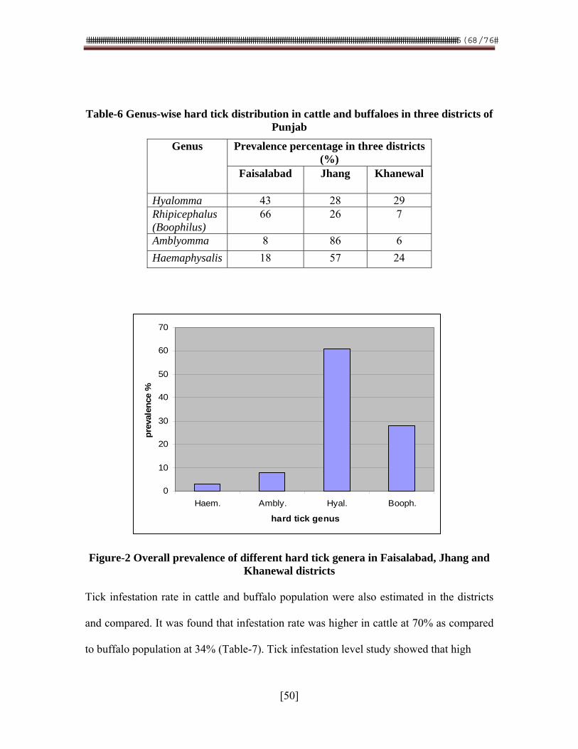
������������������������������������������������������������������������������������������������������������������������������������������������������������������������������������������
[50]
0
10
20
30
40
50
60
70
Haem. Ambly. Hyal. Booph.
hard tick genus
pre
vale
nce
%
Table-6 Genus-wise hard tick distribution in cattle and buffaloes in three districts of Punjab
Figure-2 Overall prevalence of different hard tick genera in Faisalabad, Jhang and Khanewal districts
Tick infestation rate in cattle and buffalo population were also estimated in the districts
and compared. It was found that infestation rate was higher in cattle at 70% as compared
to buffalo population at 34% (Table-7). Tick infestation level study showed that high
Genus Prevalence percentage in three districts (%)
Faisalabad Jhang Khanewal
Hyalomma 43 28 29 Rhipicephalus (Boophilus)
66 26 7
Amblyomma 8 86 6
Haemaphysalis 18 57 24
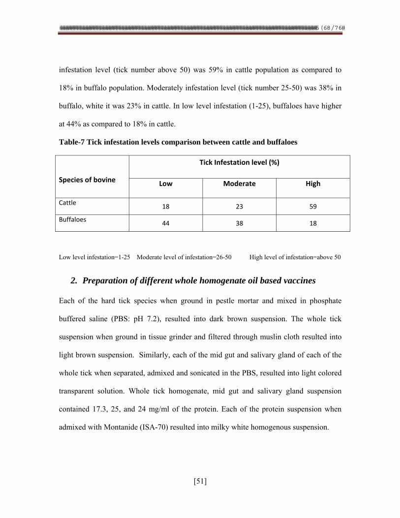
������������������������������������������������������������������������������������������������������������������������������������������������������������������������������������������
[51]
infestation level (tick number above 50) was 59% in cattle population as compared to
18% in buffalo population. Moderately infestation level (tick number 25-50) was 38% in
buffalo, white it was 23% in cattle. In low level infestation (1-25), buffaloes have higher
at 44% as compared to 18% in cattle.
Table-7 Tick infestation levels comparison between cattle and buffaloes
Species of bovine
Tick Infestation level (%)
Low Moderate High
Cattle 18 23 59
Buffaloes 44 38 18
Low level infestation=1-25 Moderate level of infestation=26-50 High level of infestation=above 50
2. Preparation of different whole homogenate oil based vaccines
Each of the hard tick species when ground in pestle mortar and mixed in phosphate
buffered saline (PBS: pH 7.2), resulted into dark brown suspension. The whole tick
suspension when ground in tissue grinder and filtered through muslin cloth resulted into
light brown suspension. Similarly, each of the mid gut and salivary gland of each of the
whole tick when separated, admixed and sonicated in the PBS, resulted into light colored
transparent solution. Whole tick homogenate, mid gut and salivary gland suspension
contained 17.3, 25, and 24 mg/ml of the protein. Each of the protein suspension when
admixed with Montanide (ISA-70) resulted into milky white homogenous suspension.
������������������������������������������������������������������������������������������������������������������������������������������������������������������������������������������
[52]
3. Monitoring sero-conversion of vaccinated rabbits and calves
Each of the whole tick homogenate vaccine prepared from either of the species of
Hyalomma, Rhipecephalus (Boophilus) or Amblyomma, when injected to rabbits (0.5 ml:
intramuscularly) induced detectable level of anti-whole tick homogenate protein agar gel
precipitating (WTH-AGP) antibodies. Each antiserum containing each tick specific
WTH-AGP antibodies cross reacted with tick specific homogenate protein antigen
(Table-8). The proteins of each of the hard ticks were antigenically similar. In the same
way, vaccine prepared from either salivary gland, mid-gut or whole tick homogenate
protein of the same species of Hyalomma induced detectable anti-tick organ specific
protein agar gel precipitating (TOSP-AGP) antibodies (Table-9). The vaccine prepared
from salivary gland induced 0, 3.2 and 6.5 geometric mean titer of TOSP-AGP antibodies
on 0, 15 and 30 days post priming, respectively. The vaccine prepared from mid gut
protein induced 0, 6.5 and 13.0 geometric mean titer of TOSP-AGP antibodies on 0, 15
and 30 days post priming, respectively. The vaccine prepared from WTHE protein
induced 0, 13 and 26 geometric mean titer of TOSP-AGP antibodies on 0, 15 and 30 days
post priming, respectively. The WTH vaccine induced higher titer of the antibodies in
rabbits as compared to that of salivary gland and mid gut protein vaccines.
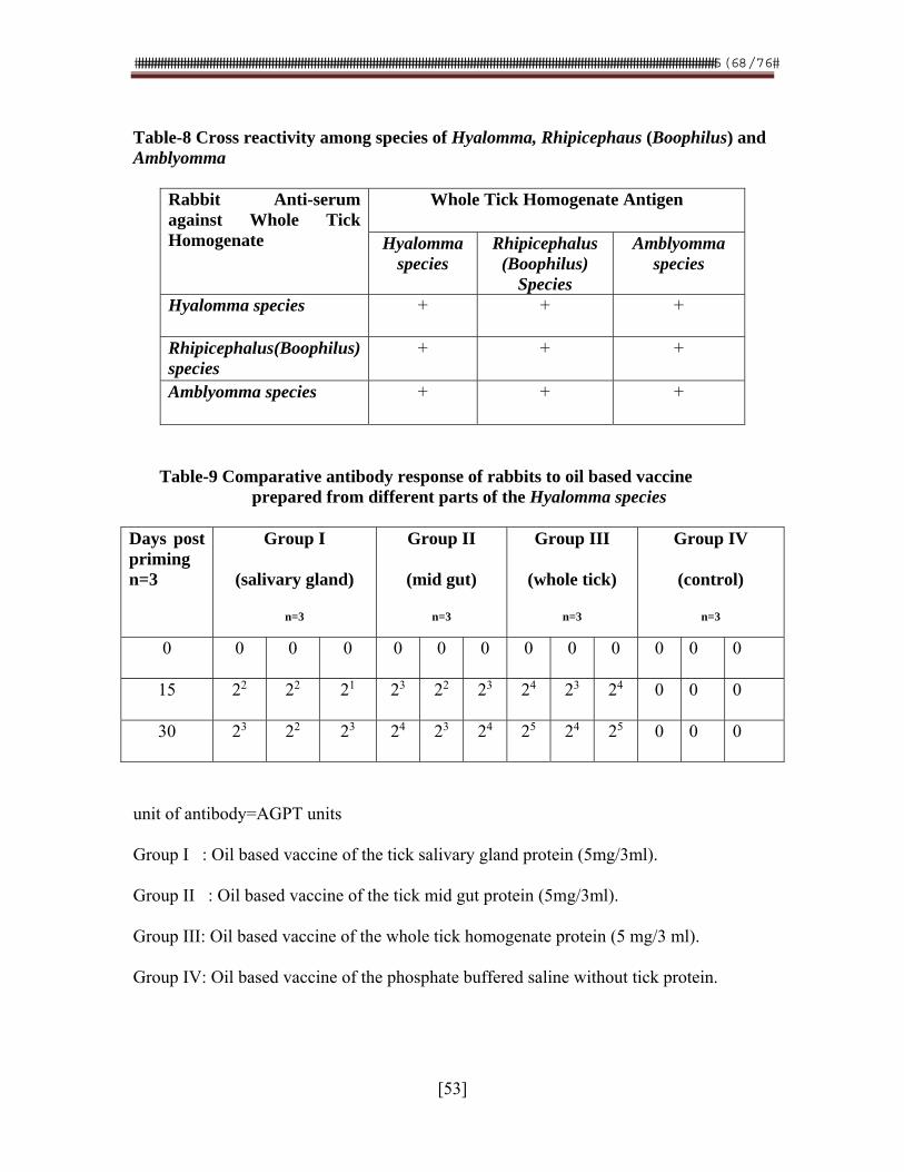
������������������������������������������������������������������������������������������������������������������������������������������������������������������������������������������
[53]
Table-8 Cross reactivity among species of Hyalomma, Rhipicephaus (Boophilus) and Amblyomma
Rabbit Anti-serum against Whole Tick Homogenate
Whole Tick Homogenate Antigen
Hyalomma species
Rhipicephalus (Boophilus)
Species
Amblyomma species
Hyalomma species + + +
Rhipicephalus(Boophilus) species
+ + +
Amblyomma species + + +
Table-9 Comparative antibody response of rabbits to oil based vaccine prepared from different parts of the Hyalomma species
Days post priming n=3
Group I
(salivary gland)
n=3
Group II
(mid gut)
n=3
Group III
(whole tick)
n=3
Group IV
(control)
n=3
0 0 0 0 0 0 0 0 0 0 0 0 0
15 22 22 21 23 22 23 24 23 24 0 0 0
30 23 22 23 24 23 24 25 24 25 0 0 0
unit of antibody=AGPT units
Group I : Oil based vaccine of the tick salivary gland protein (5mg/3ml).
Group II : Oil based vaccine of the tick mid gut protein (5mg/3ml).
Group III: Oil based vaccine of the whole tick homogenate protein (5 mg/3 ml).
Group IV: Oil based vaccine of the phosphate buffered saline without tick protein.
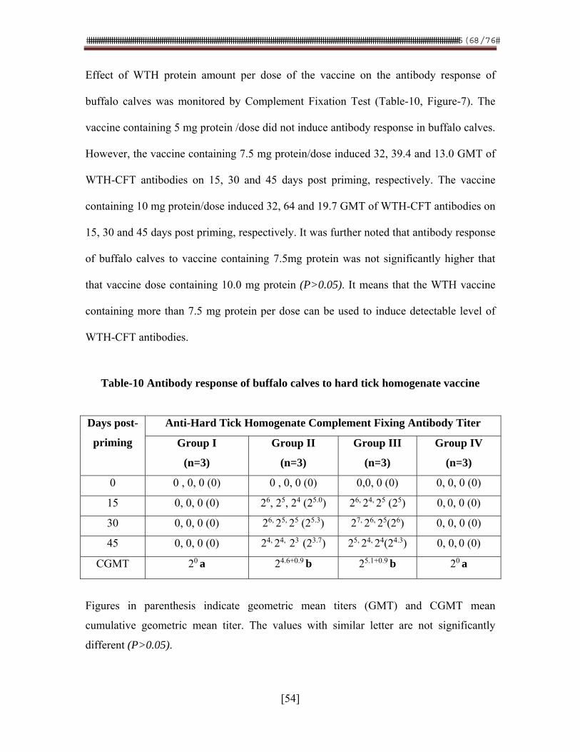
������������������������������������������������������������������������������������������������������������������������������������������������������������������������������������������
[54]
Effect of WTH protein amount per dose of the vaccine on the antibody response of
buffalo calves was monitored by Complement Fixation Test (Table-10, Figure-7). The
vaccine containing 5 mg protein /dose did not induce antibody response in buffalo calves.
However, the vaccine containing 7.5 mg protein/dose induced 32, 39.4 and 13.0 GMT of
WTH-CFT antibodies on 15, 30 and 45 days post priming, respectively. The vaccine
containing 10 mg protein/dose induced 32, 64 and 19.7 GMT of WTH-CFT antibodies on
15, 30 and 45 days post priming, respectively. It was further noted that antibody response
of buffalo calves to vaccine containing 7.5mg protein was not significantly higher that
that vaccine dose containing 10.0 mg protein (P>0.05). It means that the WTH vaccine
containing more than 7.5 mg protein per dose can be used to induce detectable level of
WTH-CFT antibodies.
Table-10 Antibody response of buffalo calves to hard tick homogenate vaccine
Days post-
priming
Anti-Hard Tick Homogenate Complement Fixing Antibody Titer
Group I
(n=3)
Group II
(n=3)
Group III
(n=3)
Group IV
(n=3)
0 0 , 0, 0 (0) 0 , 0, 0 (0) 0,0, 0 (0) 0, 0, 0 (0)
15 0, 0, 0 (0) 26, 25, 24 (25.0) 26, 24, 25 (25) 0, 0, 0 (0)
30 0, 0, 0 (0) 26, 25, 25 (25.3) 27, 26, 25(26) 0, 0, 0 (0)
45 0, 0, 0 (0) 24, 24, 23 (23.7) 25, 24, 24(24.3) 0, 0, 0 (0)
CGMT 20 a 24.6+0.9 b 25.1+0.9 b 20 a
Figures in parenthesis indicate geometric mean titers (GMT) and CGMT mean
cumulative geometric mean titer. The values with similar letter are not significantly
different (P>0.05).
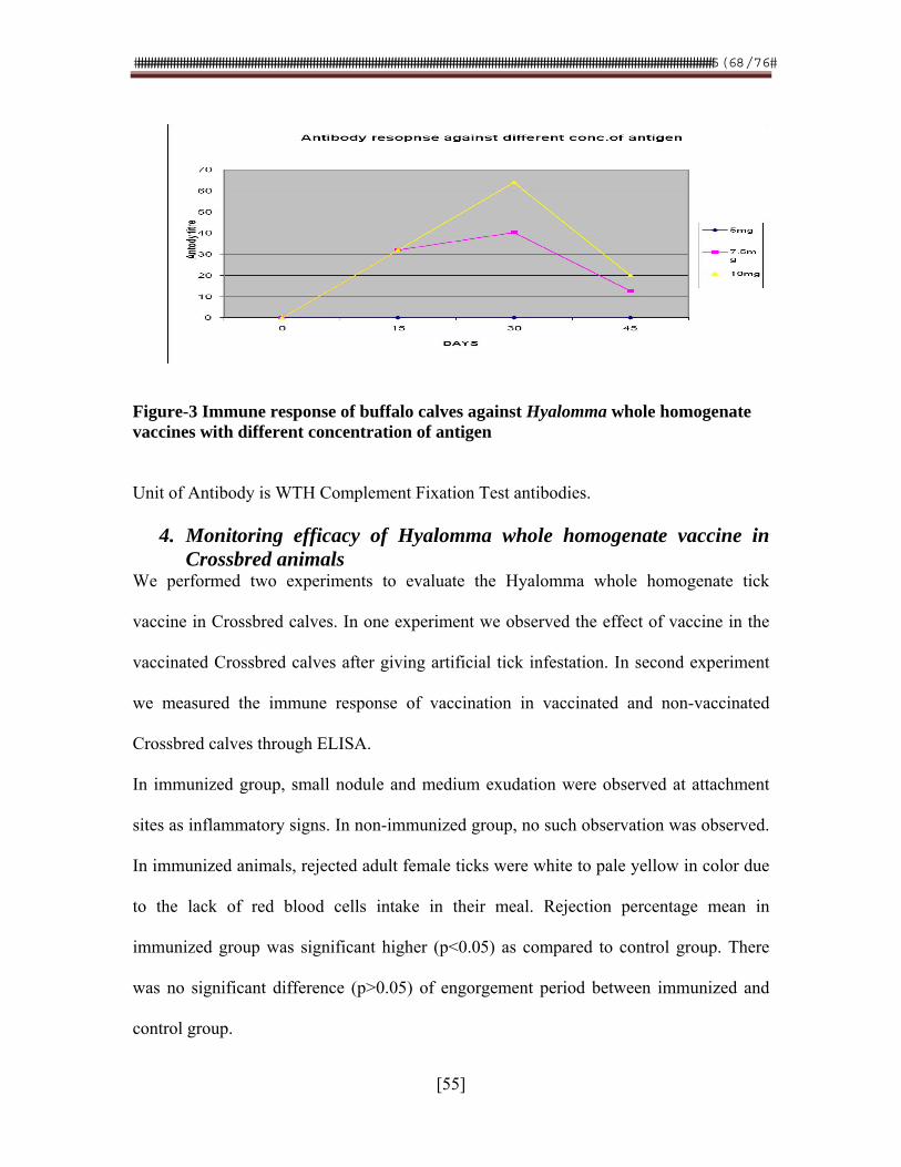
������������������������������������������������������������������������������������������������������������������������������������������������������������������������������������������
[55]
Figure-3 Immune response of buffalo calves against Hyalomma whole homogenate vaccines with different concentration of antigen
Unit of Antibody is WTH Complement Fixation Test antibodies.
4. Monitoring efficacy of Hyalomma whole homogenate vaccine in Crossbred animals
We performed two experiments to evaluate the Hyalomma whole homogenate tick
vaccine in Crossbred calves. In one experiment we observed the effect of vaccine in the
vaccinated Crossbred calves after giving artificial tick infestation. In second experiment
we measured the immune response of vaccination in vaccinated and non-vaccinated
Crossbred calves through ELISA.
In immunized group, small nodule and medium exudation were observed at attachment
sites as inflammatory signs. In non-immunized group, no such observation was observed.
In immunized animals, rejected adult female ticks were white to pale yellow in color due
to the lack of red blood cells intake in their meal. Rejection percentage mean in
immunized group was significant higher (p<0.05) as compared to control group. There
was no significant difference (p>0.05) of engorgement period between immunized and
control group.
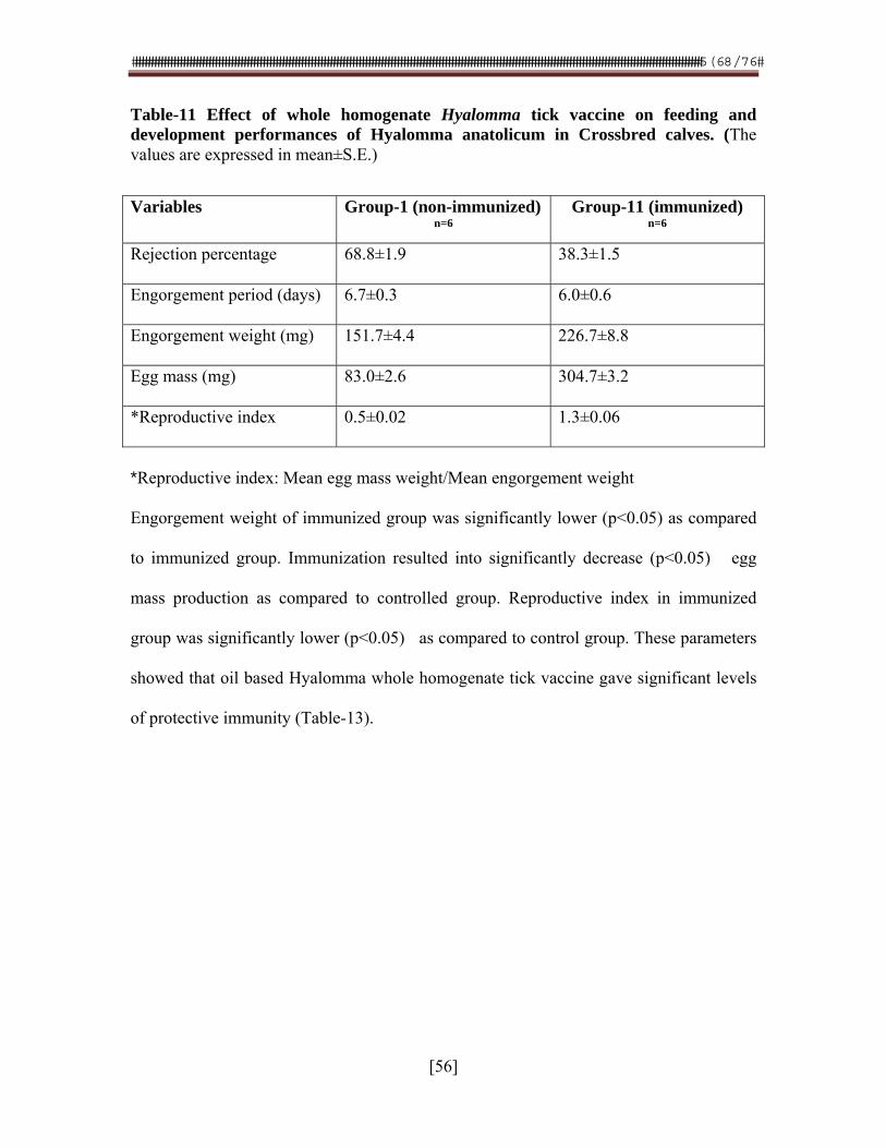
������������������������������������������������������������������������������������������������������������������������������������������������������������������������������������������
[56]
Table-11 Effect of whole homogenate Hyalomma tick vaccine on feeding and development performances of Hyalomma anatolicum in Crossbred calves. (The values are expressed in mean±S.E.)
*Reproductive index: Mean egg mass weight/Mean engorgement weight
Engorgement weight of immunized group was significantly lower (p<0.05) as compared
to immunized group. Immunization resulted into significantly decrease (p<0.05) egg
mass production as compared to controlled group. Reproductive index in immunized
group was significantly lower (p<0.05) as compared to control group. These parameters
showed that oil based Hyalomma whole homogenate tick vaccine gave significant levels
of protective immunity (Table-13).
Variables Group-1 (non-immunized)n=6
Group-11 (immunized) n=6
Rejection percentage 68.8±1.9 38.3±1.5
Engorgement period (days) 6.7±0.3 6.0±0.6
Engorgement weight (mg) 151.7±4.4 226.7±8.8
Egg mass (mg) 83.0±2.6 304.7±3.2
*Reproductive index 0.5±0.02 1.3±0.06
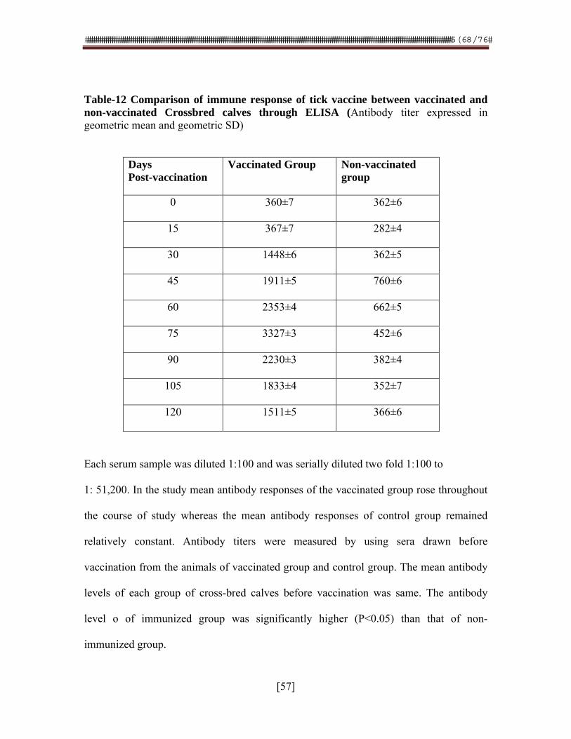
������������������������������������������������������������������������������������������������������������������������������������������������������������������������������������������
[57]
Table-12 Comparison of immune response of tick vaccine between vaccinated and non-vaccinated Crossbred calves through ELISA (Antibody titer expressed in geometric mean and geometric SD)
Days Post-vaccination
Vaccinated Group Non-vaccinated group
0 360±7 362±6
15 367±7 282±4
30 1448±6 362±5
45 1911±5 760±6
60 2353±4 662±5
75 3327±3 452±6
90 2230±3 382±4
105 1833±4 352±7
120 1511±5 366±6
Each serum sample was diluted 1:100 and was serially diluted two fold 1:100 to
1: 51,200. In the study mean antibody responses of the vaccinated group rose throughout
the course of study whereas the mean antibody responses of control group remained
relatively constant. Antibody titers were measured by using sera drawn before
vaccination from the animals of vaccinated group and control group. The mean antibody
levels of each group of cross-bred calves before vaccination was same. The antibody
level o of immunized group was significantly higher (P<0.05) than that of non-
immunized group.
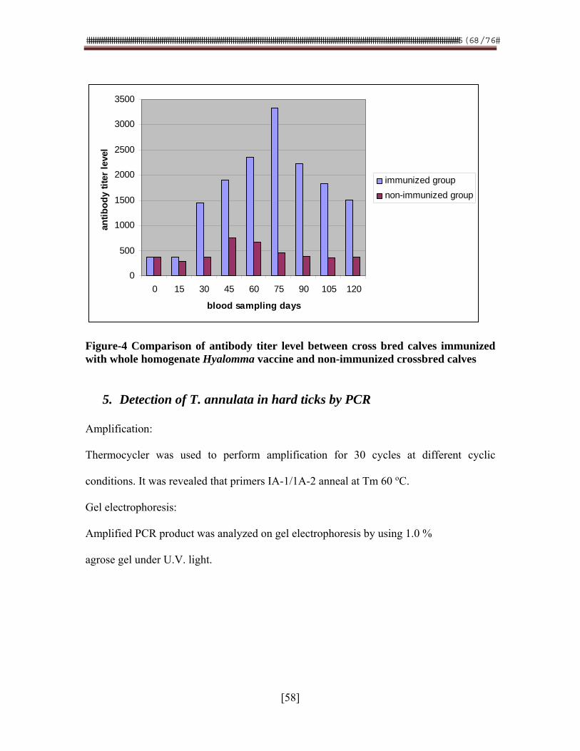
������������������������������������������������������������������������������������������������������������������������������������������������������������������������������������������
[58]
0
500
1000
1500
2000
2500
3000
3500
0 15 30 45 60 75 90 105 120
blood sampling days
anti
bo
dy
tite
r le
vel
immunized group
non-immunized group
Figure-4 Comparison of antibody titer level between cross bred calves immunized with whole homogenate Hyalomma vaccine and non-immunized crossbred calves
5. Detection of T. annulata in hard ticks by PCR
Amplification:
Thermocycler was used to perform amplification for 30 cycles at different cyclic
conditions. It was revealed that primers IA-1/1A-2 anneal at Tm 60 oC.
Gel electrophoresis:
Amplified PCR product was analyzed on gel electrophoresis by using 1.0 %
agrose gel under U.V. light.
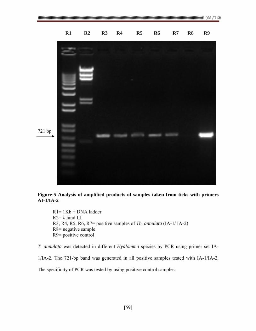
������������������������������������������������������������������������������������������������������������������������������������������������������������������������������������������
[59]
R1 R2 R3 R4 R5 R6 R7 R8 R9 721 bp Figure-5 Analysis of amplified products of samples taken from ticks with primers AI-1/IA-2
R1= 1Kb + DNA ladder R2= λ hind III
R3, R4, R5, R6, R7= positive samples of Th. annulata (IA-1/ IA-2) R8= negative sample R9= positive control T. annulata was detected in different Hyalomma species by PCR using primer set IA-
1/IA-2. The 721-bp band was generated in all positive samples tested with IA-1/IA-2.
The specificity of PCR was tested by using positive control samples.
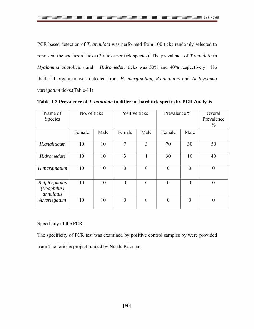
������������������������������������������������������������������������������������������������������������������������������������������������������������������������������������������
[60]
PCR based detection of T. annulata was performed from 100 ticks randomly selected to
represent the species of ticks (20 ticks per tick species). The prevalence of T.annulata in
Hyalomma anatolicum and H.dromedari ticks was 50% and 40% respectively. No
theilerial organism was detected from H. marginatum, R.annulatus and Amblyomma
variegatum ticks.(Table-11).
Table-1 3 Prevalence of T. annulata in different hard tick species by PCR Analysis
Name of Species
No. of ticks Positive ticks Prevalence % Overal Prevalence
%
Female Male Female Male Female Male
H.analiticum 10 10 7 3 70 30 50
H.dromedari 10 10 3 1 30 10 40
H.marginatum 10 10 0 0 0 0 0
Rhipicephalus (Boophilus) annulatus
10 10 0 0 0 0 0
A.variegatum 10 10 0 0 0 0 0
Specificity of the PCR:
The specificity of PCR test was examined by positive control samples by were provided
from Theileriosis project funded by Nestle Pakistan.
������������������������������������������������������������������������������������������������������������������������������������������������������������������������������������������
[61]
6. Growing of intestinal epithelial cells of hard tick as monolayer
After overnight incubation at 37 0C, Karrel flask was placed under inverted microscope
and examined for the evidence of cell growth. It was found that intestinal cells were
growing as under inverted microscope spindle shaped cells were found which the signs
for cell division are.
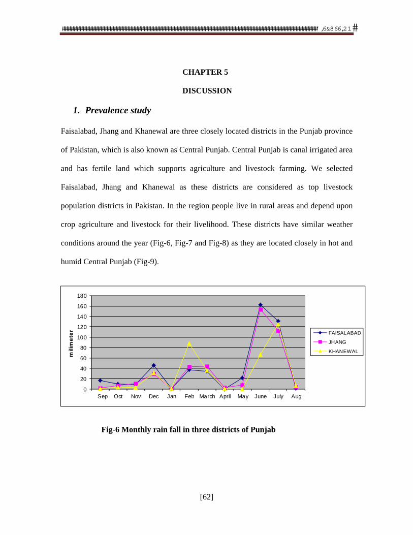
�������������������������������������������������������������������������������������������������������������������������������������������������������������������������������������
[62]
CHAPTER 5
DISCUSSION
1. Prevalence study
Faisalabad, Jhang and Khanewal are three closely located districts in the Punjab province
of Pakistan, which is also known as Central Punjab. Central Punjab is canal irrigated area
and has fertile land which supports agriculture and livestock farming. We selected
Faisalabad, Jhang and Khanewal as these districts are considered as top livestock
population districts in Pakistan. In the region people live in rural areas and depend upon
crop agriculture and livestock for their livelihood. These districts have similar weather
conditions around the year (Fig-6, Fig-7 and Fig-8) as they are located closely in hot and
humid Central Punjab (Fig-9).
Fig-6 Monthly rain fall in three districts of Punjab
0
20
40
60
80
100
120
140
160
180
Sep Oct Nov Dec Jan Feb March April May June July Aug
mili
me
ter FAISALABAD
JHANG
KHANEWAL
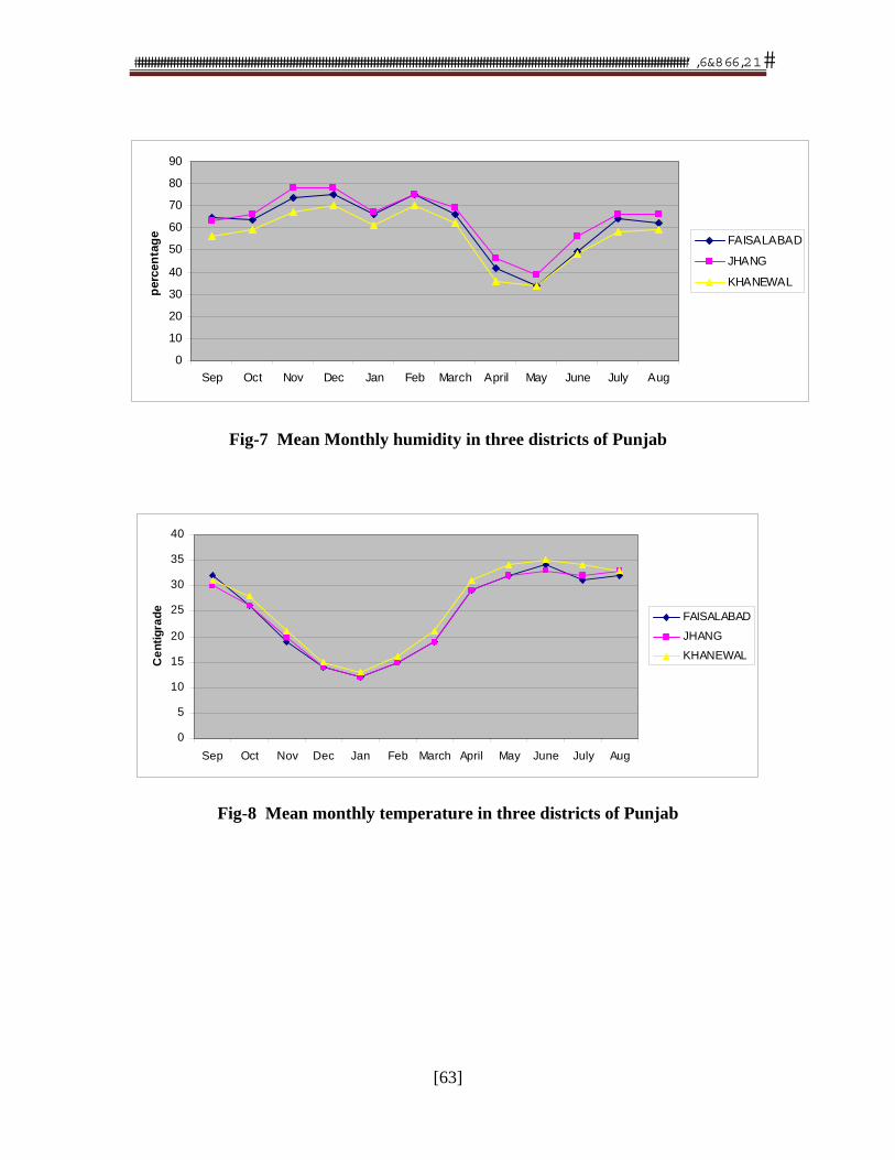
�������������������������������������������������������������������������������������������������������������������������������������������������������������������������������������
[63]
Fig-7 Mean Monthly humidity in three districts of Punjab
0
5
10
15
20
25
30
35
40
Sep Oct Nov Dec Jan Feb March April May June July Aug
Ce
nti
gra
de
FAISALABAD
JHANG
KHANEWAL
Fig-8 Mean monthly temperature in three districts of Punjab
0
10
20
30
40
50
60
70
80
90
Sep Oct Nov Dec Jan Feb March April May June July Aug
per
cen
tag
e FAISALABAD
JHANG
KHANEWAL
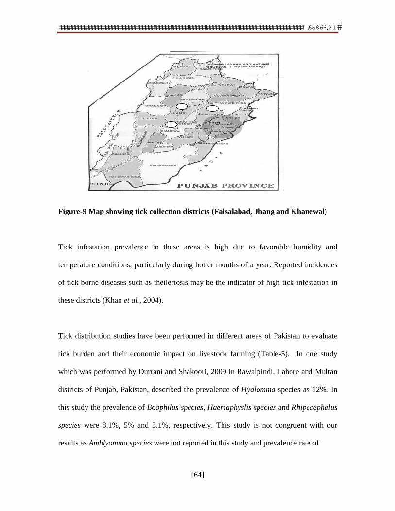
�������������������������������������������������������������������������������������������������������������������������������������������������������������������������������������
[64]
Figure-9 Map showing tick collection districts (Faisalabad, Jhang and Khanewal)
Tick infestation prevalence in these areas is high due to favorable humidity and
temperature conditions, particularly during hotter months of a year. Reported incidences
of tick borne diseases such as theileriosis may be the indicator of high tick infestation in
these districts (Khan et al., 2004).
Tick distribution studies have been performed in different areas of Pakistan to evaluate
tick burden and their economic impact on livestock farming (Table-5). In one study
which was performed by Durrani and Shakoori, 2009 in Rawalpindi, Lahore and Multan
districts of Punjab, Pakistan, described the prevalence of Hyalomma species as 12%. In
this study the prevalence of Boophilus species, Haemaphyslis species and Rhipecephalus
species were 8.1%, 5% and 3.1%, respectively. This study is not congruent with our
results as Amblyomma species were not reported in this study and prevalence rate of
�������������������������������������������������������������������������������������������������������������������������������������������������������������������������������������
[65]
Hyalomma species is significantly low as compared to our study. In lower Punjab
(Muzaffargarh and Layyah districts) H. anatolicum at 41% was highly prevalent tick
species in cattle population followed by R. sanguineus at 25.5%, while in buffalo
population H. anatolicum was at 36.3%, R. sanguineus was at 29.6% in this study
(Sajid et al., 2008). Hyalomma species prevalence in this study is higher as compared to
other tick species showing similar results to our study. Another tick infestation study was
reported by Manan et al., 2007 in Peshawar region situated in the Frontier province
(presently known as KPK) of Pakistan, showed highest prevalence of Boophilus species
at 46% followed by Hyalomma species at 31.25%, Rhipicephalus species at 17.93% and
Amblyomma species at 4.61%. Similar study was performed by Rehman et al., 2004 in
Rawalpindi and Islamabad areas which are situated in upper Punjab of Pakistan, showing
Haemaphysalis species prevalence as 74% and Hyalomma species prevalence as 26%.
Boophilus species, Amblyomma species and Rhipicephalus species were not reported in
Rawalpindi and Islamabad areas.
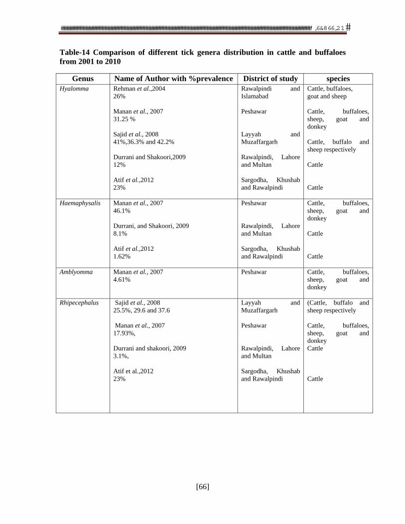
�������������������������������������������������������������������������������������������������������������������������������������������������������������������������������������
[66]
Table-14 Comparison of different tick genera distribution in cattle and buffaloes from 2001 to 2010
Genus Name of Author with %prevalence District of study species Hyalomma Rehman et al.,2004
26% Manan et al., 2007 31.25 % Sajid et al., 2008 41%,36.3% and 42.2% Durrani and Shakoori,2009 12% Atif et al.,2012 23%
Rawalpindi and Islamabad Peshawar Layyah and Muzaffargarh Rawalpindi, Lahore and Multan Sargodha, Khushab and Rawalpindi
Cattle, buffaloes, goat and sheep Cattle, buffaloes, sheep, goat and donkey Cattle, buffalo and sheep respectively Cattle Cattle
Haemaphysalis Manan et al., 2007 46.1% Durrani, and Shakoori, 2009 8.1% Atif et al.,2012 1.62%
Peshawar Rawalpindi, Lahore and Multan Sargodha, Khushab and Rawalpindi
Cattle, buffaloes, sheep, goat and donkey Cattle Cattle
Amblyomma Manan et al., 2007 4.61%
Peshawar
Cattle, buffaloes, sheep, goat and donkey
Rhipecephalus Sajid et al., 2008 25.5%, 29.6 and 37.6 Manan et al., 2007 17.93%, Durrani and shakoori, 2009 3.1%, Atif et al.,2012 23%
Layyah and Muzaffargarh Peshawar Rawalpindi, Lahore and Multan Sargodha, Khushab and Rawalpindi
(Cattle, buffalo and sheep respectively Cattle, buffaloes, sheep, goat and donkey Cattle Cattle
�������������������������������������������������������������������������������������������������������������������������������������������������������������������������������������
[67]
Our study demonstrated the occurrence of adult female ticks to be significantly higher as
compared to adult male ticks. Similar results were described by Sayin et al. (2003)
showing higher distribution of female ticks as compared to male ticks. In Hyalomma
species, prevalence of female tick population was higher than male tick population as
described by Flach et al. 1993 and Aktas et al. 2004. Ghosh et al., 2007 also reported
similar results in a study describing female population as 77% and 72% in Boophilus
species and Haemaphysalis species, respectively in one area of study in Bangladesh.
Khan et al. Salivary gland of adult female Hyalomma ticks have more type III acini as
compared to male describing their more disease transmission potential of female
Hyalomma tick as theileria parasites are only found in Type III acini (Young et al., 1983).
Moreover, histamine binding proteins which counteract pain/response of host to tick
attachment are two in female tick as compared to male ticks (Brossard and Wikel, 2004).
Similar result of tick infestation was reported by Sajid et al., (2008) describing 75.1%
cattle infested with ticks as compared to buffaloes with 40% infestation rate. These
findings are in line with our study in which we estimated tick infestation rate in cattle and
buffalo population. In cattle and buffaloes tick infestation was also observed by Rehman
et al., (2004) and Manan et al., (2007) describing double magnitude of ticks in cattle as
compared to buffaloes. In tick infestation level study high infestation level (tick number
above 50) was higher at 59% in cattle as compared to buffalo high at 18%. Moderately
infestation level (tick umber 25-50) was higher in buffaloes at 38% as compared to cattle
at 23%. Again there was a significant difference between buffalo and. In the category of
�������������������������������������������������������������������������������������������������������������������������������������������������������������������������������������
[68]
low infestation level (1-25), buffaloes has 44% as compared to cattle population at 18%.
High tick infestation rate and high tick level infestation in cattle may due to the reason
that majority of cattle in the area of study were cross bred which are more vulnerable to
tick infestation (Fesharki, 1988). Tick infestation level study in cattle and buffalo
population is not reported previous in the literature in these areas.
During study it was observed that animals tethered under trees have tremendously low
level of tick infestation than those animals which are kept in sheds. This was due to tick
eating habits of predator birds perching on the trees. In these areas small farm holders
usually keep poultry in their houses as poultry can not only pick ticks from the body of
animals but also from corner and crevices. Similar approach of tick biological control is
being used in some parts of America by rearing guinea fowl as Lyme disease tick eater
(reported in unpublished data). In our study, it was also observed that farmers spread sand
under and around their animals which is a type of physical control method of ticks.
2. Detection of T. annulata in tick species by PCR
The results of PCR Analysis showed the detection of T. Annulata in H. annatolicum and
H. dromedari but the protozoa was not detected in H. marginatum. Rhipicephalus
(Boophilus) annulatus and A.variegatum. Molecular detection survey of T. Annulata by
PCR from hard ticks has not been yet performed in Pakistan.
�������������������������������������������������������������������������������������������������������������������������������������������������������������������������������������
[69]
In conclusion, the tick population in cattle and buffaloes are higher particularly
Hyalomma species which are member of Ixodidae (hard ticks) and are potential vector for
the tropical Theileriosis in bovine and other wild animals not only in Pakistan and but
also in other regions like Europe, Africa, South Asia and Middle East (Dumanli et al.,
2005). As compared to previous studies (Table-7), higher prevalence of Hyalomma
species demands better integrated pest management measures for the control of tropical
theileriosis. Secondly, it is the first report that described the prevalence of T.annulata in
the ticks in Pakistan, which proves that ticks are spreading tick borne diseases in
livestock population of the country. Moreover disease transmission potential for other
prevalent ticks is also important like Rhipicephalus (Boophilus) (bovine babesiosis and
anaplasmosis), Haemaphysalis (theileriosis and babesiosis in sheep and goats) and
Amblyomma (Heart water in cattle).
3. Tick susceptibility due to differences in breed, sex and age of host
Holstein cows in mid-lactation period were artificially infested with tick larvae to
estimate host resistance against ticks. It was estimated that tick resistance in this breed
could be effective with out decreasing milk production (Jonson et al., 2000). Prevalence
of ticks was significantly higher in female cattle at 59.7% as compared to male at 35.83%
It was also estimated that tick infestation was more in young cattle at 46.28% as
compared to adult at 27.8% (Kabir et al., 2011). During feeding, ticks inject its saliva that
�������������������������������������������������������������������������������������������������������������������������������������������������������������������������������������
[70]
contain a variety of enzymes, toxins, enzyme inhibitors, anticoagulants and vasoacfive
substances that facilitate effective feeding . These pharmacopia in tick saliva produce
immune response in host vertebrates and make the animal resistance tick ifestation
(Abdel Wahab et al., 2000).
Non-chemical control method like immunization of susceptible animal species may have
an additive effect as artificial active acquired immunity could be a better alternate of
therapeutics application. Immunization results into reducing the number of engorged
ticks, weight and capacity of egg laying (Willadson et al., 1996), thus enhancing cattle
protection against blood sucking, immune-suppression, skin damage and tick borne
diseases (Andreoti et al., 2002). Immunoglobins produced after immunization is major
component of host serum as they can cross the tick gut and react with tissues in
hemocoele (Brossard and Wikel, 2004). In this way tick vaccine may affect the
transmission of tick borne diseases by decreasing vector capability of ticks (de la Fuente
and Kocan, 2006).
4. Effects on tick reproductive efficiency by immunization
Developmental and reproductive performances of adult H.anatolicum ticks were
observed in cross-bred calves by immunizing whole homogenate Hyalomma vaccine.
The vaccine mitigated egg mass, engorgement weight and reproductive index in
vaccinated animals because this vaccine contains exposed as well as concealed antigen.
�������������������������������������������������������������������������������������������������������������������������������������������������������������������������������������
[71]
Salivary contents as exposed antigens induce local hypersensitivity reactions which
results into restriction of blood flow to the tick. Intestinal contents as concealed antigens
provided local damage to gut resulting into gut perforation due to cell mediated lyses. In
immunized animals, rejected adult female ticks were white to pale yellow in color due to
the lack of red blood cells intake in their meal. This may suggest the decrease in
transmission of tick borne diseases due to reduced pick up of infected RBC’S.
Immunization of animals with tick vaccine although interfere the tick life cycles and their
reproductive capacity but there are no observations of high tick mortality (Rodriguez et
al., 1995 and Willadson, 1989). In our study percentage rejection of adult female
H.anatolicum tick was 68.8567±1.8687 which is significant higher than controlled group.
Das et al. (2000) purified extract of unfed larvae of H.a.anatilucm by immunoaffinity
chromatography and used for immunization of cross-bred calves. In their study rejection
percentage was 75.0±3.42 and 70.6±10.2, respectively, which is in agreement with than
that of our study. Slightly higher rejection percentage might be due to use of purified
antigen as a source of vaccine.
In engorgement period there was non-significant different between vaccinated and
controlled group. In vitro conditions egg laying mass in immunized group was
151.67±4.40 which was significantly higher as compared to controlled group. Similarly
engorged weight in immunized group was significantly high as compared to control
group. This indicates the disruption of feeding process of ticks due to immune response
�������������������������������������������������������������������������������������������������������������������������������������������������������������������������������������
[72]
induced by the vaccine in cross-bred calves. Similar parameters were used by Sangwan et
al. (1998) in their study, reproductive performance of immunized group was poor as
compared to control group but statistically there was no significant difference in these
parameters except reproductive index. Our study provides overall 69% protection of adult
ticks by immunization of whole homogenate tick vaccine in cross-bred calves, in
comparison to 55.8% protection provided by unpurified antigen of salivary gland (Saran,
1996). This variation is inline with our another study where immune response to whole
homogenate vaccine in rabbits was better as compared to vaccine prepared from salivary
gland extract.
Similar criteria for assessing the efficacy of tick vaccine was used (Rechav and Dauth,
1987; Varma et al., 1990; Opdebeeck et al., 1988a and Willadson et al., 1996) and their
results are in line with our study. We prepared whole homogenate vaccine from adult
female ticks and challenge infestation was later provided with adult female ticks of same
species. Mulenga et al. (2000) characterized a 29 kDa salivary gland associated protein
from Haemaphysalis longicornis which led to significant reduction in adult female
engorgement weight. Howerver, Varma et al., 1990; Ghosh et al.; 1998 and Ghosh and
Khan, 1999 have shown that rabbits and cattle were protected significantly against adults
of Rhipecephalus appendiculatus and Hylomma a. anatolicum following immunizing
with the immature stage. In future study, immune response of vaccine prepared from
local strain of adult tick might be monitored in the immature stages of same species.
�������������������������������������������������������������������������������������������������������������������������������������������������������������������������������������
[73]
Our rationale of applying ELISA on immunized and non-immunized group was to co-
relate the immune response produced by whole homogenate vaccine with feeding and
reproductive performance of the ticks after immunization. Up to 15 days of post
vaccination, there was no significant difference between antibody titer of immunized and
non-immunized group due to slow rise in antibody titer in serum. The result of present
study shows that tick infestation can be controlled in the area by using
immunoprophylactic measures. After boosting, rapid rise in antibody titer was detected;
this is a characteristic of memory response. On day 45, the rise in antibody titer in cross-
bred calves of control group was due to challenge infestation of ticks that was equally
provided in immunized and control group. A significant difference in ELISA titer value
was observed between immunized and non-immunized group.
It has been reported that a vaccine against ticks would progressively control ticks and tick
borne diseases in successive generations rather controlling tick population in single
generation (Patarroyo et al., 2002). The principle of such immunization is uptake of
blood meal, tissue fluid or wound exudates which inevitably carry antibodies,
complements and other components of host immune system (Tizard, 2000). There are
three ways of complement activation (Tortora, 2007), the alternative path-way of
complement activation is involved in the host-tick interaction (Wikel, 1979). Acquired
resistance to ticks may be based on circulating anti-tick IgM and IgG antibodies as well
as Th1-cell-mediated delayed type hypersensitivity reactions, modulated in part by IL-2
�������������������������������������������������������������������������������������������������������������������������������������������������������������������������������������
[74]
and IFN-g (Wikel and Bergman, 1997). Control of hard ticks has been difficult as they
have fewer enemies (de la Fuente and Kokan, 2006) and as a whole arthropod parasites
are difficult to immunize against because being ectoparasites they feed intermittently
from different host, (Opdebeeck, 1994).
5. Vaccination studies
5.1 Different tick organ tissues as candidate antigen
Tick GARD (Escherichia coli expressed BM 86 vaccine) and GAVAC (Pichia pastoris
expressed BM 86 vaccine) have been developed against infestation of B. microplus and
these vaccines may have mitigated use of acaricides (Anderotti et al., 2002). Presence of
only one immunogen in recombinant tick vaccine seems to be a plausible reason of less
attraction of the vaccine to be used against tick infestation (Mulenga et al., 2000).
However, different attempts have been made to immunize animals against the ticks by
using crude extract (Sangwan et al., 1998). Crud immunogenic extracts from different
stages of H. a. anatolicum have been found protective against challenge infestatation
(Manohar and Banerjee, 1992; Sangwan et al., 1998 and Ghosh et al., 1998) but
information about the potency or efficacy of such crude extract is scanty.
Different efforts have been made previously to immunize rabbits against H. anatolicum
using crude extract antigen prepared from different tissues of the tick. Histological
structure of hard ticks is mainly composed of chitinous coverings, legs, mouth parts, and
internal organs like salivary tissues and intestinal epithelium lining the body cavity. In
�������������������������������������������������������������������������������������������������������������������������������������������������������������������������������������
[75]
our study, organ-based vaccines were prepared from extracts of salivary glands (SGE),
intestinal (ITI) tissue and whole tick homogenate (WTH) of Hyalomma ticks. Salivary
gland is a paired organ consisting of grapelike clusters of acini extending from the level
of the peritremes along the sides to the gnathosoma, where the paired main ducts open
into the salivarium, which opens dorsally into the buccal canal. Salivary glands are
essential for the biological success of ticks and induce number of pharmacopeia during
feeding on mammalian host. These pharmacological agents are anticoagulants,
prostaglandin, vasodilators, apyrase, antiinflammatory agents, anti-histamines etc.
(Bowman and Sauer, 2004). The importance of multi-functionary salivary gland to tick
survival and vector competency makes the gland a potential target for intervention
(Bowman and Sauer, 2004) and an attractive immunogenic source (exposed antigen) for
vaccine development against TBD (Valenzuela, 2004). Tick salivary pharmacopeia also
potentiates infection with several infectious agents like tick-borne encephalitis virus,
Borellia burgdorferi sensu stricto and Franciscella tularensis through saliva-activated
transmission (SAT) (Nuttal and Labuda, 2004). Tick saliva often contains high
concentration of prostaglandins (Bowman et al., 1996) which are vasodilators (Riebero,
1995a) and inhibitors of platelet aggregation.
Intestinal tissue acting as concealed antigen can also be a source of tick antigen as
antibody engorged from successfully immunized host blood forms antigen-antibody
complex with the help of complements and causes damage to tick intestine. Muslin cloth
filtrate of whole tick homogenate contained mainly extract of intestinal epithelium and
�������������������������������������������������������������������������������������������������������������������������������������������������������������������������������������
[76]
salivary gland, so it has both the qualities of exposed antigen as well as concealed
antigen. The immune response of vaccines prepared from each of SGE, ITE and WTE
was monitored and compared by AGPT in rabbits. The antibody response of rabbits to
WTH protein was better than that of vaccine prepared from isolated salivary gland or
intestinal tissue homogenate because WTH contains salivary gland components (exposed
antigen) as well as intestinal components (concealed antigen). The reason is that
immunity induced by natural antigen (exposed) is believed to be inferior as compared to
concealed antigen due to reduced antigenicity of natural antigen (Tellam et al., 1992).
One drawback of non-salivary gland vaccine is that this
may not absolutely prevent feeding of ticks resulting into transmission of tick borne
diseases and hide damage (Ribero, 1987).Valenzuela et al., 2004 purified an anti-
complement protein from I. scapularis saliva which appears to inhibit C3b binding and
accelerate upcoupling of factor Bb from alternative pathway C3 convertase.
5.2 Cross-reactivity study
A cross-reactivity study amongst different prevalent genera was conducted. In
conclusion, whole tick homogenate (WTH) of Hyalomma species shows cross reactivity
with WTH of Boophilus and Amblyomma species. The results are in agreement to
Kawather et al., 2006; de Vos et al. 2001 and de la Fuente, 2006 who reported evidence
of cross reactivity between different hard tick species. They noted that Tick GARD which
�������������������������������������������������������������������������������������������������������������������������������������������������������������������������������������
[77]
is B. microplus derived recombinant Bm86 vaccine is effective against Hyalomma,
Rhipecephalus and Amblomma species of hard ticks. Bm86 is a membrane-bound
glycoprotein of 89kDa on the microvilli of tick gut digest cells (Willadsen et al., 1989).
Bm86 has been expressed in E. coli, insect cells using a baculovirus vector (Rand et al.,
1989) and re-cloned from an Argentine isolate of B. microplus and designated BM95
(Garcia Garcia et al., 2000). The amino acid sequence of a Bm86 homologue found in H.
a. anatolicum demonstrated the conservation of this molecule in this species. Gavac
vaccine containing the B.microplus gut antigen Bm86 showed high efficacy against
B.annulatus tick infestation in Mexico and Iran (Fragoso et al., 1998). Vaccine made
against one tick species can be used against other tick species if cross reactivity occurs
between their antigens. Moreover, existing tick vaccines could be improved by the
inclusion of additional tick antigens in case of existence of cross-protection.
Sangwan et al. (1998) performed a study in which they derived antigens from partially
engorged nymph of H. anatolicum anatolicum and used in immunization of cross-bred
cattle against adult H.a.anatolicum. This antigen provided no cross protection against the
antigen from adult stage and the results differ with our observations. Rechav et al. (1989)
artificially immunized guinea pigs and rabbits with larvae of any one of five species of
ticks, Rhipicephalus appendiculatus Neumann, Rhipicephalus evertsi evertsi Neumann,
Amblyomma hebrauem Koch, Amblyomma variegatum Fabricius and Ixodes ricinus L.
Resistanc was conferred in the animals when exposed to subsequent infestations with the
�������������������������������������������������������������������������������������������������������������������������������������������������������������������������������������
[78]
same tick species. Resistance to infestations was species specific and protection was not
observed against other tick species.
Similarly, anti-sera raised against 64 truncated recombinant proteins (TRPs), a secreted
cement protein from salivary gland of Rhipicephalus appendiculatus showed cross
reactivity with crude extract proteins of Ixodes recinus and Rhipicephalus sanguineus
(Trimnel et al., 2005). Two larval glycoproteins 34 kDa and 29 kDA were isolated from
larvae of Hyalomma anatolicum and Boophilus microplus respectively. Cross reactivity
and synergistic effects were observed between these two glycoprotein (Ghosh et al.,
2005). In another study, truncated constructs of 64P (64TRP’S), a secreted cement
protein from salivary glands of the tick Rhipicephalus appendiculatus also provided cross
reactivity against R. sanguineus and Ixodes ricinus. Apparently, this protection was
provided by making target of antigens in the mid-gut and salivary glands (Trimnell,
2005). Subolesin, a highly conserved protein discovered in I.scapularis by expression
library immunization was protective against all developmental stages of I. scarpularis
(Almazan et al., 2003).
These results suggest the feasibility of controlling infestation by multi tick species using
protective antigen from one source tick species. It can be also suggested that tick vaccine
in future may be formulated by inclusion of multiple antigens which will have broader
possible applications.
�������������������������������������������������������������������������������������������������������������������������������������������������������������������������������������
[79]
5.3 Dose of antigen
A study was conducted to optimize the concentration of tick immunogen per dose in
vaccine that would provide detectable antibodies in the immunized buffalo calves. For
this purpose CFT was performed by using guinea pig as complement source because it
contains greater concentration of all complements. Complement has the ability to cause
the hemolysis of sensitized sheep RBC’S and combines with antigen-antibody complex
causing its fixation. The immunized buffalo calves showed detectable level of anti-WTH
complement fixing antibodies in response to the oil based WTH vaccine containing more
than 7.5 mg protein/dose.
The antibody titer started increasing on day 15 post priming, reached to peak level on day
45 post-priming and declined thereafter. It was further noted that antibody response of
buffalo calves to vaccine containing 7.5 mg protein was not significantly higher than that
of the vaccine dose containing 10.0 mg protein (P<0.05). These results are in agreement
with that of Andreotti et al. (2002), who observed high level of anti- Boophilus microplus
Trypsin Inhibitors (BmT1s) antibodies in Bos indicus Nelore calves on 40 days post
priming with subcutaneous injection of the vaccine (0.1 mg protein/ dose). In their study
high level of ELISA antibodies to low amount of immunogen could be due to highly
sensitive technique for antibody detection. However, Jackson and Opdebeeck (1990)
observed significant increase in total immunoglobulin in serum of cattle immunized with
small amounts of mid-gut membrane antigens (500ug) in Quil A (Pipano et al., 2003).
�������������������������������������������������������������������������������������������������������������������������������������������������������������������������������������
[80]
Opdebeeck et al., 1988a protected cattle with 2mg and then 500ug of crude midgut
antigen from B.microplus. In our study SGE, ITE and whole homogenate vaccines with
2.5mg dose provided detectable immune response in rabbits. In guinea pigs, 200ng of
partially purified salivary gland antigen from A. americanum was inoculated. This
induced a low level of protection (23.4 %) due to lower dose of antigen (Brown and
Askenase, 1986).
We inoculated whole homogenate vaccine with 7.5mg concentration in single dose, but
vaccine inoculated in multiple and small doses have been shown better immune response
as compared to large and single dose (Murray et al., 1979 and Oldham, 1983). In future
study, 7.5mg immunogen may be inoculated in crossbred calves in multiple doses to
monitor immune response and effect of tick rejection. Vaccine efficacy study was
conducted on limited experimental scale and Crossbred animals. It is suggested that field
trials in large population may be conducted to increase the strength and precision of
results. Moreover, various bovine breeds should be included in the field trials to confirm
the potency and efficacy results. The result of present study shows that tick infestation
can be controlled in the area by using immunoprophylactic measures. The future of
research on tick vaccine development is exciting and lot of work is still to be done in the
field of identification of candidate vaccine antigens and exploiting the recombinant
technology in the synthesis of these antigens. Development of vaccine that target both
ticks and pathogen transmission would be the required goal of these research projects.
�������������������������������������������������������������������������������������������������������������������������������������������������������������������������������������
[81]
Invaluable information from tick genomics may be exploited for the development of tick
vaccine. Immunization of these tick vaccines to animal population may also restrict
transmission of tick borne pathogens to human population at risk. The results of our study
showed that the vaccine prepared in this way may be contaminated with T. Annulata –the
causative agent of theileriosis. It is therefore recommended that buparvaquone be
administered to animals before immunization of this vaccine.
6. Growth of tick intestinal cell as monolayer
The efforts were made to grow midgut cells in insect culture media after isolation the
cells from Hyalomma ticks. The gut of arthropods is composed of foregut, midgut and
hindgut divisions. The midgut may be further divided into anterior and posterior regions
and is the site of action for blood meal digestion and absorption. The majority of cells in
midgut are digestive in nature and there are four different types of these digestive cell
which have secretory vesicles (Marquardt et al., 2005). Midgut is the region where
antigen-antibody complex is formed with the help of complement which leads to
perforation of midgut. The isolated epithelium cells got confluence rate up to 40% in
seven days and then started distorted after each effort due to some contamination. There
are different sources of contamination for the tick cell culture which are extrinsic as well
extrinsic. The purpose of this experiment was to grow midgut cell and then use these cells
as a source of antigen for vaccine development. The growth rate of these cells in our
study was no optimal so the media was not split to get more cells.
��� � �������������������������������������
[82]
CHAPTER 7
SUMMARY
A study to investigate prevalence of different genera of hard ticks was carried out in three districts of the
Punjab province, Pakistan (Faisalabad, Jhang and Khanewal). Overall prevalence of Hyalomma species is
the highest at 61% as compared to other genera of hard ticks. In sex-wise distribution, it was found that
female Hyalomma species were the highest at 85% followed by Amblyomma species at 81%, while
Rhipicephalus (Boophilus) species and Haemaphysalis species were at 77%. Infestation rate in cattle at
70% was higher as compared to buffaloes at 34%. In tick infestation level study, high infestation level in
cattle at 59% was higher as compared to that of buffalo population at 18%. In cattle population, PCR
results showed the prevalence of T. annulata in H. anatolicum and H.dromedari ticks as 50% and 40%
respectively. No theilerial organism was detected in H.marginatum, Rhipicephalus (Boophilus) annulatus
and Amblyomma variegatum ticks.
Three different types of vaccines were prepared from different organs of ticks i.e., salivary gland,
intestine or whole ticks of the same species of Hyalomma and they were injected to rabbits. It
was found that vaccine prepared by grinding whole tick produced the higher level of antibody as
compared to two other vaccines. Each of the whole tick homogenate vaccine prepared from
either of the species of Hyalomma, Rhipicephalus or Amblyomma and injected to rabbits. These
vaccines produced antibody as well and cross reacted with each other showing each of the hard
ticks were antigenically similar.
Efforts were made to prepare oil based whole Hyalomma tick vaccine with three different
antigen concentration 5.0 mg, 7.5mg and 10.0 mg and evaluated its potency in buffalo calves. It
��� � �������������������������������������
[83]
was found that the vaccine dose containing 5.0 mg antigen per dose did not produced detectable
antibody in buffalo calves while the vaccine containing 7.5mg or more antigen produced
detectable antibody. Moreover, we concluded that montanide based hard tick homogenate
vaccine with more than 7.5mg protein per dose is effective in producing antibodies against tick
infestation in the dairy animals. The antibody level in vaccinated buffaloes as well as in
vaccinated rabbits reached to peak level on day 45 post vaccination and started declining
thereafter.
Capacity of vaccine in controlling tick infestation was assessed in 12 cross-bred calves. It was
found that rejection percentage in immunized group was higher as compared to control group.
There was no difference of engorgement period between immunized and control group.
Reproductive index in immunized group was lower as compared to control group.
The efforts were made to grow midgut cells insect culture media after isolation them from Hyalomma
ticks. The purpose of this experiment was to grow midgut cell and then use these cells as a source of
antigen for vaccine development. The isolated epithelium cells got confluence rate up to 40% in seven
days and then started distorted after each effort due to some contamination. There are different sources of
contamination for the tick cell culture which are extrinsic as well extrinsic. The growth rate of these cells
in our study was not optimal so the media was not splitted to get more cells.
������������������������������������������������������������������������������������������������������������������������������������������������������������������������
[84]
CHAPTER 7
LITERATURE CITED
Abdel–Wahab, K.S; S. M. Habeeb; and K. M. El Kammah (2000).Cattle Natural
immunity against the cattle tick Boophilus annulatus (Acari: Ixodidae): detection of
antibodies against salivary gland and gut proteins using ELISA. Inter. J. of Acarol.
26(4):385-389
Ahmad, M. Bukhari, A. and Awan, F. (2000). Punjab Livestock Census, Livestock and
Dairy Development Lahore, Punjab. Vol. 6 Part-II.p. 465.
Akhtar, M. (1995). Studies on the immunization of buffalo against Boophilus microplus.
Ph.D., thesis. Dept. of Parasitol., Univ. Agri. Fsd. Pakistan.
Aktas, M., Dumanli, N. and Angin, M. (2004). Cattle infestation by Hyaloma ticks and
prevalence of Theileria in hyalomma species in the East of Turkey. Vet. Parasitol.,
119(1):1-8
Almazan, C; K.M. Kocan; D.K. Bergman; J.C. Garcia-Garcia; E.F. Blouin and J. de la
Fuente (2003). Identification of protective antigens for the control of Ixodes scapularis
infestations using cDNA expression library immunization. Vaccine,21: 1492–1501
������������������������������������������������������������������������������������������������������������������������������������������������������������������������
[85]
Andreotti, R; A. Gomes; K.C. Malavazi-piza; S.D. Sasaki; C.A.M. Sampaio and A.S.
Tanaka (2002). BmTI antigens induce a bovine protective immune response against
Boophilus microplus tick. Int. Immunopharmacol. 2:557– 563
Assenga, S. P; M. You; C.H. Shy; J. Yamagishi; T. Sakaguchi; J. Zhou; M. K. Kibe;
X. Xuan and K. Fujisaki (2005) . The use of a recombinant baculovirus expressing a
chitinase from the hard tick Haemaphysalis longicornis and its potential application as a bioacaricide for tick control. Parasitol. Res. 98(2):111-118
Atif, F. A; M. S. Khan;, H. J. Iqbal; Z. Ali and S. Ullah (2012). Prevalence of cattle tick
infestation in three districts of the Punjab, Pakistan. Pak. J. Sc. 64(1) 49-53
Barbet, A. F; R. Blentlinger; Yi Jooyoung; A.M. Lundgren; E. F. Blouin and K. M.
Kocan (1999). Comparison of Surface Proteins of Anaplasma marginale Grown in Tick
Cell Culture, Tick Salivary Glands, and Cattle. Infection and Immunity, 67(1):102-107
Barker, S.C. and A. Murrell (2004). Systematics and evolution of ticks with a list of valid
genus and species names. Parasitology, 129, S15–S36
Bell-Sakyi, L; A. E.D Paxton, U. G. Munderloh, and K. J. Sumption (2000). Growth of
Cowdria ruminantium, the Causative Agent of Heartwater, in a Tick Cell Line. J. of
Clinic. Microbiol. 38(3): 1238-1240
������������������������������������������������������������������������������������������������������������������������������������������������������������������������
[86]
Bhattacharyulu, Y; R.P, Chaudhri and B.S. Gill (1975). Transstadial transmission of
T.annulata through common ixodid ticks infesting Indian cattle. Parasitology, 71:1-7
Bishop R; B. Lambson; C. Wells; P. Pandit; J. Osaso; C. Nkonge; S. Morzaria; A.
Musoke and V. Nene (2002). A cement protein of the tick Rhipicephalus appendiculatus,
located in the secretory cell granules of the type III salivary gland acini, induces strong
antibody responses in cattle. Int. J. for Parasitol. 32 (7): 833-842.
Bowman, A.S., Dillwith, J.W. and Sauer, J.R. (1996). Tick salivary prostaglandins:
presence, origin and significance. Parasitology Today. 12:388-396.
Bowman, D.D; Lynn, R.C and Eberhard M.L. (2003). Georgis’ Parasitology for
Veterinarions. Elsevier Science, 11830 Westline Industrial Drive, St. Louis, Missouri,
USA. p 52.
Bowman, A.S. and J.R. Sauer (2004). Tick salivary glands: function, physiology and
future. Parasitology, 129s:67-82.

������������������������������������������������������������������������������������������������������������������������������������������������������������������������
[87]
Brossard, M. and S.K. Wikel (2004). Tick immunology. Parasitology, 129s: 161-171
Brown, S.J and P.W. Askenase (1986). Amblyomma americanum: physiochemical
isolation of a protein derived from the tick salivary gland that is capable of inducing
immune resistance in guinea pigs. Exp. Parasitol. 62(1):40-50
Chaudhry, S. M. and S. Kamal (2006). Introduction to Statistical Theory, Ilmi Kitab
Khana, Kabir Street, Urdu Bazar, Lahore. pp: 506-509
Chhabra, M.B. and K.L Khurana (2005). Immunity and immunization against Hyalomma
tick vector of bovine tropical theilriosis. J.Vet.Parasitol. 19(1): 456-459
Das, S.S. (1994). Prevalence of Ixodid tick infestation on farm animals in Pantnager,
Terai of Utter Pardesh. J. Parasitol. and App. Anim. Bio. 3(1): 31-73
Das, G; S. Ghosh; M.h. Khan; and J.K. Sharma (2000). Immunization of cross-bred
cattle against Hyalomma anatolicum anatolicum by purified antigens. Exp Appl Acarol.
2000 24(8):645-59.
������������������������������������������������������������������������������������������������������������������������������������������������������������������������
[88]
de la Fuente, J; M. Rodriguez; M. Redondo; M. Carlos; C.G. Jose; M. Luis; S. Emerio;
V. Mario; E. Antonio; C. Mario; R. Eduardo; B. Oscar; M. Hector; L. Ricardo; C.A. de
Armass; R. Sara; J.R. Rodriguezs; A. Mirna and G. Libertad (1998). Field studies and
cost-effectiveness analysis of vaccination with Gavac against the cattle tick Boophilus
microplus. Vaccine, 16 (4): 366–373
de la Fuente J. and K. M. Kocan (2006). Strategies for development of vaccines for
control of ixodid tick . Parasite Immunol. 28: 275-283
de Oliveiraa, C; M. van der Weide; P. Jacquiet and F. Jongejana (1997). Detection of
Theileria annulata by the PCR in ticks (Acari: Ixodidae) collected from cattle in
Mauritania. Exp. and App. Acarology, 21: 279–291
De Rose R; R.V. McKenna; G. Cobon; J. Tennent; H. Zakrzewski and K. Gale (1999).
Bm86 antigen induces a protectiveimmune response against Boophilus microplus
following DNA and protein vaccination in sheep. Vet Immunol Immunopatho; 71: 151–60
de Vos, S; L. Zeinstra; O. Taoufik; P. Willadsen and F. Jongejan (2001). Evidence for
the utility of the Bm86 antigen from Boophilus microplus in vaccination against other
tick species. Exp. and App. Acarol. 25: 245–261
������������������������������������������������������������������������������������������������������������������������������������������������������������������������
[89]
Dumanli, N; M. Aktas; B. Cetinkay; A. Cakmak; C.E. Koroglu Saki; Z. Erdogmus;
S. Nalbantoglu; H. Ongor; S. Simsek; M. Karahan and K. Altay (2005). Prevalence and
distribution of tropical theileriosis in Eastern Turkey. Vet. Parasitol. 127: 9–15
Durrani, A.Z. and A.R. Shakoori (2009). Study on ecological growth conditions of cattle
Hyalomma ticks in Punjab, Pakistan. Iran. J. Parasitol. 4(1):19-25
Fesharki, R.H. (1988). Control of Theileria annulata in Iran. Parasitol. Today. 4(2):36 40
Fivaz, B.H; I. Renckon; R. Cross; S. Terry and D. Hendry (1991). Specificity and Effects
of host resistance on the African tick Rhipicephalus zambeziensis (Acarina:Ixodidae).
Exp. and Appl. Acarol. 11:223-232
Flach, E. J., H. Ouhelli, D. Waddington, and M. El-hasnaoui (1993). Prevalence of
Theileria in the tick Hyalomma detritum in the Doukkala region, Morocco. Med. and Vet.
Entomol., 7(4):343-350
Fragoso, H; P. Hoshman Rad; M. Ortiz; M. Rodriguez; M. Redondo; L. Herrera and J.
de la Fuente (1998). Protection against Boophilus annulatus infestations in cattle
vaccinated with the B. microplus BmS6-containing vaccine Gavac. Vaccine,
16(20):1990-l992
������������������������������������������������������������������������������������������������������������������������������������������������������������������������
[90]
Garcia-Garcia, J. C; S. Alejandro; N.S. Fabian; M.S. Marcela; J. Marisdania; H.Maidel;
L. Jorge and J. de la Fuente (1998). Adjuvant and immunostimulating properties of the
recombinant Bm86 protein expressed in Pichia pastors. Vaccine, 16(10):1053-1055
Garcia-Garcia, J.C; C. Montero; M. Redondo; M. Vargas; M. Canales; O. Boue; M.
Rodriguez; M. Joglar; H. Machado; I.L. Gonzalez; M. Valdes; L. Mendez and de la J.
Fuente (2000). Control of ticks resistant to immunization with Bm86 in cattle vaccinated
with the recombinant antigen Bm95 isolated from the cattle tick, Boophilus microplus.
Vaccine, 18(21): 2275–2287
Gharbi, M; L. Sassi; P. Dorchies and P. Darghouth (2006). Infection of Calves with
Theileria annulata in Tunasia: Economic analysis and Evaluation of the Potential benefit
of vaccination .Vet. Parasitol. 137:231-241
Ghosh, S; M.H. Khan and S.C. Gupta (1998). Immunization of rabbits against Hyalomma
anatolicum anatolicum using homogenates from unfed immature ticks. Indian J. Exp.
Biol. 36: 167–170
������������������������������������������������������������������������������������������������������������������������������������������������������������������������
[91]
Ghosh, S; N.K. Singh and G. Das (2005). Assessment of duration of immunity in
crossbred cattle immunized with glycoproteins isolated from Hyalomma antolicum
anatolicum and Boophilus. Parasitol. Res. 95: 319-326
Gosh, S; P. Azhahianambi and M.P. Yadav (2007). Upcomming and future strategies of
tick control: a review. J.Vect. Borne Dis. 44: 79-89
Gwakisa, P; K. Yoshihara; T. Long To; H. Gotoh; F. Amano and M. Eiichi
(2001). Salivary gland extract of Rhipicephalus appendiculatus ticks inhibits in vitro
transcription and secretion of cytokines and production of nitric oxide by LPS-stimulated
JA-4 cells. Vet. Parasitol. 99:53–61
Harrow I.D., K.A.F. Gration, N. Evans (1991) Neurobiology of arthropod parasites.
Parasitol 102:559–569
Hoogstraal H. (1956). African Ixodidae 1. Ticks of the Sudan (with special reference to
Equatorial Province and preliminary reviews of the genera Boophilus, Margaropus and
Hyalomma). Research Report NM 005.05.29.27, Department of Navy Bureau of
Medicine and Surgery, Washington D.C., 1,101 pp.
Hudson, L. and F.C Hay (1976). Practical Immunology, 1st edn, p. 134, Blackwell
Scientific Publications, London.
������������������������������������������������������������������������������������������������������������������������������������������������������������������������
[92]
Iqbal, R.Z; H. Song-hua; C. Wan-jun; G.A. Abdullah and X. Chen-wen (2006).
Importance of ticks and their chemical and immunological control in livestock. J.
Zhejiang Univ. SCIENCE B 7(11):912-921
Ismael, A.B; D. Sekkai; C. Collin; D. Bout and M. Mevelec (2003). The MIC3 Gene of
Toxoplasma gondii Is a Novel Potent Vaccine Candidate against Toxoplasmosis. Infec.
and Immunity. 71(11): 6222–6228
Jackson, L.A. and J.P. Opdebeeck (1990). Humoral immune response of Hereford cattle
vaccinated with mid-gut antigens of the cattle tick, Boophilus microplus. Parasite
Immunol. 12: 141-151
Jongejan, F. and G. Uilenberg (1994). Ticks and control methods. In: Ectoparasites of
Animals and Control Methods. Blancou, J. (ed.), Scientific and Technical Review, Office
Internationale des Epizooties (OIE), Paris, France. 13(4):1201-1226
Jongejan, F and G. Uilenberg (2004). The Global importance of ticks. Parasitology,
129s: 3-14
������������������������������������������������������������������������������������������������������������������������������������������������������������������������
[93]
Jonsson, N.N; D.G. Mayer; A.L. Matschossa; P.E. Green and J. Ansell (1998).
Production effects of cattle tick (Boophilus microplus) infestation of high yielding dairy
cows. Vet. Parasitol. 78:65-77
Jonsson, N.N; A.L Matschoss; P. Pepper; P.E. Green ande J. Ansel (2000).
Resistance of Holstein-Friesian cows to infestation by the cattle tick (Boophilus
microplus). Vet. Parasitol. 89, 297–305
Kabir, M.H.B; M.M.H. Mondal; M. Eliyas; M.A. Mannan; M. A. Hashem; N. C.
Debnath; F. Miazi; C. Mohiuddin;_M. A. Kashem; M. R. Islam and M. F. Elahi (2011).
An epidemiological survey on investigation of tick infestation in cattle at Chittagong
District, Bangladesh. African J. of Research 5(4) 346-352
Kawther, M.E; S.E.A Kouka; M.I.O Laila; and M.I.O. Andhanan (2006). Studies on
hyalomma Dromedarii (acari: ixodidae) salivary glands and gutimmunogenecity. Arab j.
Biotech., 9: 41-50
Khan, M.H; R.C. Naithani and K.S. Singh (1982). Study on the bionomics of Nosomma
monostrosum (Nuttall and Warburton) (Acari: Ixodidae). Acarologia, 23: 119–123
Khan,.M. Q; A. Zahoor; 1. M. Jahangir and M. Ashraf Mirza (2004). Prevalence of blood
parasites in cattle and buffaloes. Pak. Vet. J. 24(4):193-195
������������������������������������������������������������������������������������������������������������������������������������������������������������������������
[94]
Khan, M.N; Z. Iqbal and C. S. Hayat (1998). Evaluation of acaricidal efficacy of
Ivermectin, Diazinon, Permethrin and Coumaphos in sheep and goats. Pak. J. of
Biological Sc. 1(2):63-65
Kivaria, F.M. (2006). Estimated direct economic costs associated with tick-borne
diseases on cattle in Tanzania. Trop. Anim. Health Prod. 38 (4): 291-299
Kocan, K.K; J. de la Fuente; E.F.Blouin; J.F. Coetzee and S.A. Ewing (2010). The
natural history of Anaplasma marginale Vet. Parasitol. 167: 95–107
Konnai, S; S. Yamada; S. Imamura; M. Simuunza; M. Chembensof; A. Chota; A.
Nambota; K. Ohashi and M. Onuma (2007). Attachment Duration Required for
Rhipicephalus appendiculatus to Transmit Theileria parva to the Host. Vector-Borne and
Zoonotic Dis. 7(2):241-248
Latif, A.A. and R.G. Pegram (1992). Naturally acquired host resistance in tick control in
Africa. Insect. Sci.and App. 13(4): 54-56
Lowry, O.H; M.J. Rosebrough; A.L. Farr and R.J. Randall (1951). Protein measurement
with the Folin phenol reagent. J. Biol. Chem. 193: 265-275
������������������������������������������������������������������������������������������������������������������������������������������������������������������������
[95]
Iori, A; D. Grazioli; E. Gentile; G. Marano and G. Salvatore (2005). Acaricidal
properties of the essential oil of Melaleuca alternifolia Cheel (tea tree oil) against
nymphs of Ixodes ricinus. Vet. Parasitol. 129(1-2):173-176
Makala, L.H; P. Mangani; K. Fujisaki and H. Nagasawa (2003). The current status of
major tick born diseases in Zambia Vet. Res.34:27-45
Manan, A; Z. Khan; B. Ahmad and Abdullah (2007). Identification of ixodid tick genera
in Frontier region ,Peshawar. J. Agri. and Bio. Sci . 2(1)
Manohar, G.S. and D.P. Banerji (1992). Effects of immunization of Rabbits on
establishment surrvial and reproductive biology of the ticks Hyalomma anatolicum
anatolicum. Parasitology, 78:77-81
Marquardt, W.C; W.C. Black IV; S. Higgs; J.E. Freier; A.A. James; H.H. Hagedorn; B.
Kondratieff; J. Hemingway and C.G. Moore (2005). Biology of disease vectors. 2nd Edit.
Elsevier Academic Press, Burlington, USA.pp. 290
Martin, J.R. and J. Furlog. (2001). Avermectin resistnace of the cattle tick Boophilus
microplus in Brazil. Vet. Record. 149-164
������������������������������������������������������������������������������������������������������������������������������������������������������������������������
[96]
McCoskar, P.J. (2004). The global importance of thelerisosis. In. Thelriosis Ristie M,
Kreier JP (eds), New York: Academic Press: 1-24
Merchant, L.A. and Packer, R.A (1983). Bacteriology and Virilogy 7th Edi. CBS
publishers & distributors 485, Janin Bhawan, Bhola Nath Nagar Shahdra, Delhi-110032,
India, pp.163
Mejri, N; B. Rutti and M. Brossard (2001). Immunosuppressive effects of Ixodes ricinus
tick saliva or salivary gland extracts on innate and acquired immune response of BALB/c
mice. Parasitol. Res. 88 (3): 192 – 197
Meltzer, M. I. and R. A. I. Norval (1993). Evaluating the economic damage threshold for
bont tick (Amblyomma hebraeum) control in Zimbabw. Exp. App. Acarol. 17(3):171-185
Meltzer, M.I. (1996). A possible explanation of the apparent breed resistance in cattle to
bont leg ( Amblyomma herbraeum) infestations .Vet. Parasitol.67:275-279
������������������������������������������������������������������������������������������������������������������������������������������������������������������������
[97]
Minjauw, B and A. McLeod ( 2003). Tick-borne Diseases and Poverty. The Impact of
Ticks and Tick-borne Diseases on the Livelihoods of Small scale and Marginal Livestock
Owners in India and Eastern and Southern Africa. Research Report, DFID Animal Health
Programme, Centre for Tropical Veterinary Medicine, University of Edinburgh, UK,
p. 116
Muhammad, G; A. Naureen; M. Saqib and S. Faryal (2008). Tick control strategies in
dairy production medicine. Pak. Vet. J. 28: 43-50
Mulenga, A; C. Sugimoto; Y. Sako; K, Ohashi; A. Musoke; M. Shubash, and M. Onuma
(1999). Molecular Characterization of a Haemaphysalis longicornis Tick Salivary Gland-
Associated 29-Kilodalton Protein and Its Effect as a Vaccine against Tick Infestation in
Rabbits. Infection and immunity, 67(4):1652–1658
Mulenga, A; C. Sugimoto and M. Onuma (2000). Issues in tick vaccine development:
identification and characterization of potential candidate vaccine antigens. Microbes and
Infection. 2:1353–1361
Murray, M; P.B. Robinson; C, Grierson and R.A. Crawford (1979). Immunization
against Nippostrongylus brasiliensis in the rat. Acta Trop. 36, 297
������������������������������������������������������������������������������������������������������������������������������������������������������������������������
[98]
Nikpay, A; S. Nabian; S. Rahbari; P. Shayan and N. Alidadi (2008). Detection of
Humoral Immune Response of Calves to Boophilus annulatus by ELISA. Iran. J.
Parasitol. 3(1) 26-30
Nuttall, P.A and M. Labuda (2004). Tick-host interaction: saliva-activated transmission.
Parasitology, 129s:177-189
Nuttall, P.A; A. R. Trimnell; M. Kazimirova and M. Labuda (2006). Exposed and
concealed antigens as vaccine targets for controlling ticks and tick-born diseases.
Parasite Immunol. 28:155-163
Ogden, N.H; P. Gwakisa; E. Swai; N.P. French; J. Fitzpatrick; D. Kambarage and
M. Bryant (2003). Evaluation of PCR to detect Theileria parva in field-collected tick and
bovine samples in Tanzania. Vet. Parasitol. 112(3): 177-83
Oldham, G. (1983). Protection against Fasciola hepatica in rats with adult fluke antigen
in Freund's adjuvant: influence of antigen batch, antigen dose and number of sensitising
injections. Res. Vet. Sci.34: 240-244
������������������������������������������������������������������������������������������������������������������������������������������������������������������������
[99]
Omnia, M. K. and S. M. Habeeb (2009). Fractionation and Characterization of Different
Protein Extracts from Hyalomma dromedarii (Acaris: Ixodidae). American-Eurasian J.
Agric. and Environ. Sci.5 (1): 24-30
Opdebeeck.P; Y.M. Wongj; A. Jacksonl and C. Dobson (1988a). Vaccines to protect
Hereford cattle against the cattle tick Boophilus microplus. Inimunology, 63(3):363-367.
Opdebeeck, J.P., 1994.Vaccines against blood-sucking arthropods.Vet. Parasitol. 54:
205-222
Pegram, R.G; A.D. James and C. Bamhare (1996). Effects of immunization against
Theileria parva on beef cattle productivity and economics of control options. Trop. An.
Health and Production. 29:92-98
Patarroyo, J. H; R. W. Portela; R. O. DeCastro; J. couto Pimentel; F. Guzman; M. E.
Patarroyo; M. I. Vargas; A. A. Prates and M. A. Dias mendes (2002): Immunization of
cattle with synthetic peptides derived from the Boophilus microplus gut protein (Bm 86).
Vet. Immunol. Immunopathol. 88, 163-172
Pipano, E; E. Alekceev; F. Galke; L. Fish; M. Samish and V. Shkap (2003). Immunity
against Boophilus annulatus induced by the Bm86 (Tick-GARD) vaccine. Exp. and App.
Acarol. 29: 141–149
������������������������������������������������������������������������������������������������������������������������������������������������������������������������
[100]
Porto Neto, L.R; N.N. Jonsson; M.J. D’Occhio and W. Barendse (2011).Molecular
genetic approaches for identifying the basis of variation in resistance to tick infestation in
cattle. Vet. Parasitol. (2011) 165– 172
Rand, K., T. Moore; A. Srikatha; K. Spring; R. Tellam; P. Willadsen and G.S. Cobon,
(1989).Cloning and expression of a protective antigen from cattle tick Boophilus
microplus. Proc.Natl. Acad. Sci. USA 86: 9657–9661
Razmi, G. R; G. Meisam and S. Shaboddin (1987). Prevalence of ixodid ticks on cattle
in Mazandaran province, Iran K. J. of Parasitol. 45(4):307-10
Rechav,Y. and J. Dauth (1987). Development of resistance in rabbits to immature stages
of the Ixodid tick, Rhipicephalus appendiculatus. Med. Vet. Entomol. 1: 177–183.
Rechav Y, Heller-Haupt A, Varma MG (1989). Resistance and cross-resistance in
guinea-pigs and rabbits to immature stages of ixodid ticks. Med Vet Entomol. 1989
3(4):333-6
������������������������������������������������������������������������������������������������������������������������������������������������������������������������
[101]
Rehman, W.U; I.A. Khan; A.H. Qureshi and S. Hussain (2004). Prevalence of different
species of Ixodidae (hard-tick) in Rawalpindi and Islamabad.Pakistan J. Med.Res.43(2)
Ribeiro, J.M.C. Role of saliva in blood-feeding by arthropods (1987). Ann. Rev. Entomol.
32:463–478
Ribeiro, J.M.C. (1995a) Blood feeding arthropods: Live syringes or invertebrate
pharmacologist? Infectious agent and disease.4:143–152
Rodriguez, M; M.L. Penichet and A.E. Mouris (1995). Control of Boophilus microplus
populations in grazing cattle vaccinated with a recombinant Bm 86 antigen preparation.
Vet. Parasitol. 57: 339–349
Ronald, D.B; J.A Miller; J.E. George and R.J. Miller (2005). Therapeutic and persistent
efficacy of a single injection treatment of ivermectin and moxidectin against Boophilus
microplus (Acari: Ixodidae) on infested cattle. Exp. and appl. acarol. 35:117-129
Sajid, M.S; Z. Iqbal; M.N. Khan and G. Muhammad (2008). Point prevalence of hard
ticks (Ixodids) infesting domestic ruminants of Lower Punjab, Pakistan. Inter. J. of Agri.
and Biol. 10(3):349–351
������������������������������������������������������������������������������������������������������������������������������������������������������������������������
[102]
Sajid, M.S; Z. Iqbal; M. N. Khan and G. Muhammad(2009). In vitro and in vivo
efficacies of ivermectin and cypermethrin against the cattle tick Hyalomma anatolicum
anatolicum (Acari: Ixodidae). Parasitol. Res. 5(4):1133-1138
Sangwan, A.K; D.P. Banerjee; and N. Sangwan (1998). Immunization of cattle with
nymphal Hyalomma anatolicum anatolicum extracts: Effects on tick Biology. Trop.
Anim. Hlth. Prod. 30: 97-106
Saran, H.S; A.S. Grewal; J.K. Kondal (1996). Enhanced immunity to Hyalomma
anatolicum anatolicum ticks in cross-bred ( Bos indicus X Bos taurus) calves using
ascaris extract immunomodulator with the tick salivary gland extract antigens. Vet.
Immunol. and Immunopathol. 51:333-343
Sarataphan, N; S. Nilwarangkoon; C. Tananyuttawongese; T. Kakuda; M. Onuma and
K. Chansiri (1999). Genetic diversity of major piroplasm surface protein genes and their
allelic variants of Theileria parasite in Thai cattle. J. Ver. Med. Sci. 61 (3): 991-994.
Sayin, F; Z. Karaer; S. Dineer; A. Cakmak; B. A. Yukari; H. Eren; S. Vatansever; T.
Narbantoglu and R. Melrose (2003). A comparison of susceptibilities to infection of four
species of Hyalomma ticks with Theileria annulata. Vet. Parasitol. 113 (2): 115-121
������������������������������������������������������������������������������������������������������������������������������������������������������������������������
[103]
Samish, M. and J. Rehacek (1999). Pathogens and predators of ticks and their potential
in biocontrol. Ann. Re. Entomol. 44:159-182
Samish, M; H. Ginsberg and I. Glazer (2004). Biological control of ticks. Parasitology,
129 Supp: S 389-403
Shanmugam J, S.E. Smirnova; and M.P. Chumakor (1976). Presence of antibodies to
arboviruses of the Crimean haemorrhagic fever Congo (CHF-congo) group in human
being and domestic animals in India. Indian J Med Res 1976; 64(10): 1403–13
Sirigireddy, K.R. and R.R. Ganta (2005). Multiplex Detection of Ehrlichia and
Anaplasma Species Pathogens in Peripheral Blood by Real-Time Reverse Transcriptase-
Polymerase Chain Reaction. J. of Molecular Diagnostics, 7(2):308-316
Sonenshine D.E. (1991). Biology of ticks, vol 1. Oxford University Press, New York, pp
1–447
Soulsby, E.J.L. (2006). Helminths, Arthropods and Protozoa of Domesticated Animals.
Elsvier, a division of Reed Elsevier India Private Limited, Shri Pratap Udyog 274, Captin
Gaur Marg, Sriniwaspuri, New Delhi-110 065 (India).p372-76
������������������������������������������������������������������������������������������������������������������������������������������������������������������������
[104]
Susana, W; A. Tait; D. Brown; A. Walker; P. Beck; B. Shiels; J. Fletcher and R. Hall
(1989). Thieleria annulata sporoziite surface antigen expressed in Escherichia coli elicits
neuterlizing antibody.Proc.National Acad.Sci.USA, 86:4639-4663.
Tabar, G.R.H; G.R. Razmi and A. Naghibi (2004). Effect of tick gut extract on the
protection of cattle against Hyalomma anatolicum anatolicum. Archives of Razmi
Institute 57:105-112
Tellam, T.L; D. Smith; D.H. Kemp and P. Willadsen, Vaccination against ticks. In: W.K
Wong, Editor, Animal Parasite Control Utilising Biotechnology, CRC press Inc, Roca
Raton (1992), pp. 303–331
Tellford III, S.R. and H.K. Goethert (2004). Emerging tick borne infections:
rediscovered, or truly new? Parasitology, 129s:301-328
Trimnell, A. R; M.D. Gillian; L. Olga; S. Rosie; Hails and A.N. Patricia (2005). A cross-
reactive tick cement antigen is a candidate broad-spectrum tick vaccine. Vaccine, 23(24):
4329-4341
Tizard, I.R (2004). Veterinary Immunology. The Curtis Centre Independense Square West Philadelphia, Pennsylvania 19106. 7th Ed. p.306.
������������������������������������������������������������������������������������������������������������������������������������������������������������������������
[105]
Tortora, G.J., B.R. Funk. and C.L. Chase (2007). Microbiology, 9th Edit. Dorling
Kindersleys 482 F.I.E, Patparganj, Delhi 110092, India. pp.490
Urquhart G.M, J. Armur, J. L. Duncan and F. W. Jennings (1996). Veterinary
Parasitology. Blackwell Science Ltd. pp.181
Valenzuela, J.G. (2004). Exploring tick saliva: from biochemistry to ‘sialomes’ and
functional genomics. Parasitology, 129s:83-94
Varma, M.G.R; A. Heller-Haupt; P.K.E. Trinder and A.O. Langi (1990). Immunization
of guineapigs against Rhipicephalus appendiculatus adult ticks using homogenates from
unfed immature ticks. Immunology, 71: 133–138
Villegas, P. (1998). Biological titration. In: Isolation and identification of avian
pathogens. The American Association of Avian Pathologists, USA. pp. 248-252
Wagland, B.M. (1978). Host resistance to cattle tick (Boophilus microplus) in Brahman
(Bos indicus) cattle. The dynamics of resistance in previously unexposed and exposed
cattle. Aus. J. of Agri. Res. 29:395-400
������������������������������������������������������������������������������������������������������������������������������������������������������������������������
[106]
Walker, A.R; S.B. McKellar; L.J. Bell and C.G.D. Brown (1979). Rapid quantitative
assessment of Theileria infection in ticks. Trop. Anim. Health Prod., 11: 21-26
Wambura, P.N; P. S. Gwakisa; R. S. Silayo and E. A. Rugaimukamu (1998). Breed-
associated resistance to tick infestation in Bos indicus and their crosses with Bos taurus
Vet. Parasitol.77(1): 63-70
Wikel, S.K. (1979). Acquired resistance to ticks. Expression ofresistance by (:4 deficiet~l
guinea pigs. Am. ]. Troy. Med. Hyg. 28:586-59O
Wikel, S.K. and Bergman, D. (1997). Tick-host immunology: significant advances and
challenging opportunities. Parasitology Today, 13(10): 383-389
Willadsen, P; G.A. Riding; R.V. McKenna; D.H. Kemp; R.L. Tellam; J.N. Nielsen; J.
Lahnstein; G.S. Cobon and J.M. Gough ( 1989). Immunological control of parasitic
arthropods. Identification of a protective antigen from Boophilus microplus. J. of
Immunol.143:1346–1351
������������������������������������������������������������������������������������������������������������������������������������������������������������������������
[107]
Willadsen, P; D. Smith; G. Cobon and R.V. McKenna (1996). Comparative vaccination
of cattle against Boophilus microplus with recombinant antigen Bm86 alone or in
combination with recombinant Bm91. Parasite Immunol. 18: 241–246
Yakhchali, M. and H.S. Hasanzadehzarza (2004). Study on some ecological aspects and
prevalence of different species of hard ticks (Acarina:Ixodidae) on cattle ,buffaloes and
sheep in Oshnaveih Suburb. Paiouhesh-va-Sazandegi.In Animal and Fisheries Sciences.
17(2):30-35
Young, A.S; B.L. Leitch; D.A. Staff and T.A. Dolan (1983). Identification of Theileria
infection in living salivary glands of ticks. Parasitology, 86:519–528
Zajac, A; A.C. Gary and W.S. Margaret (2006). Veterinary clinical parasitology. Ames,
Iowa : Blackwell Pub. 210 pp.

[108]
APPENDICES
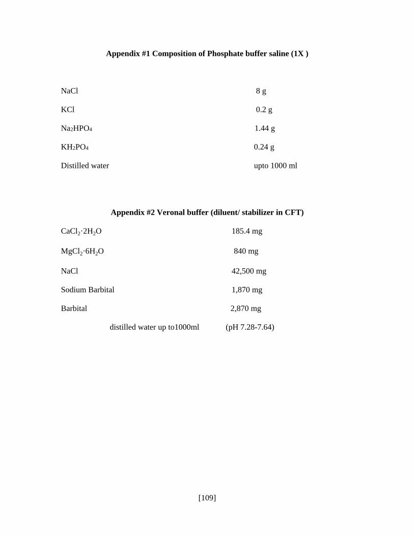
[109]
Appendix #1 Composition of Phosphate buffer saline (1X )
NaCl 8 g
KCl 0.2 g
Na2HPO4 1.44 g
KH2PO4 0.24 g
Distilled water upto 1000 ml
Appendix #2 Veronal buffer (diluent/ stabilizer in CFT) CaCl2·2H2O 185.4 mg
MgCl2·6H2O 840 mg
NaCl 42,500 mg
Sodium Barbital 1,870 mg
Barbital 2,870 mg
distilled water up to1000ml (pH 7.28-7.64)
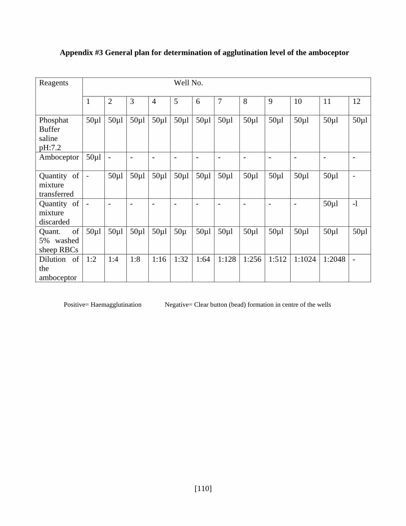
[110]
Appendix #3 General plan for determination of agglutination level of the amboceptor
Positive= Haemagglutination Negative= Clear button (bead) formation in centre of the wells
Reagents Well No.
1 2 3 4 5 6 7 8 9 10 11 12
Phosphat Buffer saline pH:7.2
50µl 50µl 50µl 50µl 50µl 50µl 50µl 50µl 50µl 50µl 50µl 50µl
Amboceptor 50µl - - - - - - - - - - -
Quantity of mixture transferred
- 50µl 50µl 50µl 50µl 50µl 50µl 50µl 50µl 50µl 50µl -
Quantity of mixture discarded
- - - - - - - - - - 50µl -l
Quant. of 5% washed sheep RBCs
50µl 50µl 50µl 50µl 50µ 50µl 50µl 50µl 50µl 50µl 50µl 50µl
Dilution of the amboceptor
1:2 1:4 1:8 1:16 1:32 1:64 1:128 1:256 1:512 1:1024 1:2048 -
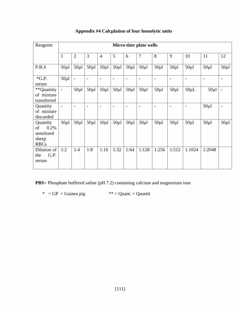
[111]
Appendix #4 Calcµlation of four hemolytic units
PBS= Phosphate buffered saline (pH 7.2) containing calcium and magnesium ions
* = GP = Guinea pig ** = Quant. = Qauntit
Reagents Micro-titer plate wells
1 2 3 4 5 6 7 8 9 10 11 12
P.B.S 50µl 50µl 50µl 50µl 50µl 50µl 50µl 50µl 50µl 50µl 50µl 50µl
*G.P. serum
50µl - - - - - - - - - - -
**Quantity of mixture transferred
- 50µl 50µl 50µl 50µl 50µl 50µl 50µl 50µl 50µL 50µl -
Quantity of mixture discarded
- - - - - - - - - - 50µl -
Quantity of 0.2% sensitized sheep RBCs
50µl 50µl 50µl 50µl 50µl 50µl 50µl 50µl 50µl 50µl 50µl 50µl
Dilution of the G.P. serum
1:2 1:4 1:8 1:16 1:32 1:64 1:128 1:256 1:512 1:1024 1:2048
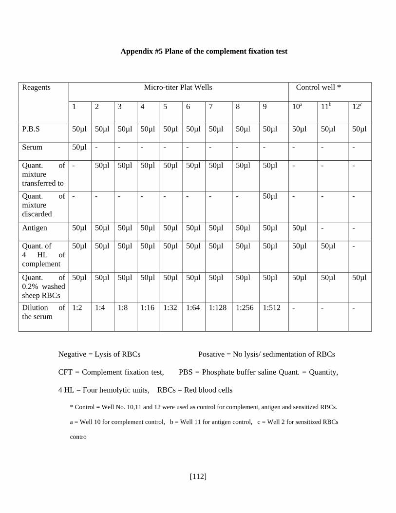
[112]
Appendix #5 Plane of the complement fixation test
Reagents Micro-titer Plat Wells Control well *
1 2 3 4 5 6 7 8 9 10a 11b 12c
P.B.S 50µl 50µl 50µl 50µl 50µl 50µl 50µl 50µl 50µl 50µl 50µl 50µl
Serum 50µl - - - - - - - - - - -
Quant. of mixture transferred to
- 50µl 50µl 50µl 50µl 50µl 50µl 50µl 50µl - - -
Quant. of mixture discarded
- - - - - - - - 50µl - - -
Antigen 50µl 50µl 50µl 50µl 50µl 50µl 50µl 50µl 50µl 50µl - -
Quant. of 4 HL of complement
50µl 50µl 50µl 50µl 50µl 50µl 50µl 50µl 50µl 50µl 50µl -
Quant. of 0.2% washed sheep RBCs
50µl 50µl 50µl 50µl 50µl 50µl 50µl 50µl 50µl 50µl 50µl 50µl
Dilution of the serum
1:2 1:4 1:8 1:16 1:32 1:64 1:128 1:256 1:512 - - -
Negative = Lysis of RBCs Posative = No lysis/ sedimentation of RBCs
CFT = Complement fixation test, PBS = Phosphate buffer saline Quant. = Quantity,
4 HL = Four hemolytic units, RBCs = Red blood cells
* Control = Well No. 10,11 and 12 were used as control for complement, antigen and sensitized RBCs.
a = Well 10 for complement control, b = Well 11 for antigen control, c = Well 2 for sensitized RBCs
contro

[113]
Appendix #6 Coating buffer for ELISA 50mM (pH: 9.6)
Sodium carbonate (Na2CO3) 10.60 gm
Sodium bircarbonate (NaHCO3) 8.4gm
Sodium azide (NaNO3) 0.20gm
Make volume up to 1000ml with distilled water and adjust pH:9.6 with HCl or NaOH.
Appendix #7 Washing buffer for Elisa
Phosphate buffer saline (1x) 1000 ml
Tween 20 0.5ml
Appendix #8 Citrate phosphate buffer (pH: 5)
Citric acid 2.14gm
Na2HPO4. 2H2O 3.54gm
Distilled water 400ml
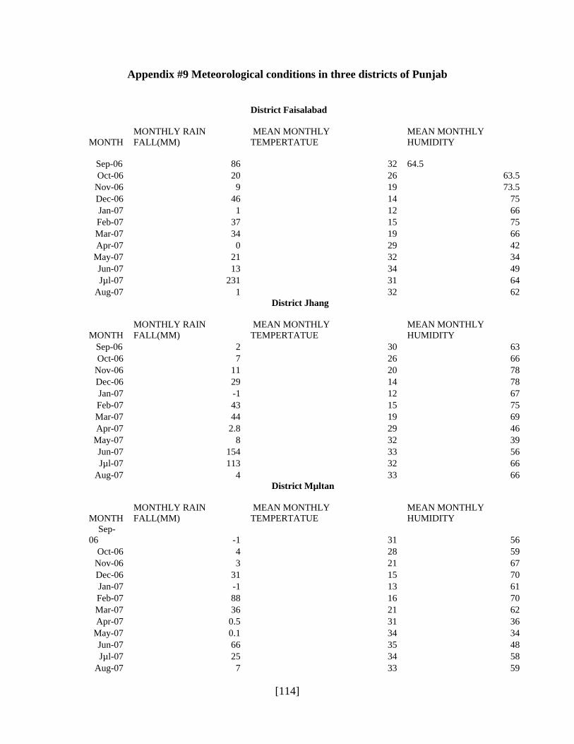
[114]
Appendix #9 Meteorological conditions in three districts of Punjab
District Faisalabad
MONTH MONTHLY RAIN FALL(MM)
MEAN MONTHLY TEMPERTATUE
MEAN MONTHLY HUMIDITY
Sep-06 86 32 64.5
Oct-06 20 26 63.5 Nov-06 9 19 73.5 Dec-06 46 14 75 Jan-07 1 12 66 Feb-07 37 15 75 Mar-07 34 19 66 Apr-07 0 29 42
May-07 21 32 34 Jun-07 13 34 49 Jµl-07 231 31 64
Aug-07 1 32 62
District Jhang
MONTH MONTHLY RAIN FALL(MM)
MEAN MONTHLY TEMPERTATUE
MEAN MONTHLY HUMIDITY
Sep-06 2 30 63 Oct-06 7 26 66
Nov-06 11 20 78 Dec-06 29 14 78 Jan-07 -1 12 67 Feb-07 43 15 75 Mar-07 44 19 69 Apr-07 2.8 29 46
May-07 8 32 39 Jun-07 154 33 56 Jµl-07 113 32 66
Aug-07 4 33 66
District Mµltan
MONTH MONTHLY RAIN FALL(MM)
MEAN MONTHLY TEMPERTATUE
MEAN MONTHLY HUMIDITY
Sep-06 -1 31 56
Oct-06 4 28 59 Nov-06 3 21 67 Dec-06 31 15 70 Jan-07 -1 13 61 Feb-07 88 16 70 Mar-07 36 21 62 Apr-07 0.5 31 36
May-07 0.1 34 34 Jun-07 66 35 48 Jµl-07 25 34 58
Aug-07 7 33 59

[115]