Embed Size (px)
DESCRIPTION
Metais Pesados
Citation preview
Modulation of metallothionein and metal partitioning in liverand kidney of Solea senegalensis after long-term acclimationto two environmental temperatures
R. Siscar a, A. Torreblanca a, J. del Ramo a, M. Solé b,n
a Departamento Biología Funcional y Antropología Física, Universitat de València, Dr. Moliner 50 Burjassot, 46100 València, Spainb Institut de Ciencies del Mar (ICM-CSIC), Passeig marítim de la Barceloneta 37-49, 08003 Barcelona, Spain
a r t i c l e i n f o
Article history:Received 28 November 2013Received in revised form10 April 2014Accepted 12 April 2014
Keywords:MetalsBenthic fishMetallothioneinSeleniumTemperature
a b s t r a c t
Juveniles of Solea senegalensis were fed with commercial pellets under controlled conditions at twoenvironmental Mediterranean temperatures (15 and 20 1C) for two months. After this period, theaccumulation of essential and non-essential metals and metallothionein (MT) levels was measured inliver and kidney by inductively coupled plasma mass spectrometry (ICP-MS) and pulse polarography,respectively. The bioaccumulation factor (BAF) for selected metals in both tissues was calculated inrelation to levels present in the feed. Tissue partitioning (liver/kidney) and molar ratios, considering themetal protective mechanisms: MT and Selenium (Se), were included for evaluating the detoxificationcapacity of each tissue. Ag, Cd, Cu and Mn were preferentially accumulated in the liver whereas Co, Fe,Hg, Se and Zn were found in larger concentrations in the kidney, and higher temperature enhanced theaccumulation of some of them, but not all. MT content in liver, but not in kidney, was also influenced bytemperature changes and by length of exposure. The BAF revealed that Cu was taken up mainly by theliver whereas Se was efficiently taken up by both tissues. The high molar ratios of MT and most metalsdenoted the kidney's remarkable spare capacity for metal detoxification through MT binding. Moreover,the potential protective role of Se was also more evident in kidney as a higher Se:Cd and Se:Ag molarratios were reached in this organ. In contrast to other fish, the storage of Cd in kidney wasparticularly low.
& 2014 Elsevier Inc. All rights reserved.
1. Introduction
The marine environment is exposed to physical and chemicalchanges that can be man-induced. Among these factors, enhancedwater temperature and increased metal presence in their habitatcan negatively affect the health status of resident organisms. Oneof these changes is due to climate alterations that are responsiblefor sustained increases in water temperature (Belkin, 2009).Modifications in temperature can report changes in the physiologyof aquatic species as well as in bioavailability and metabolism ofchemicals, including metals (Noyes et al., 2009). On the assess-ment of metal toxicity, several authors have selected fish liver asthe organ that best reflects metal storage, and some of themcompared it to other tissues, including kidney (Campana et al.,2003; Jeffree et al., 2006; Ureña et al., 2007; Adams et al., 2010; DeBoeck et al., 2010; Ghedira et al., 2010; Kamunde and MacPhail,2011; Luszczek-Trojnar et al., 2013). Nonetheless, there is
controversy on which is the preferential tissue for metal accumu-lation in fish, as both liver and kidney are considered organsinvolved in metal internal dynamics. Only a few marine fishstudies have already addressed the effect of temperature on metalhandling (Olsson et al., 1996; Guinot et al., 2012) and even fewerhave used kidney as the target tissue. However, in the species ofconcern for this study, Solea senegalensis, kidney was revealed tohave a relevant role in metal handling (Siscar et al., 2013).
To prevent the toxic effect of some metals, in fish as well as inmost aquatic organisms, two main mechanisms have beendescribed. Firstly, metallothionein (MT) constitutes a family oflow-molecular-weight, cysteine-rich proteins functioning in theregulation of essential metals such as Cu and Zn, and in thedetoxification of these and other non-essential metals such as Cdand Hg (Wood et al., 2012a, 2012b). Temperature can affect notonly the kinetics of uptake, biotransformation and elimination ofmetal but also MT synthesis in aquatic organisms including fish(Olsson et al., 1996; Baykan et al., 2007; Guinot et al., 2012).Secondly, the protective role of other metals such as selenium (Se)to prevent Hg toxicity has recently also been described in fish(Sørmo et al., 2011; Damiano et al., 2011; Branco et al., 2012,
Contents lists available at ScienceDirect
journal homepage: www.elsevier.com/locate/envres
Environmental Research
http://dx.doi.org/10.1016/j.envres.2014.04.0200013-9351/& 2014 Elsevier Inc. All rights reserved.
n Corresponding author. Fax: þ34 932 30 95 55.E-mail address: [email protected] (M. Solé).
Environmental Research 132 (2014) 197–205
Siscar et al., 2014). In fact, the molar ratio Se:Hg has been proposedas a measure of the potential capacity of Se to face toxic effects ofHg in fish muscle, liver and kidney (Burger et al., 2012, 2013). Inrats, evidence was found that Se protects against the toxic effectsof Cd and Ag alone (Newairy et al., 2007) or by forming a complexwith Zn, in liver as well as in kidney (Jihen et al., 2008).
Few studies focus on physiological responses of S. senegalensisto temperature variation (Castro et al., 2012) and metal accumula-tion (Oliva et al., 2012a; Arellano et al., 1999). This species has alsobeen broadly used as sentinel in metal pollution monitoringstudies (Usero et al., 2004; Kalman et al., 2010; Fonseca et al.,2011; Costa et al., 2011, 2012; Oliva et al., 2012a, 2012b; Galindoet al., 2012; Siscar et al., 2013). However, there is little informationon the effect of temperature on accumulation, detoxification andmetal homeostasis in S. senegalensis.
The aims of this study were (1) to determine the effect oftemperature on the accumulation and distribution of metalsbetween liver and kidney, (2) to find whether the observeddifferences, if any, are due to modifications in metal assimilationfrom food, (3) to search for the existence of temperature-drivenchanges in metal detoxification abilities at background levels ofmetals.
2. Material and methods
2.1. Source of fish and laboratory acclimation
S. senegalensis were hatched and reared under aquaculture conditions by theStolt Sea Farm SA (La Coruña, Spain). Specimens of about 150–200 g wet weightwere transported and maintained in the University of Valencia facilities. To ensurerecovery after transport and adaptation to the new laboratory conditions, animalswere kept at 20 1C for two months.
2.2. Temperature acclimation experiment
Thirty-two fish were randomly distributed into 8 round tanks of 3 m3 each.Physical water conditions were checked daily and maintained at the desiredtemperatures. One week before the beginning of the experiment, the temperatureof four of the tanks was lowered at a rate of about 1 1C/day until the desiredtemperature of 15 1C was reached, which was one day before the start of theexperiment. The water parameters from the start of the experiment (t0) and for aperiod of up to 60 days (t60) for the 15 1C group were T 15.871.8 1C; salinity29.070.9 p.s.u and O2 98.3%71.7 and for the 20 1C group T 19.170.6 1C; salinity29.671.2 p.s.u and O2 97.8%71.3. The light regime was 12:12 and fish were feddaily ad libitum by hand with commercial Le-5-Elite trout pellet, Skretting (UK),but fasted for 24 h prior to the sampling of eight fish (2 from each 4 replicate tanks)for each time and temperature conditions.
2.3. Fish sampling
Fish were sacrificed by severing the spinal cord. Fish were immediatelydissected and the organs (liver and kidney) flash frozen in liquid nitrogen. Tissueswere stored at �80 1C until analysis. Total weight and length were also measuredbefore sacrifice to calculate the condition factor (CF) as: total length/(total weight)3
and the hepatosomatic index (HSI) as: (liver weight/total weight)�100. The Q10
coefficient, the relative change of a biological activity as a consequence ofincreasing the temperature by 10 1C, was used to evaluate the influence oftemperature on growth. Q10¼(WT2/WT1)10/T2�T1, where W is the weight gainedby fish at either temperature and T1 and T2 the respective acclimation tempera-tures (15 and 20 1C). Handling of the fish was done according to national andinstitutional regulations of the Spanish Council for Scientific Research (CSIC) andDirective 2010/63/EU.
2.4. Metal content analysis
Individual fish liver and kidney samples of about 0.1 g wet weight weredigested in concentrated nitric acid 65% (Baker), at room temperature overnightand were heated at 80 1C for 2 h. Within each digestion series, appropriate blankswith ultra-pure water were also subjected to the same procedure to account forbackground contamination levels. After cooling, solutions were transferred to astandard volume with ultra-pure water. Determination of metals (Ag, Cd, Co, Cr, Cu,
Fe, Hg, Mn, Pb, Se and Zn) was undertaken using an inductively coupled plasmamass spectrometry (ICP-MS) (Elan DRC-I, Perkin-Elmer Sciex). Samples of similarweight of certified reference material (DOLT-3 and LUTS-1, National ResearchCouncil of Canada, Ottawa), were digested and analyzed during each analyticalrun. The values of all elements found were in good agreement with the certifiedvalues, with the recoveries ranging from 87% to 108%. A sample of water from eachtank was analyzed for metal content. The food used Le-5-Elite trout pellet,Skretting (UK), was also analyzed for metal content.
2.5. Metallothionein determination by differential pulse polarography
About 0.2 g wet weight portions of frozen liver and kidney were homogenizedusing an ultra-turrax in 20 mM Tris–HCl buffer, 1 mM dithiothreitol (DTT) and0.2 mM phenylmethylsulfonylfluoride (PMSF) at pH 8.6 in an ice bath. Thehomogenates were centrifuged at 30,000g for 45 min at 4 1C. The supernatantwas heated at 80 1C for 10 min in order to denaturate high molecular weightproteins, subsequently centrifuged at 30,000g for 45 min at 4 1C, and the heat-treated supernatant, containing thermally stable MT, was separated from precipi-tated proteins. MT was measured using differential pulse polarography as describedby Bebianno and Langston (1989). An aliquot of the heat-treated supernatant wasadded to the polarographic cell, containing 20 mL hexamminecobalt chloride buffer(the supporting electrolyte), together with Triton-X (0.025%v/v). The cell waspurged for 2 min with purified N2 prior to analysis. The polarographic responsewas measured during a potential scan between �1.38 V and �1.7 V (Model 757VAComputrace Analyzer, Methrom, Switzerland) in SMDE (Static Mercury DropElectrode) mode. Quantification of MT was performed using the standard additionmethod with rabbit liver MT Iþ II (Sigma). Results are expressed as mg g�1 wetweight of tissue.
2.6. Molar ratios and bioaccumulation factor calculations
Molar ratios were calculated considering an average molecular weight of6000 Da for MT and the atomic weights for the metals: Se (78.96), Hg (200.5), Cd(112.4), Zn (65.4) and Ag (107.9) were adopted for the conversion of MT and metalcontent (mg/g w.w) into molar content (moles/g w.w). In this way, the calculatedratios are expressed as moles of MT per atom of metal in liver and kidney. Thereported value ratios were calculated for each individual fish and then averaged toobtain a mean value.
The bioaccumulation factor (BAF) was calculated at the two set times in orderto study the effect of temperature on the kinetics of the ten selected metals in thetwo organs (liver and kidney). This factor was calculated as a ratio between metalconcentration in each tissue and metal content in the commercial food pellets.
2.7. Statistics
All statistical analyses were performed using the software package SPSS. Datawere checked for normality (Kolmogorof–Smirnof test) and homogeneous variance(Levene's test) and transformed, when necessary, to comply with normality andhomocedasticity assumptions. The influence of time and temperature and theirinteraction was evaluated using the two-way ANOVA test. Differences betweenpairs of experimental groups were assessed by one-way ANOVA and Bonferroni asa Post-hoc test. Spearman correlation coefficient (ρ) was calculated between metalsin liver and kidney and their respective MT levels in order to measure the strengthof association between these variables with the non-transformed data. Student'st-test was applied to contrast overall kidney and liver differences in MT content.Differences at the 5% significance level were considered significant.
3. Results
3.1. Biological parameters of the fish
In Table 1 the total weight, total length and sex ratios of the fishselected for the study at the start and after 60 days are reported. Fishincreased in length and weight (po0.05) during the two-monthexperiment, regardless of water temperature. The condition factor(CF) was not influenced by temperature but the hepatosomatic index(HSI) was significantly lower (po0.05) in fish kept at 20 1C for60 days.
3.2. Metal content in water and foodstuff
Metal content of food pellets (in mg/g w.w) was for Ag:0.00770.001, Cd: 0.1670.01, Co: 0.12370.023, Cr: 2.2970.29, Cu:
R. Siscar et al. / Environmental Research 132 (2014) 197–205198
10.4470.88, Fe: 116.0278.06, Hg: 0.03570.001, Mn: 32.7971.49, Pb: 0.6770.03, Se: 0.5470.03 and Zn: 98.0576.58. Metalcontent in the water from the tanks was similar and about or justover the LOD at the two rearing temperatures (data not presented).
3.3. Metal content in liver and kidney
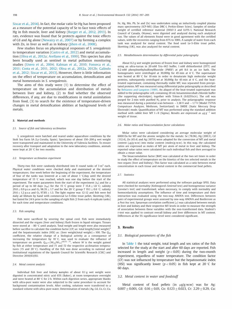
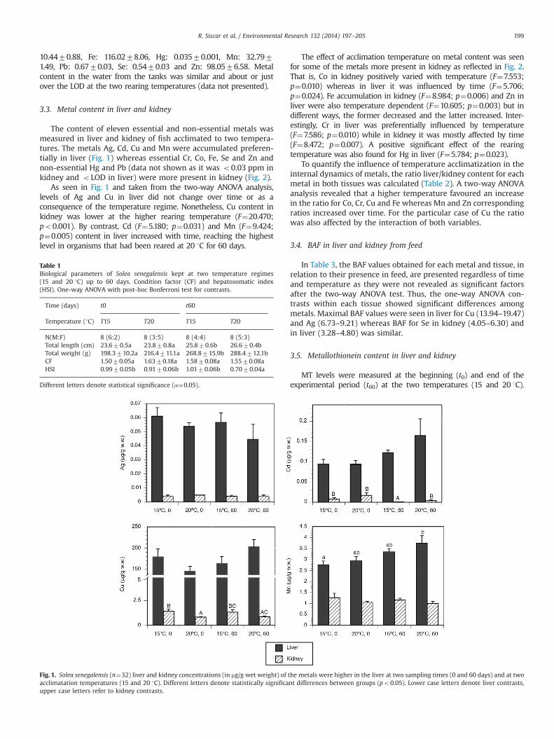
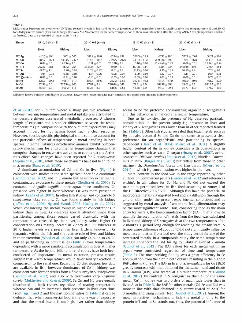
The content of eleven essential and non-essential metals wasmeasured in liver and kidney of fish acclimated to two tempera-tures. The metals Ag, Cd, Cu and Mn were accumulated preferen-tially in liver (Fig. 1) whereas essential Cr, Co, Fe, Se and Zn andnon-essential Hg and Pb (data not shown as it was o0.03 ppm inkidney and oLOD in liver) were more present in kidney (Fig. 2).
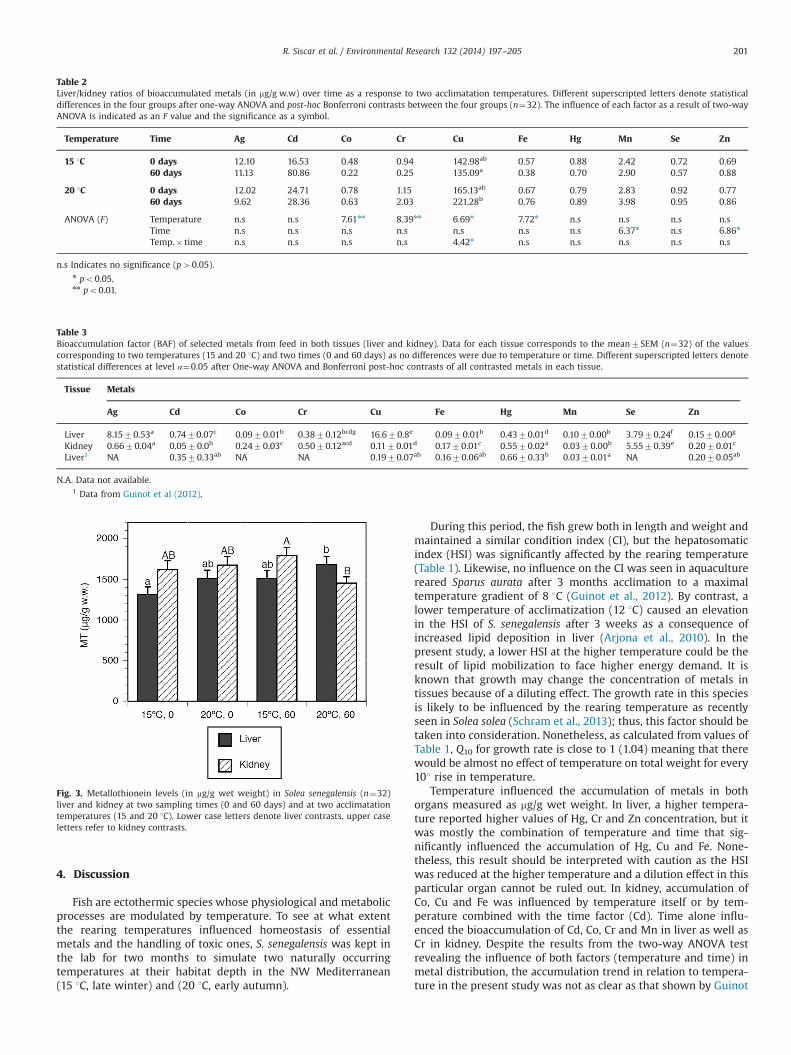
As seen in Fig. 1 and taken from the two-way ANOVA analysis,levels of Ag and Cu in liver did not change over time or as aconsequence of the temperature regime. Nonetheless, Cu content inkidney was lower at the higher rearing temperature (F¼20.470;po0.001). By contrast, Cd (F¼5.180; p¼0.031) and Mn (F¼9.424;p¼0.005) content in liver increased with time, reaching the highestlevel in organisms that had been reared at 20 1C for 60 days.
The effect of acclimation temperature on metal content was seenfor some of the metals more present in kidney as reflected in Fig. 2.That is, Co in kidney positively varied with temperature (F¼7.553;p¼0.010) whereas in liver it was influenced by time (F¼5.706;p¼0.024). Fe accumulation in kidney (F¼8.984; p¼0.006) and Zn inliver were also temperature dependent (F¼10.605; p¼0.003) but indifferent ways, the former decreased and the latter increased. Inter-estingly, Cr in liver was preferentially influenced by temperature(F¼7.586; p¼0.010) while in kidney it was mostly affected by time(F¼8.472; p¼0.007). A positive significant effect of the rearingtemperature was also found for Hg in liver (F¼5.784; p¼0.023).
To quantify the influence of temperature acclimatization in theinternal dynamics of metals, the ratio liver/kidney content for eachmetal in both tissues was calculated (Table 2). A two-way ANOVAanalysis revealed that a higher temperature favoured an increasein the ratio for Co, Cr, Cu and Fe whereas Mn and Zn correspondingratios increased over time. For the particular case of Cu the ratiowas also affected by the interaction of both variables.
3.4. BAF in liver and kidney from feed
In Table 3, the BAF values obtained for each metal and tissue, inrelation to their presence in feed, are presented regardless of timeand temperature as they were not revealed as significant factorsafter the two-way ANOVA test. Thus, the one-way ANOVA con-trasts within each tissue showed significant differences amongmetals. Maximal BAF values were seen in liver for Cu (13.94–19.47)and Ag (6.73–9.21) whereas BAF for Se in kidney (4.05–6.30) andin liver (3.28–4.80) was similar.
3.5. Metallothionein content in liver and kidney
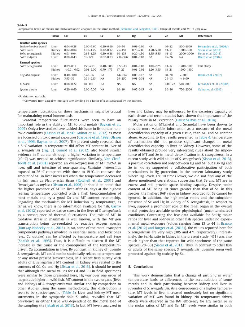
MT levels were measured at the beginning (t0) and end of theexperimental period (t60) at the two temperatures (15 and 20 1C).
Fig. 1. Solea senegalensis (n¼32) liver and kidney concentrations (in mg/g wet weight) of the metals were higher in the liver at two sampling times (0 and 60 days) and at twoacclimatation temperatures (15 and 20 1C). Different letters denote statistically significant differences between groups (po0.05). Lower case letters denote liver contrasts,upper case letters refer to kidney contrasts.
Table 1Biological parameters of Solea senegalensis kept at two temperature regimes(15 and 20 1C) up to 60 days. Condition factor (CF) and hepatosomatic index(HSI). One-way ANOVA with post-hoc Bonferroni test for contrasts.
Time (days) t0 t60
Temperature (1C) T15 T20 T15 T20
N(M:F) 8 (6:2) 8 (3:5) 8 (4:4) 8 (5:3)Total length (cm) 23.670.5a 23.870.8a 25.870.6b 26.670.4bTotal weight (g) 198.3710.2a 216.4711.1a 268.8715.9b 288.4712.1bCF 1.5070.05a 1.6370.18a 1.5870.08a 1.5570.08aHSI 0.9970.05b 0.9170.06b 1.0170.06b 0.7070.04a
Different letters denote statistical significance (α¼0.05).
R. Siscar et al. / Environmental Research 132 (2014) 197–205 199
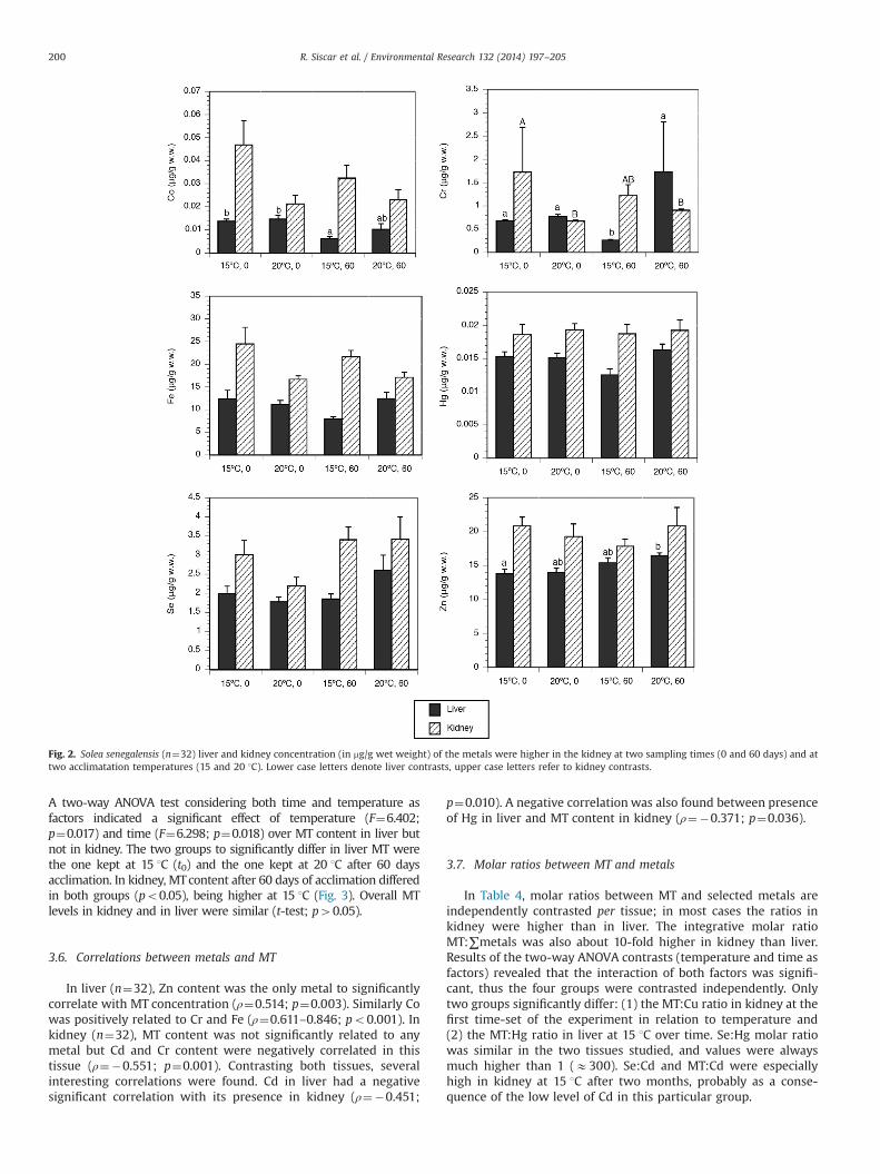
A two-way ANOVA test considering both time and temperature asfactors indicated a significant effect of temperature (F¼6.402;p¼0.017) and time (F¼6.298; p¼0.018) over MT content in liver butnot in kidney. The two groups to significantly differ in liver MT werethe one kept at 15 1C (t0) and the one kept at 20 1C after 60 daysacclimation. In kidney, MTcontent after 60 days of acclimation differedin both groups (po0.05), being higher at 15 1C (Fig. 3). Overall MTlevels in kidney and in liver were similar (t-test; p40.05).
3.6. Correlations between metals and MT
In liver (n¼32), Zn content was the only metal to significantlycorrelate with MT concentration (ρ¼0.514; p¼0.003). Similarly Cowas positively related to Cr and Fe (ρ¼0.611–0.846; po0.001). Inkidney (n¼32), MT content was not significantly related to anymetal but Cd and Cr content were negatively correlated in thistissue (ρ¼�0.551; p¼0.001). Contrasting both tissues, severalinteresting correlations were found. Cd in liver had a negativesignificant correlation with its presence in kidney (ρ¼�0.451;
p¼0.010). A negative correlation was also found between presenceof Hg in liver and MT content in kidney (ρ¼�0.371; p¼0.036).
3.7. Molar ratios between MT and metals
In Table 4, molar ratios between MT and selected metals areindependently contrasted per tissue; in most cases the ratios inkidney were higher than in liver. The integrative molar ratioMT:∑metals was also about 10-fold higher in kidney than liver.Results of the two-way ANOVA contrasts (temperature and time asfactors) revealed that the interaction of both factors was signifi-cant, thus the four groups were contrasted independently. Onlytwo groups significantly differ: (1) the MT:Cu ratio in kidney at thefirst time-set of the experiment in relation to temperature and(2) the MT:Hg ratio in liver at 15 1C over time. Se:Hg molar ratiowas similar in the two tissues studied, and values were alwaysmuch higher than 1 (E300). Se:Cd and MT:Cd were especiallyhigh in kidney at 15 1C after two months, probably as a conse-quence of the low level of Cd in this particular group.
Fig. 2. Solea senegalensis (n¼32) liver and kidney concentration (in mg/g wet weight) of the metals were higher in the kidney at two sampling times (0 and 60 days) and attwo acclimatation temperatures (15 and 20 1C). Lower case letters denote liver contrasts, upper case letters refer to kidney contrasts.
R. Siscar et al. / Environmental Research 132 (2014) 197–205200
4. Discussion
Fish are ectothermic species whose physiological and metabolicprocesses are modulated by temperature. To see at what extentthe rearing temperatures influenced homeostasis of essentialmetals and the handling of toxic ones, S. senegalensis was kept inthe lab for two months to simulate two naturally occurringtemperatures at their habitat depth in the NW Mediterranean(15 1C, late winter) and (20 1C, early autumn).
During this period, the fish grew both in length and weight andmaintained a similar condition index (CI), but the hepatosomaticindex (HSI) was significantly affected by the rearing temperature(Table 1). Likewise, no influence on the CI was seen in aquaculturereared Sparus aurata after 3 months acclimation to a maximaltemperature gradient of 8 1C (Guinot et al., 2012). By contrast, alower temperature of acclimatization (12 1C) caused an elevationin the HSI of S. senegalensis after 3 weeks as a consequence ofincreased lipid deposition in liver (Arjona et al., 2010). In thepresent study, a lower HSI at the higher temperature could be theresult of lipid mobilization to face higher energy demand. It isknown that growth may change the concentration of metals intissues because of a diluting effect. The growth rate in this speciesis likely to be influenced by the rearing temperature as recentlyseen in Solea solea (Schram et al., 2013); thus, this factor should betaken into consideration. Nonetheless, as calculated from values ofTable 1, Q10 for growth rate is close to 1 (1.04) meaning that therewould be almost no effect of temperature on total weight for every101 rise in temperature.
Temperature influenced the accumulation of metals in bothorgans measured as mg/g wet weight. In liver, a higher tempera-ture reported higher values of Hg, Cr and Zn concentration, but itwas mostly the combination of temperature and time that sig-nificantly influenced the accumulation of Hg, Cu and Fe. None-theless, this result should be interpreted with caution as the HSIwas reduced at the higher temperature and a dilution effect in thisparticular organ cannot be ruled out. In kidney, accumulation ofCo, Cu and Fe was influenced by temperature itself or by tem-perature combined with the time factor (Cd). Time alone influ-enced the bioaccumulation of Cd, Co, Cr and Mn in liver as well asCr in kidney. Despite the results from the two-way ANOVA testrevealing the influence of both factors (temperature and time) inmetal distribution, the accumulation trend in relation to tempera-ture in the present study was not as clear as that shown by Guinot
Table 3Bioaccumulation factor (BAF) of selected metals from feed in both tissues (liver and kidney). Data for each tissue corresponds to the mean7SEM (n¼32) of the valuescorresponding to two temperatures (15 and 20 1C) and two times (0 and 60 days) as no differences were due to temperature or time. Different superscripted letters denotestatistical differences at level α¼0.05 after One-way ANOVA and Bonferroni post-hoc contrasts of all contrasted metals in each tissue.
Tissue Metals
Ag Cd Co Cr Cu Fe Hg Mn Se Zn
Liver 8.1570.53a 0.7470.07c 0.0970.01b 0.3870.12bcdg 16.670.8e 0.0970.01b 0.4370.01d 0.1070.00b 3.7970.24f 0.1570.00g
Kidney 0.6670.04a 0.0570.0b 0.2470.03c 0.5070.12acd 0.1170.01d 0.1770.01c 0.5570.02a 0.0370.00b 5.5570.39e 0.2070.01c
Liver1 NA 0.3570.33ab NA NA 0.1970.07ab 0.1670.06ab 0.6670.33b 0.0370.01a NA 0.2070.05ab
N.A. Data not available.1 Data from Guinot et al (2012).
Fig. 3. Metallothionein levels (in mg/g wet weight) in Solea senegalensis (n¼32)liver and kidney at two sampling times (0 and 60 days) and at two acclimatationtemperatures (15 and 20 1C). Lower case letters denote liver contrasts, upper caseletters refer to kidney contrasts.
Table 2Liver/kidney ratios of bioaccumulated metals (in mg/g w.w) over time as a response to two acclimatation temperatures. Different superscripted letters denote statisticaldifferences in the four groups after one-way ANOVA and post-hoc Bonferroni contrasts between the four groups (n¼32). The influence of each factor as a result of two-wayANOVA is indicated as an F value and the significance as a symbol.
Temperature Time Ag Cd Co Cr Cu Fe Hg Mn Se Zn
15 1C 0 days 12.10 16.53 0.48 0.94 142.98ab 0.57 0.88 2.42 0.72 0.6960 days 11.13 80.86 0.22 0.25 135.09a 0.38 0.70 2.90 0.57 0.88
20 1C 0 days 12.02 24.71 0.78 1.15 165.13ab 0.67 0.79 2.83 0.92 0.7760 days 9.62 28.36 0.63 2.03 221.28b 0.76 0.89 3.98 0.95 0.86
ANOVA (F) Temperature n.s n.s 7.61nn 8.39nn 6.69n 7.72n n.s n.s n.s n.sTime n.s n.s n.s n.s n.s n.s n.s 6.37n n.s 6.86n
Temp.� time n.s n.s n.s n.s 4.42n n.s n.s n.s n.s n.s
n.s Indicates no significance (p40.05).n po0.05.nn po0.01.
R. Siscar et al. / Environmental Research 132 (2014) 197–205 201
et al. (2012) for S. aurata where a sharp positive relationshipbetween rearing temperature and metal uptake was attributed totemperature-driven accelerated metabolic processes. A shorterlength of exposure and a smaller difference between the testedtemperatures (in comparison with the S. aurata experiment) mayaccount in part for not having found such a clear response.However, species-specific physiological traits can also account forthe particular effects of temperature in metal handling in bothspecies. In some instances ectothermic animals exhibit compen-satory mechanisms for environmental temperature changes thatcomprise changes in enzymatic activities to counteract the Arrhe-nius effect. Such changes have been reported for S. senegalensis(Arjona et al., 2010), while those mechanisms have not been foundin S. aurata (Ibarz et al., 2010).
A dominant accumulation of Cd and Cu in liver (Fig. 1) iscoincident with studies in the same species under field conditions(Galindo et al., 2012) and in S. aurata but based on experimentalcontaminated exposure to these metals (Ghedira et al., 2010). Bycontrast, in Anguilla anguilla under aquaculture conditions, Cdpresence was higher in liver whereas Cu was more present inkidney (Ureña et al., 2007). In other fish studies, and contrary to S.senegalensis observations, Cd was found mainly in fish kidney(Jeffree et al., 2006; Ng and Wood, 2008; Huang et al., 2007).When considering the metals found in higher concentrations inkidney than in liver, Cr deserves special attention since theirpartitioning among those organs varied drastically with thetemperature as revealed by their liver/kidney ratios. Higher Crconcentration was mainly located in kidney at 15 1C whereas at20 1C higher levels were present in liver. Little is known on Crdynamics within the fish and the relative role of liver and kidneyin their excretion (Wood et al., 2012a). Not only Cr, but also Co, Cuand Fe partitioning in both tissues (Table 2) was temperature-dependent with a more significant accumulation in liver at highertemperature. As the hepato-biliar and renal routes have both beenconsidered of importance in metal excretion, present resultssuggest that warm temperatures would favor biliary excretion incomparison to the renal one. Regarding Co distribution, few dataare available in fish, however dominance of Co in kidney wascoincident with former results from a field survey in S. senegalensis(Galindo et al., 2012) and also with freshwater carp, Cyprinuscarpio (Mukherjee and Kaviraj, 2009). Se, Zn and Hg were equallydistributed in both tissues regardless of rearing temperaturewhereas Mn and Zn increased their presence in liver over time.From Figs. 1 and 2 and the partitioning ratios of Table 2 it can bededuced that when commercial food is the only way of exposure,and thus the metal intake is not high, liver rather than kidney,
seems to be the preferred accumulating organ in S. senegalensisand this behavior is enhanced at a higher temperature.
Due to its toxicity, the presence of Hg deserves particularconsideration. In the present study Hg presence in liver andkidney of S. senegalensis was lower than in other reported farmedfish (Table 5). Other fish studies revealed that toxic metals such asHg but also essential Se and Zn do not seem to present a clearpreference for an organ/tissue and partitioning is species-dependent (Usero et al., 2004; Mieiro et al., 2011). A slightlyhigher content of Hg in kidney coincides with observations inother species such as carp, C. carpio (Huang et al., 2007), zebra-seabream, Diplodus cervius (Branco et al., 2012), bluefish, Pomato-mus saltatrix (Burger et al., 2013) but differs from those in othermarine fish, Dicentrarchus labrax and Liza aurata (Mieiro et al.,2011) in which Hg concentration was higher in the liver.
Metal content in the food was in the range reported by otherauthors on commercial pellets (Guinot et al., 2012 and referenceswithin). In all, values for Cd, Hg and Pb did not exceed themaximum permitted level in fish feed according to Annex I ofthe UE Directive 2002/32/EC. Although fish have the potential toincorporate metals via ingested food and via diffusion through thegills or skin, under the present experimental conditions, and assuggested by metal analysis of water and feed, alimentation maybe the most significant route. Thus, considering food as the mainentry for metals, the bioaccumulation factor (BAF), that allows toquantify the accumulation of metals from the feed, was calculatedin liver and kidney of S. senegalensis at the two temperatures after2 months, a period long enough for reaching the steady state. Atemperature difference of about 5 1C did not significantly influencemetal accumulation from feed over the study period for any of thecontrasted metals. In a comparable study the same temperatureincrease enhanced the BAF for Hg by 3-fold in liver of S. aurata(Guinot et al., 2012). The BAF values for each metal within anorgan were contrasted regardless of time and temperature(Table 3). The most striking finding was a great efficiency in Seaccumulation from the diet in both organs, resulting in the highestBAF value in kidney. The BAF in liver of S. senegalensis for Cu (16.6)greatly contrasts with that obtained for the same metal and tissuein S. aurata (0.19) also reared at a similar temperature (Guinotet al., 2012). By contrast in S. senegalensis the BAF of the samemetal (Cu) in kidney was two orders of magnitude lower than inliver. Also in Table 3, the BAF for other metals (Cd, Fe and Zn) wasmore in line with that obtained in S. aurata reared at 22 1C for3 months and using similar feed (Guinot et al., 2012). Among themetal protective mechanisms of fish, the metal binding to theprotein MT and to Se stands out, thus, the potential influence of
Table 4Molar ratios between metallothionein (MT) and selected metals in liver and kidney of juveniles of Solea senegalensis (n¼32) acclimated to two temperatures (15 and 20 1C)for 60 days in two tissues (liver and kidney). One-way ANOVA contrasts with Bonferroni post-hoc as there was interaction after the 2-way ANOVA test (temperature and timeas factors). Data are presented as mean7SD (n¼8).
Tissue 15 1C, 0 d (n¼8) 20 1C, 0 d (n¼8) 15 1C, 60 d (n¼8) 20 1C, 60 d (n¼8)
Liver Kidney Liver Kidney Liver Kidney Liver Kidney
MT:Ag 416.7749.1 60517565 511.9736.0 62347298 506.3735.8 67337190 577.8765.0 54217209MT:Cd 288.7736.4 5333A72377 314.6740.7 7146A72929 233.4711.2 20992B7592 170.1726.8 5833A71383MT:Cu 0.0870.01 12.73A71.5 0.1170.01 20.22B71.8 0.1070.01 15.48AB70.87 0.0970.01 16.75AB70.74MT:Hg 2928a7281 28727254 3336ab7227 29297170 4179b7334 33147224 3500ab7192 26647281MT:Se 9.2771.1 7.7070.91 11.2870.82 11.3171.9 11.1570.98 7.3570.65 10.5072.2 7.2371.6MT:Zn 1.0470.08 0.8870.10 1.1470.06 0.9870.07 1.0970.04 1.1270.07 1.1170.01 0.8470.11MT:∑metals 0.0870.01 0.8170.10 0.1070.01 0.9370.06 0.0970.01 1.0370.05 0.0970.01 0.7970.10Se:Hg 326.6720.3 408.7751.7 305.4729.6 292.2733.5 392.5746.3 471.4747.0 403.0749.6 461.7787.9Se:Cd 32.9975.0 707.3A7263 27.8773.1 780.6A7347 21.9172.0 3025B7303 19.0173.7 981.8A7226Se:Ag 45.5972.9 860.27112 46.2573.6 618.6762.2 48.5876.0 971.7799.4 65.77711.9 971.77165
Different letters indicate significance at α¼0.05. Lower case letters indicate liver contrasts and upper case indicate kidney contrasts.
R. Siscar et al. / Environmental Research 132 (2014) 197–205202
temperature fluctuations on these mechanisms might be crucialfor maintaining metal homeostasis.
Seasonal temperature fluctuations were seen to have animportant role in the ability of MT to bind metals (Baykan et al.,2007). Only a few studies have tackled this issue in fish under non-toxic conditions (Olsson et al., 1996; Guinot et al., 2012) as mostare focussed on toxic metal exposures (Carpené et al., 1992; Olssonet al., 1996; Baykan et al., 2007). The present study revealed thata 5 1C variation in temperature did affect MT content in liver ofS. senegalensis (Fig. 3). Guinot et al. (2012) also found similarevidence in S. aurata, although a higher variation in temperature(10 1C) was needed to achieve significance. Similarly, Van Cleef-Toedt et al. (2001) reported an over-expression of MT mRNA inliver, gill and intestine of non-spawning Fundulus heteroclitusexposed to 26 1C compared with those to 19 1C. In contrast, theamount of MT in liver increased when the temperature decreasedin fish such as Pleuronectes flesus (Rotchell et al., 2001) andOncorhynchus mykiss (Olsson et al., 1996). It should be noted thatthe higher presence of MT in liver after 60 days at the highestrearing temperature coincided with a high bioaccumulation ofmost metals and by a good Zn and MT content relationship.Regarding the mechanism for MT induction by temperature, asfar as we know, there is no information available for fish. Castroet al. (2012) reported oxidative stress in the liver of S. senegalensisas a consequence of thermal fluctuations. The role of MT inoxidative stress in mammals is well known, with the MT gentranscription being up-regulated by reactive oxygen species(Ruttkay-Nedecky et al., 2013). In rat, some of the metal transportcomponents pathways involved in essential metal and toxic ones(e.g., Cu uptake) can be affected by temperature and cell type(Shaikh et al., 1995). Thus, it is difficult to discern if the MTincrease is the cause or the consequence of the temperature-driven Cu accumulation in liver. By contrast, in kidney of juvenileS. senegalensis, MT could not be statistically related to temperatureor any metal present. Nevertheless, in a recent field survey withadults of S. senegalensis MT content in kidney was related to thecontents of Cd, Cu and Hg (Siscar et al., 2013). It should be notedthat although the metal values for Cd and Cu in field specimenswere similar to those presented here, Hg was over one order ofmagnitude higher in wild fish. MT content in the two organs (liverand kidney) of S. senegalensis was similar and by comparison toother studies using the same methodology, this distribution isseen to be species-specific (Table 5). Liver and kidney MT mea-surements in the sympatric sole S. solea, revealed that MTprevalence in either tissue was dependent on the metal load ofthe sampling site (Jebali et al., 2013). In fact, MT levels analyzed in
liver and kidney may be influenced by the excretory capacity ofeach tissue and recent studies have shown the importance of thebiliary route in MT excretion (Hauser-Davis et al., 2014).
Molar ratios of MT:metal and Se:metal have been shown toprovide more valuable information as a measure of the metaldetoxification capacity of a given tissue, than MT and Se contentalone. According to the results presented in Table 4, temperatureof acclimation did not produce significant changes in metaldetoxification capacity in liver or kidney. However, some of theresults obtained provide very interesting clues about the impor-tance of MT and Se in metal detoxification in S. senegalensis. In arecent study with wild adults of S. senegalensis (Siscar et al., 2013),a positive correlation not only between Hg and MT but also Hg andSe in kidney supported a collaborative participation of bothmechanisms in Hg protection. In the present laboratory studywhere Hg levels are 10 times lower, we did not find any of theabove mentioned correlations, suggesting that Se and MT are inexcess and still provide spare binding capacity. Despite molarcontent of MT being 10 times greater than that of Se, in thisspecies, the highest BAF value in kidney recorded for Se cannot beignored. In addition, the high molar ratios and the coincidentpresence of Se and Hg in kidney of S. senegalensis, in respect toliver, support a prominent role of the renal organ in the overallmetal protection available under physiological (uncontaminated)conditions. Contrasting the few data available for Se:Hg molarratios for liver and kidney in other fish species under no experi-mental metal-exposure (values ranging from 13 to 44 in Brancoet al. (2012) and Burger et al. (2013)), the values reported here forS. senegalensis are very high (305 and 471, respectively). Interest-ingly, the Se:Hg ratio in kidney in the present study (471) was alsomuch higher than that reported for wild specimens of the samespecies (28–33) (Siscar et al., 2013). Thus, in contrast to other fishor adults of the same species, S. senegalensis juveniles seem wellprotected against Hg toxicity by Se.
5. Conclusions
This work demonstrates that a change of just 5 1C in watertemperature leads to differences in the accumulation of somemetals and in their partitioning between kidney and liver injuveniles of S. senegalensis. As a consequence of a higher tempera-ture, MT levels in liver increased moderately but no significantvariation of MT was found in kidney. No temperature-driveneffects were observed in the BAF efficiency for any metal, or inthe molar ratios of MT and Se. MT levels were similar in both
Table 5Comparative levels of metals and metallothionein analyzed in the same method (Bebianno and Langston, 1989). Range of metals and MT in μg/g w.w.
Tissue Cd Cu Cr Fe Hg Se Zn MT References
Benthic wild speciesLepidorhombus bosciia Liver 0.04–0.28 2.00–5.60 0.20–0.60 20–44 0.01–0.09 NA 16–32 800–1600 Fernandes et al. (2008a)Solea solea Kidney 0.02–0.04 1.00–1.75 0.32–0.37 75–150 0.70–2.80 4.20–5.30 13–18 1300–3600 Siscar et al. (2013)Solea senegalensis Kidney 0.01–0.04 0.85–1.25 0.30–0.38 60–175 0.20–1.50 3.55–5.65 14–17 2000–3000 Siscar et al. (2013)Solea vulgaris Liver 0.08–0.43 51–129 0.02–0.03 236–326 0.01–0.03 NA 15–26 NA Usero et al. (2004)
Farmed speciesSolea senegalensis Liver 0.09–0.17 150–210 0.40–1.80 6.50–13 0.01–0.02 1.80–2.75 13–17 1200–1800 This study
Kidney o0.01–0.02 0.01–2.00 0.70–1.75 17–25 0.01–0.02 2.20–3.55 18–21 1600–1800
Anguilla anguilla Liver 0.40–3.80 5.40–36 NA 147–367 0.08–0.17 NA 18–70 E700 Ureña et al. (2007)Kidney 3.95–36 0.34–2.13 NA 59–250 0.08–0.38 NA 24–43 E1400
L. boscii Liver 0.08–0.22 40–180 NA NA NA NA 6.00–22 540–800 Fernandes et. al. (2008b)
Sparus aurata Liver 0.20–0.60 2.00–7.00 NA 30–80 0.05–0.15 NA 30–80 750–2500 Guinot et al. (2012)
NA: data not available.a Converted from mg/g d.w into mg/g w.w dividing by a factor of 5 as suggested by the authors.
R. Siscar et al. / Environmental Research 132 (2014) 197–205 203
tissues but parameters related to other protective mechanisms,such as Se content and Se:Hg ratios, suggested a greater potentialHg detoxification capacity of kidney in relation to liver. Also worthnoting is the high BAF of Se in liver and kidney, revealing aremarkable potential capacity of these tissues for Hg detoxificationthrough Se binding.
Acknowledgments
This work was financed by the Ministry of Science andInnovation of Spain (Ref CTM2010-16611). S. Piñeiro and L. Cabrerafrom the SCSIE-University of Valencia are acknowledged for thefish maintenance. A. Hernández is acknowledged for hertechnical help.
References
Adams, D.H., Sonne, C., Basu, N., Dietz, R., Nam, D.H., Leifsson, P.S., Jensen, A.L., 2010.Mercury contamination in spotted seatrout, Cynoscion nebulosus: an assess-ment of liver, kidney, blood, and nervous system health. Sci. Total Environ. 408,5808–5816.
Arellano, J.M., Storch, V., Sarasquete, C., 1999. Histological changes and copperaccumulation in liver and gills of the Senegalese Sole, Solea senegalensis.Ecotoxicol. Environ. Saf. 44, 62–72.
Arjona, F.J., Ruiz-Jarabo, I., Vargas-Chacoff, L., del Rio, M.P.M., Flik, G., Mancera, J.M.,Klaren, P.H.M., 2010. Acclimation of Solea senegalensis to different ambienttemperatures: implications for thyroidal status and osmoregulation. Mar. Biol.157, 1325–1335.
Baykan, U., Atli, G., Canli, M., 2007. The effects of temperature and metal exposureson the profiles of metallothionein-like proteins in Oreochromis niloticus.Environ. Toxicol. Pharmacol. 23, 33–38.
Bebianno, M.J., Langston, W.J., 1989. Quantification of metallothionein in marineinvertebrates using differential pulse polarography. Port Electrochim. Acta 7,511–524.
Belkin, I.M., 2009. Rapid warming of large marine ecosystems. Prog. Oceanogr. 81,207–213.
Branco, V., Canário, J., Lu, J., Holmgren, A., Carvalho, C., 2012. Mercury and seleniuminteraction in vivo: effects on thioredoxin reductase and glutathione perox-idase. Free Radic. Biol. Med. 52, 781–793.
Burger, J., Gochfeld, M., Jeitner, C., Donio, M., Pittfield, T., 2012. Interspecific andintraspecific variation in selenium:mercury molar ratios in saltwater fish fromthe Aleutians: potential protection on mercury toxicity by selenium. Sci. TotalEnviron. 431, 46–56.
Burger, J., Jeitner, C., Donio, M., Pittfield, T., Gochfeld, M., 2013. Mercury andselenium levels, and selenium:mercury molar ratios of brain, muscle and othertissues in bluefish (Pomatomus saltatrix) from New Jersey, USA. Sci. TotalEnviron. 443, 278–286.
Campana, O., Sarasquete, C., Blasco, J., 2003. Effect of lead on ALA-D activity,metallothionein levels, and lipid peroxidation in blood, kidney, and liver of thetoadfish Halobatrachus didactylus. Ecotoxicol. Environ. Saf. 55, 116–125.
Carpené, E., Camatti, A., Isani, G., Cattani, O., Cortesi, P., 1992. Cd-metallothionein inliver and kidney of goldfish (Carassius auratus) – effects of temperature andsalinity. Ital. J. Biochem. 41, 273–282.
Castro, C., Pérez-Jiménez, A., Guerreiro, I., Peres, H., Castro-Cunha, M., Oliva-Teles,A., 2012. Effects of temperature and dietary protein level on hepatic oxidativestatus of Senegalese sole juveniles (Solea senegalensis). Comp. Biochem. Physiol163A, 372–378.
Costa, P.M., Neuparth, T.S., Caeiro, S., Lobo, J., Martins, M., Ferreira, A.M., Caetano,M., Vale, C., Ángel DelValls, T., Costa, M.H., 2011. Assessment of the genotoxicpotential of contaminated estuarine sediments in fish peripheral blood:laboratory versus in situ studies. Environ. Res. 111, 25–36.
Costa, P.M., Caeiro, S., Vale, C., DelValls, T.À., Costa, M.H., 2012. Can the integrationof multiple biomarkers and sediment geochemistry aid solving the complexityof sediment risk assessment? A case study with a benthic fish. Environ. Pollut.161, 107–120.
De Boeck, G., Eyckmans, M., Lardon, I., Bobbaers, R., Sinha, A.K., Blust, R., 2010. Metalaccumulation and metallothionein induction in the spotted dogfish Scyliorhinuscanicula. Comp. Biochem. Physiol. 155A, 503–508.
Damiano, S., Papetti, P., Menesatti, P., 2011. Accumulation of heavy metals to assessthe health status of swordfish in a comparative analysis of Mediterranean andAtlantic areas. Mar. Pollut. Bull. 62, 1920–1925.
Fernandes, D., Bebianno, M.J., Porte, C., 2008a. Hepatic levels of metal andmetallothioneins in two commercial fish species of the Northern Iberian shelf.Sci. Total Environ. 391, 159–167.
Fernandes, D., Zanuy, S., Bebianno, M.J., Porte, C., 2008b. Chemical and biochemicaltools to assess pollution exposure in cultured fish. Environ. Pollut. 152,138–146.
Fonseca, V.F., França, S., Serafim, A., Company, R., Lopes, B., Bebianno, M.J., Cabral, H.N., 2011. Multi-biomarker responses to estuarine habitat contamination in
three fish species: Dicentrarchus labrax, Solea senegalensis and Pomatoschistusmicrops. Aquat. Toxicol. 102, 216–227.
Galindo, M.D., Jurado, J.A., Garcia, M., de Canales, M.L.G., Oliva, M., López, F.,Granado, M.D., Espada, E., 2012. Trace metal accumulation in tissues of sole(Solea senegalensis) and the relationships with the abiotic environment. Int. J.Environ. Anal. Chem. 92, 1072–1092.
Ghedira, J., Jebali, J., Bouraoui, Z., Banni, M., Guerbej, H., Boussetta, H., 2010.Metallothionein and metal levels in liver, gills and kidney of Sparus aurataexposed to sublethal doses of cadmium and copper. Fish Physiol. Biochem. 36,101–107.
Guinot, D., Ureña, R., Pastor, A., Varó, I., Ramo, J., Torreblanca, A., 2012. Long-termeffect of temperature on bioaccumulation of dietary metals and metallothio-nein induction in Sparus aurata. Chemosphere 87, 1215–1221.
Hauser-Davis, R.A., Bastos, F.F., Tuton, B., Chávez Rocha, R., Pierre, T.S., Ziolli, R.L.,Arruda, M.A.Z., 2014. Bile and liver metallothionein behavior in copper-exposedfish. J. Trace Elem. Med. Biol. 28, 70–74.
Huang, Z.Y., Zhang, Q., Chen, J., Zhuang, Z.X., Wang, X.R., 2007. Bioaccumulation ofmetals and induction of metallothioneins in selected tissues of common carp(Cyprinus carpio L.) co-exposed to cadmium, mercury and lead. Appl. Organo-met. Chem. 21, 101–107.
Ibarz, A., Blasco, J., Gallardo, M.A., Fernandez-Borras, J., 2010. Energy reserves andmetabolic status affect the acclimation of gilthead sea bream (Sparus aurata) tocold. Comp. Biochem. Physiol. 155A, 319–326.
Jebali, J., Sabbagh, M., Banni, M., Kamel, N., Ben-Khedher, S., M’Hamdi, N., Boussetta,H., 2013. Multiple biomarkers of pollution effects in Solea solea fish on theTunisia coastline. Environ. Sci. Pollut. Res. 20, 3812–3821.
Jeffree, R.A., Warnau, M., Teyssié, J., Markich, S.J., 2006. Comparison of thebioaccumulation from seawater and depuration of heavy metals and radio-nuclides in the spotted dogfish Scyliorhinus canicula (Chondrichthys) and theturbot Psetta maxima (Actinopterygii: Teleostei). Sci. Total Environ. 368,839–852.
Jihen, E.H., Imed, M., Fatima, H., Abdelhamid, K., 2008. Protective effects ofselenium (Se) and zinc (Zn) on cadmium (Cd) toxicity in the liver and kidneyof the rat: histology and Cd accumulation. Food Chem. Toxicol. 46, 3522–3527.
Kalman, J., Riba, I., Ángel DelValls, T., Blasco, J., 2010. Comparative toxicity ofcadmium in the commercial fish species Sparus aurata and Solea senegalensis.Ecotoxicol. Environ. Saf. 73, 306–311.
Kamunde, C., MacPhail, R., 2011. Metal–metal interactionø of dietary cadmium,copper and zinc in rainbow trout, Oncorhynchus mykiss. Ecotoxicol. Environ. Saf.74, 658–667.
Luszczek-Trojnar, E., Drag-Kozak, E., Popek, W., 2013. Lead accumulation andelimination in tissues of Prussian carp, Carassius gibelio (Bloch, 1782), afterlong-term dietary exposure, and depuration periods. Environ. Sci. Pollut. Res.20, 3122–3132.
Mieiro, C.L., Bervoets, L., Joosen, S., Blust, R., Duarte, A.C., Pereira, M.E., Pacheco, M.,2011. Metallothioneins failed to reflect mercury external levels of exposure andbioaccumulation in marine fish – considerations on tissue and species specificresponses. Chemosphere 85, 114–121.
Mukherjee, S., Kaviraj, A., 2009. Evaluation of growth and bioaccumulation of cobaltin different tissues of common carp, Cyprinus carpio (actinopterygii: cyprini-formes: cyprinidae), fed cobalt-supplemented diets. Acta Ichthyol. Piscat. 39,87–93.
Newairy, A.A., El-Sharaky, A.S., Badreldeen, M.M., Eweda, S.M., Sheweita, S.A., 2007.The hepatoprotective effects of selenium against cadmium toxicity in rats.Toxicology 242, 23–30.
Ng, T.Y., Wood, C.M., 2008. Trophic transfer and dietary toxicity of Cd from theoligochaete to the rainbow trout. Aquat. Toxicol. 87, 47–59.
Noyes, P.M., McElwee, M.K., Miller, H.D., Clark, B.W., Van Tiem, L.A., Walcott, K.C.,Erwwin, K.N., Levin, E.D., 2009. The toxicology of climate change: environ-mental contaminants in a warming world. Environ. Int. 35, 971–986.
Oliva, M., Vicente, J.J., Gravato, C., Guilhermino, L., Galindo-Riano, M.D., 2012a.Oxidative stress biomarkers in Senegal sole, Solea senegalensis, to assess theimpact of heavy metal pollution in a Huelva estuary (SW Spain): seasonal andspatial variation. Ecotoxicol. Environ. Saf. 75, 151–162.
Oliva, M., Perales, J.A., Gravato, C., Guilhermino, L., Galindo-Riano, M.D., 2012b.Biomarkers responses in muscle of Senegal sole (Solea senegalensis) from aheavy metals and PAHs polluted estuary. Mar. Pollut. Bull. 64, 2097–2108.
Olsson, P., Larsson, Å., Haux, C., 1996. Influence of seasonal changes in watertemperature on cadmium inducibility of hepatic and renal metallothionein inrainbow trout. Mar. Environ. Res. 42, 41–44.
Rotchell, J.M., Clarke, K.R., Newton, L.C., Bird, D.J., 2001. Hepatic metallothionein asa biomarker for metal contamination: age effects and seasonal variation inEuropean flounders (Pleuronectes flesus) from the Severn Estuary and BristolChannel. Mar. Environ. Res. 52, 151–171.
Ruttkay-Nedecky, B., Nejdl, L., Gumulec, J., Zitka, O., Masarik, M., Eckschlager, T.,Stiborova, M., Adam, V., Kizek, R., 2013. The role of metallothionein in oxidativestress. Int. J. Mol. Sci. 14, 6044–6066.
Schram, E., Bierman, S., Teal, L.R., Haenen, O., van de Vis, H., Rijnsdorp, A.D., 2013.Thermal preference of juvenile Dover sole (Solea solea) in relation to thermalacclimation and optimal growth temperature. PLoS ONE 8 (4), e61357, http://dx.doi.org/10.1371/journal.pone.0061357.
Shaikh, Z.A., Blazka, M.E., Endo, T., 1995. Metal transport in cells: cadmium uptakeby rat hepatocytes and renal cortical epithelial cells. Environ. Health Perspect.103, 73–75.
R. Siscar et al. / Environmental Research 132 (2014) 197–205204
Siscar, R., Torreblanca, A., Palanques, A., Solé, M., 2013. Metal concentrations anddetoxification mechanisms in Solea solea and Solea senegalensis from NWMediterranean fishing grounds. Mar. Pollut. Bull. 77, 90–99.
Siscar, R., Koenig, S., Torreblanca, A., Solé, M., 2014. The role of metallothionein andselenium in metal detoxification in the liver of deep-sea fish from the NWMediterranean Sea. Sci. Total Environ. 466–467, 898–905.
Sørmo, E.G., Ciesielski, T.M., Øverjordet, I.B., Lierhagen, S., Eggen, G.S., Berg, T.,Jenssen, B.M., 2011. Selenium moderates mercury toxicity in free-rangingfreshwater fish. Environ. Sci. Technol. 45, 6561–6566.
Ureña, R., Peri, S., del Ramo, J., Torreblanca, A., 2007. Metal and metallothioneincontent in tissues from wild and farmed Anguilla anguilla at commercial size.Environ. Int. 33, 532–539.
Usero, J., Izquierdo, C., Morillo, J., Gracia, I., 2004. Heavy metals in fish (Soleavulgaris, Anguilla anguilla and Liza aurata) from salt marshes on the southernAtlantic coast of Spain. Environ. Int. 29, 949–956.
Van Cleef-Toedt, K.A., Kaplan, L.A.E., Crivello, J.F., 2001. Killifish metallothioneinmessenger RNA expression following temperature perturbation and cadmiumexposure. Cell Stress Chaperons 6, 351–359.
Wood, C.M., Farrell, A.P., Brauner, C.J., 2012a. Homeostasis and Toxicologyof Essential Metals, Fish Physiology. vol. 31A. Academic Press, Amsterdam,pp. 1–520.
Wood, C.M., Farrell, A.P., Brauner, C.J., 2012b. Homeostasis and Toxicology ofNon-Essential Metals, Fish Physiology. vol. 31B. Academic Press, Amsterdam,pp. 1–531.
R. Siscar et al. / Environmental Research 132 (2014) 197–205 205


