Embed Size (px)
Citation preview
DNA - The Molecular Basis of Inheritance
Fred Griffith (1928) – Experiments with pneumonia and bacterial transformation determined that there is a molecule that controls inheritance.
Oswald T. Avery (1944) - Transformation experiment determined that DNA was the genetic material responsible for Griffith’s results (not RNA).
Hershey-Chase Experiments (1952) – discovered that DNA from viruses can program bacteria to make new viruses.
Erwin Chargaff (1947) – noted that the the amount of A=T and G=C and an overall regularity in the amounts of A,T,C and G within species.
Important Early Discoveries
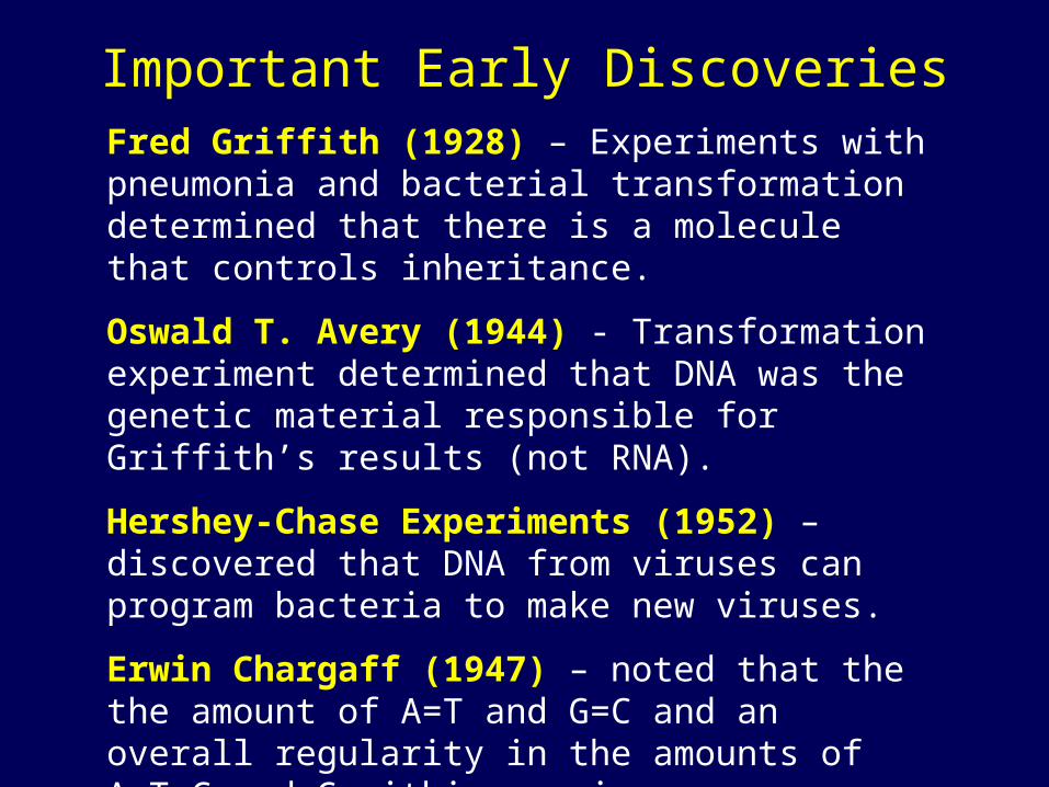
Frederick Griffith’s Transformation Experiment• The discovery of the genetic role of DNA began with research by Frederick Griffith in 1928• Griffith worked with two strains of a bacterium, a pathogenic “S” strain and a harmless “R” strain• When he mixed heat-killed remains of the pathogenic strain with living cells of the harmless
strain, some living cells became pathogenic
• He called this phenomenon transformation, now defined as a change in genotype and phenotype due to assimilation of foreign DNA
Living S cells(control)
Living R cells(control)
Heat-killedS cells (control)
Mixture of heat-killed S cells and living R cells
Mouse dies
Living S cellsare found in blood sample
Mouse healthy Mouse healthy Mouse dies
RESULTS
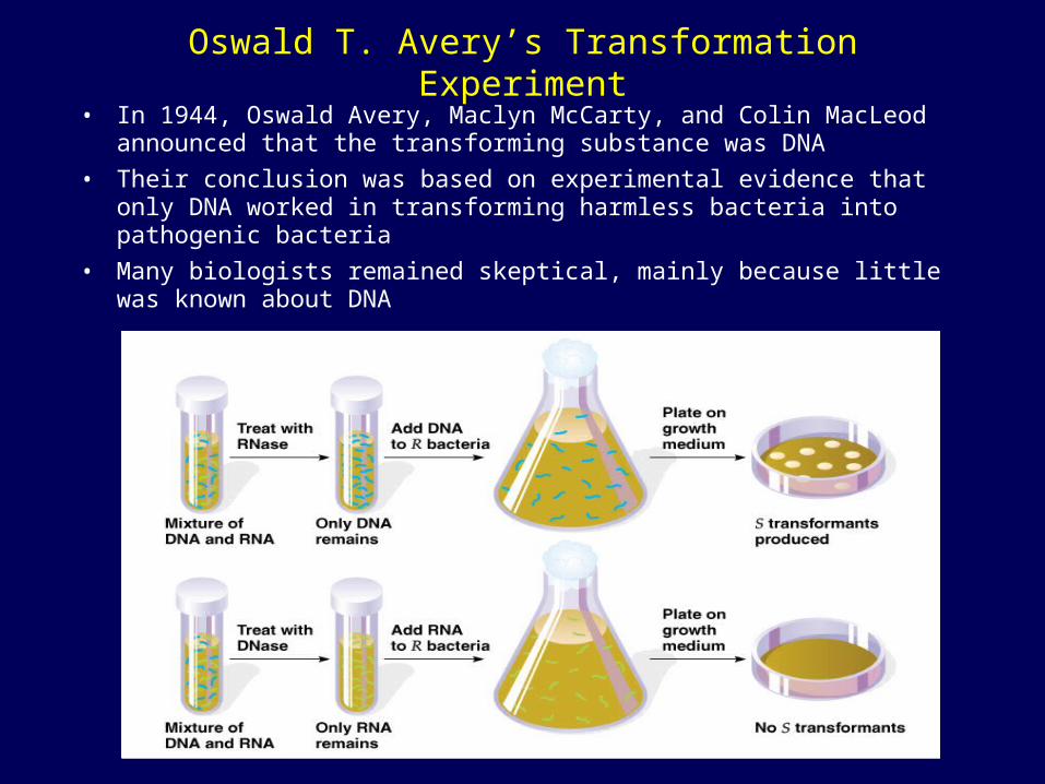
Oswald T. Avery’s Transformation Experiment
• In 1944, Oswald Avery, Maclyn McCarty, and Colin MacLeod announced that the transforming substance was DNA
• Their conclusion was based on experimental evidence that only DNA worked in transforming harmless bacteria into pathogenic bacteria
• Many biologists remained skeptical, mainly because little was known about DNA
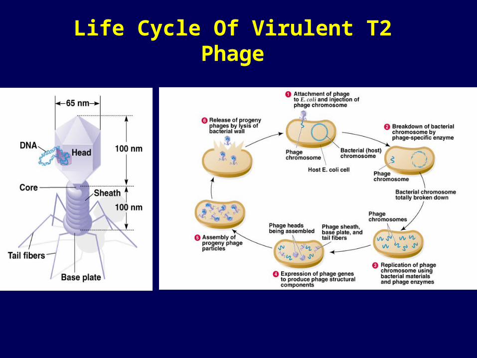
Life Cycle Of Virulent T2 Phage
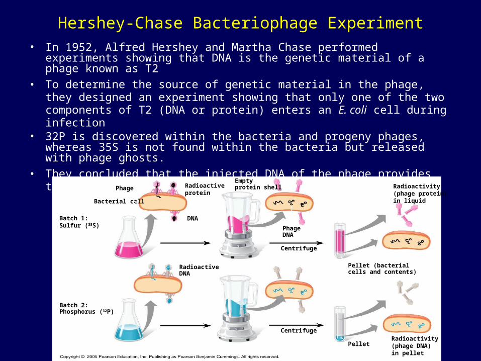
Hershey-Chase Bacteriophage Experiment
• In 1952, Alfred Hershey and Martha Chase performed experiments showing that DNA is the genetic material of a phage known as T2
• To determine the source of genetic material in the phage, they designed an experiment showing that only one of the two components of T2 (DNA or protein) enters an E. coli cell during infection
• 32P is discovered within the bacteria and progeny phages, whereas 35S is not found within the bacteria but released with phage ghosts.
• They concluded that the injected DNA of the phage provides the genetic information
Bacterial cell
Phage
DNA
Radioactiveprotein
Emptyprotein shell
PhageDNA
Radioactivity(phage protein)in liquid
Batch 1:Sulfur (35S)
RadioactiveDNA
Centrifuge
Pellet (bacterialcells and contents)
PelletRadioactivity(phage DNA)in pellet
Centrifuge
Batch 2:Phosphorus (32P)
Additional Evidence That DNA Is the Genetic Material
• In 1947, Erwin Chargaff reported that DNA composition varies from one species to the next
• This evidence of diversity made DNA a more credible candidate for the genetic material
• By the 1950s, it was already known that DNA is a polymer of nucleotides, each consisting of a nitrogenous base, a sugar, and a phosphate group
• Franklin’s X-ray crystallographic images of DNA enabled Watson to deduce that DNA was helical
• The X-ray images also enabled Watson to deduce the width of the helix and the spacing of the nitrogenous bases
• The width suggested that the DNA molecule was made up of two strands, forming a double helix
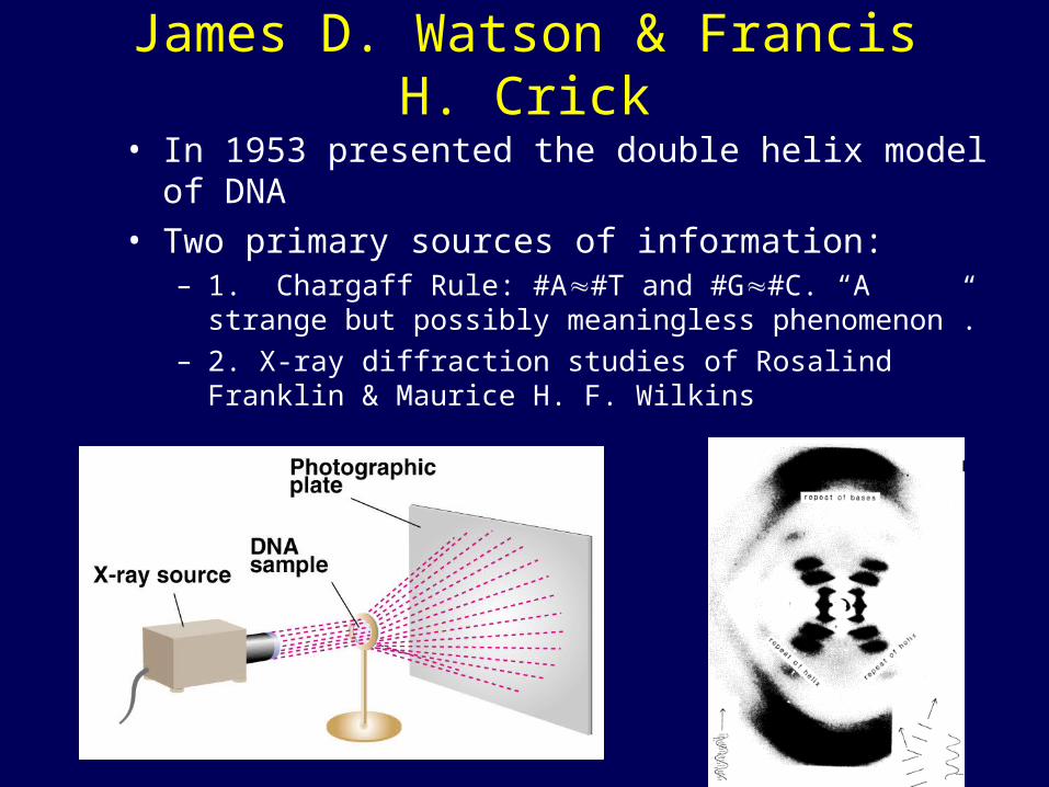
James D. Watson & Francis H. Crick
• In 1953 presented the double helix model of DNA• Two primary sources of information:
– 1. Chargaff Rule: #A#T and #G#C. “A strange but possibly meaningless phenomenon”.
– 2. X-ray diffraction studies of Rosalind Franklin & Maurice H. F. Wilkins
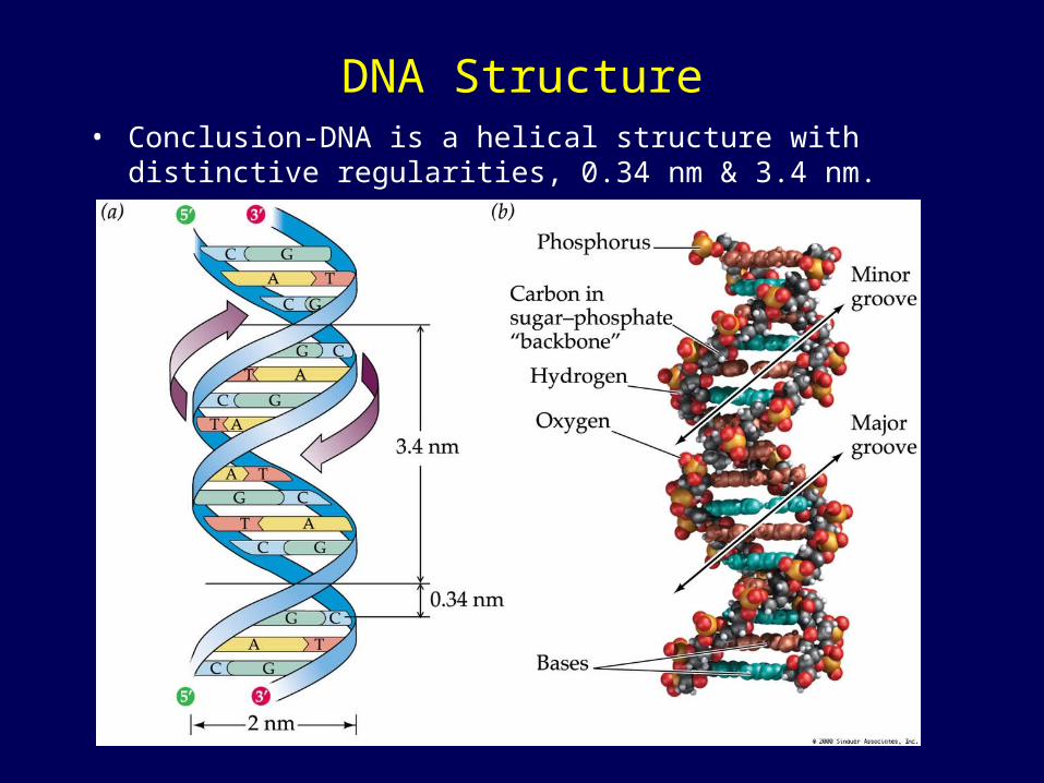
DNA Structure• Conclusion-DNA is a helical structure with distinctive
regularities, 0.34 nm & 3.4 nm.
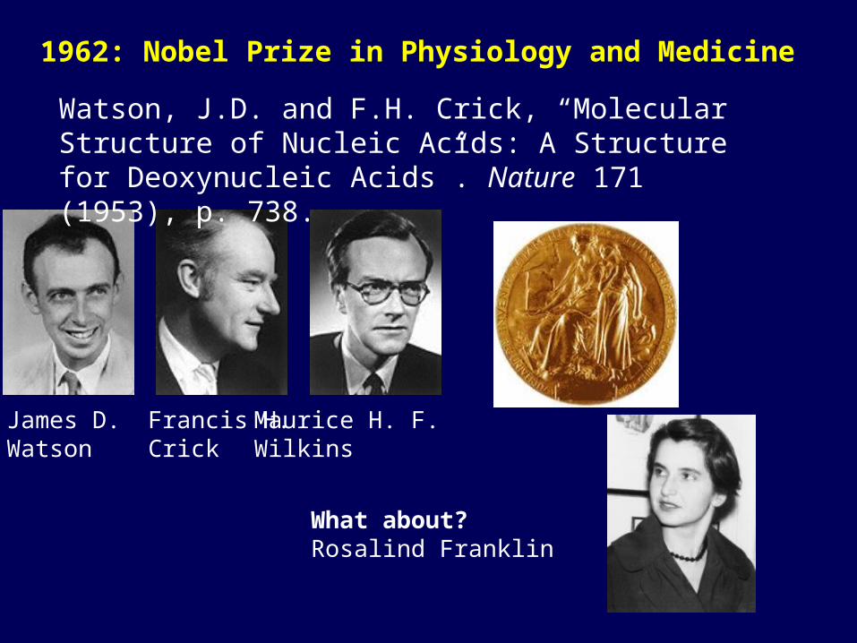
1962: Nobel Prize in Physiology and Medicine
James D.Watson
Francis H.Crick
Maurice H. F.Wilkins
What about?Rosalind Franklin
Watson, J.D. and F.H. Crick, “Molecular Structure of Nucleic Acids: A Structure for Deoxynucleic Acids”. Nature 171 (1953), p. 738.
The Structure of DNA
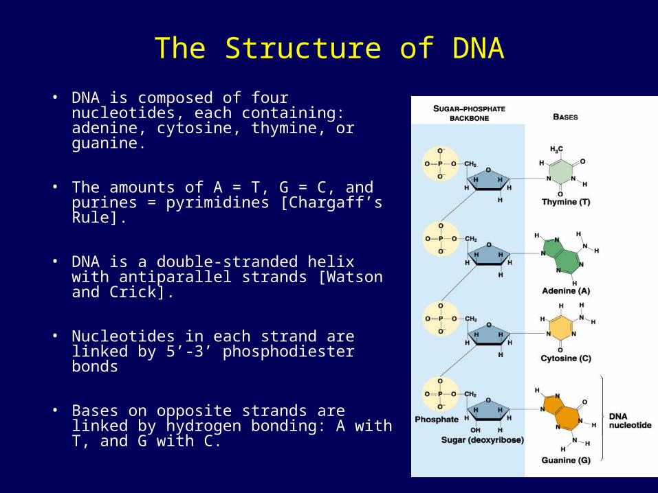
• DNA is composed of four nucleotides, each containing: adenine, cytosine, thymine, or guanine.
• The amounts of A = T, G = C, and purines = pyrimidines [Chargaff’s Rule].
• DNA is a double-stranded helix with antiparallel strands [Watson and Crick].
• Nucleotides in each strand are linked by 5’-3’ phosphodiester bonds
• Bases on opposite strands are linked by hydrogen bonding: A with T, and G with C.
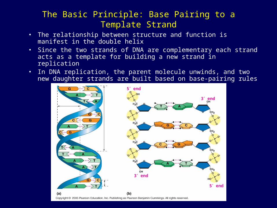
The Basic Principle: Base Pairing to a Template Strand
• The relationship between structure and function is manifest in the double helix
• Since the two strands of DNA are complementary each strand acts as a template for building a new strand in replication
• In DNA replication, the parent molecule unwinds, and two new daughter strands are built based on base-pairing rules
5 end
3 end
5 end
3 endHydrogen bond
0.34 nm
3.4 nm
1 nm
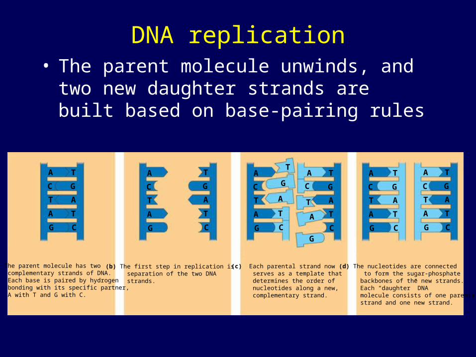
DNA replication• The parent molecule unwinds, and two new
daughter strands are built based on base-pairing rules
(a) The parent molecule has two complementary strands of DNA. Each base is paired by hydrogen bonding with its specific partner, A with T and G with C.
(b) The first step in replication is separation of the two DNA strands.
(c) Each parental strand now serves as a template that determines the order of nucleotides along a new, complementary strand.
(d) The nucleotides are connected to form the sugar-phosphate backbones of the new strands. Each “daughter” DNA molecule consists of one parental strand and one new strand.
A
C
T
A
G
A
C
T
A
G
A
C
T
A
G
A
C
T
A
G
T
G
A
T
C
T
G
A
T
C
A
C
T
A
G
A
C
T
A
G
T
G
A
T
C
T
G
A
T
C
T
G
A
T
C
T
G
A
T
C
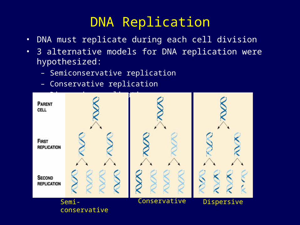
DNA Replication• DNA must replicate during each cell division• 3 alternative models for DNA replication were hypothesized:
– Semiconservative replication
– Conservative replication
– Dispersive replication
ConservativeSemi-conservative Dispersive
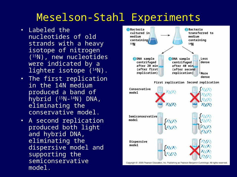
Meselson-Stahl Experiments• Labeled the nucleotides of
old strands with a heavy isotope of nitrogen (15N), new nucleotides were indicated by a lighter isotope (14N).
• The first replication in the 14N medium produced a band of hybrid (15N-14N) DNA, eliminating the conservative model.
• A second replication produced both light and hybrid DNA, eliminating the dispersive model and supporting the semiconservative model.
Bacteriacultured in mediumcontaining15N
DNA samplecentrifugedafter 20 min(after firstreplication)
DNA samplecentrifugedafter 40 min(after secondreplication)
Bacteriatransferred tomediumcontaining14N
Lessdense
Moredense
Conservativemodel
First replication
Semiconservativemodel
Second replication
Dispersivemodel
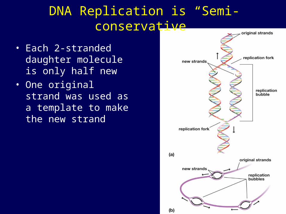
DNA Replication is “Semi-conservative”
• Each 2-stranded daughter molecule is only half new
• One original strand was used as a template to make the new strand
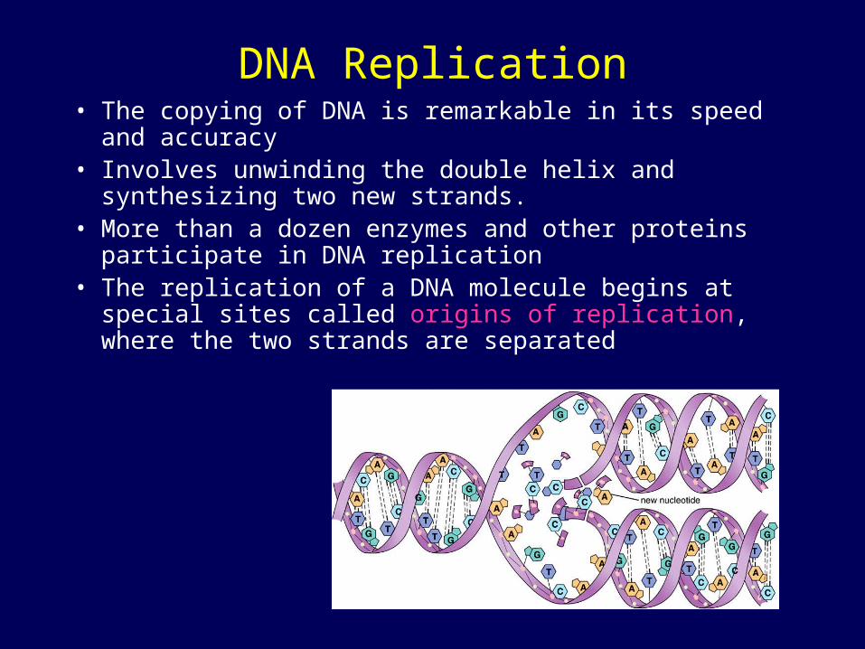
DNA Replication• The copying of DNA is remarkable in its speed and accuracy• Involves unwinding the double helix and synthesizing two
new strands.• More than a dozen enzymes and other proteins participate
in DNA replication• The replication of a DNA molecule begins at special sites
called origins of replication, where the two strands are separated
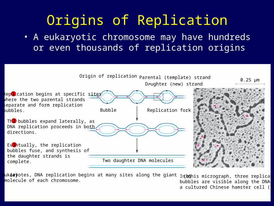
Origins of Replication• A eukaryotic chromosome may have hundreds or
even thousands of replication origins
Replication begins at specific siteswhere the two parental strandsseparate and form replicationbubbles.
The bubbles expand laterally, asDNA replication proceeds in bothdirections.
Eventually, the replicationbubbles fuse, and synthesis ofthe daughter strands iscomplete.
1
2
3
Origin of replication
Bubble
Parental (template) strand
Daughter (new) strand
Replication fork
Two daughter DNA molecules
In eukaryotes, DNA replication begins at many sites along the giantDNA molecule of each chromosome.
In this micrograph, three replicationbubbles are visible along the DNA ofa cultured Chinese hamster cell (TEM).
(b)(a)
0.25 µm
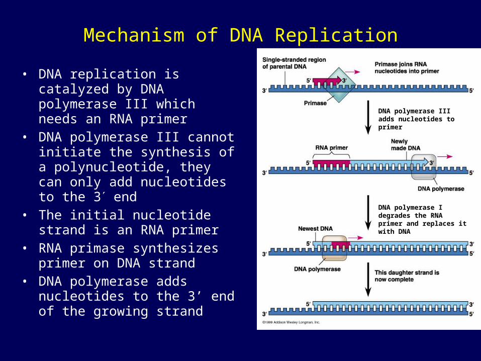
Mechanism of DNA Replication
• DNA replication is catalyzed by DNA polymerase III which needs an RNA primer
• DNA polymerase III cannot initiate the synthesis of a polynucleotide, they can only add nucleotides to the 3 end
• The initial nucleotide strand is an RNA primer
• RNA primase synthesizes primer on DNA strand
• DNA polymerase adds nucleotides to the 3’ end of the growing strand
DNA polymerase I degrades the RNA primer and replaces it with DNA
DNA polymerase III adds nucleotides to primer

Mechanism of DNA Replication• Nucleotides are added by complementary base pairing with the template strand• DNA always reads from 5’ end to 3’ end for transcription replication • During replication, new nucleotides are added to the free 3’ hydroxyl on the
growing strand• The nucleotides (deoxyribonucleoside triphosphates) are hydrolyzed as added,
releasing energy for DNA synthesis.
• The rate of elongation is about 500 nucleotides per second in bacteria and 50 per second in human cells
New strand5 end
Phosphate BaseSugar
Template strand3 end 5 end 3 end
5 end
3 end
5 end
3 end
Nucleosidetriphosphate
DNA polymerase
Pyrophosphate
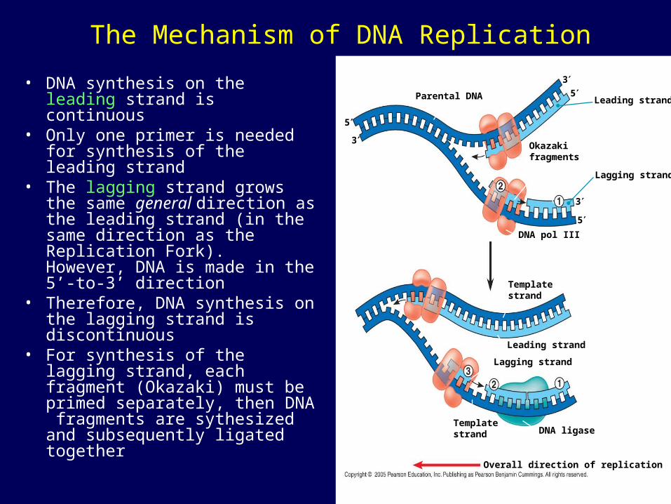
The Mechanism of DNA Replication
• DNA synthesis on the leading strand is continuous
• Only one primer is needed for synthesis of the leading strand
• The lagging strand grows the same general direction as the leading strand (in the same direction as the Replication Fork). However, DNA is made in the 5’-to-3’ direction
• Therefore, DNA synthesis on the lagging strand is discontinuous
• For synthesis of the lagging strand, each fragment (Okazaki) must be primed separately, then DNA fragments are sythesized and subsequently ligated together
Parental DNA
5
3
Leading strand
35
3
5
Okazakifragments
Lagging strand
DNA pol III
Templatestrand
Leading strand
Lagging strand
DNA ligase Templatestrand
Overall direction of replication
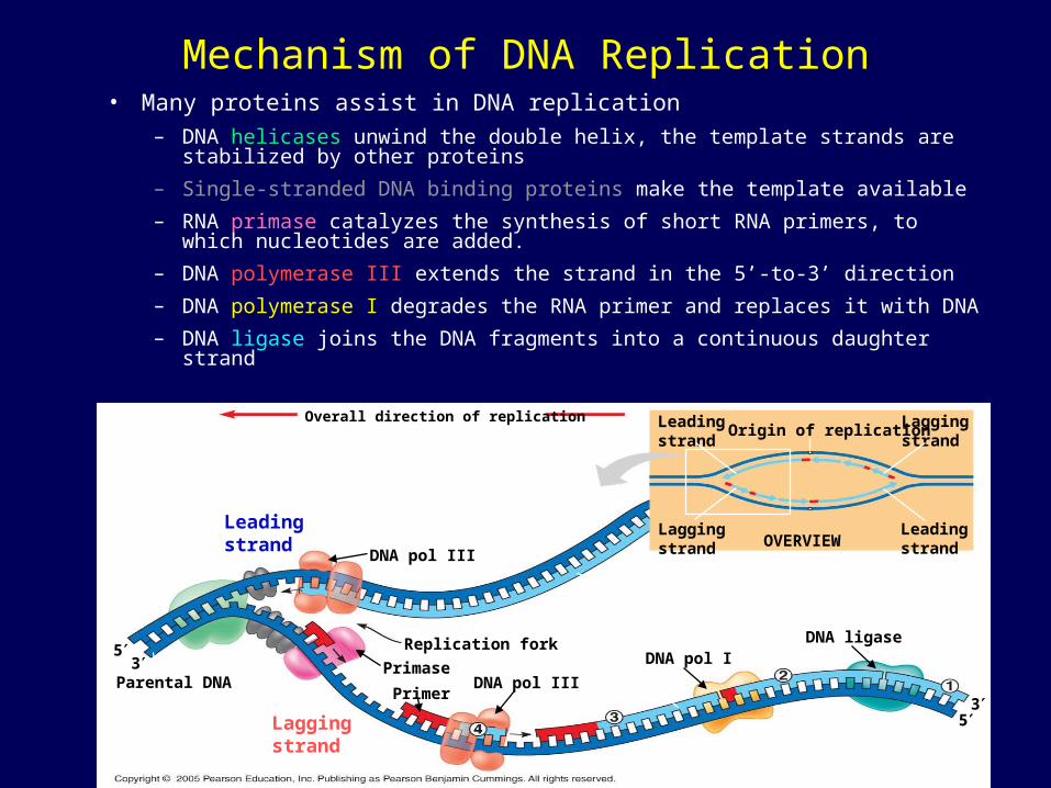
Mechanism of DNA Replication• Many proteins assist in DNA replication
– DNA helicases unwind the double helix, the template strands are stabilized by other proteins
– Single-stranded DNA binding proteins make the template available
– RNA primase catalyzes the synthesis of short RNA primers, to which nucleotides are added.
– DNA polymerase III extends the strand in the 5’-to-3’ direction
– DNA polymerase I degrades the RNA primer and replaces it with DNA
– DNA ligase joins the DNA fragments into a continuous daughter strand
5
3Parental DNA
3
5
Overall direction of replication
DNA pol III
Replication fork
Leadingstrand
DNA ligase
Primase
OVERVIEW
PrimerDNA pol III
DNA pol I
Laggingstrand
Laggingstrand
Leadingstrand
Leadingstrand
Laggingstrand
Origin of replication
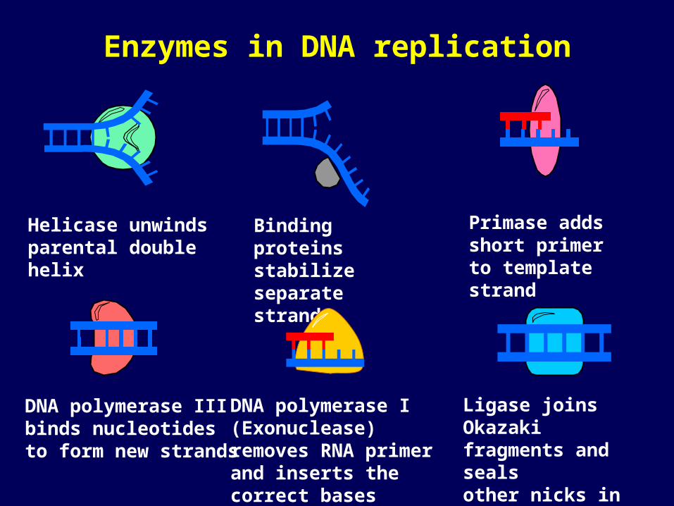
Enzymes in DNA replication
Helicase unwinds parental double helix
Binding proteinsstabilize separatestrands
DNA polymerase III binds nucleotides to form new strands
Ligase joins Okazaki fragments and seals other nicks in sugar-phosphate backbone
Primase adds short primer to template strand
DNA polymerase I (Exonuclease) removes RNA primer and inserts the correct bases
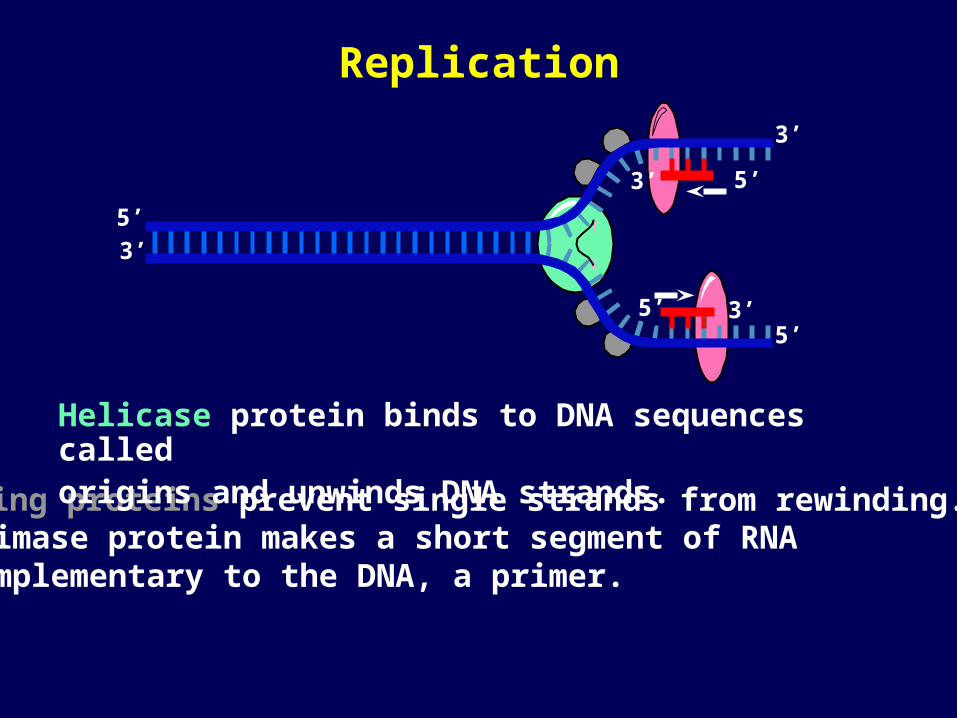
Binding proteins prevent single strands from rewinding.
Helicase protein binds to DNA sequences called origins and unwinds DNA strands.
5’
3’
5’
3’
Primase protein makes a short segment of RNA complementary to the DNA, a primer.
3’ 5’
5’ 3’
Replication
Overall directionof replication
5’ 3’
5’
3’
5’
3’
3’ 5’
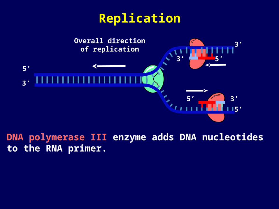
DNA polymerase III enzyme adds DNA nucleotides to the RNA primer.
Replication
5’
5’
Overall directionof replication
5’
3’
5’
3’
3’
3’
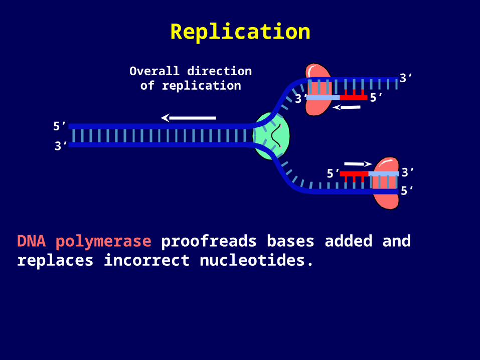
DNA polymerase proofreads bases added and replaces incorrect nucleotides.
Replication
5’
5’ 3’
5’
3’
3’
5’
3’Overall directionof replication
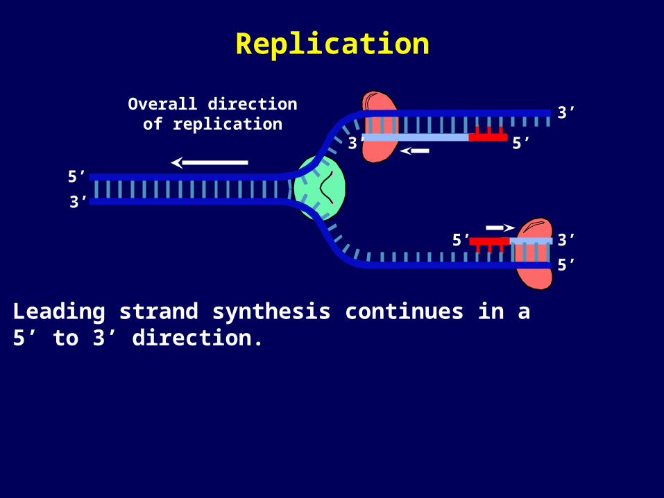
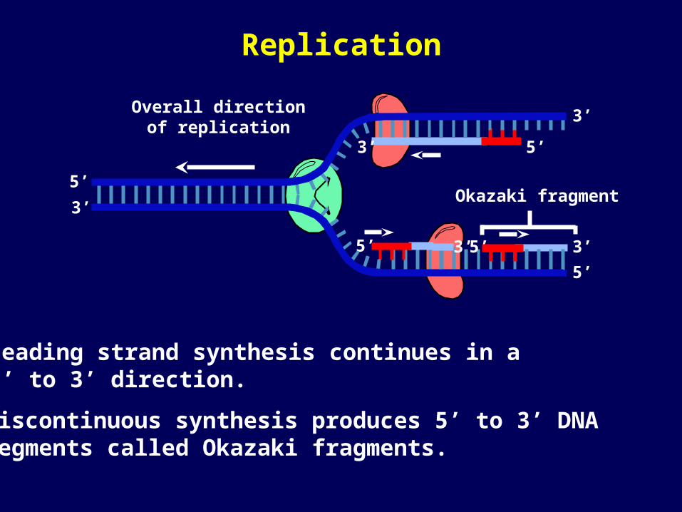
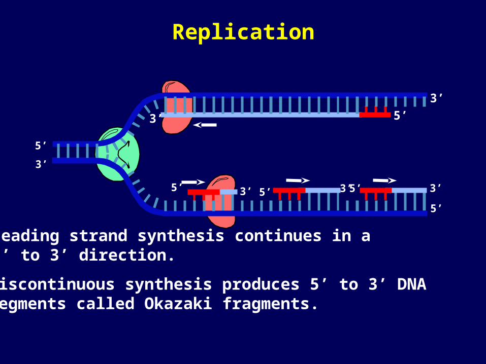
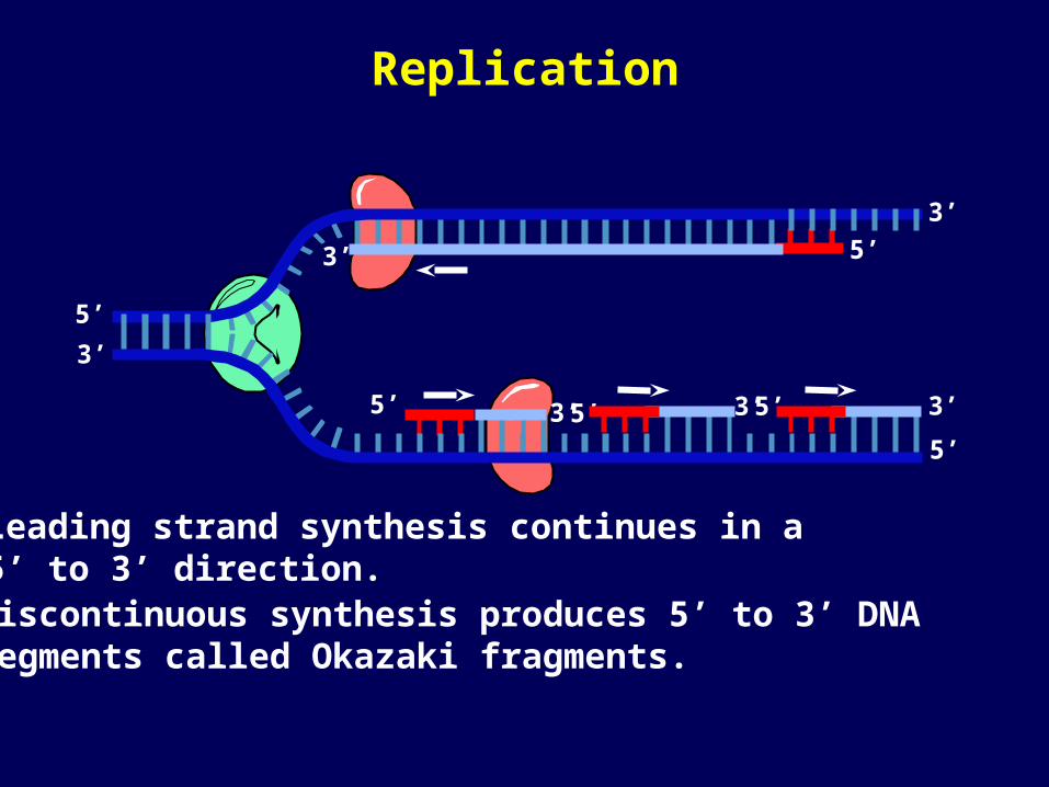
Leading strand synthesis continues in a 5’ to 3’ direction.
Replication

3’ 5’ 5’
5’ 3’
5’
3’
3’
5’
3’Overall directionof replication
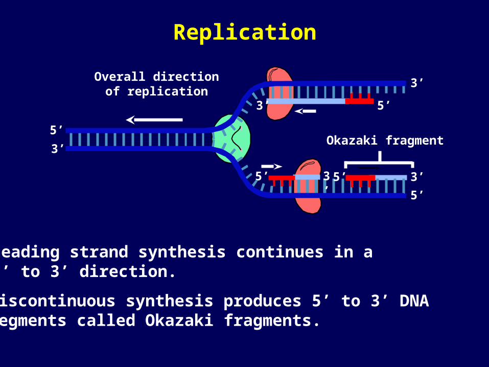
Okazaki fragment
Leading strand synthesis continues in a 5’ to 3’ direction.
Discontinuous synthesis produces 5’ to 3’ DNA segments called Okazaki fragments.
Replication
3’ 5’ 5’
5’ 3’
5’
3’
3’
5’
3’Overall directionof replication
Okazaki fragment
Leading strand synthesis continues in a 5’ to 3’ direction.
Discontinuous synthesis produces 5’ to 3’ DNA segments called Okazaki fragments.
Replication
5’ 5’
5’ 3’
5’
3’
3’
5’
3’Overall directionof replication
3’
Leading strand synthesis continues in a 5’ to 3’ direction.
Discontinuous synthesis produces 5’ to 3’ DNA segments called Okazaki fragments.
Okazaki fragment
Replication
5’
5’ 3’
5’
3’
3’
5’
3’
3’
5’ 5’ 3’
Leading strand synthesis continues in a 5’ to 3’ direction.
Discontinuous synthesis produces 5’ to 3’ DNA segments called Okazaki fragments.
Replication
3’
5’
3’
5’
5’ 3’
5’
3’
3’
5’ 5’ 3’
Leading strand synthesis continues in a 5’ to 3’ direction.Discontinuous synthesis produces 5’ to 3’ DNA segments called Okazaki fragments.
Replication
5’
5’
3’ 3’
5’
3’
5’ 3’
5’
3’
3’
5’
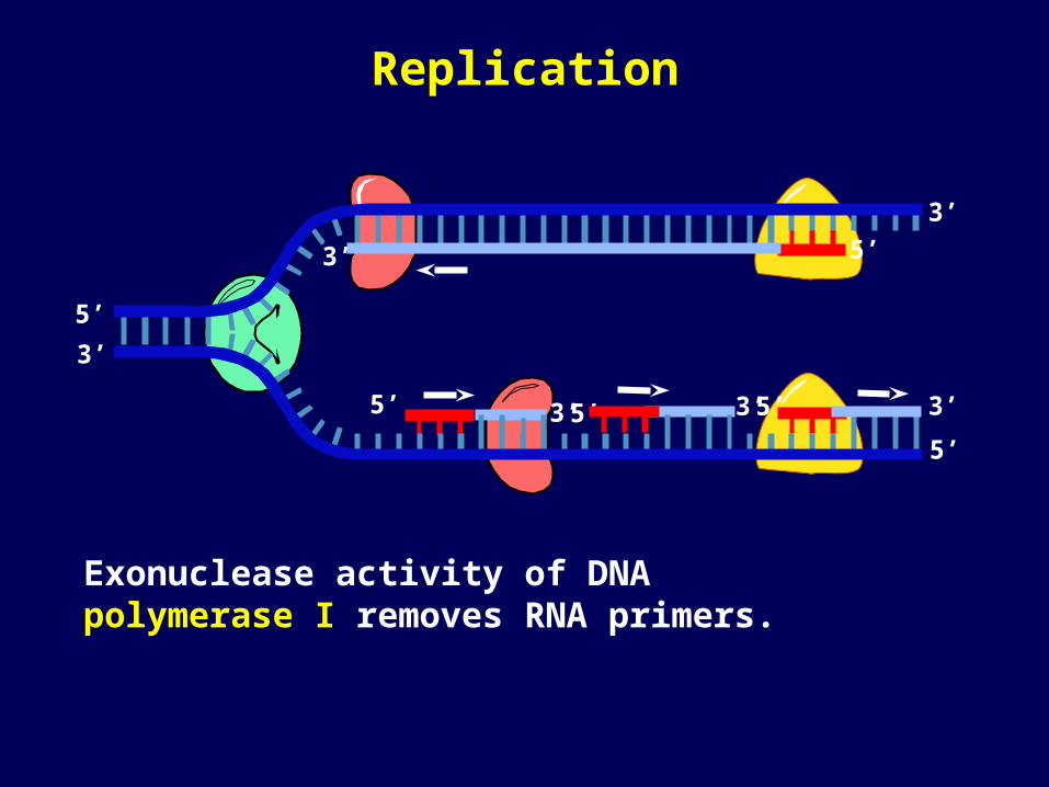
Exonuclease activity of DNA polymerase I removes RNA primers.
Replication
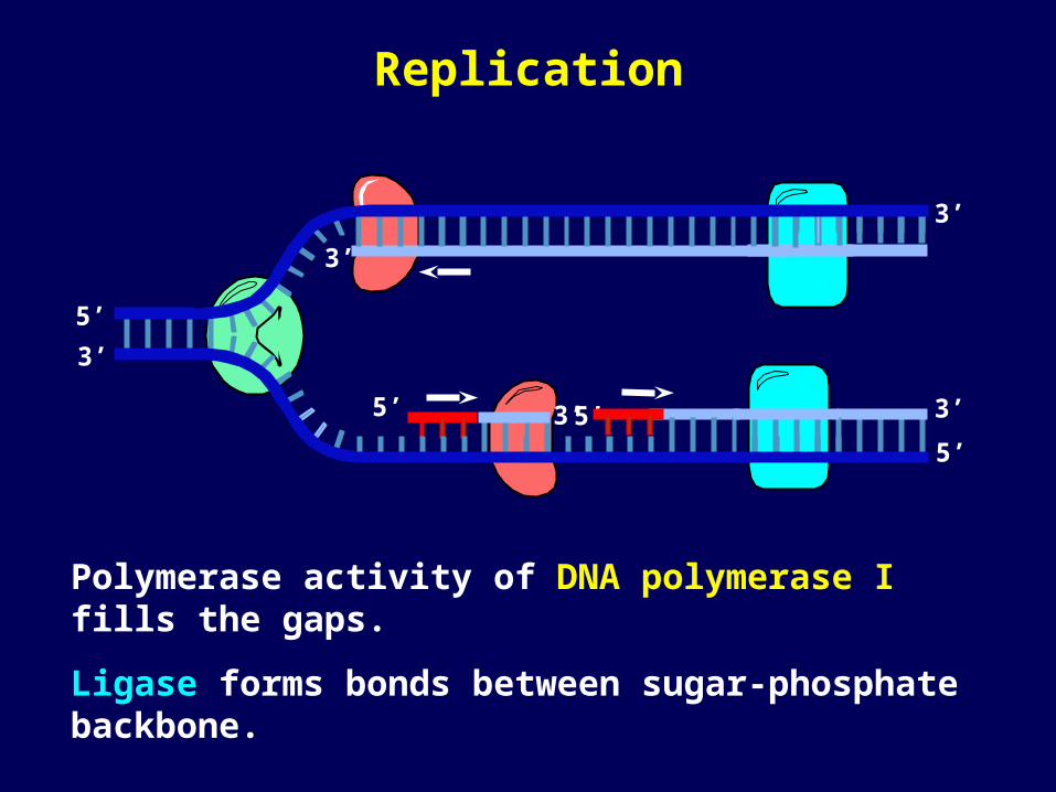
Polymerase activity of DNA polymerase I fills the gaps.
Ligase forms bonds between sugar-phosphate backbone.
3’
5’
3’
5’ 3’
5’
3’
3’
5’
Replication
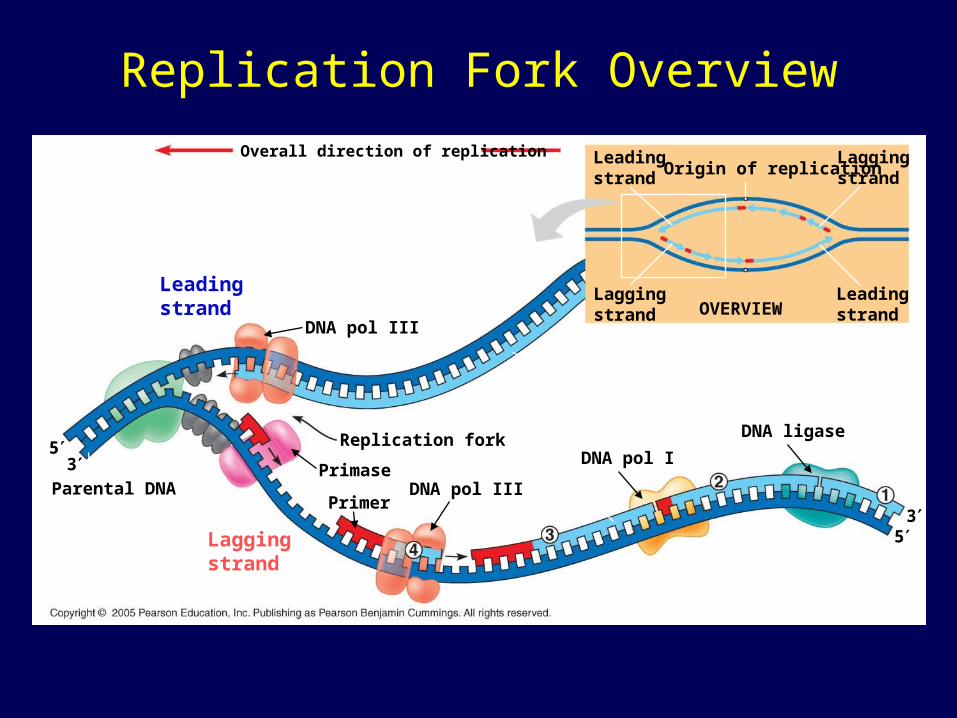
Replication Fork Overview
5
3Parental DNA
3
5
Overall direction of replication
DNA pol III
Replication fork
Leadingstrand
DNA ligase
Primase
OVERVIEW
PrimerDNA pol III
DNA pol I
Laggingstrand
Laggingstrand
Leadingstrand
Leadingstrand
LaggingstrandOrigin of replication
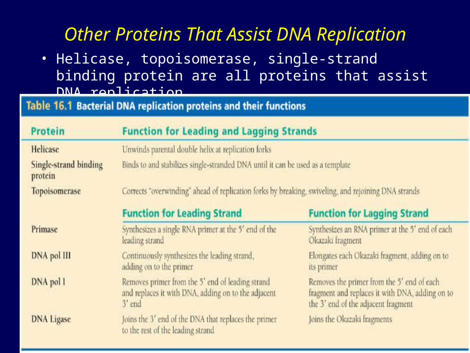
Other Proteins That Assist DNA Replication• Helicase, topoisomerase, single-strand binding
protein are all proteins that assist DNA replication
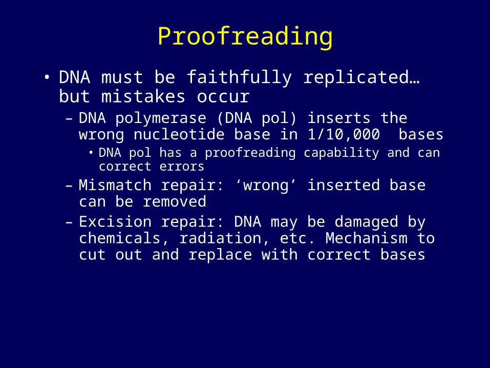
Proofreading
• DNA must be faithfully replicated…but mistakes occur– DNA polymerase (DNA pol) inserts the wrong
nucleotide base in 1/10,000 bases• DNA pol has a proofreading capability and can correct
errors
– Mismatch repair: ‘wrong’ inserted base can be removed
– Excision repair: DNA may be damaged by chemicals, radiation, etc. Mechanism to cut out and replace with correct bases
Mutations
• A mismatching of base pairs, can occur at a rate of 1 per 100,000 bases.
• DNA polymerase proofreads and repairs accidental mismatched pairs.
• Chances of a mutation occurring at any one gene is over 1 in 10,000,000,000 (billion)
• Because the human genome is so large, even at this rate, mutations add up. Each of us probably inherited 3-4 mutations!
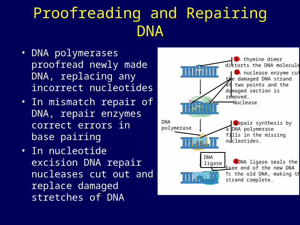
Proofreading and Repairing DNA
• DNA polymerases proofread newly made DNA, replacing any incorrect nucleotides
• In mismatch repair of DNA, repair enzymes correct errors in base pairing
• In nucleotide excision DNA repair nucleases cut out and replace damaged stretches of DNA
Nuclease
DNApolymerase
DNAligase
A thymine dimerdistorts the DNA molecule.1
A nuclease enzyme cutsthe damaged DNA strandat two points and thedamaged section isremoved.
2
Repair synthesis bya DNA polymerasefills in the missingnucleotides.
3
DNA ligase seals theFree end of the new DNATo the old DNA, making thestrand complete.
4
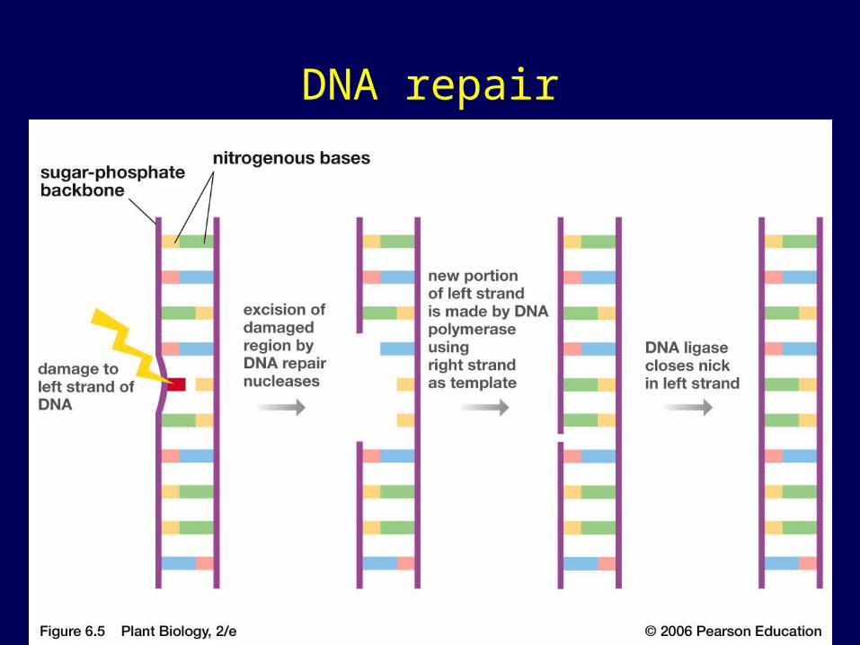
DNA repair
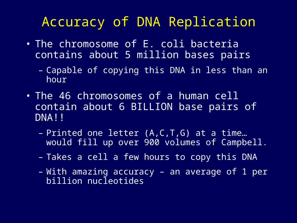
Accuracy of DNA Replication
• The chromosome of E. coli bacteria contains about 5 million bases pairs
– Capable of copying this DNA in less than an hour
• The 46 chromosomes of a human cell contain about 6 BILLION base pairs of DNA!!
– Printed one letter (A,C,T,G) at a time…would fill up over 900 volumes of Campbell.
– Takes a cell a few hours to copy this DNA
– With amazing accuracy – an average of 1 per billion nucleotides
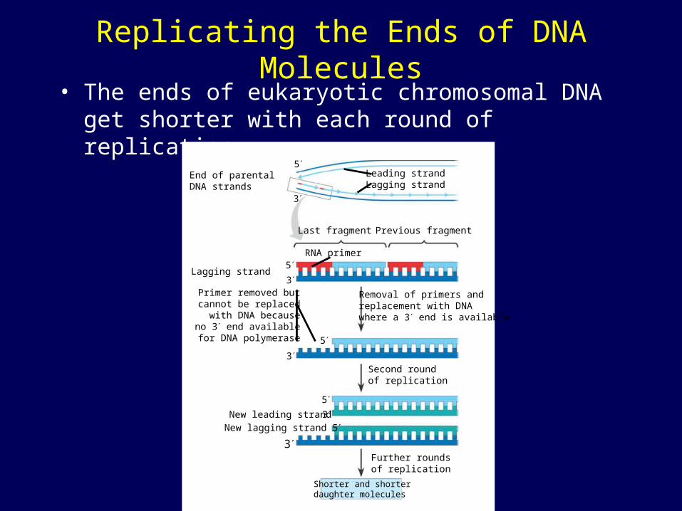
Replicating the Ends of DNA Molecules• The ends of eukaryotic chromosomal DNA get
shorter with each round of replication
End of parentalDNA strands
Leading strandLagging strand
Last fragment Previous fragment
RNA primer
Lagging strand
Removal of primers andreplacement with DNAwhere a 3 end is available
Primer removed butcannot be replacedwith DNA becauseno 3 end available
for DNA polymerase
Second roundof replication
New leading strand
New lagging strand 5
Further roundsof replication
Shorter and shorterdaughter molecules
5
3
5
3
5
3
5
3
3

Telomeres• Eukaryotic chromosomal DNA molecules
have at their ends nucleotide sequences, called telomeres, that postpone the erosion of genes near the ends of DNA molecules
1 µm
Telomerases
• If the chromosomes of germ cells became shorter in every cell cycle essential genes would eventually be missing from the gametes they produce
• An enzyme called telomerase catalyzes the lengthening of telomeres in germ cells