Embed Size (px)
Citation preview
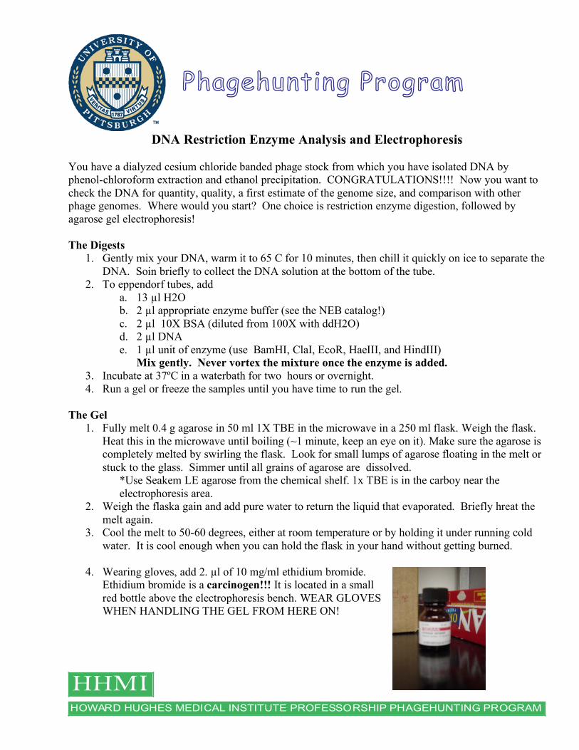
HOWARD HUGHES MEDICAL INSTITUTE PROFESSORSHIP PHAGEHUNTING PROGRAM
HHMI
Phagehunting Program
DNA Restriction Enzyme Analysis and Electrophoresis You have a dialyzed cesium chloride banded phage stock from which you have isolated DNA by phenol-chloroform extraction and ethanol precipitation. CONGRATULATIONS!!!! Now you want to check the DNA for quantity, quality, a first estimate of the genome size, and comparison with other phage genomes. Where would you start? One choice is restriction enzyme digestion, followed by agarose gel electrophoresis! The Digests
1. Gently mix your DNA, warm it to 65 C for 10 minutes, then chill it quickly on ice to separate the DNA. Soin briefly to collect the DNA solution at the bottom of the tube.
2. To eppendorf tubes, add a. 13 µl H2O b. 2 µl appropriate enzyme buffer (see the NEB catalog!) c. 2 µl 10X BSA (diluted from 100X with ddH2O) d. 2 µl DNA e. 1 µl unit of enzyme (use BamHI, ClaI, EcoR, HaeIII, and HindIII)
Mix gently. Never vortex the mixture once the enzyme is added. 3. Incubate at 37ºC in a waterbath for two hours or overnight. 4. Run a gel or freeze the samples until you have time to run the gel.
The Gel
1. Fully melt 0.4 g agarose in 50 ml 1X TBE in the microwave in a 250 ml flask. Weigh the flask. Heat this in the microwave until boiling (~1 minute, keep an eye on it). Make sure the agarose is completely melted by swirling the flask. Look for small lumps of agarose floating in the melt or stuck to the glass. Simmer until all grains of agarose are dissolved.
*Use Seakem LE agarose from the chemical shelf. 1x TBE is in the carboy near the electrophoresis area.
2. Weigh the flaska gain and add pure water to return the liquid that evaporated. Briefly hreat the melt again.
3. Cool the melt to 50-60 degrees, either at room temperature or by holding it under running cold water. It is cool enough when you can hold the flask in your hand without getting burned.
4. Wearing gloves, add 2. µl of 10 mg/ml ethidium bromide.
Ethidium bromide is a carcinogen!!! It is located in a small red bottle above the electrophoresis bench. WEAR GLOVES WHEN HANDLING THE GEL FROM HERE ON!

5. Put an inner tray in a casting tray. Pour the gel in to the casting tray. 6. Put a comb with the appropriate number of wells in the liquid gel.
7. Add 5 ml 1X TBE, allow the gel to cool and harden.
Then remove the comb and pour the buffer into the gel box and remove the gel from the casting tray. Rinse off the comb and casting tray, and put them on the test tube rack to dry.
8. Put the gel on the tray into an electrophoresis chamber (gel box) with the wells to the left side.
9. Add enough 1X TBE with 0.5 g/ml Ethidium Bromide to cover the wells.
10. Add 2 µl ficoll dye to each sample.
The ficoll dyes are located above the electrophoresis area.
11. Mix 2 µl uncut DNA with 10 µl TE and 2 µl ficoll dye in an eppendorf tube.
12. Also mix 1 µl 1kb DNA Ladder with 9 µl TE and 2 µl ficoll dye. The 1 kb DNA ladders are in the freezer next to Dr. Hatfull’s office in the drawer next to “Communal DNA Regrows, etc.” Make sure the ladder is completely thawed and mixed before using.
13. Load the samples in the gel in the following way:
Note: Gel is loaded ladder, uncut, alphabetically ordered enzyme digest name. When reading gels, the gel’s orientation is that the wells are placed at the top and the order can be read left to right.
14. Gently place the top on the electrophoresis chamber so your samples don’t rock out of the wells. Make sure the black (negative) electrode is on the left (DNA is negatively charged, so it will move towards the red (positive) electrode). Make sure black and red wires are connected to the power supply correctly.
HindIII
HaeIII
EcoRI
ClaI
BamHI
Uncut
1 Kb Ladder

15. Turn the power supply on and set it to constant voltage. Put it on the low setting and turn the dial until the voltmeter reads 100 volts. Run the gel at this voltage until you have good separation. There are two dyes in ficoll dye and they will separate as you run the gel. Both dyes should be on the gel when you are done. This will take ~30-45 minutes.
16. Turn the power source off and remove the chamber lid. (If another person is running a gel on
the same power source quickly unhook your cables and turn the power supply back on!)
17. Place the gel on saran wrap and take it to the gel doc. (Do not touch the gel doc or the computer with an ethidium bromide buffer contaminated glove!!!!) Push the gel out of the
inner tray into the gel doc and close the door. Hit “Live Focus” on the software and focus on the gel with the knob on top of the gel doc. Frame the gel so that the gel fills the screen. Push the trans-luminator button on and adjust the intensity if needed.
18. Video print two copies of your gel (one for
your notebook and one for the digest book) and save the file on the Hatfull lab folder in a subfolder with your name. Be sure to label the file accurately.
19. Remove your gel from the gel doc. Wipe down the surface with DI water and KimWipes.
20. The picture of your gel should be taped in the digest
book as shown here with well labeled wells and your name, your phage’s name, and the date.
The Hints!
1. The enzymes are in the -20º C freezer…THEY MUST STAY COLD… (Therefore, prepare the other ingredients the digest uses at your bench and keep on ice if necessary.) The enzymes are in blue racks in the freezer. Take out the enzymes you need and put them in your ice bucket and return promptly to minimize the time they are out of the freezer!!!!
2. Mix the reactions well, but don’t vortex once the enzyme is added! 3. Make sure the tubes are fully sealed before putting them in the water bath 4. Wear gloves with ethidium bromide gels!!! It is a carcinogen! 5. Note which manufacturer of DNA ladder you use. 6. Keep track of what is loaded in each lane – diagram it in your notebook. 7. If you are running more than one phage, load the gel with the 2 phages with the same enzymes in
lanes next to each other, not all one phage with its enzymes then the other phage. 8. If gel doc is not up and running, select the Quantity One icon from the desktop. 9. Make sure you clean everything. Rinse off all electrophoresis equipment, wipe off gel doc
platform, throw the gel in the proper chemical waste disposal bag, etc.