Embed Size (px)
Citation preview

Chapter 16
Pages 292-301
DNA Replication & Repair


Cell Cycle
http://scientopia.org/img-archive/scicurious/img_862.png

Models of DNA replication
Fig. 16.8

Models of DNA replication
Conservative model Semiconservative model Dispersive model
Daughter duplex
made of 2 newly
synthesized strands. Parent duplex
conserved.
Daughter duplexes are
made up of one
parental strand and one
newly synthesized
strand
Daughter duplexes are
made up of segments
of parental DNA and
newly synthesized
DNA

Matthew Meselson
& Franklin Stahl (1958) Performed an experiment to determine which
model of DNA replication was true See first half of video up to CsCl centrifugation: http://highered.mcgraw-hill.com/olc/dl/120076/bio22.swf
http://www.pnas.org/content/101/52/17889/F1.medium.gif

Meselson & Stahl Experiment Step 1
DNA contains nitrogen
atoms (bases)
First grew E. Coli cells in
heavy nitrogen (15N)
When cells are
centrifuged the DNA
will show up heavy

Density Gradient Centrifugation

Density Gradient Centrifugation

Meselson & Stahl Experiment Step 2
Then transferred E. Coli cells into a medium with
light nitrogen (14N)
Cells were allowed one round of replication
Then they were centrifuged

Meselson & Stahl Experiment Step 2
■ Predict what the
centrifuge tube would
show with each
model
Conservative
Semi- conservative
Dispersive

Meselson & Stahl Experiment Step 2
■ Which one did he see?
Conservative
Semi- conservative
Dispersive

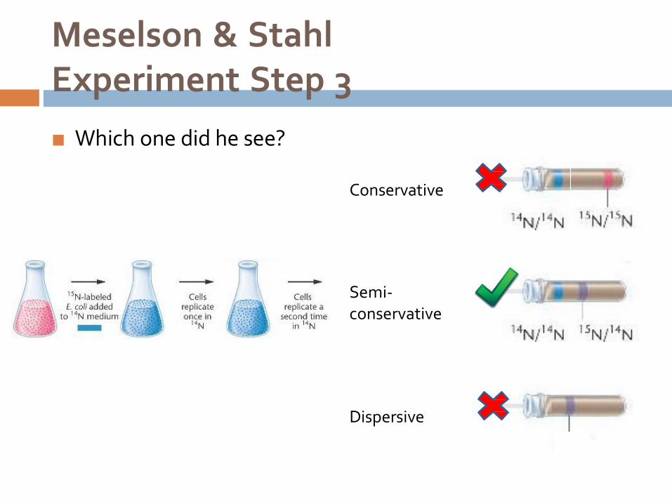
Meselson & Stahl Experiment Step 3
Cells were then allowed to grow for a second
round of replication in the light medium (14N)
And they were once again centrifuged

Meselson & Stahl Experiment Step 3
■ Predict what the
centrifuge tube would
show with each
model
Conservative
Semi- conservative
Dispersive
Conservative
Semi- conservative
Dispersive
Meselson & Stahl Experiment Step 3
■ Which one did he see?

Meselson & Stahl Experiment
Summary
15N
14N
14N

Round of replication
Conservative
model
Semiconservative
model Dispersive
model First replication
Two bands
Heavy: 15N15N
Light: 14N14N
Two bands
Heavy: 15N15N
Light: 14N14N
One band
medium: 15N14N One band medium: 15N14N
Second
replication
Two bands
Medium: 15N14N
Light: 14N14N
One band at a weight that is in between the medium
and light band weights
Meselson & Stahl Experiment
Summary Start with 15N
Add 14N Add
14N

Meselson & Stahl Experiment
Summary
Model Conservative
Generation I
Semi- Dispersive
conservative
Generation II

Meselson & Stahl Experiment

http://www.hhmi.org/biointeractive/dna/DNAi_replication_schematic.html
Basic concept of DNA replication (video)

Basic concept of DNA replication
During DNA replication, base pairing enables
existing DNA strands to serve as templates
for new complimentary strands
Fig. 16.7

2. DNA Replication Mechanism
Initiation
Elongation
Termination

DNA replication: Initiation
Origins of replication (ori): Special sites on DNA
where replication begins

Replication Initiation: Prokaryote
Replication begins at one fixed origin (only 1
ori)
Replication proceeds
bidirectionally until the DNA is replicated

Replication Initation: Eukaryote
More than one origin of replication (thousands of origin sites per chromosome)

Replication forks and bubbles
At the origin sites, the DNA strands separate
forming a replication “bubble” with replication forks
at each end.
Fig. 16.10

Replication forks and bubbles
The replication
bubbles
elongate as the
DNA is
replicated and
eventually fuses.
Fig. 16.10

Proteins in Replication Initiation
Sticky Tack Demo to show action of helicase, SSBP and topoisomerase
Yarn
Sticky tack or tape
scissors
■ Watch first half of video (before the fork): http://highered.mcgraw-hill.com/olc/dl/120076/bio23.swf

Proteins in Replication Initiation
http://eprots.pdbj.org/eprots/index_en.cgi?PDB%3A3BEP

Proteins in Replication Initiation
■ Helicase: enzyme that disrupts H bonds between two
strands of DNA to separate the template DNA strands
at the replication fork.
■ Single-strand binding proteins (SSBPs): proteins that bind to unwound single-stranded regions of DNA to
keep the template strands apart during replication
■ Topoisomerases: enzymes that can break bonds in
DNA and then reforms the bonds
■ Purpose is to release the twists in DNA that are generated
during DNA replication
■ Example of a topoisomerase: DNA gyrase

Priming DNA for replication
■ Blue line: DNA to be copied
■ Pink line: RNA nucleotides added = Primer
■ Light pink blob: Enzyme that adds RNA = RNA polymerase
Fig. 16.13

Priming DNA for replication
Primer: a short segment of RNA needed to initiate
DNA replication
Note: all nucleic acids are formed in the 5’ to 3’ direction, even RNA (thus primers)
Primase: an RNA polymerase (RNAP) which
synthesizes the primer by adding ribonucleotides
that are complementary to the DNA template
Polymerase: enzyme that makes polymers

Why is priming required?
Fig. 16.13

Why is priming required?
Due to the different abilities of RNA polymerase (RNAP) versus DNA polymerase (DNAP)
RNAP: can start a new chain without an existing end
All it needs is a template
E.g. primase
DNAP: can only add nucleotides to the end of an existing chain
can never start a new chain because it needs the 3’ OH

DNA Polymerase (DNAP)
Enzyme which synthesizes nucleotide chains
Prokaryotes:
DNA polymerase I, II, III, IV & V
Eukaryotes:
over 15 different types named with Greek letters (e.g. DNAP )

DNA Replication: Elongation
Fig. 16.11

DNA Replication: Elongation
DNA polymerase III (DNAP III) catalyzes the
elongation of DNA molecules by adding
nucleotides to the 3’ end of a pre-existing
nucleotide
As each nucleotide is added, the last two
phosphate groups are hydrolyzed to form
pyrophosphate.
Pyrophosphate is broken down into two phosphates
NTP NMP + 2P

DNA Elongation

DNA Polymerase
DNA polymerase I (DNAP I) replaces the RNA
primer with DNA complementary to the
template

DNA Elongation
DNAP III: elongates
DNA strand
DNAP I: replaces
RNA with DNA
Fig. 16.14
DNA polymerase III
DNA polymerase I

The problem at the fork
Due to antiparallel nature of DNA
One parental strand has
its 3’ end at the fork
while the other parental strand has its
5’ end at the fork.
But DNA synthesis can
only proceed in a 5’ 3’ direction.

Directionality
A strand of DNA can only
add nucleotides onto its
3’ end
DNA elongation only
proceeds in the 5’ to 3’ direction
DNAP must move along
the template strand’s 3’ to 5’ direction.

The leading and lagging strands
Leading strand: is synthesized continuously
Lagging strand: is synthesized in short, discontinuous segments of 1000-2000 nucleotides
called Okazaki fragments

Replication Fork
Fig. 16.16 http://kvhs.nbed.nb.ca/gallant/biology/replication_overview.jpg

Lagging strand: Okazaki fragments
DNAP III synthesizes the DNA
DNAP I replaces the RNA primer with DNA complementary to the template
DNA ligase joins broken pieces of DNA by catalyzing the formation of phosphodiester bonds

DNA Ligase
http://fhs-bio-wiki.pbworks.com/f/DNA%20Ligase%20reaction.jpg

Animations: Replication at the
Fork, Leading & Lagging
Strands Second half of video: http://highered.mcgraw- hill.com/olc/dl/120076/bio23.swf
Video: http://highered.mcgraw- hill.com/olc/dl/120076/micro04.swf

DNA Replication Machinery
III
I I
III
Fig. 16.15

The DNA Replication Complex
Proteins involved in DNA replication form a single
large complex anchored to the fibers in the
nucleus.
Stationary complex of DNA polymerase molecules
pull in the parental DNA and produce newly
made daughter DNA molecules.
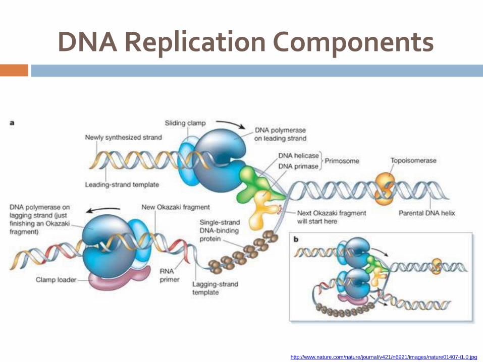
DNA Replication Components
http://www.nature.com/nature/journal/v421/n6921/images/nature01407-i1.0.jpg


http://www.hhmi.org/biointeractive/dna/DNAi_replication_vo2.html
Video: 3D model visualization
and explanation of replication

DNA replication: Termination
■ DNA replication ends when:
■ Reach the end of the
chromosome
■ Replication bubble / fork meets
another replication bubble/ fork

DNA Replication Tutorials
http://www.stolaf.edu/people/giannini/flashanimat/molgenetics/dn a-rna2.swf
http://www.wiley.com/legacy/college/boyer/0470003790/animation s/replication/replication.swf
http://www.wiley.com/college/pratt/0471393878/student/animation s/dna_replication/index.html
http://www.mcb.harvard.edu/Losick/images/TromboneFINALd.swf
http://www.johnkyrk.com/DNAreplication.html

HW Questions
Write out the point form summary of the steps in
DNA replication. Include all the enzymes in the
correct order.
Since DNAP can only polymerize off an available
3’ end, where does DNAP I (the one that removes the RNA primer and replaces it with
DNA) get that 3’ end from?
Compare prokaryotic and eukaryotic replication
and repair mechanisms by describing the
differences. Use a comparative chart if possible.

3. DNA Repair: Proofreading
Error Rate
Types of Errors
Exonuclease
Endonuclease

Proofreading DNA
Error rate
Average human chromosome has 150,000,000 bp
Initial pairing error: 1 in 10,000 bp = 15,000 errors per replication… that’s a LOT
Final error: 1 in 1,000,000,000 bp (1 in 109)
Mechanisms in place to proofread errors as DNA
is being replicated (exonuclease)
Cell also continuously monitors and repairs DNA
outside of replication (endonuclease)

Types of Errors in DNA
Mismatch mutations:
incorrectly paired bases
one of the most common types of errors during
replication
Missing bases
Fused bases

UV light
A common cause of DNA damage
Produces pyrimidine dimers (a type of fused base)
http://www.nature.com/scitable/nated/content/19185/pierce_17_24_FULL.jpg

Xeroderma Pigmentosum (XP)
A condition where individual is unable to repair damage caused by UV light
Individuals may need to avoid sunlight completely
(“children of the night”)
Leads to early skin cancer

Repair by Nuclease
Nuclease: an enzyme that can break
phosphodiester bonds in DNA thus excising out the nucleotide
Exonuclease: binds to ends of nucleotide chain (5’ or 3’)
Endonuclease: binds to the middle of a nucleotide
chain

Exonuclease Proofreading
Instantaneous repair:
Occurs as the DNA is replicating
Due to errors during elongation at the 3’ end
DNAP III and DNAP I both have exonuclease
activity

Exonuclease Proofreading
Mechanism of repair:
DNAP instantly recognize mismatches during replication
hydrolyze the phosphodiester bond releasing the last nucleotide that was just added (exonuclease activity)
replaces with the correct nucleotide (polymerase
activity)
Note: one enzyme (DNAP) does both the nuclease
and polymerase function

Endonuclease
Proofreading
Repair often occurs after DNA is already
replicated
Mechanism of repair known as nucleotide
excision repair (NER)
http://www.nature.com/scitable/content/36086/pierce-17_30_FULL.jpg

Endonuclease Proofreading: NER
■ Endonuclease:
■ recognizes and binds to
error
■ nicks the strand by breaking
phosphodiester bonds
■ error is excised (removed)
■ Polymerase: replaces the
gap with the correct nucleotides
■ Ligase: seals the nick
Fig. 16.17

4. Telomeres
Problem at the ends
Aging
Telomerase
http://images.sciencedaily.com/2009/10/091005110401-large.jpg

The problem with replication at
the ends of linear DNA
Fig. 16.18
DNA gets
progressively
shorter with each
round of replication
Prokaryotes avoid
problem by having
circular DNA

■ Structure:
■ DNA found at the ends of eukaryotic chromosomes
■ Noncoding (no genes)
■ Consists of multiple repeats of a short genetic sequence (humans: TTAGGG)
■ Function:
■ protect chromosomes from being eroded through multiple rounds of DNA replication
■ Less about preserving genetic information and more about serving as
a protective cap to prevent unwinding because
■ uncapping is sensed by cells
■ leads to cellular aging where cells stop growing and dividing
(senescence) and/or programmed self-destruction (apoptosis) http://www.dreva.com/shop/images/telomeres.jpg
Telomere

Telomere
http://science.nasa.gov/media/medialibrary/2006/03/16/22mar_telomeres_resources/chattps:./g/mif edia.npr.org/assets/img/2012/11/19/telomere-f87b8e7c2652d4f2d403da52beac222ce534b929-s6-c10.jpg

Telomere and Aging
At conception, cells have telomeres that are
about 15,000 bases long
At birth, cells in the infant have telomeres that are
10,000 bases long
Humans die from old age when telomere length
shortens to about 5000 bases
You’re born with the longest telomeres you’ll ever have.
It only gets shorter with age.

Telomere and Aging
Weng, Nan-ping. "Telomere and adaptive immunity."
Mechanisms of Ageing and Development 129.1-2 (2008): 60-66. Fig. 1. Model of telomere attrition in T and B cells with
age. Loss of telomere length is rapid during the first decade of life
and decreases during most of adult life. At advanced age, the
rate of telomere shortening may increase. The graph projects
the telomere attrition in CD4, CD8, and B cells in vivo based
on the cross-sectional analysis of telomere length in
lymphocytes with age. Whether significantly shortened
telomeres in advanced age cause declined function of lymphocytes will need further study.
http://www.sierrasci.com/telomere/index.html The time remaining on this "telomere clock"
can be measured from our blood cells. When
such measurements are taken, a significant correlation is found between a person's age
and the number of "ticks" remaining on the
person's clock.

Telomere
Question:
■When are telomeres added
to the chromosome when
the length of the telomere is
decreasing even at conception?
Answer:
■Telomeres added onto the
ends of chromosomes prior to birth, prior to
conception, prior to
fertilization

Telomere
Question:
■In what cell types would adding of the telomeres occur in if it happens before conception and fertilization?
Answer:
■In germ line cells which give rise to sex cells (gametes: sperm & egg)
■Also seen in cancer cells
http://cueflash.com/cardimages/answers/thumbnails/3/9/5493130.jpg

Telomerase
Enzyme responsible for adding telomeres to
chromosomes
A ribonucleoprotein that extends the ends of chromosomes using the enzymatic action of reverse transcriptase

Telomerase
Activity
Video clip:
http://www.youtube.com/watch?v=AJNoTm
WsE0s&feature=related
o-a-med/elizabeth-blackburn.html
Comprehensive series of lectures on
telomeres and telomerase by
Elizabeth Blackburn (discovered the
molecular nature of telomeres and
telomerase):
http://www.ibioseminars.org/lectures/cell-bi
Lecture 1 – Telomeres & Telomerase (48:27)
Lecture 2 – Telomeres & Telomerase in Human
Stem Cells & Cancer (26:58)
Lecture 3 – Stress, Telomeres & Telomerase in
Humans (45:58)

Telomerase Activity
Binds to 3’ end
(parental strand)
Extends the chain by
reverse transcribing
off its internal RNA
template (repeats)
Primase adds primer
DNAP elongates
Ligase seals the chain
Fig. 16.19

Telomerase
Ribonucleoprotein:
an enzyme that contains protein and RNA
human telomerase has an RNA component that contains
the internal sequence AAUCCC
Reverse transcriptase:
synthesis of complementary DNA from an RNA template
uses its internal RNA sequence as a template to make a complementary DNA strand with sequence TTAGGG
extends the 3’ end of the chromosome

Telomerase and Aging
Telomerase is only found in certain cells
Germ line cells
Cancer cells
Cells that have telomerase live for a longer period
of time
The lack of telomerase in most cells may explain
why cells have a finite lifespan
Example: DNA of dividing somatic cells (non-sex cells) tend to be shorter in older individuals

Telomerase and Aging
■ Why would germ line
cells need telomerase?
■ What would happen if germ line cells don’t have telomerase?
Scientist suspect that one of the
reasons why Dolly (the first mammal cloned from an adult somatic cell) didn’t live as long was because its telomeres were the
length of the adult that it was cloned from (as opposed to being
as long as a typical newly born
sheep)
http://1.bp.blogspot.com/-E1HNtngaNio/T_uldNBEfeI/AAAAAAAAA0c/dRTiYl12efM/s1600/Baby+Daddy+(final).jpg

Videos: Telomere and Aging
■ http://www.youtube.com/watch?v=IzinjLhZXpA (BBC
TV "Don't grow old", 3:41)
■ http://www.youtube.com/watch?v=xI70O69EZY8
(Today Show "How to live to 100", 5:59)
■ http://www.youtube.com/watch?v=m3qqUy880dQ
(Isagenix, 11:08)
■ http://www.youtube.com/watch?v=lBngws_cWho
(TedMed, 12:42)
■ http://www.youtube.com/watch?v=-bmMv6dcsgE
(Independent Pharmacy Business Growth Conference, February 23, 2012 in Orlando, FL, 1:44:06)
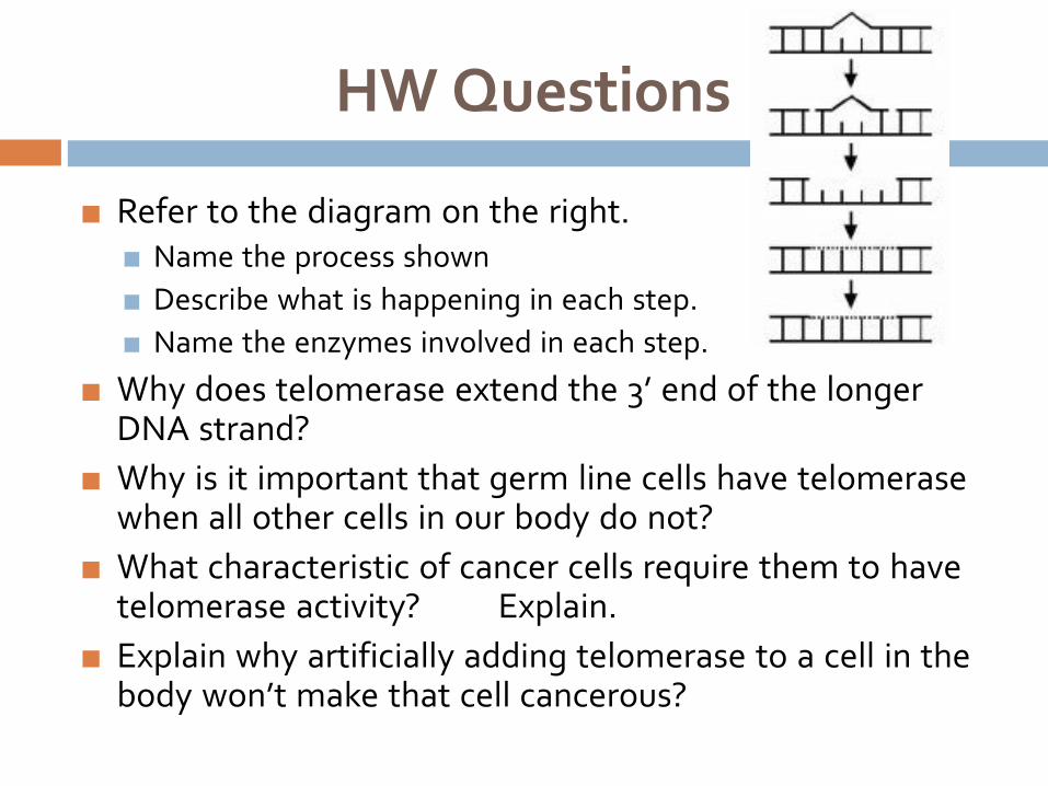
HW Questions
■ Refer to the diagram on the right. ■ Name the process shown
■ Describe what is happening in each step.
■ Name the enzymes involved in each step.
■ Why does telomerase extend the 3’ end of the longer DNA strand?
■ Why is it important that germ line cells have telomerase when all other cells in our body do not?
■ What characteristic of cancer cells require them to have telomerase activity? Explain.
■ Explain why artificially adding telomerase to a cell in the body won’t make that cell cancerous?

DNA Replication and Repair Machinery
Helicase
SSBPs
Topoisomerase (gyrase)
Primase (a RNAP)
DNAP III
DNAP I
DNA ligase
Nuclease
Telomerase