Embed Size (px)
Citation preview

DNA Replication
AP BiologyChapter 16 part 2

Watson and Crick
• In a second paper Watson and Crick published their hypothesis for how DNA replicates.– Essentially, because each strand is complementary
to each other, each can form a template when separated.
– The order of bases on one strand can be used to add in complementary bases and therefore duplicate the pairs of bases exactly.
• When a cell copies a DNA molecule, each strand serves as a template for ordering nucleotides into a new complimentary strand.– One at a time, nucleotides line up along the template strand
according to the base-pairing rules.– The nucleotides are linked to form new strands.

• Watson and Crick’s model, semiconservative replication, predicts that when a double helix replicates each of the daughter molecules will have one old strand and one newly made strand.
• There were some other competing models, the conservative model and the dispersive model.
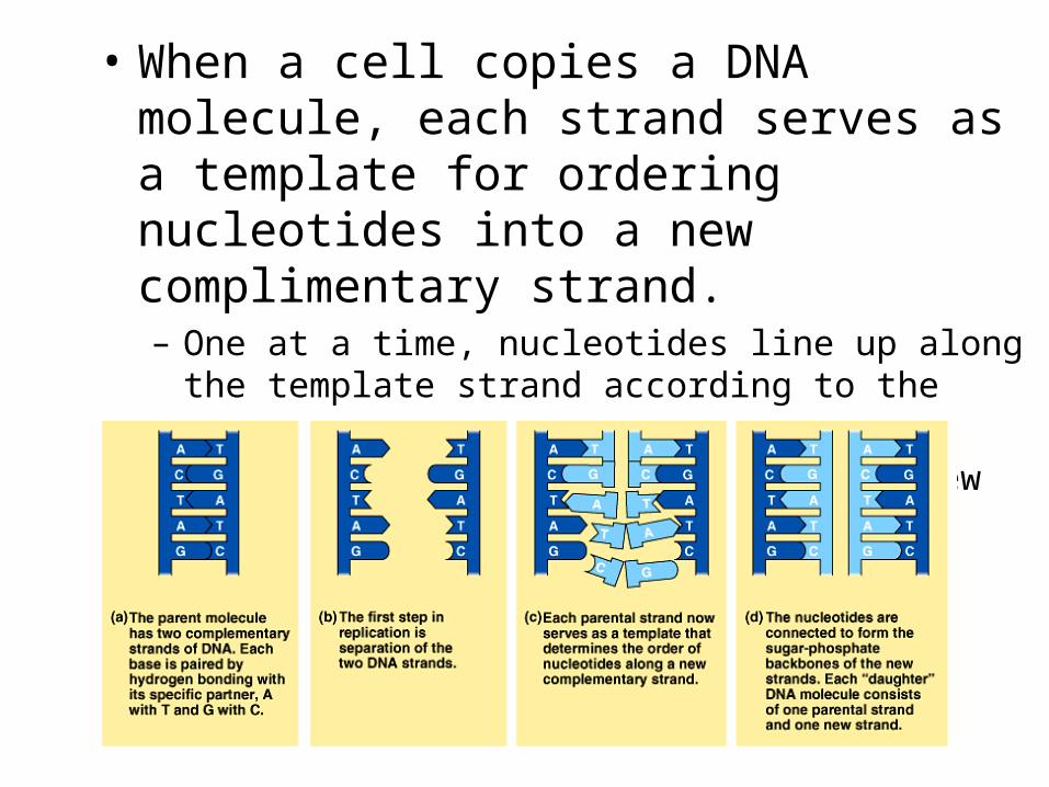
The Semiconservative Model• Experiments in the late 1950s by Matthew Meselson
and Franklin Stahl supported the semiconservative model, proposed by Watson and Crick, over the other two models.– They labeled the nucleotides of the old strands with a
heavy isotope of nitrogen (15N) while any new nucleotides would be indicated by a lighter isotope (14N).
– Replicated strands could be separated by density in a centrifuge.
– Each model: the semi-conservative model, the conservative model, and the dispersive model, made specific predictions on the density of replicated DNA strands.

– The first replication in the 14N medium produced a band of hybrid (15N-14N) DNA, eliminating the conservative model.
– A second replication produced both light and hybrid DNA, eliminating the dispersive model and supporting the semiconservative model.
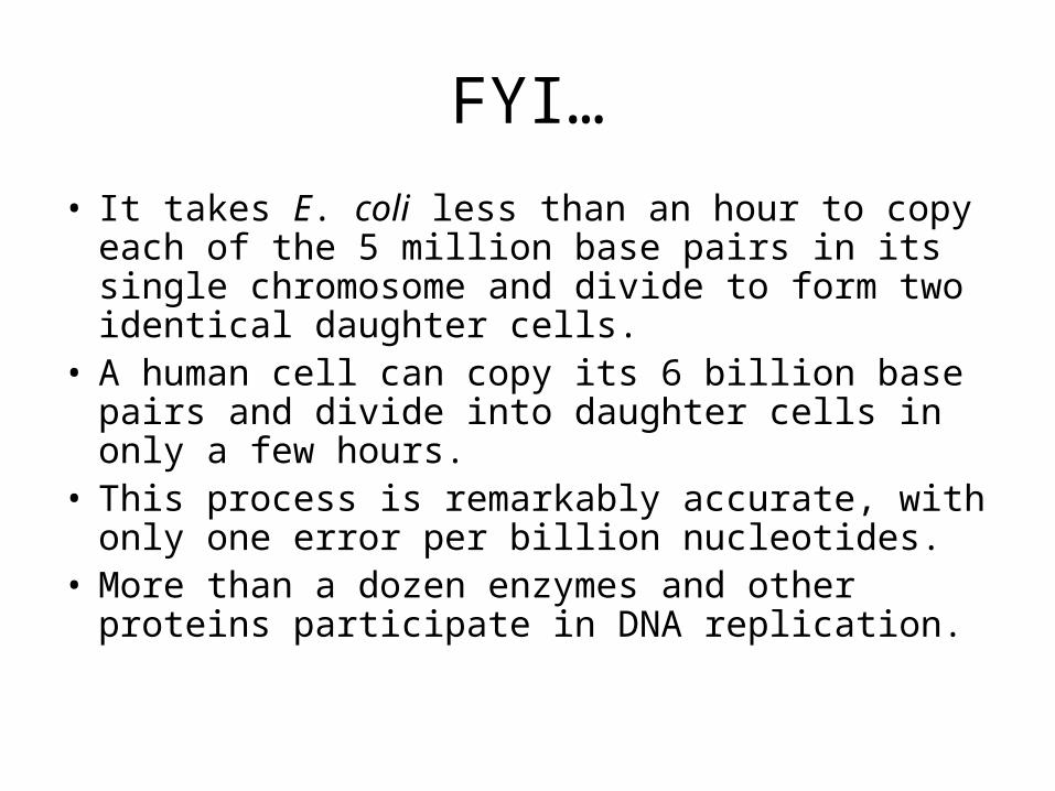
FYI…
• It takes E. coli less than an hour to copy each of the 5 million base pairs in its single chromosome and divide to form two identical daughter cells.
• A human cell can copy its 6 billion base pairs and divide into daughter cells in only a few hours.
• This process is remarkably accurate, with only one error per billion nucleotides.
• More than a dozen enzymes and other proteins participate in DNA replication.

How Replication Begins
• The replication of a DNA molecule begins at the origins of replication.
• In bacteria, this is a single specific sequence of nucleotides that is recognized by the replication enzymes.– These enzymes separate the strands, forming a
replication “bubble”.– Replication proceeds in both directions until the
entire molecule is copied.

• In eukaryotes, there may be hundreds or thousands of origin sites per chromosome. – At the origin sites, the DNA strands separate
forming a replication “bubble” with replication forks at each end.
– The replication bubbles elongate as the DNA is replicated and eventually fuse.

Stuff Needed for Replication• DNA polymerases starts the elongation of new DNA
at a replication fork.• As nucleotides match with complementary bases
along the template strand, they are added to the growing end of the new strand by the polymerase.– The rate of elongation is about 500 nucleotides per second
in bacteria and 50 per second in human cells.• The raw nucleotides are nucleoside triphosphates.– Each has a nitrogen base, deoxyribose, and a triphosphate
tail.

• As each nucleotide is added, the last two phosphate groups are broken off to form pyrophosphate.– The exergonic hydrolysis of pyrophosphate to two inorganic
phosphate molecules drives the polymerization of the nucleotide to the new strand.

Antiparallel• The strands in the double
helix are antiparallel.• The sugar-phosphate back-
bones run in opposite directions.– Each DNA strand has a 3’
end with a free hydroxyl group attached to deoxyribose and a 5’ end with a free phosphate group attached to deoxyribose.
– The 5’ 3’ direction of one strand runs counter to the 3’ 5’ direction of the other strand.

How DNA Strands Grow• DNA polymerases can only add nucleotides to the
free 3’ end of a growing DNA strand.• A new DNA strand can only elongate in the 5’ to 3’
direction.• This creates a problem at the replication fork
because one parental strand is oriented 3’5’ into the fork, while the other antiparallel parental strand is oriented 5’3’ into the fork.
• At the replication fork, one parental strand (3’ 5’ into the fork), the leading strand, can be used by polymerases as a template for a continuous complimentary strand.

• The other parental strand (5’3’ into the fork), the lagging strand, is copied away from the fork in short segments (Okazaki fragments).
• Okazaki fragments, each about 100-200 nucleotides, are joined by DNA ligase to form the sugar-phosphate backbone of a single DNA strand.

Primers• DNA polymerases cannot initiate synthesis of a
polynucleotide because they can only add nucleotides to the end of an existing chain that is base-paired with the template strand.
• To start a new chain requires a primer, a short segment of RNA.– The primer is about 10 nucleotides long in eukaryotes.
• Primase, an RNA polymerase, links ribonucleotides that are complementary to the DNA template into the primer.– RNA polymerases can start an RNA chain from a single
template strand.

Finishing Up Replication• After formation of the
primer, DNA polymerases can add deoxyribonucleotides to the 3’ end of the ribonucleotide chain.
• Another DNA polymerase later replaces the primer ribonucleotides with deoxyribonucleotides complimentary to the template.

The Lagging Strand
• Returning to the original problem at the replication fork, the leading strand requires the formation of only a single primer as the replication fork continues to separate.
• The lagging strand requires formation of a new primer as the replication fork progresses.
• After the primer is formed, DNA polymerase can add new nucleotides away from the fork until it runs into the previous Okazaki fragment.
• The primers are converted to DNA before DNA ligase joins the fragments together.
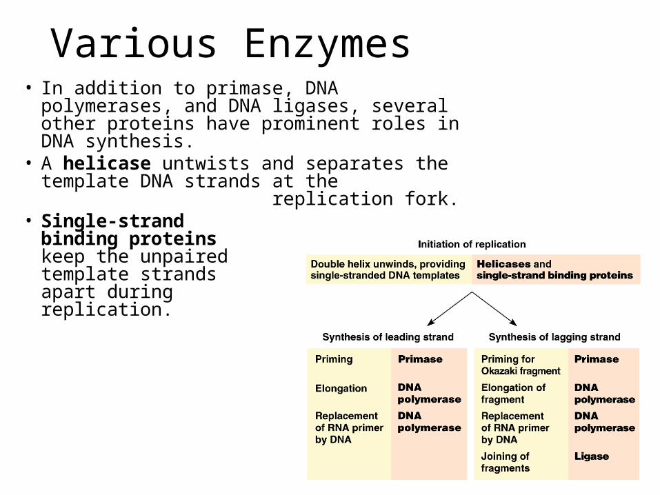
Various Enzymes• In addition to primase, DNA polymerases, and
DNA ligases, several other proteins have prominent roles in DNA synthesis.
• A helicase untwists and separates the template DNA strands at the replication fork.
• Single-strand binding proteins keep the unpaired template strands apart during replication.

• To summarize, at the replication fork, the leading stand is copied continuously into the fork from a single primer.
• The lagging strand is copied away from the fork in short segments, each requiring a new primer.

What About Mistakes?• Mistakes during the initial pairing of template
nucleotides and complementary nucleotides occur at a rate of one error per 10,000 base pairs.
• DNA polymerase proofreads each new nucleotide against the template nucleotide as soon as it is added.
• If there is an incorrect pairing, the enzyme removes the wrong nucleotide and then resumes synthesis.
• The final error rate is only one per billion nucleotides.

Mutagens• DNA molecules are constantly subject to potentially
harmful chemical and physical agents.– Reactive chemicals, radioactive emissions, X-rays, and
ultraviolet light can change nucleotides in ways that can affect encoded genetic information.
– DNA bases often undergo spontaneous chemical changes under normal cellular conditions.
• Mismatched nucleotides that are missed by DNA polymerase or mutations that occur after DNA synthesis is completed can often be repaired.– Each cell continually monitors and repairs its genetic
material, with over 130 repair enzymes identified in humans.

Fixing the Mistakes• In mismatch repair, special enzymes fix incorrectly
paired nucleotides.– A hereditary defect in
one of these enzymesis associated with a form of colon cancer.
• In nucleotide excision repair, a nuclease cuts out a segment of a damaged strand.– The gap is filled in by
DNA polymerase and ligase.

• Limitations in the DNA polymerase create problems for the linear DNA of eukaryotic chromosomes.
• The usual replication machinery provides no way to complete the 5’ ends of daughter DNA strands.– Repeated rounds of
replication produce shorter and shorter DNA molecules.

Telomeres• Telomerase is not present in most cells of
multicellular organisms.• Therefore, the DNA of dividing somatic cells and
cultured cells does tend to become shorter.• Thus, telomere length may be a limiting factor in the
life span of certain tissues and the organism.• Telomerase is present in germ-line cells, ensuring
that zygotes have long telomeres.• Active telomerase is also found in cancerous somatic
cells.– This overcomes the progressive shortening that would
eventually lead to self-destruction of the cancer.