Embed Size (px)
Citation preview
1 3
Extremophiles (2016) 20:195–205DOI 10.1007/s00792-016-0814-1
ORIGINAL PAPER
DNA Gyrase of Deinococcus radiodurans is characterized as Type II bacterial topoisomerase and its activity is differentially regulated by PprA in vitro
Swathi Kota1 · Yogendra S. Rajpurohit1 · Vijaya K. Charaka1,4 · Katsuya Satoh2 · Issay Narumi3 · Hari S. Misra1
Received: 8 October 2015 / Accepted: 20 January 2016 / Published online: 5 February 2016 © Springer Japan 2016
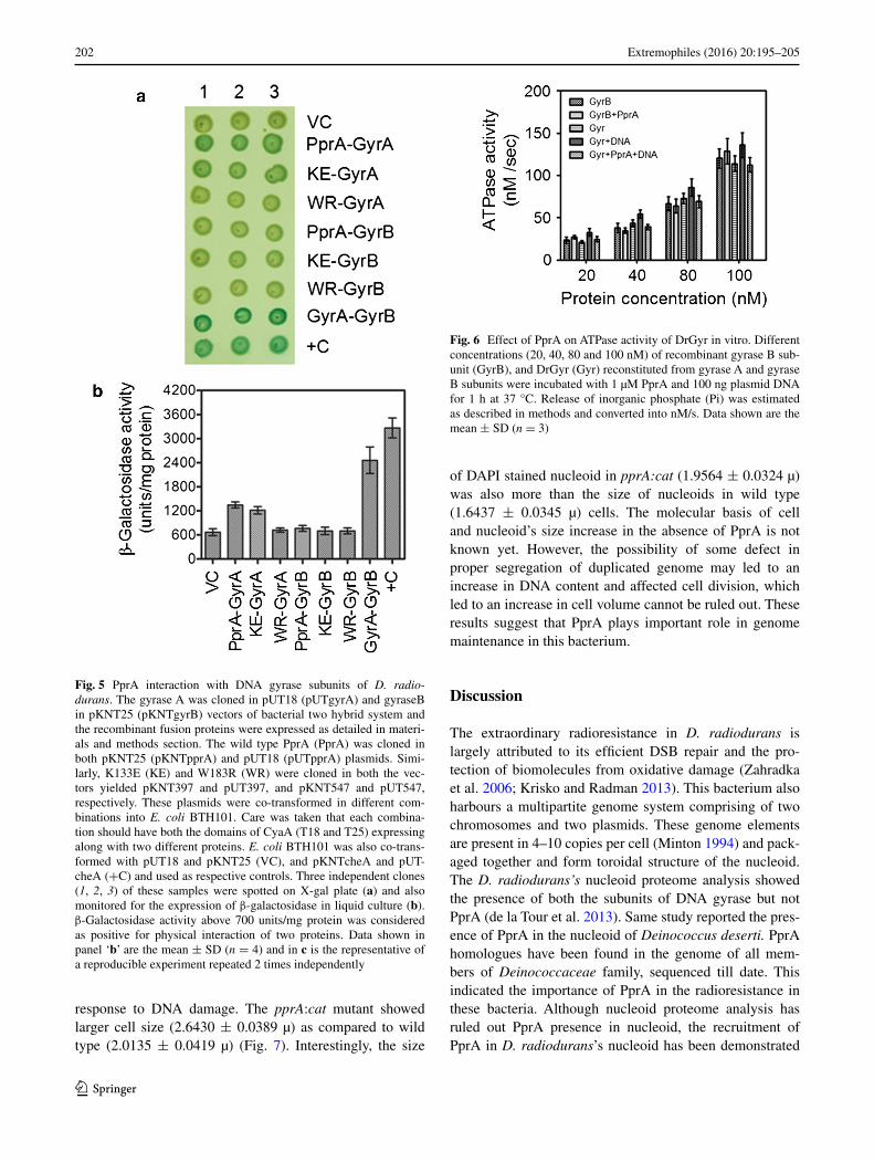
W183R, which formed relatively short oligomers did not interact with GyrA. The size of nucleoid in PprA mutant (1.9564 ± 0.324 µm) was significantly bigger than the wild type (1.6437 ± 0.345 µm). Thus, we showed that DrGyr confers all three activities of bacterial type IIA family DNA topoisomerases, which are differentially regulated by PprA, highlighting the significant role of PprA in DrGyr activity regulation and genome maintenance in D. radiodurans.
Keywords Deinococcus · DNA gyrase · Genome maintenance · PprA · Radioresistance
Introduction
Deinococcus radiodurans is a radioresistant bacterium that is known for its efficient DNA double strand break (DSB) repair (Zahradka et al. 2006; Ishino and Narumi 2015) and an extraordinary tolerance to oxidative stress (Daly et al. 2010; Tian and Hua 2010; Misra et al. 2013). It contains a multipartite genome system comprising of chromosome I, chromosome II and plasmids. Each of these genome ele-ments are present in multiple copies (Minton 1994) and packaged in the form of a doughnut shaped toroidal struc-ture (Levin-Zaidman et al. 2003). A number of proteins including DNA topoisomerases have been found associ-ated with the nucleoid of this bacterium (de la Tour et al. 2013). In Escherichia coli, DNA topoisomerases roles in maintaining correct DNA topology as well as in the res-olution of duplicated intertwined circular chromosome have been reported (Buck and Zechiedrich 2004). It has been shown that DNA topoisomerase IV (TopoIV) but not DNA gyrase is responsible for decatenation of site-spe-cific recombination intermediates in E. coli chromosome (Zechiedrich et al. 1997). More recently, it is observed that
Abstract The multipartite genome of Deinococcus radio-durans forms toroidal structure. It encodes topoisomerase IB and both the subunits of DNA gyrase (DrGyr) while lacks other bacterial topoisomerases. Recently, PprA a plei-otropic protein involved in radiation resistance in D. radio-durans has been suggested for having roles in cell division and genome maintenance. In vivo interaction of PprA with topoisomerases has also been shown. DrGyr constituted from recombinant gyrase A and gyrase B subunits showed decatenation, relaxation and supercoiling activities. Wild type PprA stimulated DNA relaxation activity while inhib-ited supercoiling activity of DrGyr. Lysine133 to glutamic acid (K133E) and tryptophane183 to arginine (W183R) replacements resulted loss of DNA binding activity in PprA and that showed very little effect on DrGyr activi-ties in vitro. Interestingly, wild type PprA and its K133E derivative continued interacting with GyrA in vivo while
Communicated by L. Huang.
Electronic supplementary material The online version of this article (doi:10.1007/s00792-016-0814-1) contains supplementary material, which is available to authorized users.
* Hari S. Misra [email protected]
1 Molecular Biology Division, Bhabha Atomic Research Centre, Mumbai 400084, India
2 Ion Beam Mutagenesis Research Group, Quantum Beam Science Center, Japan Atomic Energy Agency, Takasaki, Gunma 370-1292, Japan
3 Radiation Microbiology Laboratory, Department of Life Sciences, Toyo University, Itakura, Gunma 374-0193, Japan
4 Present Address: Department of Radiation Oncology, Houston Methodist Research Institute, Houston Methodist Hospital, Houston, TX 77030, USA
196 Extremophiles (2016) 20:195–205
1 3
Topo IV is actually knots and unknots the sister duplexes of DNA during replication in E. coli (Lopez et al. 2012). The mechanisms underlying the regulation of mutu-ally incompatible (knot and unknot) activities of TopoIV in vivo are not known yet. The D. radiodurans genome does not encode TopoIV but encodes DNA topoisomerase IB (DraTopoIB) and DNA gyrase (DrGyr) (White et al. 1999). PprA (named after pleiotropic protein promot-ing DNA repair) has been known for its role in radiation resistance in D. radiodurans (Narumi et al. 2004). Earlier, PprA was shown to contribute in DSB repair by stimu-lating DNA ends joining activities of T4 DNA ligase, E. coli DNA ligase and a deinococcal DNA ligase (LigB) (Narumi et al. 2004; Kota et al. 2010). PprA and DraT-opoIB were found in a multiprotein DNA processing com-plex characterized from this bacterium (Kota and Misra 2008). Molecular mechanisms underlying the packaging of multipartite genome in toroidal structure, its remodel-ling during replication, transcription, recombination and segregation, and faithful inheritance are largely unexplored in D. radiodurans. Recently, the identification of centro-meric sequences in the chromosome I of D. radiodurans and functional characterization of its partitioning pro-teins has been reported (Charaka and Misra 2012). Also, the interaction with DNA gyrase A subunit (GyrA) and DraTopoIB with PprA and the stimulation of DraTopoIB activity by this protein have been demonstrated (Kota et al. 2014a, b). Therefore, the involvement of deinococcal DNA topoisomerases and their possible interaction with PprA in the maintenance of multipartite genome struc-ture and functions would be worth understanding. Here, we report the functional characterization of recombinant DNA gyrase of D. radiodurans. Using purified recombi-nant gyrase A and gyrase B (GyrB) subunits of D. radio-durans an active DNA gyrase enzyme was reconstituted. Interestingly, DrGyr showed all three activities like DNA relaxation, supercoiling and decatenation of bacterial type IIA family DNA topoisomerases. PprA and its DNA binding mutant K133E interacted with GyrA. However, W183R mutant that had compromised both DNA binding and polymerization functions of PprA did not interact with GyrA. The relaxation activity of DrGyr was stimulated by wild type PprA but not by K133E and W183R derivatives. Interestingly, the DNA supercoiling activity of DrGyr was inhibited in the presence of wild type PprA but not mutants. These results suggested that DrGyr seems to be a sole multifunctional type IIA family DNA topoisomer-ase in D. radiodurans, and the DNA binding characteristic of PprA is crucial for its regulation of DrGyr activities at least in vitro. The possibility of DrGyr and PprA interac-tion contributing in dynamic regulation of the chromo-some functions during growth of this bacterium may be suggested.
Materials and methods
Bacterial strains and plasmids
D. radiodurans R1 (ATCC13939) was a gift from Profes-sor J. Ortner, Germany (Schaefer et al. 2000) and is main-tained in TGY (0.5 % Bacto Tryptone, 0.3 % Bacto Yeast Extract, and 0.1 % glucose) medium at 32 °C. E. coli strain DH5α is used for maintaining cloned genes on plasmids while E. coli BL21 (DE3) pLysS is used for the expression of recombinant protein. E. coli BL21 cells containing pET vectors (Novogen Inc.) and their derivatives while E. coli BTH101 containing pKNT25 and pUT18 plasmids (Kari-mova et al. 1998) and their derivatives were grown in LB broth with appropriate antibiotics. All recombinant tech-niques used in this study were as described earlier (Sam-brook and Russell 2001).
Generation of PprA mutants by random mutagenesis
The pprA gene was mutagenized by error-prone PCR or hydroxylamine treatment. For error-prone PCR, the pprA gene was amplified using a GeneMorph PCR Mutagen-esis Kit (Stratagene, Inc) with a set of primers that were used to generate the pprA expression plasmid pET3pprAwt (Narumi et al. 2004). For hydroxylamine treatment, pET-3pprAwt was treated with 0.1 M potassium phosphate (pH 6.0) containing 0.4 M hydroxylamine and 5 mM EDTA at 68 °C for 1 h, and purified using a MinElute Reaction Cleanup Kit (Qiagen, Inc). The purified plasmids were transformed into E. coli BMH 71-18 to efficiently fix muta-tions. Plasmids were digested with NdeI and BamHI and mutated inserts were ligated into pET3a at NdeI and BamHI site, and introduced into E. coli BL21 (DE3) to select mutant clones. Large colonies grown on LB agar plates were selected as candidate clones containing a mutation in the pprA gene. The presence of mutation was checked by DNA sequencing and pprA A397G (K133E) and pprA T547A (W183R) mutants have been used in this study.
Construction of expression plasmids
The ORF DR_1913 (gyrA) encoding GyrA and DR_0906 (gyrB) GyrB subunits of DrGyr in the D. radiodurans genome were PCR amplified from genomic DNA using the gene specific primers gryAF (5′ CGGGATCCATGACCGG AATTCAACCT 3′) and gyrAR (5′ AGCAAGCTTTTACAG CTCGTCTTCCTTGCGA 3′) for the gyrA, and gyrBF (5′ C GGGATCCATGAGCTTTTCCCATGC 3′) and gyrBR (5′ A GCAAGCTTTCAGACGCTGATTTCAGCGA 3′) for the gyrB, respectively. PCR amplified gyrA (2439 bp) and gyrB (1992 bp) were cloned in pET28a+ vector at BamHI and HindIII sites to generate recombinant plasmids pETgyrA
197Extremophiles (2016) 20:195–205
1 3
and pETgyrB. The PprA expression plasmids were con-structed as described earlier (Narumi et al. 2004; Kota and Misra 2006). The coding sequences of DNA bind-ing mutants K133E and W183R of pprA were cloned in pET28a+ vector at BamHI and HindIII sites and recom-binant plasmids pETK133E and pETW183R were gener-ated. These plasmids were transferred into E. coli BL21 (DE3) pLysS and the recombinant cells were induced with 200 μM IPTG. The expression of recombinant proteins was ascertained on SDS-PAGE and native PAGE.
Protein purification
Recombinant PprA and its derivatives were purified from recombinant E. coli expressing these proteins separately, as described earlier (Kota and Misra 2006). Recombinant GyrA and GyrB were purified by Ni2+ affinity chromatog-raphy using modified protocol of kit manufacturer (Qiagen, Inc). In brief, the recombinant E. coli cells expressing these proteins were lysed in buffer A (20 mM Tris–HCl, pH 8.0, 50 mM NaCl, 1 mM EDTA and 1 mM PMSF) containing 200 µg/ml lysozyme followed by 0.5 % Triton X-100 and 0.5 % NP40. Lysate was sonicated for 10 min at 30 s pulse with 1 min intermittent cooling at ice temperature with 50 % duty cycle. Clear cell free extract containing recom-binant proteins was obtained by centrifugation at 20,000×g and dialyzed in buffer A containing 5 mM imidazole and passed through NiNTA agarose column (Qiagen, Inc) pre-equilibrated with buffer A. Column was washed extensively with buffer A containing 20 mM imidazole till unbound or loosely bound proteins stopped coming from the column. The recombinant proteins were eluted by applying step gradients of 100, 200, and 300 mM imidazole in buffer A. All the fractions were analysed on SDS-PAGE and the fractions containing pure protein were pooled, dialysed in buffer A and concentrated using 50 kDa cut off filters (Amicon, Inc.). Histidine tag was removed with thrombin and the mixture was further purified through ion-exchange column chromatography. Fractions showing single protein band between 150 and 200 mM NaCl on SDS-PAGE were pooled and dialysed overnight in buffer A containing 50 % glycerol and stored at −70 °C for subsequent uses.
Gel shift assay
The DNA binding activity of wild type and mutant pro-teins was performed by agarose gel electrophoresis as described previously (Adachi et al. 2014). In brief, 100 ng of EcoRI-digested pUC19 plasmid DNA was added to TA buffer (40 mM Tris–acetate, pH 8.0, 20 mM sodium ace-tate) containing 1 mM MgCl2. To this 6.5 μM or 13 μM PprA protein was added and volume was made to 10 μl. Assay was carried out for 10 min at 37 °C, and subjected to
0.8 % agarose gel electrophoresis using TA buffer. Gel shift assay was also employed for checking the physical inter-action of purified PprA and DrGyr subunits. For that plas-mid (pSK+) DNA was incubated with GyrA, GyrB, and PprA separately and in different combinations. Nucleopro-tein complexes were analysed on agarose gel as described above.
Enzyme activity assay
DNA gyrase activity assays were performed as described previously (Karkare et al. 2012). In brief, purified recom-binant GyrA and GyrB subunits were reconstituted in 1:2 molar ratio 15 min prior to reaction for obtaining an active DNA gyrase complex as suggested earlier (Manjunatha et al. 2002). Superhelical pBluescript SK+ plasmid DNA was gel purified from a carefully made plasmid prepara-tion and relaxed pUC19 plasmid substrate was procured commercially (Cat. No. N0471S, New England Biolabs). For supercoiling assay, 500 ng of relaxed plasmid DNA was added to buffer containing 40 mM Tris–HCl (pH 7.9), 25 mM KCl, 1 mM DTT, 0.1 mg/ml tRNA, 100 mM potas-sium glutamate, 0.36 mg/ml BSA, 6 mM MgCl2, 2 mM spermidine and 1 mM ATP. To this 150 nM reconstituted DNA gyrase was added and volume was made to 30 μl. Assay was carried out for 60 min at 37 °C. DNA nicking assays were performed with different concentrations of reconstituted DNA gyrase in a similar way, except ATP and spermidine were omitted and the substrate was 400 ng superhelical form of pBluescript SK+ plasmid DNA pre-pared indigenously. Decatenation assay was done in 30 μl volume with 300 ng kinetoplast DNA (kDNA) (TopoGEN, Inc. Florida) in 40 mM Tris–HCl (pH 7.9), 25 mM KCl, 4 mM DTT, 100 mM potassium glutamate, 0.36 mg/ml BSA, 6 mM MgCl2 and 1 mM ATP for 60 min at 37 °C. The effects of PprA and its mutant derivatives on gyrase activity was monitored by adding the required amount of these proteins to reconstituted DrGyr and enzymatic activi-ties were carried out as above. All the reactions were ter-minated with 2 μl of 2 % sodium dodecyl sulphate (SDS), followed by proteinase K treatment for 20 min at 37 °C. Samples were extracted once with an equal volume of chloroform:isoamyl alcohol (24:1) and mixed with 0.1 vol-ume of loading dye (50 % glycerol, 0.025 % bromophenol blue). Similarly, E. coli DNA gyrase was procured com-mercially (New England Biolabs, Inc) and all three activi-ties like DNA relaxation, supercoiling and decatenation were measured as described above. For supercoiling assay and DNA nicking assay, products were analysed by 1 % agarose gel and ethidium bromide stained DNA bands were visualized, and quantified densitometrically as required. For decatenation assay, products were analyzed on 1 % agarose gel containing ethidium bromide. ATPase activity
198 Extremophiles (2016) 20:195–205
1 3
of DrGyr was monitored as described earlier (Modi and Misra 2014). The Pi released was measured and enzyme activity has been represented as nM/sec.
Protein–protein interaction studies
In vivo protein–protein interaction was studied using BACTH [Bacterial Adenylate Cyclase (CyaA)-based Two Hybrid] bacterial two-hybrid system (Karimova et al. 1998) as described earlier (Kota et al. 2014b). pUT18 and pUT18C expressing T18 the C-terminal domain while pKNT25 and pKT25 expressing T25 the N termi-nal domain of CyaA were procured and used in this study for generating translation fusion with target proteins. T25 and T18 domains of CyaA were fused separately with tar-get proteins and co-expressed in cyaA− strain of E. coli BHT101. Whenever, the two target proteins interact, both T18 and T25 domains of CyaA would have interacted too, resulting in the reconstitution of an active CyaA enzyme in cyaA mutant and thus the expression of β-galactosidase would be seen. In this study, the coding sequences of pprA mutants, pprA397 (for K133E) and pprA547 (for W183R), were cloned in pKNT25 vector at BamHI and KpnI sites yielded pKNTK133E and pKNTW183R plasmids, respec-tively. Similarly, the cloning of gyrA, gyrB and wild type pprA were carried out in respective bacterial two hybrid system plasmids as described earlier (Kota et al. 2014a). These plasmids were transformed into E. coli BTH-101 in different combinations. Recombinant clones harbour-ing both the plasmids, as monitored by co-expression of respective antibiotic resistance, were grown overnight and spotted on LB agar plate containing 200 µM IPTG. Plate was incubated at 30 °C overnight and the appearance of blue colour colonies was recorded. In parallel, these cells were grown overnight at 30 °C in the presence of IPTG in liquid culture, and β-galactosidase activity was measured in whole cells using standard protocols (Sambrook and Rus-sell 2001) and the specific activity of enzyme as U/mg pro-tein was calculated as described earlier (Kota et al. 2014b).
Microscopic studies
Microscopic studies were carried out as described ear-lier (Charaka and Misra 2012). Both wild type and pprA mutant (Narumi et al. 2004) were grown overnight under normal conditions. Cells were treated with 6.5 kGy gamma radiation and allowed to recover for 2 h post irradia-tion. Cells were stained with Nile red (4 μg/ml) and with 4′,6-diamidine-2′-phenylindole dihydrochloride (DAPI) (0.2 μg/ml) whenever required. These cells were mounted on agarose-coated slides (1.2 %) and observed under flu-orescence microscope (Axio Imager CM5, Carl Zeiss AG). Images were recorded under phase contrast, and
by excitation at 516 and 350 nm for Nile red and DAPI, respectively. Cell and nucleoid sizes were measured using Axiovision 4.8 software and statistically analysed using Graphpad Prism software.
Results
DNA Gyrase of D. radiodurans acts like bacterial Type II DNA topoisomerase
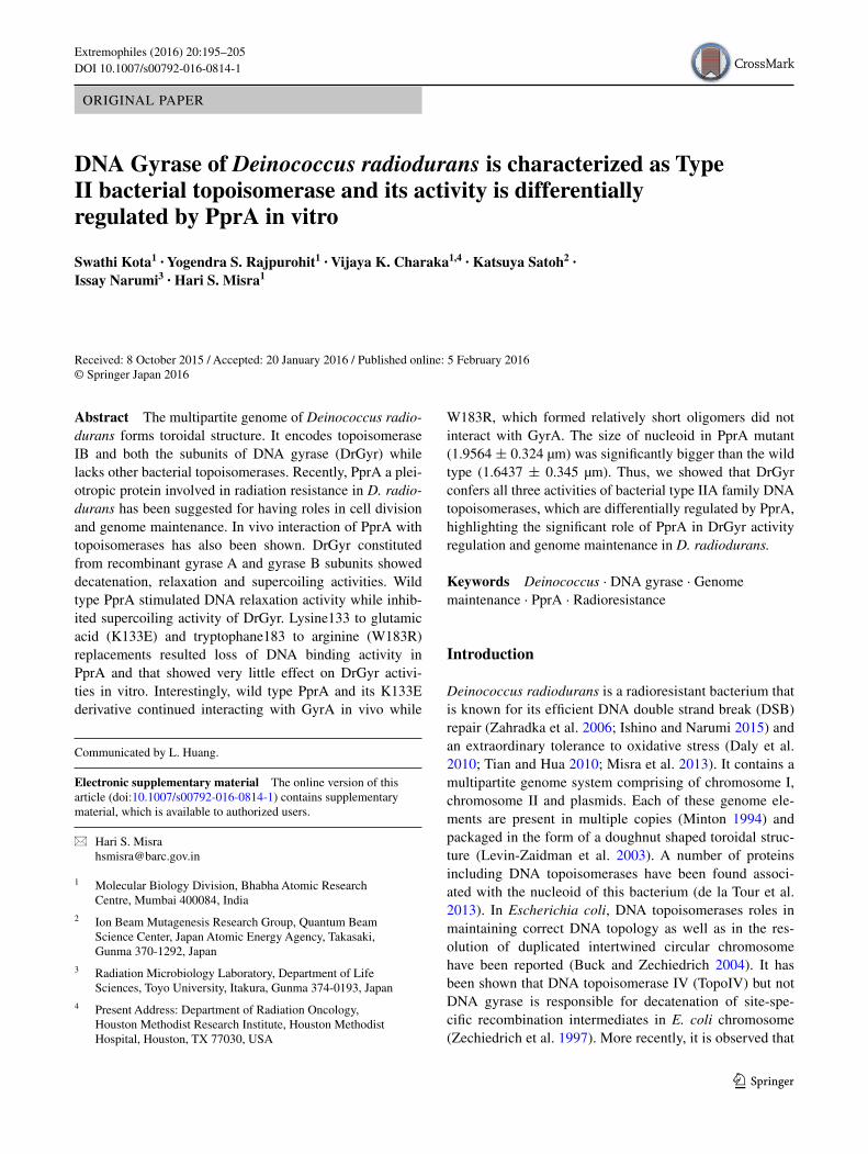
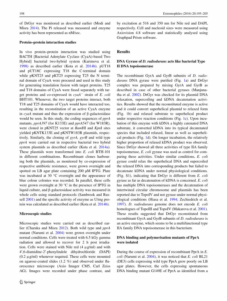
The recombinant GyrA and GyrB subunits of D. radio-durans DNA gyrase were purified (Fig. 1a) and DrGyr complex was prepared by mixing GyrA and GyrB as described in case of other bacterial gyrases (Manjuna-tha et al. 2002). DrGyr was checked for its plasmid DNA relaxation, supercoiling and kDNA decatenation activi-ties. Results showed that the reconstituted enzyme is active and it could convert superhelical plasmid to relaxed form (Fig. 1b) and relaxed substrate to superhelical product under respective reaction conditions (Fig. 1c). Upon incu-bation of this enzyme with kDNA a highly catenated DNA substrate, it converted kDNA into its typical decatenated species that included relaxed, linear as well as superheli-cal products (Fig. 1d). On longer incubation, the relatively higher proportion of relaxed kDNA product was observed. Since DrGyr showed all three activities of type IIA family topoisomerase, E. coli gyrase was used as control for com-paring these activities. Under similar conditions, E. coli gyrase could relax the superhelical DNA and supercoiled the relaxed DNA into corresponding products but failed to decatenate kDNA under normal physiological conditions. (Fig. S1), indicating that DrGyr is different from E. coli gyrase as far as decatenation of kDNA is concerned. E. coli has multiple DNA topoisomerases and the decatenation of intertwined circular chromosome and plasmids has been reported due to TopoIV and not gyrase under normal physi-ological conditions (Hiasa et al. 1994; Zechiedrich et al. 1997). D. radiodurans genome does not encode E. coli homologues of TopoIII and TopoIV (Makarova et al. 2001). These results suggested that DrGyr reconstituted from recombinant GyrA and GyrB subunits of D. radiodurans is an active enzyme, which seems to be a multifunctional type IIA family DNA topoisomerase in this bacterium.
DNA binding and polymerisation mutants of PprA were isolated
During the course of expression of recombinant PprA in E. coli (Narumi et al. 2004), it was noticed that E. coli BL21 (DE3) cells expressing wild type PprA grew poorly on LB agar plates. However, the cells expressing spontaneous DNA binding mutant G149E of PprA as identified from a
199Extremophiles (2016) 20:195–205
1 3
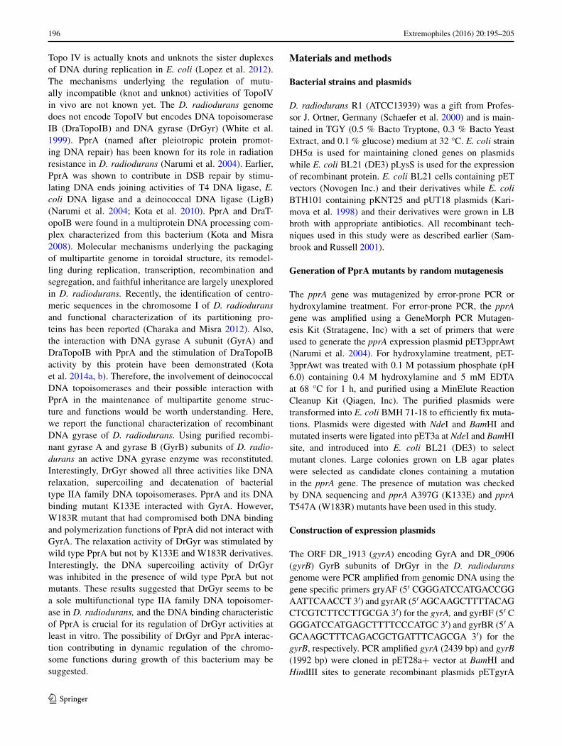
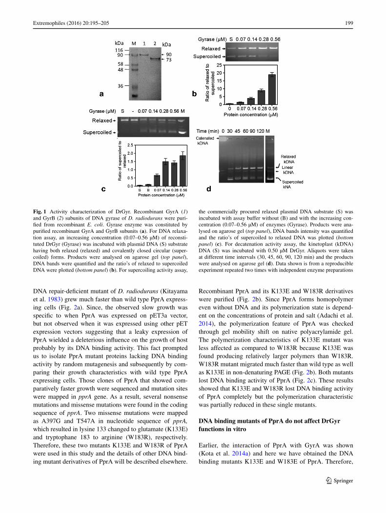
DNA repair-deficient mutant of D. radiodurans (Kitayama et al. 1983) grew much faster than wild type PprA express-ing cells (Fig. 2a). Since, the observed slow growth was specific to when PprA was expressed on pET3a vector, but not observed when it was expressed using other pET expression vectors suggesting that a leaky expression of PprA wielded a deleterious influence on the growth of host probably by its DNA binding activity. This fact prompted us to isolate PprA mutant proteins lacking DNA binding activity by random mutagenesis and subsequently by com-paring their growth characteristics with wild type PprA expressing cells. Those clones of PprA that showed com-paratively faster growth were sequenced and mutation sites were mapped in pprA gene. As a result, several nonsense mutations and missense mutations were found in the coding sequence of pprA. Two missense mutations were mapped as A397G and T547A in nucleotide sequence of pprA, which resulted in lysine 133 changed to glutamate (K133E) and tryptophane 183 to arginine (W183R), respectively. Therefore, these two mutants K133E and W183R of PprA were used in this study and the details of other DNA bind-ing mutant derivatives of PprA will be described elsewhere.
Recombinant PprA and its K133E and W183R derivatives were purified (Fig. 2b). Since PprA forms homopolymer even without DNA and its polymerization state is depend-ent on the concentrations of protein and salt (Adachi et al. 2014), the polymerization feature of PprA was checked through gel mobility shift on native polyacrylamide gel. The polymerization characteristics of K133E mutant was less affected as compared to W183R because K133E was found producing relatively larger polymers than W183R. W183R mutant migrated much faster than wild type as well as K133E in non-denaturing PAGE (Fig. 2b). Both mutants lost DNA binding activity of PprA (Fig. 2c). These results showed that K133E and W183R lost DNA binding activity of PprA completely but the polymerization characteristic was partially reduced in these single mutants.
DNA binding mutants of PprA do not affect DrGyr functions in vitro
Earlier, the interaction of PprA with GyrA was shown (Kota et al. 2014a) and here we have obtained the DNA binding mutants K133E and W183E of PprA. Therefore,
Fig. 1 Activity characterization of DrGyr. Recombinant GyrA (1) and GyrB (2) subunits of DNA gyrase of D. radiodurans were puri-fied from recombinant E. coli. Gyrase enzyme was constituted by purified recombinant GyrA and GyrB subunits (a). For DNA relaxa-tion assay, an increasing concentration (0.07–0.56 µM) of reconsti-tuted DrGyr (Gyrase) was incubated with plasmid DNA (S) substrate having both relaxed (relaxed) and covalently closed circular (super-coiled) forms. Products were analysed on agarose gel (top panel), DNA bands were quantified and the ratio’s of relaxed to supercoiled DNA were plotted (bottom panel) (b). For supercoiling activity assay,
the commercially procured relaxed plasmid DNA substrate (S) was incubated with assay buffer without (B) and with the increasing con-centration (0.07–0.56 µM) of enzymes (Gyrase). Products were ana-lysed on agarose gel (top panel), DNA bands intensity was quantified and the ratio’s of supercoiled to relaxed DNA was plotted (bottom panel) (c). For decatenation activity assay, the kinetoplast (kDNA) DNA (S) was incubated with 0.50 µM DrGyr. Aliquots were taken at different time intervals (30, 45, 60, 90, 120 min) and the products were analysed on agarose gel (d). Data shown is from a reproducible experiment repeated two times with independent enzyme preparations
200 Extremophiles (2016) 20:195–205
1 3
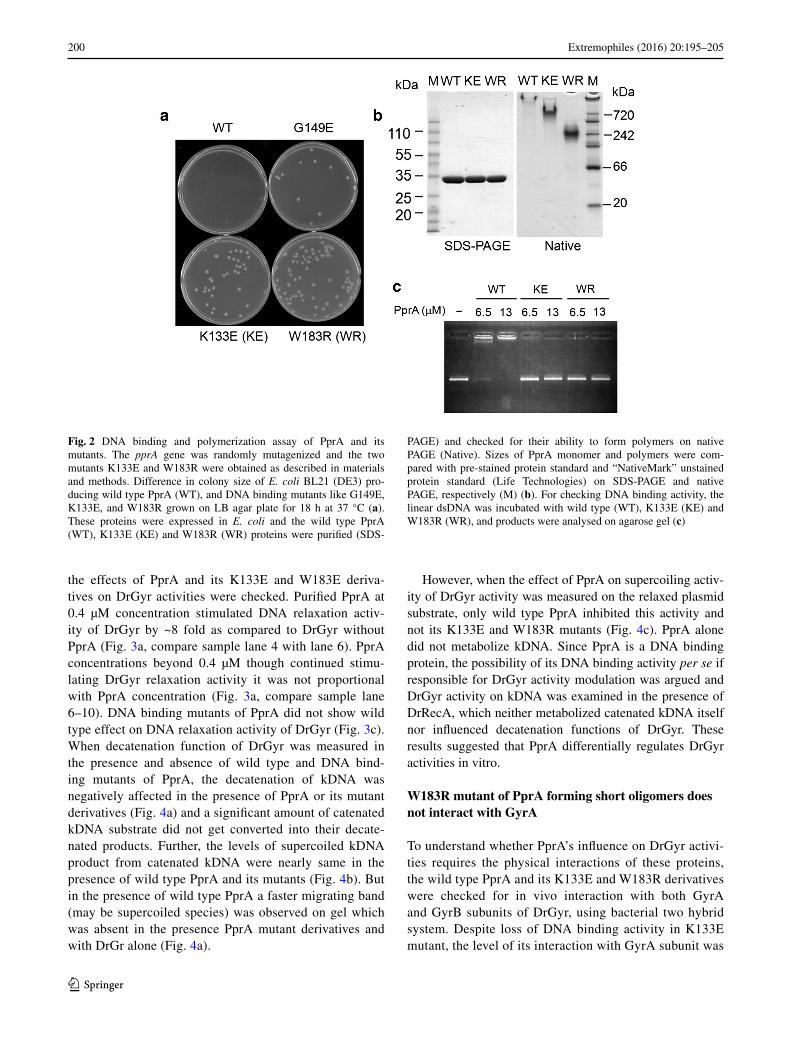
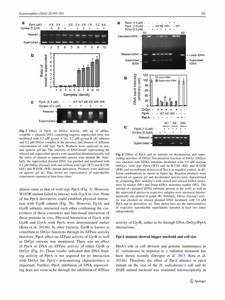
the effects of PprA and its K133E and W183E deriva-tives on DrGyr activities were checked. Purified PprA at 0.4 µM concentration stimulated DNA relaxation activ-ity of DrGyr by ~8 fold as compared to DrGyr without PprA (Fig. 3a, compare sample lane 4 with lane 6). PprA concentrations beyond 0.4 µM though continued stimu-lating DrGyr relaxation activity it was not proportional with PprA concentration (Fig. 3a, compare sample lane 6–10). DNA binding mutants of PprA did not show wild type effect on DNA relaxation activity of DrGyr (Fig. 3c). When decatenation function of DrGyr was measured in the presence and absence of wild type and DNA bind-ing mutants of PprA, the decatenation of kDNA was negatively affected in the presence of PprA or its mutant derivatives (Fig. 4a) and a significant amount of catenated kDNA substrate did not get converted into their decate-nated products. Further, the levels of supercoiled kDNA product from catenated kDNA were nearly same in the presence of wild type PprA and its mutants (Fig. 4b). But in the presence of wild type PprA a faster migrating band (may be supercoiled species) was observed on gel which was absent in the presence PprA mutant derivatives and with DrGr alone (Fig. 4a).
However, when the effect of PprA on supercoiling activ-ity of DrGyr activity was measured on the relaxed plasmid substrate, only wild type PprA inhibited this activity and not its K133E and W183R mutants (Fig. 4c). PprA alone did not metabolize kDNA. Since PprA is a DNA binding protein, the possibility of its DNA binding activity per se if responsible for DrGyr activity modulation was argued and DrGyr activity on kDNA was examined in the presence of DrRecA, which neither metabolized catenated kDNA itself nor influenced decatenation functions of DrGyr. These results suggested that PprA differentially regulates DrGyr activities in vitro.
W183R mutant of PprA forming short oligomers does not interact with GyrA
To understand whether PprA’s influence on DrGyr activi-ties requires the physical interactions of these proteins, the wild type PprA and its K133E and W183R derivatives were checked for in vivo interaction with both GyrA and GyrB subunits of DrGyr, using bacterial two hybrid system. Despite loss of DNA binding activity in K133E mutant, the level of its interaction with GyrA subunit was
Fig. 2 DNA binding and polymerization assay of PprA and its mutants. The pprA gene was randomly mutagenized and the two mutants K133E and W183R were obtained as described in materials and methods. Difference in colony size of E. coli BL21 (DE3) pro-ducing wild type PprA (WT), and DNA binding mutants like G149E, K133E, and W183R grown on LB agar plate for 18 h at 37 °C (a). These proteins were expressed in E. coli and the wild type PprA (WT), K133E (KE) and W183R (WR) proteins were purified (SDS-
PAGE) and checked for their ability to form polymers on native PAGE (Native). Sizes of PprA monomer and polymers were com-pared with pre-stained protein standard and “NativeMark” unstained protein standard (Life Technologies) on SDS-PAGE and native PAGE, respectively (M) (b). For checking DNA binding activity, the linear dsDNA was incubated with wild type (WT), K133E (KE) and W183R (WR), and products were analysed on agarose gel (c)
201Extremophiles (2016) 20:195–205
1 3
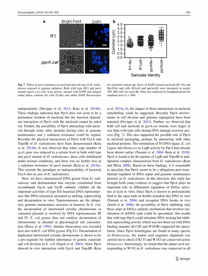
almost same as that of wild type PprA (Fig. 5). However, W183R mutant failed to interact with GyrA in vivo. None of the PprA derivatives could establish physical interac-tion with GyrB subunit (Fig. 5b). However, GyrA and GyrB subunits interacted each other confirming the cor-rectness of these constructs and functional interaction of these proteins in vivo. Physical interaction of GyrA with GyrB and GyrA with PprA were demonstrated earlier (Kota et al. 2014b). In other bacteria, GyrB is known to contribute to DrGyr functions through its ATPase activity therefore; PprA effect on ATPase activity of GyrB as well as DrGyr enzyme was monitored. There was no effect of PprA or DNA on ATPase activity of either GyrB or DrGyr (Fig. 6). These results indicated that DNA bind-ing activity of PprA is not required for its interaction with DrGyr but PprA’s polymerizing characteristics is important. Further, PprA inhibition of DNA supercoil-ing does not seem to be through the inhibition of ATPase
activity of GyrB, rather to be through DNA–DrGyr/PprA interactions.
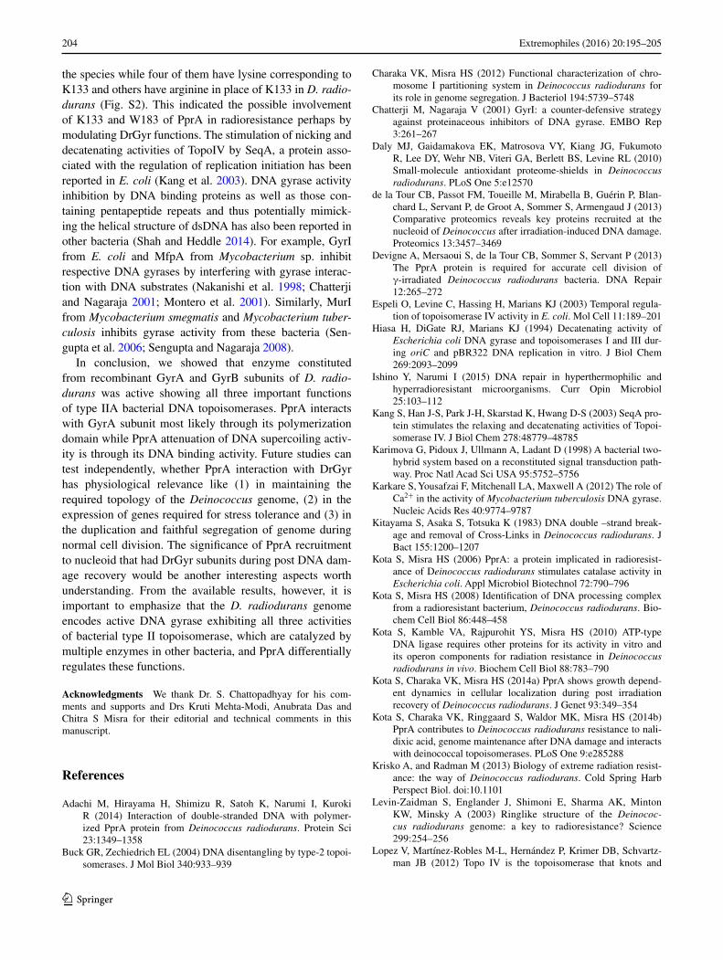
PprA mutant showed bigger nucleoid and cell size
PprA’s role in cell division and genome maintenance in D. radiodurans in response to γ radiation treatment has been shown recently (Devigne et al. 2013; Kota et al. 2014b). Therefore, the effect of PprA absence in pprA mutant on the size of the D. radiodurans’s cell and its DAPI stained nucleoid was examined microscopically in
Fig. 3 Effect of PprA on DrGyr activity. 400 ng of pBlue-scriptSK + plasmid DNA containing majority supercoiled form was incubated with 0.2 µM gyrase A (A), 0.2 µM gyrase B (B) subunits and 0.2 µM DrGyr complex in the presence and absence of different concentration of wild type PprA. Products were analysed on nor-mal agarose gel (a). The intensity of DNA bands representing the relaxed and supercoiled species were quantified densitometrically and the ratios of relaxed to supercoiled species were plotted (b). Simi-larly, the supercoiled plasmid DNA was purified and incubated with 0.2 µM DrGyr (Gyrase) with and without wild type (WT) and K133E (KE) and W183R (WR) mutant derivatives. Products were analysed on agarose gel (c). Data shown are representative of reproducible experiments repeated at least three times
Fig. 4 Effect of PprA and its mutants on decatenation and super-coiling activities of DrGyr. Decatenation function of DrGyr (DrGyr) was checked with kDNA substrate incubated with 0.5 µM enzyme (DrGyr), wild type PprA (WT) and its K133E (KE) and W183R (WR) and recombinant deinococcal RecA as negative control, in dif-ferent combinations as shown in figure (a). Reaction products were analysed on agarose gel and decatenated species were characterized by comparing their mobility’s with nicked and relaxed kDNA mono-mers in marker (M1) and linear kDNA monomer marker (M2). The amount of catenated kDNA substrate present in the wells as well as the supercoiled species in respective samples were measured densito-metrically and plotted in graph (b). Similarly, DrGyr (Gyrase) activ-ity was checked on relaxed plasmid DNA incubated with 1.0 µM PprA and its derivatives (c). Data shown here are the representatives of respective reproducible experiments repeated at least two times independently
202 Extremophiles (2016) 20:195–205
1 3
response to DNA damage. The pprA:cat mutant showed larger cell size (2.6430 ± 0.0389 µ) as compared to wild type (2.0135 ± 0.0419 µ) (Fig. 7). Interestingly, the size
of DAPI stained nucleoid in pprA:cat (1.9564 ± 0.0324 µ) was also more than the size of nucleoids in wild type (1.6437 ± 0.0345 µ) cells. The molecular basis of cell and nucleoid’s size increase in the absence of PprA is not known yet. However, the possibility of some defect in proper segregation of duplicated genome may led to an increase in DNA content and affected cell division, which led to an increase in cell volume cannot be ruled out. These results suggest that PprA plays important role in genome maintenance in this bacterium.
Discussion
The extraordinary radioresistance in D. radiodurans is largely attributed to its efficient DSB repair and the pro-tection of biomolecules from oxidative damage (Zahradka et al. 2006; Krisko and Radman 2013). This bacterium also harbours a multipartite genome system comprising of two chromosomes and two plasmids. These genome elements are present in 4–10 copies per cell (Minton 1994) and pack-aged together and form toroidal structure of the nucleoid. The D. radiodurans’s nucleoid proteome analysis showed the presence of both the subunits of DNA gyrase but not PprA (de la Tour et al. 2013). Same study reported the pres-ence of PprA in the nucleoid of Deinococcus deserti. PprA homologues have been found in the genome of all mem-bers of Deinococcaceae family, sequenced till date. This indicated the importance of PprA in the radioresistance in these bacteria. Although nucleoid proteome analysis has ruled out PprA presence in nucleoid, the recruitment of PprA in D. radiodurans’s nucleoid has been demonstrated
Fig. 5 PprA interaction with DNA gyrase subunits of D. radio-durans. The gyrase A was cloned in pUT18 (pUTgyrA) and gyraseB in pKNT25 (pKNTgyrB) vectors of bacterial two hybrid system and the recombinant fusion proteins were expressed as detailed in materi-als and methods section. The wild type PprA (PprA) was cloned in both pKNT25 (pKNTpprA) and pUT18 (pUTpprA) plasmids. Simi-larly, K133E (KE) and W183R (WR) were cloned in both the vec-tors yielded pKNT397 and pUT397, and pKNT547 and pUT547, respectively. These plasmids were co-transformed in different com-binations into E. coli BTH101. Care was taken that each combina-tion should have both the domains of CyaA (T18 and T25) expressing along with two different proteins. E. coli BTH101 was also co-trans-formed with pUT18 and pKNT25 (VC), and pKNTcheA and pUT-cheA (+C) and used as respective controls. Three independent clones (1, 2, 3) of these samples were spotted on X-gal plate (a) and also monitored for the expression of β-galactosidase in liquid culture (b). β-Galactosidase activity above 700 units/mg protein was considered as positive for physical interaction of two proteins. Data shown in panel ‘b’ are the mean ± SD (n = 4) and in c is the representative of a reproducible experiment repeated 2 times independently
Fig. 6 Effect of PprA on ATPase activity of DrGyr in vitro. Different concentrations (20, 40, 80 and 100 nM) of recombinant gyrase B sub-unit (GyrB), and DrGyr (Gyr) reconstituted from gyrase A and gyrase B subunits were incubated with 1 µM PprA and 100 ng plasmid DNA for 1 h at 37 °C. Release of inorganic phosphate (Pi) was estimated as described in methods and converted into nM/s. Data shown are the mean ± SD (n = 3)
203Extremophiles (2016) 20:195–205
1 3
independently (Devigne et al. 2013; Kota et al. 2014b). These findings indicated that PprA does not seem to be a permanent resident of nucleoid but the function depend-ent interaction of PprA with the nucleoid cannot be ruled out. Further, the possibility of PprA interacting with nucle-oid through some other proteins having roles in genome maintenance and γ radiation resistance could be argued. Recently the physical interactions of PprA with GyrA and TopoIB of D. radiodurans have been demonstrated (Kota et al. 2014b). It was observed that when copy number of gyrA gene was reduced to a certain level in both wild type and pprA mutant of D. radiodurans, these cells debilitated under normal conditions, and there was no further loss in γ radiation resistance of pprA mutant (Kota et al. 2014a). This extends the paradigm on indispensability of bacterial GyrA also in case of D. radiodurans.
Here, we have characterized DNA gyrase from D. radi-odurans and demonstrated that enzyme constituted from recombinant GyrA and GyrB subunits exhibits all the important activities of type IIA bacterial DNA topoisomer-ases like DNA relaxation measured as nicking, supercoiling and decatenation in vitro. Topoisomerases are the ubiqui-tous genome maintenance enzymes in bacteria. In E. coli, the decatenation of intertwined circular chromosome or catenated plasmid is resolved by DNA topoisomerase III and IV. E. coli gyrase does not catalyze decatenation of chromosome or plasmid at physiological salt concentra-tion (Hiasa et al. 1994). Similar observation was recorded here also with E. coli DNA gyrase (Fig S1). Decatenation of duplicated intertwined circular chromosome is shown to be a pre-requisite for faithful inheritance of genetic materials and cell division in E. coli (Espeli et al. 2003). Since PprA showed in vivo interaction with GyrA and TopoIB (Kota
et al. 2014a, b), the impact of these interactions in nucleoid remodelling could be suggested. Recently PprA involve-ments in cell division and genome segregation have been reported (Devigne et al. 2013). Further, we observed that both cell and nucleoid in pprA:cat mutant were larger in size than wild type cells during DNA damage recovery pro-cess (Fig. 7). This also supported the possible role of PprA in nucleoid packaging, perhaps by interacting with other nucleiod proteins. The stimulation of T4 DNA ligase, E. coli Ligase and Deinococcus LigB activity by PprA had already been shown earlier (Narumi et al. 2004; Kota et al. 2010). PprA is found to be the partner of LigB and TopoIB in mul-tiprotein complex characterized from D. radiodurans (Kota and Misra 2008). Based on these findings, it is reasonable to speculate that PprA seems to be a ubiquitous post trans-lational regulator of DNA repair and genome maintenance proteins in D. radiodurans. In this direction, this study has brought forth some evidence to suggest that PprA plays an important role in differential regulation of DrGyr activi-ties at least in vitro. Since PprA is known to preferentially bind to the open ends of double-stranded (ds) DNA in vitro (Narumi et al. 2004) and recognize DNA breaks in vivo (Satoh et al. 2006), the possibility of PprA inhibiting only those steps in DrGyr catalytic mechanism that involves reu-tilization of dsDNA ends could be speculated. Our results that wild type PprA could stimulate DNA nicking but inhib-ited supercoiling activity which was not observed with DNA binding mutants (K133E and W183R) supported the specu-lation. Since PprA homologues are found in many species of Deinococcus, the amino acid sequence alignment was carried out to check if K133 and W183 are conserved across Deinococci. Interestingly, we found that the amino acid cor-responding to W183 in D. radioduans was conserved in all
Fig. 7 Effect of pprA mutation on nucleoid and cell size in D. radio-durans exposed to gamma radiation. Both wild type (R1) and pprA mutant (pprA:cat) cells were grown, stained with DAPI and imaged under phase contrast for cells (Cells) and under DAPI fluorescence
for nucleoid content (a). Sizes of DAPI stained nucleoid (R1-Nu and PprANu) and cells (R1cell and pprAcell) were measured in nearly 300–500 cells for each (b). Data was analysed in Graphpad prism for standard error (n > 300)
204 Extremophiles (2016) 20:195–205
1 3
the species while four of them have lysine corresponding to K133 and others have arginine in place of K133 in D. radio-durans (Fig. S2). This indicated the possible involvement of K133 and W183 of PprA in radioresistance perhaps by modulating DrGyr functions. The stimulation of nicking and decatenating activities of TopoIV by SeqA, a protein asso-ciated with the regulation of replication initiation has been reported in E. coli (Kang et al. 2003). DNA gyrase activity inhibition by DNA binding proteins as well as those con-taining pentapeptide repeats and thus potentially mimick-ing the helical structure of dsDNA has also been reported in other bacteria (Shah and Heddle 2014). For example, GyrI from E. coli and MfpA from Mycobacterium sp. inhibit respective DNA gyrases by interfering with gyrase interac-tion with DNA substrates (Nakanishi et al. 1998; Chatterji and Nagaraja 2001; Montero et al. 2001). Similarly, MurI from Mycobacterium smegmatis and Mycobacterium tuber-culosis inhibits gyrase activity from these bacteria (Sen-gupta et al. 2006; Sengupta and Nagaraja 2008).
In conclusion, we showed that enzyme constituted from recombinant GyrA and GyrB subunits of D. radio-durans was active showing all three important functions of type IIA bacterial DNA topoisomerases. PprA interacts with GyrA subunit most likely through its polymerization domain while PprA attenuation of DNA supercoiling activ-ity is through its DNA binding activity. Future studies can test independently, whether PprA interaction with DrGyr has physiological relevance like (1) in maintaining the required topology of the Deinococcus genome, (2) in the expression of genes required for stress tolerance and (3) in the duplication and faithful segregation of genome during normal cell division. The significance of PprA recruitment to nucleoid that had DrGyr subunits during post DNA dam-age recovery would be another interesting aspects worth understanding. From the available results, however, it is important to emphasize that the D. radiodurans genome encodes active DNA gyrase exhibiting all three activities of bacterial type II topoisomerase, which are catalyzed by multiple enzymes in other bacteria, and PprA differentially regulates these functions.
Acknowledgments We thank Dr. S. Chattopadhyay for his com-ments and supports and Drs Kruti Mehta-Modi, Anubrata Das and Chitra S Misra for their editorial and technical comments in this manuscript.
References
Adachi M, Hirayama H, Shimizu R, Satoh K, Narumi I, Kuroki R (2014) Interaction of double-stranded DNA with polymer-ized PprA protein from Deinococcus radiodurans. Protein Sci 23:1349–1358
Buck GR, Zechiedrich EL (2004) DNA disentangling by type-2 topoi-somerases. J Mol Biol 340:933–939
Charaka VK, Misra HS (2012) Functional characterization of chro-mosome I partitioning system in Deinococcus radiodurans for its role in genome segregation. J Bacteriol 194:5739–5748
Chatterji M, Nagaraja V (2001) GyrI: a counter-defensive strategy against proteinaceous inhibitors of DNA gyrase. EMBO Rep 3:261–267
Daly MJ, Gaidamakova EK, Matrosova VY, Kiang JG, Fukumoto R, Lee DY, Wehr NB, Viteri GA, Berlett BS, Levine RL (2010) Small-molecule antioxidant proteome-shields in Deinococcus radiodurans. PLoS One 5:e12570
de la Tour CB, Passot FM, Toueille M, Mirabella B, Guérin P, Blan-chard L, Servant P, de Groot A, Sommer S, Armengaud J (2013) Comparative proteomics reveals key proteins recruited at the nucleoid of Deinococcus after irradiation-induced DNA damage. Proteomics 13:3457–3469
Devigne A, Mersaoui S, de la Tour CB, Sommer S, Servant P (2013) The PprA protein is required for accurate cell division of γ-irradiated Deinococcus radiodurans bacteria. DNA Repair 12:265–272
Espeli O, Levine C, Hassing H, Marians KJ (2003) Temporal regula-tion of topoisomerase IV activity in E. coli. Mol Cell 11:189–201
Hiasa H, DiGate RJ, Marians KJ (1994) Decatenating activity of Escherichia coli DNA gyrase and topoisomerases I and III dur-ing oriC and pBR322 DNA replication in vitro. J Biol Chem 269:2093–2099
Ishino Y, Narumi I (2015) DNA repair in hyperthermophilic and hyperradioresistant microorganisms. Curr Opin Microbiol 25:103–112
Kang S, Han J-S, Park J-H, Skarstad K, Hwang D-S (2003) SeqA pro-tein stimulates the relaxing and decatenating activities of Topoi-somerase IV. J Biol Chem 278:48779–48785
Karimova G, Pidoux J, Ullmann A, Ladant D (1998) A bacterial two-hybrid system based on a reconstituted signal transduction path-way. Proc Natl Acad Sci USA 95:5752–5756
Karkare S, Yousafzai F, Mitchenall LA, Maxwell A (2012) The role of Ca2+ in the activity of Mycobacterium tuberculosis DNA gyrase. Nucleic Acids Res 40:9774–9787
Kitayama S, Asaka S, Totsuka K (1983) DNA double –strand break-age and removal of Cross-Links in Deinococcus radiodurans. J Bact 155:1200–1207
Kota S, Misra HS (2006) PprA: a protein implicated in radioresist-ance of Deinococcus radiodurans stimulates catalase activity in Escherichia coli. Appl Microbiol Biotechnol 72:790–796
Kota S, Misra HS (2008) Identification of DNA processing complex from a radioresistant bacterium, Deinococcus radiodurans. Bio-chem Cell Biol 86:448–458
Kota S, Kamble VA, Rajpurohit YS, Misra HS (2010) ATP-type DNA ligase requires other proteins for its activity in vitro and its operon components for radiation resistance in Deinococcus radiodurans in vivo. Biochem Cell Biol 88:783–790
Kota S, Charaka VK, Misra HS (2014a) PprA shows growth depend-ent dynamics in cellular localization during post irradiation recovery of Deinococcus radiodurans. J Genet 93:349–354
Kota S, Charaka VK, Ringgaard S, Waldor MK, Misra HS (2014b) PprA contributes to Deinococcus radiodurans resistance to nali-dixic acid, genome maintenance after DNA damage and interacts with deinococcal topoisomerases. PLoS One 9:e285288
Krisko A, and Radman M (2013) Biology of extreme radiation resist-ance: the way of Deinococcus radiodurans. Cold Spring Harb Perspect Biol. doi:10.1101
Levin-Zaidman S, Englander J, Shimoni E, Sharma AK, Minton KW, Minsky A (2003) Ringlike structure of the Deinococ-cus radiodurans genome: a key to radioresistance? Science 299:254–256
Lopez V, Martínez-Robles M-L, Hernández P, Krimer DB, Schvartz-man JB (2012) Topo IV is the topoisomerase that knots and
205Extremophiles (2016) 20:195–205
1 3
unknots sister duplexes during DNA replication. Nucleic Acid Res 40:3563–3573
Makarova KS, Aravind L, Wolf YI, Tatusov RL, Minton KW, Koonin EV, Daly MJ (2001) Genome of extremely radiation-resistant bacterium Deinococcus radiodurans viewed from the per-spectives of comparative genomics. Microbiol Mol Biol Rev 65:44–79
Manjunatha UH, Dalal M, Chatterji M, Radha DR, Visweswariah SS, Nagaraja V (2002) Functional characterisation of mycobac-terial DNA gyrase: an efficient decatenase. Nucleic Acid Res 30(10):2144–2153
Minton KW (1994) DNA repair in the extremely radioresistant bacte-rium Deinococcus radiodurans. Mol Microbiol 13:9–15
Misra HS, Rajpurohit YS, Kota S (2013) Physiological and molecu-lar basis of extreme radioresistance in Deinococcus radiodurans. Curr Sci 104:194–205
Modi K, Misra HS (2014) Dr-FtsA, an actin homologue in Deinococ-cus radiodurans differentially affects Dr-FtsZ and Ec-FtsZ func-tions in vitro. PLoS One 9:e115918
Montero C, Mateu G, Rodriguez R, Takiff H (2001) Intrinsic resist-ance of Mycobacterium smegmatis to fluoroquinolones may be influenced by new pentapeptide protein MfpA. Antimicrob Agents Chemother 45:3387–3392
Nakanishi A, Oshida T, Matsushita T, Imajoh-Ohmi S, Ohnuki T (1998) Identification of DNA gyrase inhibitor (GyrI) in Escheri-chia coli. J Biol Chem 273:1933–1938
Narumi I, Satoh K, Cui S, Funayama T, Kitayama S, Watanabe H (2004) PprA: a novel protein from Deinococcus radiodurans that stimulates DNA ligation. Mol Microbiol 54:278–285
Sambrook J, Russell DW (2001) Molecular cloning: a laboratory manual, 3rd edn. Cold Spring Harbor Laboratory Press, Cold Spring Harbor
Satoh K, Wada S, Kikuchi M, Funayama T, Narumi I, Kobayashi Y (2006) Method for detecting DNA strand breaks in mammalian cells using the Deinococcus radiodurans PprA protein. Mutat Res 596:36–42
Schaefer M, Schmitz C, Facius R, Horneck G, Milow B, Funken K-H, Ortner J (2000) Systematic study of parameters influencing the action of Rose Bengal with visible light on bacterial cells: com-parison between biological effect and singlet-oxygen production. Photochem Photobiol 71:514–523
Sengupta S, Nagaraja V (2008) Inhibition of DNA gyrase activ-ity by Mycobacterium smegmatis MurI. FEMS Microbiol Lett 279:40–47
Sengupta S, Shah M, Nagaraja V (2006) Glutamate racemase from Mycobacterium tuberculosis inhibits DNA gyrase by affecting its DNA-binding. Nucleic Acid Res 34:5567–5576
Shah S, Heddle JG (2014) Squaring up to DNA: pentapeptide repeat proteins and DNA mimicry. Appl Microbiol Biotech 98:9545–9560
Tian B, Hua Y (2010) Carotenoid biosynthesis in extremophilic Deinococcus-Thermus bacteria. Trends Microbiol 18:512–520
White O, Eisen JA, Heidelberg JF, Hickey EK, Peterson JD, Dod-son RJ, Haft DH, Gwinn ML, Nelson WC et al (1999) Genome sequence of the radioresistant bacterium Deinococcus radio-durans R1. Science 286:1571–1577
Zahradka K, Slade D, Bailone A, Sommer S, Averbeck D, Petranovic M, Linder AB, Radman M (2006) Reassembly of shattered chro-mosomes in Deinococcus radiodurans. Nature 443:569–573
Zechiedrich EL, Khodursky AB, Cozzarelli NR (1997) Topoisomer-ase IV, not gyrase, decatenates products of site-specific recombi-nation in Escherichia coli. Genes Dev 11:2580–2592