Embed Size (px)
Citation preview
11
DNA Damage Protection and Induction of Repair by Dietary Phytochemicals and
Cancer Prevention: What Do We Know?
Alice A. Ramos1, Cristóvão F. Lima2 and Cristina Pereira-Wilson1 1CBMA – Molecular and Environmental Biology Centre
2CITAB – Centre for the Research and Technology of Agro-Environmental and Biological Sciences
Department of Biology, School of Sciences, University of Minho, Campus de Gualtar, Braga,
Portugal
1. Introduction
DNA damage accumulates in cells over time as a result of exposure to a variety of exogenous and endogenous agents. These damages, if not repaired properly, can generate mutations in somatic or germline cells, which are involved in the pathogenesis of many diseases, such as cancer. To maintain genomic integrity generation after generation, organisms possess multiple mechanisms such as inhibition of carcinogen uptake into the cells, induction of detoxification enzymes, increased cellular defenses that prevent DNA damage, enhancement of DNA repair, increased anti-inflammatory activity, inhibition of cell proliferation, and modulation of apoptosis through effects on signal transduction pathways (de Kok et al., 2008); (Pan et al., 2008). In the last decades, several dietary constituents have been shown to modulate all these processes. Epidemiological studies as well as laboratory data suggest that consumption of fruits and vegetables is associated with a reduced risk of developing a wide range of cancers. It has been estimated that 30-40% of all tumours can be prevented with a correct lifestyle and diet, in particular colon cancer (Rajamanickam and Agarwal, 2008). Multiple mechanisms have been proposed to explain the chemopreventive effects of phytochemicals. Protection of DNA from damage and modulation of DNA repair assume an important role on prevention of mutations and consequently of the carcinogenic process. The comet assay or single cell gel electrophoresis (SCGE) assay is a rapid, sensitive and relatively simple method for assessing DNA damage and its repair in individual cells. The standard comet assay measures DNA breaks and alkali-labile sites that are converted to strand breaks. With its widespread use, several modifications on the comet assay have been made that allow the quantification of other types of DNA damage as well as DNA repair rates. With the inclusion of an extra step on the comet assay by using specific DNA repair enzymes, different base lesions can be identified by the introduction of breaks at sites of base damage. In this regard, Endonuclease III, FPG and AlkA have been used to detect oxidized pyrimidines, modified purines and alkylpurines bases, respectively. With these

Selected Topics in DNA Repair
238
modifications the comet assay can be used to estimate oxidative and alkylating DNA damage. Cells’ DNA repair capacity can also be measured by using modified protocols of the comet assay, such as following the repair kinetics by the cellular repair assay, or in vitro by using cellular substrates with specific damages to measure base excision repair (BER) and nucleotide excision repair (NER) (Collins et al., 2001b; Collins, 2004). These different modifications of the comet assay have been successfully used to evaluate the chemopreventive potential of several phytochemicals present in our diet. In this review, we will show evidence that dietary agents can protect DNA from oxidative and alkylating agents as well modulate DNA repair in eukaryotic cells, by using the comet assay. Data from different experimental systems, including primary cell cultures, human cell lines, animal models and human biomonitoring studies, will be discussed in order to provide an overview of effects of dietary phytochemicals on DNA damage and repair with particular emphasis on colon cancer chemoprevention. Recently, we have shown that dietary phytochemicals such as quercetin, rutin, rosmarinic acid, luteolin and others not only protect DNA damage but also stimulate DNA repair in liver and colon cell lines (Lima et al., 2006; Ramos et al., 2008; Ramos et al., 2010b; Ramos et al., 2010a). These effects may contribute to their anti-carcinogenic effects. Effects of phytochemicals through DNA repair modulation and their interaction with alkylating agents used as chemotherapeutic drugs will also be referred.
2. DNA damage and genomic stability
Cells of all organisms are under continual attack from reactive oxygen species (ROS) and alkylanting species generated by environmental pollutants, drugs, radiation, cigarette smoke and endogenous metabolism. Theses endogenous and exogenous agents can induce harmful effects if the cell’s defense mechanisms are not enough to maintain cellular redox homeostasis. Protection and repair of DNA damage represent two important mechanisms to maintain genomic stability.
2.1 Oxidative DNA damage Endogenous and exogenous agents can induce disruption of the cellular redox homeostasis in favor of oxidant state. This imbalance is described as oxidative stress. As a consequence, different types of molecules such as proteins, lipids and nucleic acids, can be damaged, resulting in severe metabolic dysfunction (including lipid peroxidation, protein oxidation, membrane disruption and DNA damage) (Aherne et al., 2007). Oxidative stress has been involved in the development of several pathologies such as certain cancers, once it may lead to mutations that activate oncogenes or inactivate tumor suppressor genes (Allen and Tresini, 2000; Maynard et al., 2009). In tumor cells, oxidative stress can act as a selective factor in favour of these cells, through induction of DNA damage that may generate more mutations; activation of growth-promoting transcription factors and modulation of genes involved in apoptosis and proliferation (Allen and Tresini, 2000; Karihtala and Soini, 2007). A significant consequence of oxidative stress is DNA damage, which may result in genomic instability. It has been estimated that around 104 lesions are induced in a mammalian cell genome every day (Hegde et al., 2008). There are several types of oxidative DNA damage, such as oxidized bases, abasic sites (also called apurinic/apyrimidinic (AP)) and DNA strand breaks. Hydroxylation of guanine at C-8 position, 8-oxoGua (8-Oxo-7,8-

DNA Damage Protection and Induction of Repair by Dietary Phytochemicals and Cancer Prevention: What Do We Know?
239
dihydroguanine) is one of the most abundant forms of DNA oxidation and can cause G to T transversions through mispairing in replication. Tumor cells are characterized by high levels of ROS. Some studies showed that human tumor cells have an increase of levels of 8-oxoG relative to normal cells. Also, low levels of antioxidant have been found in tumor cells (Maynard et al., 2009).
2.2 Alkylating DNA damage The removal of oxidative lesions by cellular repair processes is essential for maintaining genome integrity and survival limiting mutagenesis. However, there are a number of studies indicating that oxidative DNA damage could not account entirely by itself for tumor development, since elevated levels of 8-oxoGua have been shown not to reflect reflect increased cancer rates. In fact, oxidative damage is the most studied DNA damage, however, alkylating DNA damage is not less important. Human exposure to alkylating agents can arise from diet (e.g. presence of heterocyclic amines on food), environment (e.g. exposure to cigarette smoke and fuel combustion), or produced endogenously (e.g. nitrosation of amides and amines mediated by enteric bacteria) (Wirtz et al., 2010). Alkylating agents can cause a wide spectrum of DNA adducts, including N-alkylated adducts, such as N7-methylguanine (N7MeG), N3-methyladenine (N3MeA) and N3-methylguanine (N3MeG), and O-alkylated adducts, such as O6-methylguanine (O6MeG) and O4-methylthymine (O4MeT). N-alkylated adducts correspond to more than 80% of alkylated bases and exhibit different stabilities. For example, N7MeG (correspond aprox. 70%) is the most stable N-methylation adduct in vitro with 80hr of half-life. N3MeA and N3MeG are less abundant with 9 and 2%, respectively, of total methylation adducts. O6MeG vary from 0.3% (for methyl methanesulfonate) to 8% (for methylnitrosourea) of the total DNA methyl adducts and it is stable in the absence of the DNA repair protein O6-methylguanine-DNA methyltransferase (MGMT). O4MeT is produced in very low amounts (<0.4% of the total DNA methyl adducts) and its mutagenicity and cytotoxicity are unclear. In general, O-alkylations are highly mutagenic and genotoxic, whereas N-alkylations are cytotoxic, but less mutagenic (Drablos et al., 2004; Kondo et al., 2010). O6MeG is the major pre-mutagenic, pre-carcinogenic and pre-cytotoxic DNA lesion induced by methylating agents (Wirtz et al., 2010). Prevention of DNA damage and modulation of DNA repair by dietary phytochemicals phytochemicals is the main focus of this review but first a brief overview of DNA repair mechanisms will be presented. Effects on chemoprevention of colon cancer will be addressed.
3. Mechanisms of DNA repair
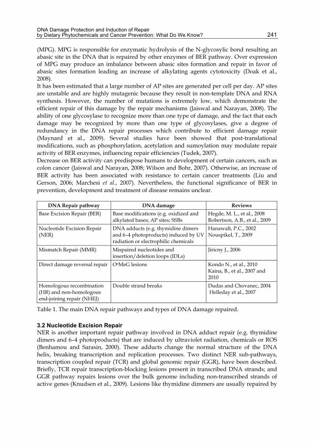
Maintainance of genomic integrity is complex due to great diversity of damage that can occur in DNA. In contrast to other biomolecules, DNA cannot be replaced, only repaired. To avoid the deleterious consequences of damage accumulation, cells have a variety of DNA repair pathways, each recognize and repair specific types of DNA damage. Base excision repair (BER), nucleotide excision repair (NER), mismatch repair (MMR), homologous recombination (HR), non-homologous end-joining repair (NHEJ) and direct damage reversal repair are some of the most important pathways used by cells to repair oxidative and alkylating DNA damage (Table I). These cellular repair pathways are not completely independent from one another. Some studies have show physical interactions between some

Selected Topics in DNA Repair
240
repair proteins from different repair pathways, suggesting that regulation of DNA repair involves protein cross-talk (Knudsen et al., 2009). Protein expression, pos-translational modifications and nuclear translocation of DNA repair proteins have been referred as essential in the regulation of DNA repair activity and to maintain genomic stability. However, these subjects need to be clarified in the future (Knudsen et al., 2009; Tudek, 2007). When DNA damage is not repaired, the cell by a complex network that collectively forms the DNA damage response (DDR) machinery delays cell-cycle progression, acting on cell cycle checkpoints. This delay gives the DNA repair machinery more time, allowing correct repair of the damage. DDR machinery can induce other mechanisms such as apoptosis or necrosis to avoid that altered cells continue to proliferate and result in disease (Bartek et al., 2007; Maynard et al., 2009).
3.1 Base Excision Repair BER is the major pathway involved in the repair of oxidation and alkylation DNA damage and occurs in both the nucleus and mitochondria (D'Errico et al., 2008). BER recognizes and repairs AP sites, DNA SSBs and different types of base modifications, such as oxidized/reduced bases (e.g. 8-oxoG or formamidopyrimidines), alkylated bases, deaminated bases (e.g. uracil) or base mismatches (Maynard et al., 2009). BER pathway involves steps as recognition, excision, filling and ligation that are carried out by four or five enzymes. Briefly, the repair process is initiated by one of several DNA glycosylases, each recognizing a specific DNA lesion (e.g. OGG1, NTH, NEIL and MYH recognize oxidation damage; deamination damages are recognized by UDG, MED1, UNG, and TDG, and MPG initiate alkylation repair) (Knudsen et al., 2009). These DNA glycosylases excise the damaged base (by cleave of N-glycosidic bond between the sugar and the base), generating an AP site. An AP endonuclease, as APE1, cleaves the AP site, to generate 3′ OH and 5′ deoxyribose phosphate (dRP) terminus. The third step is carried through DNA polymerase that fills the nucleotide gap generated due to lesion base removal. Finally, DNA ligase seals the nick on DNA (Hegde et al., 2008; Evans et al., 2004). Several other protein factors have been identified as interacting with the essential BER proteins and/or the DNA to modulate BER activity. BER pathway is described in detail in the several reviews (Fortini et al., 2003; Hegde et al., 2008; Robertson et al., 2009; Fortini et al., 2003). Hydroxylation of guanine at C-8 position, 8-oxoGua (8-Oxo-7,8-dihydroguanine) is one of the most abundant forms of DNA oxidation and the most studied because of its mutagenic potential. If not properly repaired, 8-oxoG can pair with cytosine or adenine. Replication of 8-oxoG paired with C by DNA polymerases is a non-mutagenic process. However, replication of 8-oxoG paired with A results in GC to TA or TA to GC transversions that are strong mutagenic DNA lesions (Pastoriza Gallego and Sarasin, 2003). In mammalian cells 8-oxoGua is predominantly recognized and excised by a specific DNA glycosylases. hOGG1 removes 8-oxoG when paired with a cytosine and the glycosylase hMYH removes adenine when mispaired with 8-oxoG, both through the BER pathway. Activity of OGG1 is enhanced by cofactors such as human apurinic/apyrimidinic endonuclease (APE1), Xeroderma Pigmentosum complementation group C (XPC) and human endonuclease VIII-like (NEIL1) (D'Errico et al., 2008). N7alkylG adducts can be repaired by spontaneous depurination resulting in abasic sites that are often correctly repaired by BER. If not repaired, abasic sites may result in single-stranded gaps that can stall replication forks resulting in double stranded breaks. N7alkylG adducts are also recognized and removed from DNA by N-methylpurine-DNAglycoslase
DNA Damage Protection and Induction of Repair by Dietary Phytochemicals and Cancer Prevention: What Do We Know?
241
(MPG). MPG is responsible for enzymatic hydrolysis of the N-glycosylic bond resulting an abasic site in the DNA that is repaired by other enzymes of BER pathway. Over expression of MPG may produce an imbalance between abasic sites formation and repair in favor of abasic sites formation leading an increase of alkylating agents cytotoxicity (Doak et al., 2008). It has been estimated that a large number of AP sites are generated per cell per day. AP sites are unstable and are highly mutagenic because they result in non-template DNA and RNA synthesis. However, the number of mutations is extremely low, which demonstrate the efficient repair of this damage by the repair mechanisms (Jaiswal and Narayan, 2008). The ability of one glycosylase to recognize more than one type of damage, and the fact that each damage may be recognized by more than one type of glycosylases, give a degree of redundancy in the DNA repair processes which contribute to efficient damage repair (Maynard et al., 2009). Several studies have been showed that post-translational modifications, such as phosphorylation, acetylation and sumoylation may modulate repair activity of BER enzymes, influencing repair efficiencies (Tudek, 2007). Decrease on BER activity can predispose humans to development of certain cancers, such as colon cancer (Jaiswal and Narayan, 2008; Wilson and Bohr, 2007). Otherwise, an increase of BER activity has been associated with resistance to certain cancer treatments (Liu and Gerson, 2006; Marchesi et al., 2007). Nevertheless, the functional significance of BER in prevention, development and treatment of disease remains unclear.
DNA Repair pathway DNA damage Reviews Base Excision Repair (BER) Base modifications (e.g. oxidized and
alkylated bases; AP sites; SSBs Hegde, M. L., et al., 2008 Robertson, A.B., et al., 2009
Nucleotide Excision Repair (NER)
DNA adducts (e.g. thymidine dimers and 6–4 photoproducts) induced by UV radiation or electrophilic chemicals
Hanawalt, P.C., 2002 Nouspikel, T., 2009
Mismatch Repair (MMR) Mispaired nucleotides and insertion/deletion loops (IDLs)
Jiricny J., 2006
Direct damage reversal repair O6MeG lesions Kondo N., et al., 2010 Kaina, B., et al., 2007 and 2010
Homologous recombination (HR) and non-homologous end-joining repair (NHEJ)
Double strand breaks Dudas and Chovanec, 2004 Helleday et al., 2007
Table 1. The main DNA repair pathways and types of DNA damage repaired.
3.2 Nucleotide Excision Repair NER is another important repair pathway involved in DNA adduct repair (e.g. thymidine dimers and 6–4 photoproducts) that are induced by ultraviolet radiation, chemicals or ROS (Benhamou and Sarasin, 2000). These adducts change the normal structure of the DNA helix, breaking transcription and replication processes. Two distinct NER sub-pathways, transcription coupled repair (TCR) and global genomic repair (GGR), have been described. Briefly, TCR repair transcription-blocking lesions present in transcribed DNA strands; and GGR pathway repairs lesions over the bulk genome including non-transcribed strands of active genes (Knudsen et al., 2009). Lesions like thymidine dimmers are usually repaired by

Selected Topics in DNA Repair
242
TCR, while other lesions as 6–4 photoproducts are efficiently repaired by GGR (for review see Hanawalt, 2002; Nouspikel, 2009).
3.3 Mismatch Repair MMR is a post-replicative DNA repair mechanism that mainly corrects base–base mismatches which are caused by errors of DNA polymerases and insertion/deletion loops (IDLs). Two complexes are responsible for the repair initiation, MutSα (MSH2/MSH6) and MutSβ (MSH2/MSH3) complexes. MutSα recognize base–base mismatches and small IDLs (with one or two extrahelical nucleotides) while MutSβ recognize the larger IDLs. Repair of the new synthesized strand give the DNA polymerase the chance to generate an error-free copy of the template sequence, protecting cells from point mutations and possibly cancer development (Jiricny, 2006; Knudsen et al., 2009). Loss of MMR function prevents the correction of replicative errors, leading to instability of the genome (Davis and Milner, 2007). As more details about NER and MMR pathways are known, relation between deficiencies on these pathways and cancer become stronger (Hegde et al., 2008). DNA repair pathways have an important role during all carcinogenic process and in its treatment. Defects in DNA-repair pathways, like MMR, BER and NER, lead to an accumulation of mutations and consequently to carcinogenesis (Jiricny and Marra, 2003). Some of these pathways are inactivated due to mutation or epigenetic modifications in some human cancer, for instance, loss of MMR is observed in 15% of sporadic colorectal cancers (Casorelli et al., 2008).
3.4 Direct damage reversal repair Human cells have several DNA repair mechanisms that are capable of correcting specific types of alkylating damage. O6MeG lesions are repaired by direct damage reversal repair carried out by MGMT also referred to as alkylguanine transferase (AGT). Cells with deficient repair of O6MeG by MGMT are hypersensitive to chromosome aberration induced by alkylating agents (Armstrong and Galloway, 1997). MGMT is a key suicide enzyme that efficiently repairs O6MeG before replication, through direct transfer of the adducted methyl group from the oxygen in the guanine to a cysteine residue in the catalytic site of MGMT. O6MeG is highly mutagenic and carcinogenic because it possess a high potential to mispair with thymine during replication. This lesion is read as adenine by DNA polymerases causing GC-AT transitions (Eker et al., 2009). The toxicity of the O6MeG lesion is attributed to the recognition of O6MeG:T mispairs by the MMR pathway that remove the new thymine. In the next round of replication another thymine mispairs with O6MeG that will be removed by MMR. Recognition by MMR creates a gap in DNA by incision on the new replicated strand. If O6MeG remains in one of the template strands the repair process will be repeated, creating a “futile repair loop”. This loop will eventually result in toxic double-strand breaks leading to chromosomal aberrations, cell-cycle arrest or apoptosis (Bugni et al., 2009; Kaina et al., 2007; Kaina et al., 2010; Kondo et al., 2010). When both of these systems fail to repair, O6MeG results in point mutations that can possibly initiate the carcinogenic process. Beyond the ability to remove methyl adducts, MGMT can also remove larger adducts such as, ethyl, propyl and butyl adducts, however at a lower efficiency (Doak et al., 2008). Some authors mention the important role of MGMT in protection against sporadic human colorectal cancer, once that epigenetic silencing of MGMT gene was observed in 50% of these cancers (Lind et al., 2004). MGMT expression in tumor cells have been related with the

DNA Damage Protection and Induction of Repair by Dietary Phytochemicals and Cancer Prevention: What Do We Know?
243
resistance of tumors to alkylating agents toxicity (Eker et al., 2009; Nystrom and Mutanen, 2009). Removal of the methyl group from O6MeG by MGMT is dependent on the rate of MGMT syntheses which is induced in response to DNA damage (Doak et al., 2008). Depletion of MGMT by reducing MGMT activity or decreasing gene expression can occur using a specific inhibitor O6-benzylguanine (BG) or through epigenetic silencing, (Eker et al., 2009). Inhibition of MGMT with BG in rats increases azoxymethane (AOM)-induced colon tumors, and transgenic expression of MGMT in mice protects against AOM-induced aberrant crypt foci (ACF) (Bugni et al., 2009; Wali et al., 1999).
3.5 Homologous recombination and Non-homologous end-joining repair In mammalian cells double strand breaks (DSBs), one of the most deleterious damage, can be repaired by two different types of mechanism: 1) non-homologous end-joining (NHEJ) that rejoins the two broken ends in a template independent way with concomitant loss of sequence information. After overlapping of the two DNA ends, the ligase IV complex start the ligation process of the two broken ends; and 2) homologous recombination repair (HR) that uses a homologue undamaged DNA sequence (sister-chromatid or homologous chromosome) to repair the missing sequence between the two DNA ends. HR is an error-free process (Dudas and Chovanec, 2004; Helleday et al., 2007). If not properly repaired DSB can cause loss of chromosomes and consequently generate mutations with or without induction of cell death (Dudas and Chovanec, 2004). Single-strand breaks repair (SSBR) is a DNA repair pathway extremely important to avoid the deleterious effects of single-strand breaks (for more detail see the review Caldecott, 2007). Since DNA damage is recognized as the initial step in chemical carcinogenesis, inhibition of DNA damage and/or induction of repair would be the first line of defense against cancer caused by carcinogens. Chemoprevention by diet and dietary constituints against oxidative and alkylating agents will be covered by this review.
4. Chemopreventive activities of dietary phytochemicals
Diet and lifestyle play crucial roles in cancer aetiology. Nowadays, the idea that prevention of any disease is preferable over treatment is accepted by all. In this context, in the last decades, several studies suggest that regular consumption of fruits, vegetables and spices have health benefits including risk reduction of developing a cancer, namely, colon cancer (Terry et al., 2001). Much of the protective effects have been attributed to phytochemicals such as polyphenols, terpenes and alkaloids, present in low levels in plants (Barth et al., 2005). For instance, flavonoids (polyphenolic compounds) have been reported to possess potential on prevention of several cancers specially cancers of gastrointestinal tract, like oral cavity and colon cancer (Kuo, 1996). The World Cancer Research Fund (WCRF) in its report about diet and prevention of cancer in 2007, mentioned that fruits and vegetables in general probably protect against cancers of the mouth, pharynx, larynx, oesophagus, lung, and stomach and there are limited evidences that suggest protective effects of fruits against cancers of the nasopharynx, pancreas, liver, and colorectum (WCRF, 2007). The use of plants for the prevention of diseases is an ancient practice. However, it was in the last decades that scientific community started to show interest in the medicinal properties of plants. The first scientific evidences showing that vegetables and fruits might be protective against some cancers emerged in the 1990s. Nevertheless, twenty year on no consensus

Selected Topics in DNA Repair
244
exists about the real role of diet on cancer prevention, and many questions remain to be answered. Which component(s) of the diet is (are) responsible for the protective effects? Are the protective effects the result of the interactions between different components? What type of interactions exists between them (e.g. synergistic, antagonistic interaction)? What is the mechanism by which they prevent cancer? Dietary agents have different structural features that are responsible for a great variety of biological activities such as anti-inflammatory, antioxidant, free radical scavenging, anti-mutagenic and enzyme modulating activities. These activities may be responsible for the possible chemopreventive effects of natural compound. Modulation of diet can be used as a possible cancer chemoprevention strategy (Heo et al., 2001; WCRF, 2007). Chemoprevention is the process of using natural or synthetic compounds to block, reverse, or prevent the development of cancers through the action on multiple cellular mechanisms. Generally, these cellular mechanisms can be grouped in two: 1) Anti-mutagenesis, that includes the inhibition of the uptake, formation/activation of carcinogens, their detoxification, the blockage of carcinogen–DNA binding, and the enhancement of fidelity of DNA repair; 2) Anti-proliferation/anti-progression, that includes modification of signal transduction pathways, inhibition of oncogene activity, promotion of the cellular differentiation, enhancement of apoptosis, inhibition of inflammation and angiogenesis, and modulation of hormone/growth factor activity (Davis, 2007; Moon Y. Yeo et al., 2001). Phytochemicals may alter multiple molecular targets within a specific biological process related with cancer and when in combination with other natural compounds can have an additive or synergistic effect as well as antagonistic interactions. Nowadays, it is accepted that the combination of foods and/or multiple natural compounds may offer increased chemoprevention against cancer as compared to isolated compounds. However, the interactions between the different compounds within the food or with other foods need to be clarified. Furthermore, active compounds of many plants remain uncharacterized, which restrict the knowledge about the role of diet on cancer prevention (Davis and Milner, 2007; Mehta et al., 2010). In chemoprevention studies several experimental models can be used. However, experience shows that the results may be different depending on the experimental model used and whether the whole plants evaluated or only isolated compounds. Data from cultured cells and animal models may not reflect the response in humans. Also, plants and their isolated compounds may not have similar biological effects (Davis and Milner, 2007; Mehta et al., 2010). Chemopreventive effect of food and/or its compounds depend on absorption, metabolism, distribution and excretion of phytochemicals. Phytochemicals’ absorption is dependent on source and the method of food processing. In the same plant species the phytochemicals contents may change depending on the plant genotype, the season of the year and the place where the plant was grown. Intensity and duration of the exposure to dietary components also influence the cellular response. Thus, dose and duration of exposure become fundamental considerations in interpreting findings from nutritional studies (Davis and Milner, 2007). During the last decades, some long-term intervention studies have been performed to understand the contribution of diet on prevention of diseases. However, this type of studies has the inconvenience of the high time consumption and cost. Several biomarkers have been validated to predict cancer risk and to evaluate the potential chemopreventive effect of food and/or its compounds (Davis and Milner, 2007). In general, biomarkers can be divided in three major types: biomarkers of exposure, which allow the evaluation of whether the intake
DNA Damage Protection and Induction of Repair by Dietary Phytochemicals and Cancer Prevention: What Do We Know?
245
of dietary components is sufficient to lead to a certain biological response; biomarkers of effect, which give information about the mechanisms of action of dietary components; and biomarkers of susceptibility, which indicate which individuals are susceptible to specific dietary exposures (Davis and Milner, 2007). In this review, we will be focus in one biomarker of exposure assessed by comet assay.
5. The comet assay
The comet assay, also called the single cell gel electrophoresis (SCGE) assay was first introduced by Ostling and Johanson in 1984 as a microelectrophoretic technique for the direct visualization of DNA damage in individual cells. In this assay, cells embedded in agarose are placed on a microscope slide, lysed by detergents in high salt solution and submitted to electrophoresis under neutral conditions. It usually accepted that, in neutral condition, DNA migration is due to presence of double-strand breaks (DSB). However, it was demonstrated that DSB as well as single strand breaks (SSB) were detected in this conditions (Collins et al., 1997a; Gedik et al., 1992; Ostling and Johanson, 1984). Singh et al., (1988) introduced the electrophoresis under alkaline (pH >13) conditions for detecting DNA damage in single cells. At alkaline conditions, DNA migration is associated with the presence of strand breaks (single and/or double strand), SSB associated with incomplete excision repair sites, and alkali-labile sites (ALS). The alkaline version of comet assay had more success because it allows the detection of a wide spectrum of damages, and in fact almost all genotoxic agents induce more SSB and/or ALS than DSB (Fairbairn et al., 1995; Tice et al., 2000). Among the several methods to measure DNA damage including classical cytogenetic tests such as chromosome aberrations, micronuclei and sister chromatid exchanges, the comet assay has become the most commonly used. This assay shows some advantages relatively to other genotoxicity assays such as: 1) evaluates DNA damage at individual cell; 2) requires a small number of cells per sample; 3) any animal tissue can be used, since single cell/nucleus suspension can be obtained; 4) proliferating or non-proliferating cells can be used; 5) detects low levels of DNA damage (high sensitivity); 6) needs small amounts of a test substance; 7) detects several classes of DNA damage such as DSB, SSB, ALS, incomplete repair of a-basic sites and cross-links; 8) low costs; 9) simple and fast tool (Hartmann et al., 2003; Speit et al., 2003). Despite great advantages, some limitations have been attributed to the comet assay: it does not detect high level of DNA damage and DNA fragments smaller than 50 kb, and therefore apoptotic cells detection is very difficult (Nossoni, 2008). The comet assay done with lymphocytes is an important biomarker for early biological effects of exposure to environmental mutagenic agents (Dusinska and Collins, 2008). Angerer et al., (2007) in a review about human biomonitoring refer, however, some problems that should be kept in mind when lymphocytes are used. The major difficulty is the interpretation of data, because the damage levels and capacity to repair of these cells may be different from cells of others tissues. Usually lymphocytes repair their damage very slowly and not all the damage to cells and organs are detectable using lymphocytes. Furthermore, a great intra-individual and inter-individual variability of the basal level of DNA damage it was found that is influenced by a variety of factors such as lifestyle, diet, medication, air pollution, season, climate or exercise. Lymphocytes also show limited survival in vitro, requiring incubation with a mitogen such as phytohaemagglutinin (Collins et al., 2008). In the last decade, scientific community demonstrated an increasing interest in the alkaline version of comet assay that has brought a rapid increase in the number of papers and reports

Selected Topics in DNA Repair
246
published using this assay. Comet assay is now used in different research areas such as human and environmental biomonitoring, mechanistic studies of DNA repair, genetic toxicology, nutrition and clinical studies. Below is a detailed description of the standard comet assay and new modifications to detect different DNA damages and DNA repair capacities (fig.1).
Fig. 1. General steps of the standard comet assay and its modifications.
5.1 Standard comet assay Among the various versions of the comet assay, the alkaline (pH of the unwinding and electrophoresis buffer >13) is the most used.The first step is to cover microscope slides with 1% of normal melting point (NMP) agarose and dry it at room temperature. A second layer composed by cells embedded in 0.5% LMP agarose (at 37ºC) is spread above the first layer, covering it with a cover glass and keeping at 4ºC during few minutes. After removing cover glass, slides are lysed at least for one hour up to 24h in lysing solution containing detergent and high molarity NaCl (2.5M NaCl, 100mM Na2EDTA, 10mM Tris Base, pH 10 plus 1% Triton X-100). This solution removes membranes and soluble cellular (300mM NaOH, 1mM Na2EDTA, pH >13) constituents, as well as histones, producing nucleoids of supercoiled DNA attached to the nuclear matrix. DNA unwinding occur in alkaline conditions (pH>13) immersing slides in alkaline buffer at 4ºC. The time required for unwinding changes based on the cells being examined. In this step, breaks in the DNA relax the supercoiling and allow DNA loops to expand. Electrophoresis occurs under alkaline condition at 0.8V/cm and 300 mA for 20 min.
DNA Damage Protection and Induction of Repair by Dietary Phytochemicals and Cancer Prevention: What Do We Know?
247
During electrophoresis, DNA loops containing breaks migrate towards the anode forming in the end DNA structures like a comet, with a head (the nuclear region) and a tail that contain DNA loops that extended during electrophoresis due to breaks. DNA migration is dependent of several parameters, such as, concentration of agarose in the gel, pH, temperature and duration of alkaline unwinding, temperature, voltage, and duration of electrophoresis. After electrophoresis, slides are neutralized using neutralization buffer, stained with fluorescent agent (e.g. ethidium bromide, SYBRGold), and analyzed (scored) using a fluorescent microscope. The scoring may be done by visual scoring or by computer programs. In visual scoring the researcher scores at least one hundred comets using the following classification: 0 to comet without DNA in tail; 1, 2 and 3 with increasing amount of DNA in tail and 4 to comet were DNA is almost all in the tail. In the end the score of each sample changes between 0 and 400 (arbitrary units). An alternative methodology are the computer programs that allow to measure different parameters of the comets such as tail intensity, tail length, intensity of head, and tail moment. Tail intensity corresponds to the percentage of DNA in the tail of the comet and is the most used parameter. Intensity of tail fluorescence indicates the extent of damage. It is important to use positive and negative controls as well as to blind scoring (Collins et al., 2008; Nossoni, 2008). Comet assay under standard conditions reflects endogenous DNA damage such as single and double strand breaks and apurinic/apyrimidinic (AP) sites in almost any eukaryotic cell population. There are other modifications that make it even more sensitive and allow to measure oxidised pyrimidines and purines and alkylation DNA damage. These modifications will be explained below.
5.2 The use of lesion-specific enzymes Alkaline version (described above) measures strand DNA breaks and AP sites (that are converted to strand breaks). However, genotoxic agents not only induce breaks and AP sites, but also DNA damage such as base oxidation and others base modifications, that are generated in large scale in cells. Several DNA repair enzymes recognize damaged bases, introducing breaks at sites of the base damage. Thus, inclusion of an extra step of nucleoid DNA digestion with lesion-specific enzymes following lysis, allow detection of modified bases increasing the sensitivity and specificity of the comet assay (Collins, 2009; Hoelzl et al., 2009). Endonuclease III (EndoIII) was the first enzyme used to recognize oxidized pyrimidines in DNA and to remove them, leaving an AP site that is subsequently converted in breaks at pH13. These breaks that occur at sites of base oxidation increase comet tail intensity (Collins et al., 1993). Formamidopyrimidine DNA glycosylase (FPG) recognizes and breaks modified purines as well as 8-oxoguanine and also ring-opened purines, or formamidopyrimidines (Fapy) (Dusinska and Collins, 1996). T4 endonuclease V recognise UV-induced cyclobutane pyrimidine dimers (Collins et al., 1997b). AlkA is a bacterial repair enzyme whose main substrate is the N3-MeA, an alkylated base and converts it to AP sites (Collins et al., 2001a). The use of repair enzymes has been particularly useful in estimating oxidative damage of certain pollutants and drugs in several experimental models and in biomonitoring studies, for example to evaluate the role of dietary agents in protection against oxidative DNA damage. However, the specificity of the enzymes is limited, for instance FPG recognizes 8-oxoGua but also detects alkylation damage (N7 MeG) (Speit et al., 2004). After lysis, in parallel with a slide incubated with a lesion-specific enzyme, a slide incubated without enzyme (only with buffer) is used as a control. Subtraction of control (which contain

Selected Topics in DNA Repair
248
SBs and AP sites) to the condition treated with enzyme gives a value that correspond to the damage recognized by the enzyme and is usually referred as ‘netenzyme-sensitive sites’.
5.3 DNA repair assays Beside effects on protection against DNA damage, comet assay was also developed to assess DNA repair ability of the cells and effects of diet on DNA damage repair rates. For that, two different methodologies are frequently used: 1. The “cellular repair assay” that measures the ability of cells to rejoin strand breaks
induced by a DNA damaging agent. In this assay two different treatment regimens can be used: (A) Pretreatment of cells with dietary agent followed by exposure to DNA damaging agent, and cells allowed to recover in fresh culture medium at 37ºC. At different time points, samples are taken for analysis with the standard comet assay. Thus, we evaluate the effect of preincubation with test extract/compound on the ability of cells to rejoin SBs; and (B) treatment with DNA damaging agent treatment is done before cells are incubated with the test extract/compounds. In this case the aim is to test effects on nonenzymatic repair. Inclusion of lesion-specific enzymes allow to assess repair ability of others damages beyond strand breaks and AP sites.
2. The “in vitro repair assay” was developed by (Collins et al., 2001b) to measure excision repair activity of cell extracts, such as lymphocytes collected in dietary intervention studies or from cells treated with dietary agents. These cell extracts are used as an extra step (similar to the use of lesion-specific enzymes) in DNA substrates containing a specific damage. The first application of this assay was the measurement of repair rates of oxidized bases, called “in vitro BER assay”. In this case substrate cells are treated with the photosensitiser Ro 19-8022 (Roche) plus visible light to induce 8-oxoGua, and then cells are embedded in agarose on a slide. After lysis, substrate nucleoids are incubated with a cell extract (e.g. cells incubated with dietary agent) that have enzymes that recognize 8-oxoGua and cut at the place of the lesion. This assay allows to measure the activity of the repair enzyme 8-oxoguanine DNA glycosylase OGG1present in cell extract. A modification of the “in vitro repair assay” was introduced by Langie et al., (2006) to assess nucleotide excision repair (NER), the “in vitro NER assay”. In this version cells were treated with benzo(a)pyrene diolepoxide and bulky adducts were formed. In 2009, Gaivao et al., (2009) exposed cells to UVC irradiation inducing cyclobutane pyrimidine dimers and 6-4 photoproducts. Irradiated cells are embedded in agarose on a slide, and lysed to expose the DNA, which is then incubated with the cell extract. Incision at damage sites is detected using the alkaline comet assay and indicates repair ability. In these assays, higher DNA intensity in the tail indicates higher DNA repair activity of the cell extracts. Cell extracts of cells incubated with vehicle are used as control (basal) repair abilities.
5.4 Other modifications The format of comet assay most used is 2 gels per slide. To large numbers of samples new formats have been developed, such as 12 gels per slide, that have several advantages: 1) reduces the number of cells required for each gel (approx. 200 cells/gel); 2) allows different conditions in the same slide (different genotoxic chemicals; enzymes or cell extracts); 3) requires low volume of solutions; 4) increases the number of samples processed at one time. This new format has been used in human biomonitoring studies with a great number of

DNA Damage Protection and Induction of Repair by Dietary Phytochemicals and Cancer Prevention: What Do We Know?
249
samples, and also when is necessary to use several repair enzymes to detect different kinds of damage (Shaposhnikov et al., 2010).
6. Application of the comet assay in chemoprevention studies
Application of the comet assay to the study of the protective effects of diet against oxidative/alkylating DNA damage and repair will be summarized bellow. For more details see the following revisions, (Hoelzl et al., 2009; Moller and Loft, 2002; Wasson et al., 2008 and Wong et al., 2005).
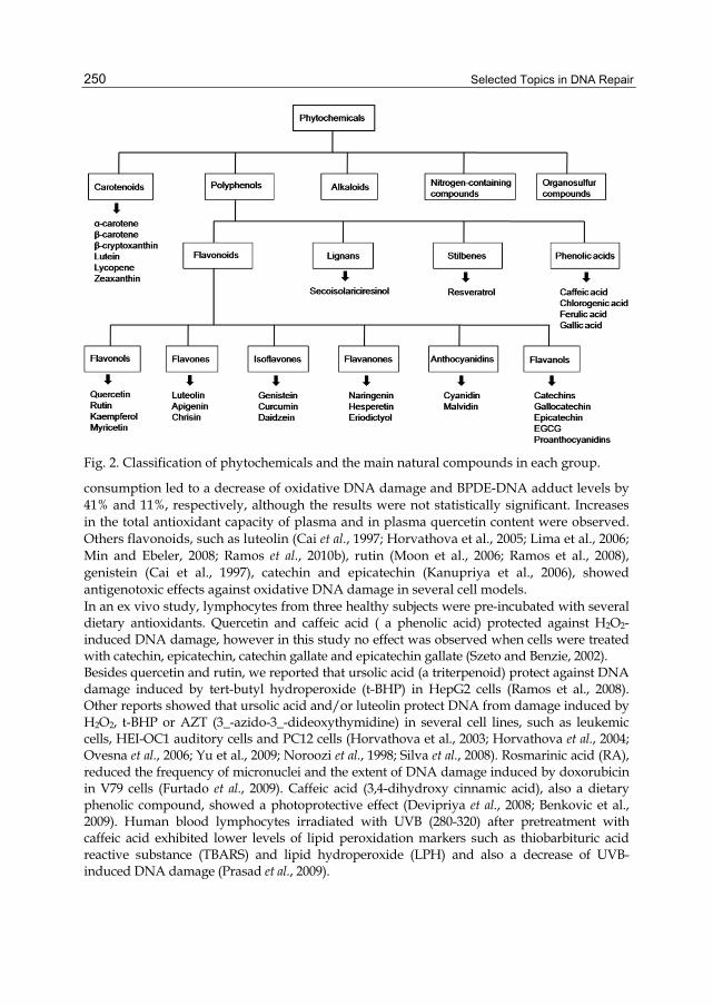
6.1 Effect of diet on prevention of oxidative DNA damage Prevention of DNA damage is one of the cellular mechanisms that may prevent cancer. Diet plays an important role in regulating DNA damage for instance by modulating the antioxidant/oxidant balance. The protective effect observed in many studies could be due, in part, to the presence of phenolic and/or non-phenolic constituents that have ability to act as antioxidant by free radical scavenging and chelating metal ions (Anderson et al., 2000); (Ross and Kasum, 2002). They can also act as indirect antioxidant by increasing levels of antioxidants such as glutathione (GSH) and/or by increasing the activity of antioxidant enzymes such as catalase (CAT), glutathione peroxidase (GPX) and superoxide dismutase (SOD) (Alia et al., 2005; Lima et al., 2005; Lima et al., 2006; Frei and Higdon, 2003). Phytochemicals can also modulate phase I (activating) enzymes and phase II (detoxifying) enzymes involved in xenobiotic metabolism (Chen and Kong, 2004; Ferguson et al., 2004; Moon et al., 2006; Ross and Kasum, 2002). Phytochemicals are bioactive compounds present in plants where they are produced as secondary metabolites to protect themselves from several pathogenic agents. Their consumption by humans confers protection against some diseases. Most bioactive phytochemicals belong to one of 5 groups: polyphenols, carotenoids, alkaloids, nitrogen-containing compounds, and organosulfur compounds. Polyphenols and carotenoids include several hundreds of compounds and are the most studied groups. Polyphenols may be further classified into 4 groups, according with the number of phenol rings that they contain: Flavonois, phenolic acids, stilbenes, and lignans. The flavonoids may themselves be classified into the follow subgroups: flavonols, flavones, isoflavones, flavanones, flavanols and anthocyanidins (Fig.2).
6.1.1 Polyphenols Numerous studies have shown the antioxidant and DNA protective properties of polyphenols. Quercetin is a flavonoid found in a variety of foods including apples, onions, wine and tea. Several studies, including our own showed the protective effects of quercetin against oxidative DNA damage in HepG2 cells (Lima et al., 2006; Ramos et al., 2008), Caco-2 cells (Aherne and O'Brien, 1999), HMB-2 cells (Horvathova et al., 2005), human macrophage (U-937 cells) (Kanupriya et al., 2006; Moon et al., 2006); human lymphocytes (healthy volunteers) (Wilms et al., 2007), and murine leukemia L1210 cells (Horvathova et al., 2003). Wilms et al., (2005) reported the protective effects of quercetin against the formation of oxidative DNA damage and bulky DNA adducts in human lymphocytes, induced by H2O2 and benzo(a)pyrene (B(a)P, respectively. In the same study, results obtained in an in vivo experiments showed that four weeks of quercetin-rich blueberry/apple juice mixture
Selected Topics in DNA Repair
250
Fig. 2. Classification of phytochemicals and the main natural compounds in each group.
consumption led to a decrease of oxidative DNA damage and BPDE-DNA adduct levels by 41% and 11%, respectively, although the results were not statistically significant. Increases in the total antioxidant capacity of plasma and in plasma quercetin content were observed. Others flavonoids, such as luteolin (Cai et al., 1997; Horvathova et al., 2005; Lima et al., 2006; Min and Ebeler, 2008; Ramos et al., 2010b), rutin (Moon et al., 2006; Ramos et al., 2008), genistein (Cai et al., 1997), catechin and epicatechin (Kanupriya et al., 2006), showed antigenotoxic effects against oxidative DNA damage in several cell models. In an ex vivo study, lymphocytes from three healthy subjects were pre-incubated with several dietary antioxidants. Quercetin and caffeic acid ( a phenolic acid) protected against H2O2-induced DNA damage, however in this study no effect was observed when cells were treated with catechin, epicatechin, catechin gallate and epicatechin gallate (Szeto and Benzie, 2002). Besides quercetin and rutin, we reported that ursolic acid (a triterpenoid) protect against DNA damage induced by tert-butyl hydroperoxide (t-BHP) in HepG2 cells (Ramos et al., 2008). Other reports showed that ursolic acid and/or luteolin protect DNA from damage induced by H2O2, t-BHP or AZT (3_-azido-3_-dideoxythymidine) in several cell lines, such as leukemic cells, HEI-OC1 auditory cells and PC12 cells (Horvathova et al., 2003; Horvathova et al., 2004; Ovesna et al., 2006; Yu et al., 2009; Noroozi et al., 1998; Silva et al., 2008). Rosmarinic acid (RA), reduced the frequency of micronuclei and the extent of DNA damage induced by doxorubicin in V79 cells (Furtado et al., 2009). Caffeic acid (3,4-dihydroxy cinnamic acid), also a dietary phenolic compound, showed a photoprotective effect (Devipriya et al., 2008; Benkovic et al., 2009). Human blood lymphocytes irradiated with UVB (280-320) after pretreatment with caffeic acid exhibited lower levels of lipid peroxidation markers such as thiobarbituric acid reactive substance (TBARS) and lipid hydroperoxide (LPH) and also a decrease of UVB- induced DNA damage (Prasad et al., 2009).

DNA Damage Protection and Induction of Repair by Dietary Phytochemicals and Cancer Prevention: What Do We Know?
251
Naringin, a citrus grapefruit flavonone, showed antigenotoxic properties in V79 cells (Jagetia et al., 2007). 4-Coumaric acid, a phenolic acid present in many foods and drinks, such as wine, tea, berries, apples, spinach and cereals reduced basal oxidative DNA damage in rat colonic mucosa when evaluated by comet assay (Guglielmi et al., 2003). Resveratrol, found in substantial amounts in several types of beverages including red wine and fruits, showed genoprotective effects under conditions of oxidative stress induced by H2O2 in C6 glioma cells (Quincozes-Santos et al., 2007). Apigenin, a flavonoid widely distributed in many herbs, fruits, and vegetables, has shown chemopreventive activities in several biological systems. Jeyabal et al., (2005) reported the protective role of apigenin against the oxidative stress caused by N-nitrosodiethylamine and phenobarbital in Wistar albino rats. Rusak et al., (2010), showed genoprotective effects of apigenin and other flavonoids (luteolin and kaempferol) in human peripheral lymphocytes against oxidative damage induced by hydrogen peroxide.
6.1.2 Carotenoids Tomato is one of the main sources of lycopene, a carotenoid with antioxidant properties. Pretreatment of rat hepatocytes with lycopene (1.86, 9.31 and 18.62 µM) in culture, showed a significant decrease in the levels of TBARS and DNA damage induced by gamma-radiation. Antioxidant enzymes such as superoxide dismutase (SOD), catalase (CAT) and glutathione peroxidase (GPx), as well as, the levels of GSH, vitamins A, E, C, increased significantly when hepatocytes were pretreated with the carotenoid (Srinivasan et al., 2007). At physiological concentrations (0.3-10 µM) lycopene and beta-carotene also protect Chinese hamster lung fibroblasts against DNA damage induced by 3-morpholinosydnonimine (SIN-1) (Muzandu et al., 2006). In a human intervention study, healthy volunteers were submitted to a supplementation with lycopene during 8 weeks. Consumption of lycopene decreased oxidative DNA damage in lymphocytes and urinary 8-hydroxy deoxoguanosine levels when compared with the basal levels (Devaraj et al., 2008). Also, Zhao et al., (2006) evaluated the protective effect of lycopene and others carotenoids against DNA damage in humans. Here, thirty-seven healthy, nonsmoking postmenopausal women consumed for 56 days a daily dose of mixed carotenoids (lycopene, lutein and beta-carotene; 4 mg each), 12 mg of a single carotenoid (lycopene, lutein or beta-carotene), or placebo. At the end the lymphocytes were isolated and DNA damage measured by comet assay. The results showed that all groups that consumed carotenoids had significantly lower endogenous DNA damage than that found on baseline measurements. No differences were found in placebo group. Lutein also decreased DNA damage induced by cisplatin in mice when evaluated by comet assay and increased GSH levels when compared with a negative control group (Serpeloni et al., 2010). Lorenzo et al., (2009) reported that β-cryptoxanthin at low concentrations, close to those found in plasma, protects Caco-2 and HeLa cells from oxidative DNA damage induced by H2O2 or by visible light in the presence of a photosensitizer. Consumption of β-carotene decreased the number of strand breaks induced by H2O2 in lymphocytes (Panayiotidis and Collins, 1997). Pre-treatment with astaxanthin, a red carotenoid used as a dietary additive, at 12.5, 25 and 50 mg/kg/day for 5 days before cyclophosphamide treatment resulted in the amelioration of antioxidant defenses (glutathione and superoxide dismutase) in the liver and decreased DNA damage evaluated by standard comet assay and using specific-enzymes (FPG and EndoIII) in bone marrow cells and peripheral blood lymphocytes isolated from mice. This
Selected Topics in DNA Repair
252
carotenoid also reduced the frequency of chromosomal breakage and micronucleus formation in the mouse bone marrow cells and peripheral blood. Astaxanthin, also showed antigenotoxic effects against cyclophosphamide in germ cell from male mice (Tripathi and Jena, 2008).
6.1.3 Whole foods Several studies, including our own with colon cancer cells, have found that polyphenol rich plant extracts inhibit formation of SBs and 8-oxoGua induced by oxidantive agents (Ramos et al., 2010a). Some plants, such as Salvia officinalis L. (sage), Rosmarinus officinalis L. (rosemary) and Origanum vulgare L. (oregano) have antioxidant properties that confer protection against oxidative DNA damage in colon cells as demonstrated by Aherne et al., (2007). Green tea is rich in polyphenolic antioxidants and their effects on health are the subject of several studies. Green tea decreased DNA oxidation in lymphocytes, colonocytes and hepatocytes when rats ingested 6.5 mg/kg bw per day, 5 days of tea extract (Kager et al., 2010). Evidences of genoprotective effects of green tea appear not only from in vitro and in vivo studies, but also from human supplementation trials. Human lymphocytes isolated from healthy volunteers that took 2 x 150 ml/d of 1% (w/v) green tea showed a decrease of basal oxidation-induced DNA damage with and without FPG enzyme (Han et al., 2011). One of the main constituents of green tea is (-)-epigallocatechin-3-gallate (EGCG) that could be responsible for their beneficial effects. In human peripheral leucocytes treatement with low EGCG concentrations (2-100µM) decreased both bleomycin-induced breaks and endonuclease III sensitive sites (Glei and Pool-Zobel, 2006). Apple is one of the most consumed fruit, therefore is an important source of polyphenols in humans. Their chemoprotective effects have been shown in vitro and in vivo studies (Koch et al., 2009; Veeriah et al., 2008). Apple juice has been found to possess antioxidant and antiproliferative activities as well as the ability to increase the expression of phase II gene glutathione S-transferase T2 (GSTT2) in human colon cells (Petermann et al., 2009). Apple extract can also protect against oxidative-induced DNA damage in human colon cells, such as LT97 (Miene (Miene et al., 2009), HT29, HCT115 and CaCo-2 cells (McCann et al., 2007; Schaefer et al., 2006). Also, grape juices exhibit antigenotoxic activity (Dani et al., 2009). The anticarcinogenic properties of olive oil have been attributed to the presence of phenolic compounds. Fabiani et al., (2008) reported that some of its isolated compounds (e.g. hydroxytyrosol, [3,4-dyhydroxyphenyl-ethanol (3,4-DHPEA)] and a complex mixture of phenols extracted from virgin olive oil) protected against H2O2 in human peripheral blood mononuclear cells and promyelocytic leukemia cells (HL60). These results have a great impact not only because of the high level of protection observed (between 80 and 90%) but also because, according to the authors, the concentrations tested could easily be reached with normal intake of olive oil. Annatto is a native shrub from Tropical America, whose seeds are a rich source of carotenoids, such as bixin, norbixin and phytoene and have antigenotoxic effects against oxidative DNA damage (Kovary et al., 2001). Results from human intervention trials have demonstrated the protective effect of isolated compounds as well as fruits and vegetables in peripheral lymphocytes. Pool-Zobel et al., (1997) showed a decrease in pyrimidine oxidation during supplementation with carrot juice. Porrini and Riso, (2000) reported that supplementation with tomato that is rich in lycopene,
DNA Damage Protection and Induction of Repair by Dietary Phytochemicals and Cancer Prevention: What Do We Know?
253
increase protection against H2O2-induced DNA damage in lymphocytes. Also a diet rich in fruit and vegetables for 14 days showed a DNA protection from oxidative damage in lymphocytes (Thompson et al., 1999). Broccoli intake also decreases oxidative DNA damage in smokers and nonsmokers (Riso et al., 2009). In a recent study, Sprague-Dawley rats fed with a wild-blueberry diet or a control diet for four or eight weeks were used to assess the effect of the consumption of this fruit on the resistance to H2O2-induced DNA damage. After treatment period, lymphocytes were exposed, ex vivo, to H2O2 and it was observed that wild-blueberry diet did not change antioxidant capacity in lymphocytes after eight weeks of treatment, but increased DNA protection against oxidative damage (Del Bo et al., 2010). Dulebohn et al., (2008) using the same animal model, reported that blueberries consumption for 3 weeks increase GST activity and decrease oxidative DNA damage in the liver. However, contrarily to in vitro studies, blueberries consumption did not significantly increase phase II enzyme activities in short-term supplementation times. As described above, many isolated compounds and some plants showed genoprotective effects in several experimental models, however it is important to keep in mind that these dietary agents can also induce DNA damage in certain conditions. The balance between the genoprotective and genotoxic effects of dietary agents is dependent on their concentration, incubation period and types of cells (Rusak et al., 2010).
6.2 Effect of diet on prevention of alkylating DNA damage Alkylation of DNA can be an important initial step in cancer formation. High levels of alkylating damage have been found in human colorectal DNA where high incidence of tumours have been observed (Hall et al., 1991; Povey et al., 2000). To assess antigenotoxic effects of diet against alkylating damage, several experimental models have been developed. Among them, colon tumours induction in rodent models, with carcinogenic chemicals such as 1,2-dimethylhydrazine (DMH) or azoxymethane (AOM) are the most used, and they are believed to be representative of colon carcinogenesis in humans (Barth et al., 2005).
6.2.1 Phytochemicals Dolara et al., (2005) reported that red wine polyphenols (50 mg/kg) administered to F344 rats for 16 weeks inhibited colon carcinogenesis induced by AOM or DMH. Wine polyphenols also decreased basal level of DNA oxidative damage of the colon mucosa. Supplementation of Wistar male rats with resveratrol showed to significantly decrease DMH-induced leukocyte DNA damage. In this study, an increase of levels of enzymic and non-enzymic antioxidant defense and a decrease in the extent of lipid peroxidation markers were also observed (Sengottuvelan et al., 2009). Other chemopreventive agents, such as quercetin, rutin, curcumin, silymarin, lycopene and farnesol, with antioxidant properties, have been found to inhibit DMH- and AOM-induced colon carcinogenesis and DNA damage in animal models (Kim et al., 1998; Volate et al., 2005). Lupeol, a pentacyclic triterpene present in mango, also protected against DMBA induced DNA alkylation damage in Swiss albino mice (Nigam et al., 2007).
6.2.2 Whole foods Many whole foods and plant crude extracts have also been found to protect against alkylating damage. The water extract of Salvia officinalis prevent formation of aberrant crypt foci (ACFs, a pre-carcinogenic lesion) induced by AOM in rat colon and also protected DNA

Selected Topics in DNA Repair
254
of colonocytes (unpublished observations). Using the same experimental model, Sengupta et al., (2004 and 2003) reported that tomato and garlic prevent ACFs, induced by AOM in rat colon. Tomato also decreased incidence and progression of 9,10-dimethyl benzanthracene (DMBA)-induced mouse skin tumours (De and Das, 2001). Intake of beer reduced ACF formation and protected against DNA damage induced by AOM in the rat colon mucosa (Nozawa et al., 2004). de Lima et al., (2005) evaluated the effect of aqueous extract of propolis on the formation of DMH-induced ACF and DNA damage in rat colon. Propolis had no effect on ACF formation, however, modulation of DMH-induced DNA damage in colon cells was observed. At lower concentrations (12, 34 and 108 mg/Kg bw/day) aqueous extract of propolis decrease the level of DNA damage. However, the highest concentration (336mg/Kg bw/day) induced DNA damage in rat colon. Dietary agents such as, ginseng, lemon grass and propolis, have been found to inhibit DMH- and AOM-induced colon carcinogenesis and DNA damage in animal model (Bazo et al., 2002; Suaeyun et al., 1997; Volate et al., 2005). Consumption of onion, blueberries (Boateng et al., 2007), and garbanzo beans (Murillo et al., 2004) also decreased the number of AOM-induced ACFs in rats and mice, respectively. Some studies reported antigenotoxic effects of diet against alkylating DNA damage using cytogenetic assays (micronucleus assay) (Azevedo Bentes Monteiro Neto et al., 2011; Gurbuz et al., 2009). Edenharder et al., (1998) reported that sweet cherries, strawberries, cucumber, tomatoes, bananas, oranges, asparagus, yellow red peppers and specially spinach had a protective effect against clastogenicity of cyclosphosphamide (an alkylating drug) in mice. Using comet assay some in vivo studies have shown the antigenotoxic effects of dietary agents, namely, artepillin C (a polyphenolic acid found in Brazilian green propolis and Baccharis dracunculifolia) (Azevedo Bentes Monteiro Neto et al., 2011), safranal, (a constituent of Crocus sativus L. stigmas) (Hosseinzadeh and Sadeghnia, 2007), orange juice (Franke et al., 2005b) and vitamin C (Franke et al., 2005a) against DNA damage induced by methyl methanesulfonate (MMS). Also lemongrass protected leukocytes from DNA damage induced by N-methyl-N-nitrosurea (MNU) (Bidinotto et al., 2010). Data from application of comet assay in the assessment of genoprotective effects of diet against alkylating DNA damage is limited. First, AlkA is, as far as we know, the only repair enzyme used for detection of alkylating DNA damage by the comet assay (Collins et al., 2001a). Alk A recognises 3-MeA in DNA, but its specificity is low, detecting other modified bases, some of which are also other alkylated bases. Furthermore, 3-MeA is not the most abundant alkylating damage and it does not represent the alkylating damage with more mutagenic/carcinogenic potential. Other alkylating lesions, like N7MeG, the most abundant lesion, and O6MeG, the most mutagenic, are until now undectectable by comet assay.
6.3 Effect of diet on induction of DNA repair DNA damage can arrest cell cycle progression to allow DNA repair, preventing, therefore, replication of errors, or to induce apoptosis to eliminate cells severely damaged. Defective DNA repair is usually linked to human cancer development. Therefore, enhancement of DNA repair can be understood as a prevention strategy against cancer and an important molecular target for dietary phytochemicals (Davis and Milner, 2007). However, few studies have investigated whether DNA repair activity can be modified by diet in humans. New modifications of the comet assay have been developed to assess effects of dietary agents on DNA repair ability (as described above). Briefly, the “cellular repair assay”

DNA Damage Protection and Induction of Repair by Dietary Phytochemicals and Cancer Prevention: What Do We Know?
255
measures the ability of cells to rejoin strand breaks induced by a DNA damaging agent; while the “in vitro repair assay” measures the excision repair activity of a protein extract prepared from cells treated with dietary agents, incubating with a DNA substrate containing a specific type of damage.
6.3.1 Polyphenols Recently, we reported that luteolin and luteolin-7-glucoside increased rejoining of strand breaks after treatment with H2O2 in Caco-2 cells. Luteolin-7-glucoside also had a BER inductive effect increasing incision activity in Caco-2 cells (Ramos et al., 2010a; Ramos et al., 2010b). Quercetin also increased rejoining of strand breaks induced by t-BHP in HepG2 (Ramos et al., 2008). Moreover, dietary agents such as flavonoids, vitamins E and C had been reported as inducers of oxidative DNA damage repair activity (Davis and Milner, 2007).
6.3.2 Carotenoids An in vitro study using HeLa and Caco-2 cells reported that -cryptoxanthin increased rejoining of strand breaks induced by H2O2 and increase the repair of oxidised bases. However, increase of repair activity was not related with increase of hOGG1 or APE1expression (Lorenzo et al., 2009). The lack of correlation between activity and mRNA expression of OGG1 and APE1 has also been demonstrated in other studies (Collins et al., 2003; Silva et al., 2009). -carotene, lutein and lycopene also enhanced strand breaks rejoining in lymphocytes (Fillion et al., 1998; Torbergsen and Collins, 2000). Paz-Elizur et al., (2007) measured OGG1 activity and mRNA expression in 120 healthy individuals and a poor correlation between activity and mRNA was found.
6.3.3 Whole foods Water extracts of Salvia species increased rejoining of strand breaks after treatment with H2O2 in Caco-2 cells. These water extracts also increased incision activity of a Caco-2 cell extract on a DNA substrate containing specific oxidative damage (8-oxoGua) (Ramos et al., 2010a). Nakamura et al., (2000) reported that aqueous fractions of Fushimi sweet pepper increase repair against ultraviolet-induced cyclobutane pyrimidine dimers in human fibroblasts. Collins et al., (2003), in a human intervention study, showed that 3 weeks of a dietary supplementation with kiwifruit increased DNA repair capacity measured by “in vitro repair assay”. Also Freese, (2006) showed that kiwifruit consumption increased DNA repair capacity in human lymphocytes. Brevik et al., (2011) in a human biomonitoring study, reported that consumption of kiwifruits and antioxidant-rich plant products reduced DNA strand breaks in lymphocytes. Increase of BER activity was observed in the group that consumed antioxidant-rich plant products. However, a reduction of NER activity was observed in both groups. No explanations have been found for this decrease in NER pathway. Diet supplementation with cooked carrots, during 3 weeks, increased in vitro repair activity and strand break rejoining in lymphocytes (Astley et al., 2004). High dietary folate intake has been associated with a decreased risk of cancer development, such as colorectal cancer. In vitro, rodent and human studies demonstrated that low folate intake increases uracil misincorporation leading to increase of DNA damage, chromosomal breakage and malignant transformation; modulates DNA repair by inhibiting thymidine

Selected Topics in DNA Repair
256
and purine biosynthesis and induces epigenetic changes leading to global DNA hypomethylation and/or changes in gene-specific methylation and protooncogene activation (Duthie and McMillan, 1997; Duthie, 2010; Duthie et al., 2000; Melnyk et al., 1999). In vivo, folate deficiency induced DNA repair, such as increase of OGG1 and MGMT (Duthie et al., 2010). However, remains unclear if increasing folate intake improve DNA repair. Generaly, the protective role of folate against carcinogenesis is not completely understood.
7. Effects of phytochemicals through DNA repair modulation and their interaction with alkylating agents used as chemotherapeutic
Some DNA-damaging agents (specially alkylating agents) are used in cancer therapy due to their ability to induce DNA damage and subsequently apoptosis of tumor cells (Maynard et al., 2009). The efficacy of DNA damage-based cancer therapy is modulated by DNA repair pathways. Therefore these pathways may, attenuate the therapeutic effects of alkylating agents. These drugs are usually classified in two classes: monofunctional (e.g., N-methyl-N_-nitro-N-nitrosoguanidine [MNNG], temozolomide [TMZ] and dacarbazine) and bifunctional alkylating agents (e.g., carmustine [BCNU], cyclophosphamide, lomustine [CCNU] and fotemustine). BCNU, induces several kinds of DNA damage such as cross-linking, strand breaks and modified bases (Kondo et al., 2010). Between alkylation damage, N7 alkyl-guanine is the most abundant (around 90% of the total alkylation events) and O6 alkyl-guanine is the less frequent. O6 alkyl-guanine, if not repaired before cell division, can form base pairs with T, generating GCAT transitions mutations by action of MMR pathway. O6 alkyl-guanine is the lesion mainly responsible for the cytotoxic and mutagenic effects of these alkylating drugs. However O6 alkyl-guanine can be repaired by MGMT. In human colorectal adenoma, reduced MGMT activity has been found. Therefore, more mutations occur when cells are treated with alkylating agent (Lees et al., 2004; Lees et al., 2002). However, high levels of MGMT where found in some other tumors (Baer et al., 1993). In the context of therapy with alkylating agents, inhibition of MGMT activity in tumor cells is desirable. The coadjuvant drug O6-benzylguanine (O6-BG) inactivates MGMT, acting as a pseudosubstrate. Effect of O6-BG has been investigated when in combination with an alkylating drug to increase its efficacy (Liu et al., 2002). N7-alkylG is the most frequent alkylation damage however is not considered to be as mutagenic as O6-alkylG because it is efficiently repaired by BER pathway. N-methylpurine-DNAglycoslase (MPG), the only glycosylase that recognizes alkylation lesions in animal cells, removes N7-alkylG by hydrolysis of the N-glycosylic bond creating an AP site that is repaired by the other enzymes of BER pathway (Drablos et al., 2004). An overexpression of MPG increases the production of AP sites. If the levels of the other enzymes of BER pathway remain unaltered, repair of AP sites is low and accumulation of these lesions becomes cytotoxic and mutagenic. Therefore, an overexpression of MPG or a decrease in other enzymes involved in the subsequent steps in BER pathway, increases cytotoxicity of alkylating drugs. This cytoxicity becomes more relevant when cells are resistant to cytotoxicity from O6-alkylation. MGMT and MMR have contrasting effects on DNA O6-alkylG. While MGMT is an efficient mechanism of repair, MMR in contrast, does not remove the alkylated base but introduce more lesions like strand break in an attempt to repair the mismatch. Accumulation of strand

DNA Damage Protection and Induction of Repair by Dietary Phytochemicals and Cancer Prevention: What Do We Know?
257
breaks may activate apoptotic pathways, leading to cell death. In some cells, resistance to alkylating agents can be mediated by MGMT and MMR. Active MGMT and loss of MMR pathway protect cells against the cell death induced by methylating chemotherapeutic drugs (Allan and Travis, 2005). Depletion of MGMT activity (for example, by O6-BG or by epigenetic silencing of the MGMT gene) and intact MMR system results in reversion of resistance with high sensitivity to the cytotoxic effects of alkylating drugs (Casorelli et al., 2008; Esteller et al., 2000; Hegi et al., 2005). Tumors with low MGMT level are more sensitive to alkylating chemotherapy (Bandres et al., 2005). Therefore,DNA repair mechanisms may be understood as a promising target to develop new cancer treatments (Helleday et al., 2008; Jiricny, 2006). Different strategies have been developed to enhance the efficacy of chemotherapy using alkylating agents. In tumor cells, inhibition of MGMT activity and/or BER pathway decreases resistance to alkylating drugs (Drablos et al., 2004; Jaiswal et al., 2011; Middleton and Margison, 2003). Downregulation of DNA repair pathways (except MMR) may increase efficacy of alkylating agents, decreasing the amount of drug needed for chemotherapy and consequently reduction of the side effects (Kondo et al., 2010). Dietary agents that modulate MGMT expression and/or BER pathway may play an important role in chemotherapy when in combination with alkylating agents. However this subject, in contrast to chemoprevention, has received little attention. Some studies have been that dietary agents may increase MGMT activity. Niture et al., (2006), investigated the potential ability of some Indian medicinal plants extracts to modulate MGMT activity and expression in human peripheral blood lymphocytes and cancer cell lines. The results showed that both the ethanolic and aqueous extracts from neem (Azadirachta indica), holy basil (Ocimum sanctum), winter cherry (Withania somnifera), and oregano (Origanum majorana) increased MGMT expression and its activity. Extracts from gooseberry (Emblica officinalis), common basil (Ocimum basilicum), and spearmint (Mentha viridis) also increased MGMT levels, however to a smaller extent. Later, the same author reported that some phytochemicals such as curcumin, silymarin and resveratrol increase protein expression as well as activity of MGMT in lymphocytes and cancer cell lines (Niture et al., 2007). In tumor cells a number of genes are abnormally methylated. Some dietary agents, such as genistein and epigallocatechin-3-gallate showed the ability to reactivate some methylation-silenced genes in cancer cells like MGMT due to a direct inhibition of DNA methyltransferase (Fang et al., 2005; Fang et al., 2003). Recently, Billson et al., (2009), demonstrated that a high vegetable intake in humans decreases MGMT activity in normal colorectal mucosa. To understand the real role of dietary agents on chemotherapy when in combination with alkylating drugs more studies are need.
8. Conclusion
Prevention of DNA damage and/or enhanced DNA repair activity by dietary agents constitute an important strategy to prevent mutations and consequently inhibit the carcinogenic process. Diet supplementation with phytochemicals, with beneficial effects, increases their concentrations in the organism. However, effects of supra-physiological concentrations need to be evaluated in each case since a safe phytochemical at physiological concentrations can be toxic at higher concentrations. The comet assay is a powerful tool to assess the effects of diet on DNA. The immense literature (more than 5500 papers in Pubmed since 1995) that use comet assay (standard and modified versions) demonstrates the real potential of this assay. Some dietary agents have
Selected Topics in DNA Repair
258
shown ability to prevent DNA damage (oxidative and/or alkylanting) in several experimental models from in vitro to human intervention studies. Less is known about the effect of diet on DNA repair modulation. However, the modifications of comet assay (e.g. use specific-enzymes) gives an opportunity to enhance the knowledge in this field. In spite of the large number of publications much remains to be done. An emergent field is the effect of combinations between diet and drugs used in chemotherapy. In our view, comet assay can be a useful approach also to understand the role of dietary constituents on chemotherapy.
9. Acknowledgment
Ramos A.A. is supported by the Foundation for Science and Technology, Portugal, grant SFRH/BD/35672/2007.
10. References
Aherne, S. A., Kerry, J. P. & O'Brien, N. M. (2007). Effects of plant extracts on antioxidant status and oxidant-induced stress in Caco-2 cells. Br J Nutr, 97, 321-328.
Aherne, S. A. & O'Brien, N. M. (1999). Protection by the flavonoids myricetin, quercetin, and rutin against hydrogen peroxide-induced DNA damage in Caco-2 and Hep G2 cells. Nutr Cancer, 34, 160-166.
Alia, M., Ramos, S., Mateos, R., Bravo, L. & Goya, L. (2005). Response of the antioxidant defense system to tert-butyl hydroperoxide and hydrogen peroxide in a human hepatoma cell line (HepG2). J Biochem Mol Toxicol, 19, 119-128.
Allan, J. M. & Travis, L. B. (2005). Mechanisms of therapy-related carcinogenesis. Nat Rev Cancer, 5, 943-955.
Allen, R. G. & Tresini, M. (2000). Oxidative stress and gene regulation. Free Radic Biol Med, 28, 463-499.
Anderson, R. F., Amarasinghe, C., Fisher, L. J., Mak, W. B. & Packer, J. E. (2000). Reduction in free-radical-induced DNA strand breaks and base damage through fast chemical repair by flavonoids. Free Radic Res, 33, 91-103.
Angerer, J., Ewers, U. & Wilhelm, M. (2007). Human biomonitoring: state of the art. Int J Hyg Environ Health, 210, 201-228.
Armstrong, M. J. & Galloway, S. M. (1997). Mismatch repair provokes chromosome aberrations in hamster cells treated with methylating agents or 6-thioguanine, but not with ethylating agents. Mutat Res, 373, 167-178.
Astley, S. B., Elliott, R. M., Archer, D. B. & Southon, S. (2004). Evidence that dietary supplementation with carotenoids and carotenoid-rich foods modulates the DNA damage: repair balance in human lymphocytes. Br J Nutr, 91, 63-72.
Azevedo Bentes Monteiro Neto, M., Souza Lima, I. M., Furtado, R. A., Bastos, J. K., Silva Filho, A. A. & Tavares, D. C. (2011). Antigenotoxicity of artepillin C in vivo evaluated by the micronucleus and comet assays. J Appl Toxicol.
Baer, J. C., Freeman, A. A., Newlands, E. S., Watson, A. J., Rafferty, J. A. & Margison, G. P. (1993). Depletion of O6-alkylguanine-DNA alkyltransferase correlates with potentiation of temozolomide and CCNU toxicity in human tumour cells. Br J Cancer, 67, 1299-1302.

DNA Damage Protection and Induction of Repair by Dietary Phytochemicals and Cancer Prevention: What Do We Know?
259
Bandres, E., Andion, E., Escalada, A., Honorato, B., Catalan, V., Cubedo, E., Cordeu, L., Garcia, F., Zarate, R., Zabalegui, N. & Garcia-Foncillas, J. (2005). Gene expression profile induced by BCNU in human glioma cell lines with differential MGMT expression. J Neurooncol, 73, 189-198.
Bartek, J., Bartkova, J. & Lukas, J. (2007). DNA damage signalling guards against activated oncogenes and tumour progression. Oncogene, 26, 7773-7779.
Barth, S. W., Fahndrich, C., Bub, A., Dietrich, H., Watzl, B., Will, F., Briviba, K. & Rechkemmer, G. (2005). Cloudy apple juice decreases DNA damage, hyperproliferation and aberrant crypt foci development in the distal colon of DMH-initiated rats. Carcinogenesis, 26, 1414-1421.
Bazo, A. P., Rodrigues, M. A., Sforcin, J. M., de Camargo, J. L., Ribeiro, L. R. & Salvadori, D. M. (2002). Protective action of propolis on the rat colon carcinogenesis. Teratog Carcinog Mutagen, 22, 183-194.
Benhamou, S. & Sarasin, A. (2000). Variability in nucleotide excision repair and cancer risk: a review. Mutat Res, 462, 149-158.
Benkovic, V., Knezevic, A. H., Orsolic, N., Basic, I., Ramic, S., Viculin, T., Knezevic, F. & Kopjar, N. (2009). Evaluation of radioprotective effects of propolis and its flavonoid constituents: in vitro study on human white blood cells. Phytother Res, 23, 1159-1168.
Bidinotto, L. T., Costa, C. A., Salvadori, D. M., Costa, M., Rodrigues, M. A. & Barbisan, L. F. (2010). Protective effects of lemongrass (Cymbopogon citratus STAPF) essential oil on DNA damage and carcinogenesis in female Balb/C mice. J Appl Toxicol.
Billson, H. A., Harrison, K. L., Lees, N. P., Hall, C. N., Margison, G. P. & Povey, A. C. (2009). Dietary variables associated with DNA N7-methylguanine levels and O6-alkylguanine DNA-alkyltransferase activity in human colorectal mucosa. Carcinogenesis, 30, 615-620.
Boateng, J., Verghese, M., Shackelford, L., Walker, L. T., Khatiwada, J., Ogutu, S., Williams, D. S., Jones, J., Guyton, M., Asiamah, D., Henderson, F., Grant, L., DeBruce, M., Johnson, A., Washington, S. & Chawan, C. B. (2007). Selected fruits reduce azoxymethane (AOM)-induced aberrant crypt foci (ACF) in Fisher 344 male rats. Food Chem Toxicol, 45, 725-732.
Brevik, A., Karlsen, A., Azqueta, A., Tirado, A. E., Blomhoff, R. & Collins, A. (2011). Both base excision repair and nucleotide excision repair in humans are influenced by nutritional factors. Cell Biochem Funct, 29, 36-42.
Bugni, J. M., Meira, L. B. and Samson, L. D. (2009) Alkylation-induced colon tumorigenesis in mice deficient in the Mgmt and Msh6 proteins. Oncogene, 28, 734-741.
Cai, Q., Rahn, R. O. & Zhang, R. (1997). Dietary flavonoids, quercetin, luteolin and genistein, reduce oxidative DNA damage and lipid peroxidation and quench free radicals. Cancer Lett, 119, 99-107.
Caldecott, K. W. (2007). Mammalian single-strand break repair: mechanisms and links with chromatin. DNA Repair (Amst), 6, 443-453.
Casorelli, I., Russo, M. T. & Bignami, M. (2008). Role of mismatch repair and MGMT in response to anticancer therapies. Anticancer Agents Med Chem, 8, 368-380.
Chen, C. and Kong, A. N. (2004). Dietary chemopreventive compounds and ARE/EpRE signaling. Free Radic Biol Med, 36, 1505-1516.

Selected Topics in DNA Repair
260
Collins, A. R. (2004). The comet assay for DNA damage and repair: principles, applications, and limitations. Mol Biotechnol, 26, 249-261.
Collins, A. R. (2009). Investigating oxidative DNA damage and its repair using the comet assay. Mutat Res, 681, 24-32.
Collins, A. R., Dobson, V. L., Dusinska, M., Kennedy, G. & Stetina, R. (1997a). The comet assay: what can it really tell us? Mutat Res, 375, 183-193.
Collins, A. R., Dusinska, M. & Horska, A. (2001a). Detection of alkylation damage in human lymphocyte DNA with the comet assay. Acta Biochim Pol, 48, 611-614.
Collins, A. R., Dusinska, M., Horvathova, E., Munro, E., Savio, M. & Stetina, R. (2001b). Inter-individual differences in repair of DNA base oxidation, measured in vitro with the comet assay. Mutagenesis, 16, 297-301.
Collins, A. R., Duthie, S. J. & Dobson, V. L. (1993). Direct enzymic detection of endogenous oxidative base damage in human lymphocyte DNA. Carcinogenesis, 14, 1733-1735.
Collins, A. R., Harrington, V., Drew, J. & Melvin, R. (2003). Nutritional modulation of DNA repair in a human intervention study. Carcinogenesis, 24, 511-515.
Collins, A. R., Mitchell, D. L., Zunino, A., de Wit, J. & Busch, D. (1997b). UV-sensitive rodent mutant cell lines of complementation groups 6 and 8 differ phenotypically from their human counterparts. Environ Mol Mutagen, 29, 152-160.
Collins, A. R., Oscoz, A. A., Brunborg, G., Gaivao, I., Giovannelli, L., Kruszewski, M., Smith, C. C. & Stetina, R. (2008). The comet assay: topical issues. Mutagenesis, 23, 143-151.
D'Errico, M., Parlanti, E. & Dogliotti, E. (2008). Mechanism of oxidative DNA damage repair and relevance to human pathology. Mutat Res, 659, 4-14.
Dani, C., Oliboni, L. S., Umezu, F. M., Pasquali, M. A., Salvador, M., Moreira, J. C. & Henriques, J. A. (2009). Antioxidant and antigenotoxic activities of purple grape juice--organic and conventional--in adult rats. J Med Food, 12, 1111-1118.
Davis, C. D. (2007). Nutritional interactions: credentialing of molecular targets for cancer prevention. Exp Biol Med (Maywood), 232, 176-183.
Davis, C. D. & Milner, J. A. (2007). Biomarkers for diet and cancer prevention research: potentials and challenges. Acta Pharmacol Sin, 28, 1262-1273.
de Kok, T. M., van Breda, S. G. & Manson, M. M. (2008). Mechanisms of combined action of different chemopreventive dietary compounds: a review. Eur J Nutr, 47 Suppl 2, 51-59.
de Lima, R. O., Bazo, A. P., Said, R. A., Sforcin, J. M., Bankova, V., Darros, B. R. & Salvadori, D. M. (2005). Modifying effect of propolis on dimethylhydrazine-induced DNA damage but not colonic aberrant crypt foci in rats. Environ Mol Mutagen, 45, 8-16.
De, S. & Das, S. (2001). Protective Effects of Tomato Juice on Mouse Skin Carcinogenesis. Asian Pac J Cancer Prev, 2, 43-47.
Del Bo, C., Martini, D., Vendrame, S., Riso, P., Ciappellano, S., Klimis-Zacas, D. & Porrini, M. (2010). Improvement of lymphocyte resistance against H(2)O(2)-induced DNA damage in Sprague-Dawley rats after eight weeks of a wild blueberry (Vaccinium angustifolium)-enriched diet. Mutat Res, 703, 158-162.
Devaraj, S., Mathur, S., Basu, A., Aung, H. H., Vasu, V. T., Meyers, S. & Jialal, I. (2008). A dose-response study on the effects of purified lycopene supplementation on biomarkers of oxidative stress. J Am Coll Nutr, 27, 267-273.

DNA Damage Protection and Induction of Repair by Dietary Phytochemicals and Cancer Prevention: What Do We Know?
261
Devipriya, N., Sudheer, A. R. & Menon, V. P. (2008). Caffeic acid protects human peripheral blood lymphocytes against gamma radiation-induced cellular damage. J Biochem Mol Toxicol, 22, 175-186.
Doak, S. H., Brusehafer, K., Dudley, E., Quick, E., Johnson, G., Newton, R. P. & Jenkins, G. J. (2008). No-observed effect levels are associated with up-regulation of MGMT following MMS exposure. Mutat Res, 648, 9-14.
Dolara, P., Luceri, C., De Filippo, C., Femia, A. P., Giovannelli, L., Caderni, G., Cecchini, C., Silvi, S., Orpianesi, C. & Cresci, A. (2005). Red wine polyphenols influence carcinogenesis, intestinal microflora, oxidative damage and gene expression profiles of colonic mucosa in F344 rats. Mutat Res, 591, 237-246.
Drablos, F., Feyzi, E., Aas, P. A., Vaagbo, C. B., Kavli, B., Bratlie, M. S., Pena-Diaz, J., Otterlei, M., Slupphaug, G. & Krokan, H. E. (2004). Alkylation damage in DNA and RNA--repair mechanisms and medical significance. DNA Repair (Amst), 3, 1389-1407.
Dudas, A. & Chovanec, M. (2004). DNA double-strand break repair by homologous recombination. Mutat Res, 566, 131-167.
Dulebohn, R. V., Yi, W., Srivastava, A., Akoh, C. C., Krewer, G. & Fischer, J. G. (2008). Effects of blueberry (Vaccinium ashei) on DNA damage, lipid peroxidation, and phase II enzyme activities in rats. J Agric Food Chem, 56, 11700-11706.
Dusinska, M. & Collins, A. R. (1996). Detection of oxi-dised purines and UV-induced photoproducts in DNA of single cells, by inclusion of lesion-specific enzymes in the comet assay. Alternative Laboratory Animal, 24, 405-411.
Dusinska, M. & Collins, A. R. (2008). The comet assay in human biomonitoring: gene-environment interactions. Mutagenesis, 23, 191-205.
Duthie, S. J. (2010) Folate and cancer: how DNA damage, repair and methylation impact on colon carcinogenesis. J Inherit Metab Dis, 34, 101-109.
Duthie, S. J., Grant, G., Pirie, L. P., Watson, A. J. and Margison, G. P. (2010) Folate deficiency alters hepatic and colon MGMT and OGG-1 DNA repair protein expression in rats but has no effect on genome-wide DNA methylation. Cancer Prev Res (Phila), 3, 92-100.
Duthie, S. J. and McMillan, P. (1997) Uracil misincorporation in human DNA detected using single cell gel electrophoresis. Carcinogenesis, 18, 1709-1714.
Duthie, S. J., Narayanan, S., Blum, S., Pirie, L. and Brand, G. M. (2000) Folate deficiency in vitro induces uracil misincorporation and DNA hypomethylation and inhibits DNA excision repair in immortalized normal human colon epithelial cells. Nutr Cancer, 37, 245-251.
Edenharder, R., Frangart, J., Hager, M., Hofmann, P. and Rauscher, R. (1998) Protective effects of fruits and vegetables against in vivo clastogenicity of cyclosphosphamide or benzo[a]pyrene in mice. Food Chem Toxicol, 36, 637-645.
Eker, A. P., Quayle, C., Chaves, I. and van der Horst, G. T. (2009) DNA repair in mammalian cells: Direct DNA damage reversal: elegant solutions for nasty problems. Cell Mol Life Sci, 66, 968-980.
Esteller, M., Garcia-Foncillas, J., Andion, E., Goodman, S. N., Hidalgo, O. F., Vanaclocha, V., Baylin, S. B. and Herman, J. G. (2000) Inactivation of the DNA-repair gene MGMT and the clinical response of gliomas to alkylating agents. N Engl J Med, 343, 1350-1354.

Selected Topics in DNA Repair
262
Evans, M. D., Dizdaroglu, M. and Cooke, M. S. (2004) Oxidative DNA damage and disease: induction, repair and significance. Mutat Res, 567, 1-61.
Fabiani, R., Rosignoli, P., De Bartolomeo, A., Fuccelli, R., Servili, M., Montedoro, G. F. and Morozzi, G. (2008) Oxidative DNA damage is prevented by extracts of olive oil, hydroxytyrosol, and other olive phenolic compounds in human blood mononuclear cells and HL60 cells. J Nutr, 138, 1411-1416.
Fairbairn, D. W., Olive, P. L. and O'Neill, K. L. (1995) The comet assay: a comprehensive review. Mutat Res, 339, 37-59.
Fang, M. Z., Chen, D., Sun, Y., Jin, Z., Christman, J. K. and Yang, C. S. (2005) Reversal of hypermethylation and reactivation of p16INK4a, RARbeta, and MGMT genes by genistein and other isoflavones from soy. Clin Cancer Res, 11, 7033-7041.
Fang, M. Z., Wang, Y., Ai, N., Hou, Z., Sun, Y., Lu, H., Welsh, W. and Yang, C. S. (2003) Tea polyphenol (-)-epigallocatechin-3-gallate inhibits DNA methyltransferase and reactivates methylation-silenced genes in cancer cell lines. Cancer Res, 63, 7563-7570.
Ferguson, L. R., Philpott, M. and Karunasinghe, N. (2004) Dietary cancer and prevention using antimutagens. Toxicology, 198, 147-159.
Fillion, L., Collins, A. and Southon, S. (1998) Beta-carotene enhances the recovery of lymphocytes from oxidative DNA damage. Acta Biochim Pol, 45, 183-190.
Fortini, P., Pascucci, B., Parlanti, E., D'Errico, M., Simonelli, V. and Dogliotti, E. (2003) The base excision repair: mechanisms and its relevance for cancer susceptibility. Biochimie, 85, 1053-1071.
Franke, S. I., Pra, D., da Silva, J., Erdtmann, B. and Henriques, J. A. (2005a) Possible repair action of Vitamin C on DNA damage induced by methyl methanesulfonate, cyclophosphamide, FeSO4 and CuSO4 in mouse blood cells in vivo. Mutat Res, 583, 75-84.
Franke, S. I., Pra, D., Erdtmann, B., Henriques, J. A. and da Silva, J. (2005b) Influence of orange juice over the genotoxicity induced by alkylating agents: an in vivo analysis. Mutagenesis, 20, 279-283.
Freese, R. (2006) Markers of oxidative DNA damage in human interventions with fruit and berries. Nutr Cancer, 54, 143-147.
Frei, B. and Higdon, J. V. (2003) Antioxidant activity of tea polyphenols in vivo: evidence from animal studies. J Nutr, 133, 3275S-3284S.
Furtado, R. A., de Araujo, F. R., Resende, F. A., Cunha, W. R. and Tavares, D. C. (2009) Protective effect of rosmarinic acid on V79 cells evaluated by the micronucleus and comet assays. J Appl Toxicol, 30, 254-259.
Gaivao, I., Piasek, A., Brevik, A., Shaposhnikov, S. and Collins, A. R. (2009) Comet assay-based methods for measuring DNA repair in vitro; estimates of inter- and intra-individual variation. Cell Biol Toxicol, 25, 45-52.
Gedik, C. M., Ewen, S. W. and Collins, A. R. (1992) Single-cell gel electrophoresis applied to the analysis of UV-C damage and its repair in human cells. Int J Radiat Biol, 62, 313-320.
Glei, M. and Pool-Zobel, B. L. (2006) The main catechin of green tea, (-)-epigallocatechin-3-gallate (EGCG), reduces bleomycin-induced DNA damage in human leucocytes. Toxicol In Vitro, 20, 295-300.

DNA Damage Protection and Induction of Repair by Dietary Phytochemicals and Cancer Prevention: What Do We Know?
263
Guglielmi, F., Luceri, C., Giovannelli, L., Dolara, P. and Lodovici, M. (2003) Effect of 4-coumaric and 3,4-dihydroxybenzoic acid on oxidative DNA damage in rat colonic mucosa. Br J Nutr, 89, 581-587.
Gurbuz, N., Ozkul, A. and Burgaz, S. (2009) Effects of vitamin C and N-acetylcysteine against cyclophosphamide-induced genotoxicity in exfoliated bladder cells of mice in vivo. J BUON, 14, 647-652.
Hall, C. N., Badawi, A. F., O'Connor, P. J. and Saffhill, R. (1991) The detection of alkylation damage in the DNA of human gastrointestinal tissues. Br J Cancer, 64, 59-63.
Han, K. C., Wong, W. C. and Benzie, I. F. (2011) Genoprotective effects of green tea (Camellia sinensis) in human subjects: results of a controlled supplementation trial. Br J Nutr, 105, 171-179.
Hanawalt, P. C. (2002) Subpathways of nucleotide excision repair and their regulation. Oncogene, 21, 8949-8956.
Hartmann, A., Agurell, E., Beevers, C., Brendler-Schwaab, S., Burlinson, B., Clay, P., Collins, A., Smith, A., Speit, G., Thybaud, V. and Tice, R. R. (2003) Recommendations for conducting the in vivo alkaline Comet assay. 4th International Comet Assay Workshop. Mutagenesis, 18, 45-51.
Hegde, M. L., Hazra, T. K. and Mitra, S. (2008) Early steps in the DNA base excision/single-strand interruption repair pathway in mammalian cells. Cell Res, 18, 27-47.
Hegi, M. E., Diserens, A. C., Gorlia, T., Hamou, M. F., de Tribolet, N., Weller, M., Kros, J. M., Hainfellner, J. A., Mason, W., Mariani, L., Bromberg, J. E., Hau, P., Mirimanoff, R. O., Cairncross, J. G., Janzer, R. C. and Stupp, R. (2005) MGMT gene silencing and benefit from temozolomide in glioblastoma. N Engl J Med, 352, 997-1003.
Helleday, T., Lo, J., van Gent, D. C. and Engelward, B. P. (2007) DNA double-strand break repair: from mechanistic understanding to cancer treatment. DNA Repair (Amst), 6, 923-935.
Helleday, T., Petermann, E., Lundin, C., Hodgson, B. and Sharma, R. A. (2008) DNA repair pathways as targets for cancer therapy. Nat Rev Cancer, 8, 193-204.
Heo, M. Y., Sohn, S. J. and Au, W. W. (2001) Anti-genotoxicity of galangin as a cancer chemopreventive agent candidate. Mutat Res, 488, 135-150.
Hoelzl, C., Knasmuller, S., Misik, M., Collins, A., Dusinska, M. and Nersesyan, A. (2009) Use of single cell gel electrophoresis assays for the detection of DNA-protective effects of dietary factors in humans: recent results and trends. Mutat Res, 681, 68-79.
Horvathova, K., Chalupa, I., Sebova, L., Tothova, D. and Vachalkova, A. (2005) Protective effect of quercetin and luteolin in human melanoma HMB-2 cells. Mutat Res, 565, 105-112.
Horvathova, K., Novotny, L., Tothova, D. and Vachalkova, A. (2004) Determination of free radical scavenging activity of quercetin, rutin, luteolin and apigenin in H2O2-treated human ML cells K562. Neoplasma, 51, 395-399.
Horvathova, K., Novotny, L. and Vachalkova, A. (2003) The free radical scavenging activity of four flavonoids determined by the comet assay. Neoplasma, 50, 291-295.
Hosseinzadeh, H. and Sadeghnia, H. R. (2007) Effect of safranal, a constituent of Crocus sativus (saffron), on methyl methanesulfonate (MMS)-induced DNA damage in mouse organs: an alkaline single-cell gel electrophoresis (comet) assay. DNA Cell Biol, 26, 841-846.
Selected Topics in DNA Repair
264
Jagetia, A., Jagetia, G. C. and Jha, S. (2007) Naringin, a grapefruit flavanone, protects V79 cells against the bleomycin-induced genotoxicity and decline in survival. J Appl Toxicol, 27, 122-132.
Jaiswal, A. S., Banerjee, S., Aneja, R., Sarkar, F. H., Ostrov, D. A. and Narayan, S. (2011) DNA polymerase beta as a novel target for chemotherapeutic intervention of colorectal cancer. PLoS One, 6, e16691.
Jaiswal, A. S. and Narayan, S. (2008) A novel function of adenomatous polyposis coli (APC) in regulating DNA repair. Cancer Lett, 271, 272-280.
Jeyabal, P. V., Syed, M. B., Venkataraman, M., Sambandham, J. K. and Sakthisekaran, D. (2005) Apigenin inhibits oxidative stress-induced macromolecular damage in N-nitrosodiethylamine (NDEA)-induced hepatocellular carcinogenesis in Wistar albino rats. Mol Carcinog, 44, 11-20.
Jiricny, J. (2006) The multifaceted mismatch-repair system. Nat Rev Mol Cell Biol, 7, 335-346. Jiricny, J. and Marra, G. (2003) DNA repair defects in colon cancer. Curr Opin Genet Dev, 13,
61-69. Kager, N., Ferk, F., Kundi, M., Wagner, K. H., Misik, M. and Knasmuller, S. (2010)
Prevention of oxidative DNA damage in inner organs and lymphocytes of rats by green tea extract. Eur J Nutr, 49, 227-234.
Kaina, B., Christmann, M., Naumann, S. and Roos, W. P. (2007) MGMT: key node in the battle against genotoxicity, carcinogenicity and apoptosis induced by alkylating agents. DNA Repair (Amst), 6, 1079-1099.
Kaina, B., Margison, G. P. and Christmann, M. (2010) Targeting O-methylguanine-DNA methyltransferase with specific inhibitors as a strategy in cancer therapy. Cell Mol Life Sci, 67, 3663-3681.
Kanupriya, Dipti, P., Sharma, S. K., Sairam, M., Ilavazhagan, G., Sawhney, R. C. and Banerjee, P. K. (2006) Flavonoids protect U-937 macrophages against tert-butylhydroperoxide induced oxidative injury. Food Chem Toxicol, 44, 1024-1030.
Karihtala, P. and Soini, Y. (2007) Reactive oxygen species and antioxidant mechanisms in human tissues and their relation to malignancies. APMIS, 115, 81-103.
Kim, J. M., Araki, S., Kim, D. J., Park, C. B., Takasuka, N., Baba-Toriyama, H., Ota, T., Nir, Z., Khachik, F., Shimidzu, N., Tanaka, Y., Osawa, T., Uraji, T., Murakoshi, M., Nishino, H. and Tsuda, H. (1998) Chemopreventive effects of carotenoids and curcumins on mouse colon carcinogenesis after 1,2-dimethylhydrazine initiation. Carcinogenesis, 19, 81-85.
Knudsen, N. O., Andersen, S. D., Lutzen, A., Nielsen, F. C. and Rasmussen, L. J. (2009) Nuclear translocation contributes to regulation of DNA excision repair activities. DNA Repair (Amst), 8, 682-689.
Koch, T. C., Briviba, K., Watzl, B., Fahndrich, C., Bub, A., Rechkemmer, G. and Barth, S. W. (2009) Prevention of colon carcinogenesis by apple juice in vivo: impact of juice constituents and obesity. Mol Nutr Food Res, 53, 1289-1302.
Kondo, N., Takahashi, A., Ono, K. and Ohnishi, T. (2010) DNA damage induced by alkylating agents and repair pathways. J Nucleic Acids, 2010, 543531.
Kovary, K., Louvain, T. S., Costa e Silva, M. C., Albano, F., Pires, B. B., Laranja, G. A., Lage, C. L. and Felzenszwalb, I. (2001) Biochemical behaviour of norbixin during in vitro DNA damage induced by reactive oxygen species. Br J Nutr, 85, 431-440.

DNA Damage Protection and Induction of Repair by Dietary Phytochemicals and Cancer Prevention: What Do We Know?
265
Kuo, S. M. (1996) Antiproliferative potency of structurally distinct dietary flavonoids on human colon cancer cells. Cancer Lett, 110, 41-48.
Langie, S. A., Knaapen, A. M., Brauers, K. J., van Berlo, D., van Schooten, F. J. and Godschalk, R. W. (2006) Development and validation of a modified comet assay to phenotypically assess nucleotide excision repair. Mutagenesis, 21, 153-158.
Lees, N. P., Harrison, K. L., Hall, C. N., Margison, G. P. and Povey, A. C. (2004) Reduced MGMT activity in human colorectal adenomas is associated with K-ras GC->AT transition mutations in a population exposed to methylating agents. Carcinogenesis, 25, 1243-1247.
Lees, N. P., Harrison, K. L., Hill, E., Hall, C. N., Margison, G. P. and Povey, A. C. (2002) Longitudinal variation in O(6)-alkylguanine DNA-alkyltransferase activity in the human colon and rectum. Br J Cancer, 87, 168-170.
Lima, C. F., Andrade, P. B., Seabra, R. M., Fernandes-Ferreira, M. and Pereira-Wilson, C. (2005) The drinking of a Salvia officinalis infusion improves liver antioxidant status in mice and rats. J Ethnopharmacol, 97, 383-389.
Lima, C. F., Fernandes-Ferreira, M. and Pereira-Wilson, C. (2006) Phenolic compounds protect HepG2 cells from oxidative damage: relevance of glutathione levels. Life Sci, 79, 2056-2068.
Lind, G. E., Thorstensen, L., Lovig, T., Meling, G. I., Hamelin, R., Rognum, T. O., Esteller, M. and Lothe, R. A. (2004) A CpG island hypermethylation profile of primary colorectal carcinomas and colon cancer cell lines. Mol Cancer, 3, 28.
Liu, L. and Gerson, S. L. (2006) Targeted modulation of MGMT: clinical implications. Clin Cancer Res, 12, 328-331.
Liu, L., Schwartz, S., Davis, B. M. and Gerson, S. L. (2002) Chemotherapy-induced O(6)-benzylguanine-resistant alkyltransferase mutations in mismatch-deficient colon cancer. Cancer Res, 62, 3070-3076.
Lorenzo, Y., Azqueta, A., Luna, L., Bonilla, F., Dominguez, G. and Collins, A. R. (2009) The carotenoid beta-cryptoxanthin stimulates the repair of DNA oxidation damage in addition to acting as an antioxidant in human cells. Carcinogenesis, 30, 308-314.
Marchesi, F., Turriziani, M., Tortorelli, G., Avvisati, G., Torino, F. and De Vecchis, L. (2007) Triazene compounds: mechanism of action and related DNA repair systems. Pharmacol Res, 56, 275-287.
Maynard, S., Schurman, S. H., Harboe, C., de Souza-Pinto, N. C. and Bohr, V. A. (2009) Base excision repair of oxidative DNA damage and association with cancer and aging. Carcinogenesis, 30, 2-10.
McCann, M. J., Gill, C. I., G, O. B., Rao, J. R., McRoberts, W. C., Hughes, P., McEntee, R. and Rowland, I. R. (2007) Anti-cancer properties of phenolics from apple waste on colon carcinogenesis in vitro. Food Chem Toxicol, 45, 1224-1230.
Mehta, R. G., Murillo, G., Naithani, R. and Peng, X. (2010) Cancer chemoprevention by natural products: how far have we come? Pharm Res, 27, 950-961.
Melnyk, S., Pogribna, M., Miller, B. J., Basnakian, A. G., Pogribny, I. P. and James, S. J. (1999) Uracil misincorporation, DNA strand breaks, and gene amplification are associated with tumorigenic cell transformation in folate deficient/repleted Chinese hamster ovary cells. Cancer Lett, 146, 35-44.
Middleton, M. R. and Margison, G. P. (2003) Improvement of chemotherapy efficacy by inactivation of a DNA-repair pathway. Lancet Oncol, 4, 37-44.
Selected Topics in DNA Repair
266
Miene, C., Klenow, S., Veeriah, S., Richling, E. and Glei, M. (2009) Impact of apple polyphenols on GSTT2 gene expression, subsequent protection of DNA and modulation of proliferation using LT97 human colon adenoma cells. Mol Nutr Food Res, 53, 1254-1262.
Min, K. and Ebeler, S. E. (2008) Flavonoid effects on DNA oxidation at low concentrations relevant to physiological levels. Food Chem Toxicol, 46, 96-104.
Moller, P. and Loft, S. (2002) Oxidative DNA damage in human white blood cells in dietary antioxidant intervention studies. Am J Clin Nutr, 76, 303-310.
Moon, H. K., Yang, E. S. and Park, J. W. (2006) Protection of peroxynitrite-induced DNA damage by dietary antioxidants. Arch Pharm Res, 29, 213-217.
Murillo, G., Choi, J. K., Pan, O., Constantinou, A. I. and Mehta, R. G. (2004) Efficacy of garbanzo and soybean flour in suppression of aberrant crypt foci in the colons of CF-1 mice. Anticancer Res, 24, 3049-3055.
Muzandu, K., Ishizuka, M., Sakamoto, K. Q., Shaban, Z., El Bohi, K., Kazusaka, A. and Fujita, S. (2006) Effect of lycopene and beta-carotene on peroxynitrite-mediated cellular modifications. Toxicol Appl Pharmacol, 215, 330-340.
Nakamura, Y., Tomokane, I., Mori, T., Tanaka, A., Koutani, J., Matsuo, T., Okamoto, S., Sato, K. and Ohtsuki, K. (2000) DNA repair effect of traditional sweet pepper Fushimi-togarashi: seen in suppression of UV-induced cyclobutane pyrimidine dimer in human fibroblast. Biosci Biotechnol Biochem, 64, 2575-2580.
Nigam, N., Prasad, S. and Shukla, Y. (2007) Preventive effects of lupeol on DMBA induced DNA alkylation damage in mouse skin. Food Chem Toxicol, 45, 2331-2335.
Niture, S. K., Rao, U. S. and Srivenugopal, K. S. (2006) Chemopreventative strategies targeting the MGMT repair protein: augmented expression in human lymphocytes and tumor cells by ethanolic and aqueous extracts of several Indian medicinal plants. Int J Oncol, 29, 1269-1278.
Niture, S. K., Velu, C. S., Smith, Q. R., Bhat, G. J. and Srivenugopal, K. S. (2007) Increased expression of the MGMT repair protein mediated by cysteine prodrugs and chemopreventative natural products in human lymphocytes and tumor cell lines. Carcinogenesis, 28, 378-389.
Noroozi, M., Angerson, W. J. and Lean, M. E. (1998) Effects of flavonoids and vitamin C on oxidative DNA damage to human lymphocytes. Am J Clin Nutr, 67, 1210-1218.
Nossoni, F. (2008) Single-Cell Gel Electrophoresis (Comet Assay): Methodology, Potential Applications, and Limitations in Cancer Research. MMG 445 Basic Biotechnology eJournal, 4, 30-35.
Nouspikel, T. (2009) DNA repair in mammalian cells : Nucleotide excision repair: variations on versatility. Cell Mol Life Sci, 66, 994-1009.
Nozawa, H., Yoshida, A., Tajima, O., Katayama, M., Sonobe, H., Wakabayashi, K. and Kondo, K. (2004) Intake of beer inhibits azoxymethane-induced colonic carcinogenesis in male Fischer 344 rats. Int J Cancer, 108, 404-411.
Nystrom, M. and Mutanen, M. (2009) Diet and epigenetics in colon cancer. World J Gastroenterol, 15, 257-263.
Ostling, O. and Johanson, K. J. (1984) Microelectrophoretic study of radiation-induced DNA damages in individual mammalian cells. Biochem Biophys Res Commun, 123, 291-298.
DNA Damage Protection and Induction of Repair by Dietary Phytochemicals and Cancer Prevention: What Do We Know?
267
Ovesna, Z., Kozics, K. and Slamenova, D. (2006) Protective effects of ursolic acid and oleanolic acid in leukemic cells. Mutat Res, 600, 131-137.
Pan, M. H., Ghai, G. and Ho, C. T. (2008) Food bioactives, apoptosis, and cancer. Mol Nutr Food Res, 52, 43-52.
Panayiotidis, M. and Collins, A. R. (1997) Ex vivo assessment of lymphocyte antioxidant status using the comet assay. Free Radic Res, 27, 533-537.
Pastoriza Gallego, M. and Sarasin, A. (2003) Transcription-coupled repair of 8-oxoguanine in human cells and its deficiency in some DNA repair diseases. Biochimie, 85, 1073-1082.
Paz-Elizur, T., Elinger, D., Leitner-Dagan, Y., Blumenstein, S., Krupsky, M., Berrebi, A., Schechtman, E. and Livneh, Z. (2007) Development of an enzymatic DNA repair assay for molecular epidemiology studies: distribution of OGG activity in healthy individuals. DNA Repair (Amst), 6, 45-60.
Petermann, A., Miene, C., Schulz-Raffelt, G., Palige, K., Holzer, J., Glei, M. and Bohmer, F. D. (2009) GSTT2, a phase II gene induced by apple polyphenols, protects colon epithelial cells against genotoxic damage. Mol Nutr Food Res, 53, 1245-1253.
Pool-Zobel, B. L., Bub, A., Muller, H., Wollowski, I. and Rechkemmer, G. (1997) Consumption of vegetables reduces genetic damage in humans: first results of a human intervention trial with carotenoid-rich foods. Carcinogenesis, 18, 1847-1850.
Porrini, M. and Riso, P. (2000) Lymphocyte lycopene concentration and DNA protection from oxidative damage is increased in women after a short period of tomato consumption. J Nutr, 130, 189-192.
Povey, A. C., Hall, C. N., Badawi, A. F., Cooper, D. P. and O'Connor, P. J. (2000) Elevated levels of the pro-carcinogenic adduct, O(6)-methylguanine, in normal DNA from the cancer prone regions of the large bowel. Gut, 47, 362-365.
Prasad, N. R., Jeyanthimala, K. and Ramachandran, S. (2009) Caffeic acid modulates ultraviolet radiation-B induced oxidative damage in human blood lymphocytes. J Photochem Photobiol B, 95, 196-203.
Quincozes-Santos, A., Andreazza, A. C., Nardin, P., Funchal, C., Goncalves, C. A. and Gottfried, C. (2007) Resveratrol attenuates oxidative-induced DNA damage in C6 Glioma cells. Neurotoxicology, 28, 886-891.
Rajamanickam, S. and Agarwal, R. (2008) Natural products and colon cancer: current status and future prospects. Drug Dev Res, 69, 460-471.
Ramos, A. A., Azqueta, A., Pereira-Wilson, C. and Collins, A. R. (2010a) Polyphenolic compounds from Salvia species protect cellular DNA from oxidation and stimulate DNA repair in cultured human cells. J Agric Food Chem, 58, 7465-7471.
Ramos, A. A., Lima, C. F., Pereira, M. L., Fernandes-Ferreira, M. and Pereira-Wilson, C. (2008) Antigenotoxic effects of quercetin, rutin and ursolic acid on HepG2 cells: evaluation by the comet assay. Toxicol Lett, 177, 66-73.
Ramos, A. A., Pereira-Wilson, C. and Collins, A. R. (2010b) Protective effects of ursolic acid and luteolin against oxidative DNA damage include enhancement of DNA repair in Caco-2 cells. Mutat Res, 692, 6-11.
Riso, P., Martini, D., Visioli, F., Martinetti, A. and Porrini, M. (2009) Effect of broccoli intake on markers related to oxidative stress and cancer risk in healthy smokers and nonsmokers. Nutr Cancer, 61, 232-237.
Selected Topics in DNA Repair
268
Robertson, A. B., Klungland, A., Rognes, T. and Leiros, I. (2009) DNA repair in mammalian cells: Base excision repair: the long and short of it. Cell Mol Life Sci, 66, 981-993.
Ross, J. A. and Kasum, C. M. (2002) Dietary flavonoids: bioavailability, metabolic effects, and safety. Annu Rev Nutr, 22, 19-34.
Rusak, G., Piantanida, I., Masic, L., Kapuralin, K., Durgo, K. and Kopjar, N. (2010) Spectrophotometric analysis of flavonoid-DNA interactions and DNA damaging/protecting and cytotoxic potential of flavonoids in human peripheral blood lymphocytes. Chem Biol Interact, 188, 181-189.
Schaefer, S., Baum, M., Eisenbrand, G., Dietrich, H., Will, F. and Janzowski, C. (2006) Polyphenolic apple juice extracts and their major constituents reduce oxidative damage in human colon cell lines. Mol Nutr Food Res, 50, 24-33.
Sengottuvelan, M., Deeptha, K. and Nalini, N. (2009) Resveratrol ameliorates DNA damage, prooxidant and antioxidant imbalance in 1,2-dimethylhydrazine induced rat colon carcinogenesis. Chem Biol Interact, 181, 193-201.
Sengupta, A., Ghosh, S. and Das, S. (2003) Tomato and garlic can modulate azoxymethane-induced colon carcinogenesis in rats. Eur J Cancer Prev, 12, 195-200.
Sengupta, A., Ghosh, S. and Das, S. (2004) Modulatory influence of garlic and tomato on cyclooxygenase-2 activity, cell proliferation and apoptosis during azoxymethane induced colon carcinogenesis in rat. Cancer Lett, 208, 127-136.
Serpeloni, J. M., Grotto, D., Mercadante, A. Z., de Lourdes Pires Bianchi, M. and Antunes, L. M. (2010) Lutein improves antioxidant defense in vivo and protects against DNA damage and chromosome instability induced by cisplatin. Arch Toxicol, 84, 811-822.
Shaposhnikov, S., Azqueta, A., Henriksson, S., Meier, S., Gaivao, I., Huskisson, N. H., Smart, A., Brunborg, G., Nilsson, M. and Collins, A. R. (2010) Twelve-gel slide format optimised for comet assay and fluorescent in situ hybridisation. Toxicol Lett, 195, 31-34.
Silva, J. P., Gomes, A. C. and Coutinho, O. P. (2008) Oxidative DNA damage protection and repair by polyphenolic compounds in PC12 cells. Eur J Pharmacol, 601, 50-60.
Silva, J. P., Gomes, A. C., Proenca, F. and Coutinho, O. P. (2009) Novel nitrogen compounds enhance protection and repair of oxidative DNA damage in a neuronal cell model: comparison with quercetin. Chem Biol Interact, 181, 328-337.
Singh, N. P., McCoy, M. T., Tice, R. R. and Schneider, E. L. (1988) A simple technique for quantitation of low levels of DNA damage in individual cells. Exp Cell Res, 175, 184-191.
Speit, G., Schutz, P., Bonzheim, I., Trenz, K. and Hoffmann, H. (2004) Sensitivity of the FPG protein towards alkylation damage in the comet assay. Toxicol Lett, 146, 151-158.
Speit, G., Witton-Davies, T., Heepchantree, W., Trenz, K. and Hoffmann, H. (2003) Investigations on the effect of cigarette smoking in the comet assay. Mutat Res, 542, 33-42.
Srinivasan, M., Sudheer, A. R., Pillai, K. R., Kumar, P. R., Sudhakaran, P. R. and Menon, V. P. (2007) Lycopene as a natural protector against gamma-radiation induced DNA damage, lipid peroxidation and antioxidant status in primary culture of isolated rat hepatocytes in vitro. Biochim Biophys Acta, 1770, 659-665.
Suaeyun, R., Kinouchi, T., Arimochi, H., Vinitketkumnuen, U. and Ohnishi, Y. (1997) Inhibitory effects of lemon grass (Cymbopogon citratus Stapf) on formation of

DNA Damage Protection and Induction of Repair by Dietary Phytochemicals and Cancer Prevention: What Do We Know?
269
azoxymethane-induced DNA adducts and aberrant crypt foci in the rat colon. Carcinogenesis, 18, 949-955.
Szeto, Y. T. and Benzie, I. F. (2002) Effects of dietary antioxidants on human DNA ex vivo. Free Radic Res, 36, 113-118.
Terry, P., Giovannucci, E., Michels, K. B., Bergkvist, L., Hansen, H., Holmberg, L. and Wolk, A. (2001) Fruit, vegetables, dietary fiber, and risk of colorectal cancer. J Natl Cancer Inst, 93, 525-533.
Thompson, H. J., Heimendinger, J., Haegele, A., Sedlacek, S. M., Gillette, C., O'Neill, C., Wolfe, P. and Conry, C. (1999) Effect of increased vegetable and fruit consumption on markers of oxidative cellular damage. Carcinogenesis, 20, 2261-2266.
Tice, R. R., Agurell, E., Anderson, D., Burlinson, B., Hartmann, A., Kobayashi, H., Miyamae, Y., Rojas, E., Ryu, J. C. and Sasaki, Y. F. (2000) Single cell gel/comet assay: guidelines for in vitro and in vivo genetic toxicology testing. Environ Mol Mutagen, 35, 206-221.
Torbergsen, A. C. and Collins, A. R. (2000) Recovery of human lymphocytes from oxidative DNA damage; the apparent enhancement of DNA repair by carotenoids is probably simply an antioxidant effect. Eur J Nutr, 39, 80-85.
Tripathi, D. N. and Jena, G. B. (2008) Astaxanthin inhibits cytotoxic and genotoxic effects of cyclophosphamide in mice germ cells. Toxicology, 248, 96-103.
Tudek, B. (2007) Base excision repair modulation as a risk factor for human cancers. Mol Aspects Med, 28, 258-275.
Veeriah, S., Balavenkatraman, K. K., Bohmer, F., Kahle, K., Glei, M., Richling, E., Scheppach, W. and Pool-Zobel, B. L. (2008) Intervention with cloudy apple juice results in altered biological activities of ileostomy samples collected from individual volunteers. Eur J Nutr, 47, 226-234.
Volate, S. R., Davenport, D. M., Muga, S. J. and Wargovich, M. J. (2005) Modulation of aberrant crypt foci and apoptosis by dietary herbal supplements (quercetin, curcumin, silymarin, ginseng and rutin). Carcinogenesis, 26, 1450-1456.
Wali, R. K., Skarosi, S., Hart, J., Zhang, Y., Dolan, M. E., Moschel, R. C., Nguyen, L., Mustafi, R., Brasitus, T. A. and Bissonnette, M. (1999) Inhibition of O(6)-methylguanine-DNA methyltransferase increases azoxymethane-induced colonic tumors in rats. Carcinogenesis, 20, 2355-2360.
Wasson, G. R., McKelvey-Martin, V. J. and Downes, C. S. (2008) The use of the comet assay in the study of human nutrition and cancer. Mutagenesis, 23, 153-162.
WCRF (2007) Food, Nutrition, Physical Activity and the Prevention of Cancer: A Global Perspective. American Institute for Cancer Research, London.
Wilms, L. C., Claughton, T. A., de Kok, T. M. and Kleinjans, J. C. (2007) GSTM1 and GSTT1 polymorphism influences protection against induced oxidative DNA damage by quercetin and ascorbic acid in human lymphocytes in vitro. Food Chem Toxicol, 45, 2592-2596.
Wilms, L. C., Hollman, P. C., Boots, A. W. and Kleinjans, J. C. (2005) Protection by quercetin and quercetin-rich fruit juice against induction of oxidative DNA damage and formation of BPDE-DNA adducts in human lymphocytes. Mutat Res, 582, 155-162.
Wilson, D. M., 3rd and Bohr, V. A. (2007) The mechanics of base excision repair, and its relationship to aging and disease. DNA Repair (Amst), 6, 544-559.

Selected Topics in DNA Repair
270
Wirtz, S., Nagel, G., Eshkind, L., Neurath, M. F., Samson, L. D. and Kaina, B. (2010) Both base excision repair and O6-methylguanine-DNA methyltransferase protect against methylation-induced colon carcinogenesis. Carcinogenesis, 31, 2111-2117.
Wong, V. W. C., Szeto, Y. T., Collins, A. R. and Benzie, I. F. F. (2005) THE COMET ASSAY: a biomonitoring tool for nutraceutical research. Current Topics in Nutraceutical Research, 3, 1-14.
Yu, H. H., Hur, J. M., Seo, S. J., Moon, H. D., Kim, H. J., Park, R. K. and You, Y. O. (2009) Protective effect of ursolic acid from Cornus officinalis on the hydrogen peroxide-induced damage of HEI-OC1 auditory cells. Am J Chin Med, 37, 735-746.
Zhao, X., Aldini, G., Johnson, E. J., Rasmussen, H., Kraemer, K., Woolf, H., Musaeus, N., Krinsky, N. I., Russell, R. M. and Yeum, K. J. (2006) Modification of lymphocyte DNA damage by carotenoid supplementation in postmenopausal women. Am J Clin Nutr, 83, 163-169.