Embed Size (px)
Citation preview

Use of Mechanistic Modeling to Assess Interindividual Variabilityand Interspecies Differences in Active Uptake in Human and
Rat Hepatocytes□S
Karelle Menochet, Kathryn E. Kenworthy, J. Brian Houston, and Aleksandra Galetin
Centre for Applied Pharmacokinetic Research, School of Pharmacy and Pharmaceutical Sciences, University of Manchester,Manchester, United Kingdom (K.M., J.B.H., A.G.); and Department of Drug Metabolism and Pharmacokinetics,
GlaxoSmithKline, Ware, United Kingdom (K.E.K.)
Received April 10, 2012; accepted June 4, 2012
ABSTRACT:
Interindividual variability in activity of uptake transporters is evi-dent in vivo, yet limited data exist in vitro, confounding in vitro-invivo extrapolation. The uptake kinetics of seven organic anion-transporting polypeptide substrates was investigated over a con-centration range in plated cryopreserved human hepatocytes. Ac-tive uptake clearance (CLactive, u), bidirectional passive diffusion(Pdiff), intracellular binding, and metabolism were estimated forbosentan, pitavastatin, pravastatin, repaglinide, rosuvastatin,telmisartan, and valsartan in HU4122 donor using a mechanistictwo-compartment model in Matlab. Full uptake kinetics of rosuv-astatin and repaglinide were also characterized in two additionaldonors, whereas for the remaining drugs CLactive, u was estimatedat a single concentration. The unbound affinity constant (Km, u) andPdiff values were consistent across donors, whereas Vmax was onaverage up to 2.8-fold greater in donor HU4122. Consistency in
Km, u values allowed extrapolation of single concentration uptakeactivity data and assessment of interindividual variability in CLactive
across donors. The maximal contribution of active transport tototal uptake differed among donors, for example, 85 to 96% and 68to 87% for rosuvastatin and repaglinide, respectively; however, inall cases the active process was the major contributor. In vitro-invivo extrapolation indicated a general underprediction of hepaticintrinsic clearance, an average empirical scaling factor of 17.1 wasestimated on the basis of seven drugs investigated in three hepa-tocyte donors, and donor-specific differences in empirical fac-tors are discussed. Uptake Km, u and CLactive, u were on average4.3- and 7.1-fold lower in human hepatocytes compared with ourpreviously published rat data. A strategy for the use of ratuptake data to facilitate the experimental design in human hepa-tocytes is discussed.
Introduction
Over the past decade human hepatocytes have become the tool ofchoice to study metabolism of new chemical entities (NCEs) becauseof the expression of phase I and II metabolizing enzymes alongsidetransporters (Hewitt et al., 2007; Soars et al., 2007; Hallifax et al.,2010; Ohtsuki et al., 2012). As the quality and characterization of thecells available have improved, investigation of hepatic disposition of
new NCEs in donors expressing various levels of enzyme and trans-port is becoming more plausible (Parker and Houston, 2008; Chiba etal., 2009; Badolo et al., 2011; Ulvestad et al., 2011; Jones et al., 2012).However, interdonor variability in transporter expression and activityhas yet to be addressed; experience with metabolic clearance predic-tions (Hallifax and Houston, 2009) indicates that this may be prob-lematic. Reports have recently been published on characterization ofactive uptake in reasonably large sets of human hepatocyte donorsusing typical OATP probes (Badolo et al., 2011; De Bruyn et al.,2011). Badolo et al. (2011) reported variation of 53% in the uptakeclearance of estradiol-17�-glucuronide when studied in six lots ofcryopreserved human hepatocytes. De Bruyn et al. (2011) observed a30-fold range in estrone-3-sulfate uptake when measured in 14 humanhepatocyte donors. However, to our knowledge, there has been noinvestigation to date of the uptake of a substantial set of drugs in thesame human hepatocyte donors and following a standardized exper-imental approach.
In the present study, uptake of seven OATP substrates was char-acterized over a range of concentration and time points in platedcryopreserved human hepatocytes (donor HU4122). Rosuvastatin,
This work was supported by GlaxoSmithKline, Ware, UK (Ph.D. studentship toK.M.) and the Biotechnology and Biological Sciences Research Council, UK.
Parts of this work were previously presented at the following conference:Menochet K, Kenworthy KE, Houston JB, and Galetin A (2010) Contribution ofactive uptake to the hepatic clearance of seven OATP substrates in rat and humanplated hepatocytes. Proceedings of the 9th International ISSX Meeting; 2010 Sept4–8; Istanbul, Turkey. International Society for the Study of Xenobiotics, http://www.issx.org/.
Article, publication date, and citation information can be found athttp://dmd.aspetjournals.org.
http://dx.doi.org/10.1124/dmd.112.046193.□S The online version of this article (available at http://dmd.aspetjournals.org)
contains supplemental material.
ABBREVIATIONS: NCE, new chemical entity; ABT, 1-aminobenzotriazole; OATP, organic anion-transporting polypeptide; DDI, drug-druginteraction; DPBS, Dulbecco’s phosphate-buffered saline; UGT, UDP-glucuronosyltransferase; LC, liquid chromatography; MS/MS, tandem massspectrometry; CV, coefficients of variation; rmse, root mean square error; gmfe, geometric mean fold error; HPLC, high-performance liquidchromatography.
1521-009X/12/4009-1744–1756$25.00DRUG METABOLISM AND DISPOSITION Vol. 40, No. 9Copyright © 2012 by The American Society for Pharmacology and Experimental Therapeutics 46193/3787567DMD 40:1744–1756, 2012
1744
http://dmd.aspetjournals.org/content/suppl/2012/06/04/dmd.112.046193.DC1Supplemental material to this article can be found at:
at ASPE
T Journals on July 10, 2018
dmd.aspetjournals.org
Dow
nloaded from

pravastatin, pitavastatin, telmisartan, valsartan, repaglinide, andbosentan were selected, because these drugs have been reported tointeract with at least one of the major OATPs expressed in the liver(OATP1B1 or OATP1B3), in either in vitro systems and/or in clinicalstudies involving patients with polymorphic variants of these trans-porters (Niemi et al., 2005; Ieiri et al., 2009; Watanabe et al., 2009;Yabe et al., 2011). These drugs have also been involved in clinicaldrug-drug interactions (DDIs) due to inhibition of active transport,either in isolation or in conjunction with a reduction in metabolism(Niemi et al., 2003; Simonson et al., 2004; Kajosaari et al., 2005). Inaddition, rosuvastatin, pravastatin, and pitavastatin have been recom-mended by the International Transporter Consortium as probe sub-strates to assess the potential risk of DDIs involving OATPs of NCEin development (Giacomini et al., 2010). Therefore, a mechanisticcharacterization of their in vitro uptake kinetics is required. In thisstudy, advantages and limitations associated with each of these sub-strates have been highlighted.
In the case of rosuvastatin and repaglinide, full kinetic profiles wereassessed in three human hepatocyte donors. Rosuvastatin was selectedas a representative of a drug undergoing minimal metabolism,whereas repaglinide allowed the simultaneous assessment of uptakeand metabolism. For the remaining drugs, uptake was assessed at asingle low concentration in two additional hepatocyte donors. Inthose cases, a priori information obtained from extensive rat he-patocyte studies (Menochet et al., 2012) and detailed kinetic datafrom the donor HU4122 were applied in the mechanistic model toestimate uptake kinetic parameters and assess interindividual vari-ability in active uptake across hepatocyte donors.
In a recent study, we have proposed a mechanistic two-compart-ment model for simultaneous assessment of uptake, passive diffusion,intracellular binding, and metabolism in vitro (Menochet et al., 2012).Application of the mechanistic model developed to describe theinterplay among multiple processes was previously assessed in rathepatocytes and extended here to human hepatocytes; hence, theinterspecies differences in uptake kinetics of seven OATP substrateswere investigated. This investigation is of particular relevance be-cause the data generated in rat hepatocytes are valuable to improveour understanding of mechanisms driving the disposition of the com-pound using physiologically based pharmacokinetic models and sub-sequent translation to human transporter-mediated pharmacokinetics(Poirier et al., 2009; Watanabe et al., 2009). Although the similaritiesand differences in metabolizing enzymes between rat and human areacknowledged (Martignoni et al., 2006; Hagenbuch and Gui, 2008;Kotani et al., 2011), a limited number of studies have elucidated thedetails of species differences in substrate specificities and activity ofuptake transporters (Hagenbuch and Meier, 2004; Nakakariya et al.,2008).
The aim of the present study was to apply the mechanistic two-compartment model to estimate uptake kinetics of seven selectedsubstrates in human hepatocytes. The use of single concentrationexperiments in conjunction with the mechanistic model was exploredfor characterization of uptake kinetics of these substrates in threehuman hepatocyte donors; subsequently, the impact of interindividualvariability in the uptake activity on the prediction of hepatic clearancein vivo was investigated. Finally, comparison of uptake kinetic pa-rameters obtained in rat and human hepatocytes was performed for thedrugs selected.
Materials and Methods
Chemicals. Bosentan, pitavastatin, pravastatin, rosuvastatin, telmisartan,and valsartan were obtained from Sequoia Research Products (Pangbourne,UK). Repaglinide, mibefradil, verapamil, and indomethacin were purchased
from Sigma-Aldrich (Poole, UK). Telmisartan acyl-�-D-glucuronide, 2-despi-peridyl-2-amino repaglinide (M1), 2-despiperidyl-2(5-carboxypentylamine)repaglinide (M2), 3�-hydroxy repaglinide (M4), and repaglinide acyl-�-D-glucuronide were obtained from Toronto Research Chemicals (North York,ON, Canada). All other reagents were purchased from Sigma-Aldrich and wereof the highest grade available.
Preparation of Human Hepatocytes. The human biological samples weresourced ethically, and their research use was in accord with the terms of theinformed consents. Cryopreserved human hepatocytes from three donors (lotsHU4122, HU4199, and HU8089) were purchased from Invitrogen (Paisley,UK). The hepatocytes were removed from liquid nitrogen storage, immediatelythawed at 37°C for 90 s, and transferred in prewarmed Cryopreserved Hepa-tocyte Recovery Medium (AP Sciences, Columbia, MD). Cells were centri-fuged at 100g for 10 min and resuspended in Cryopreserved HepatocytePlating Medium at 4°C. Cells were counted under a microscope using ahemocytometer, and the viability was assessed using the trypan blue exclusionmethod. Only cell suspensions with viability �80% were used. Cell suspen-sion was diluted to a density of 750,000 (donor HU4122) or 800,000 cells/ml(donors HU4199 and HU8089), and 0.5 ml of cell suspension was added toeach well of 24-well collagen I-coated plates (BD Biosciences, Oxford, UK).Cells were incubated for 6 h at 37°C in an atmosphere containing 5% CO2 toallow to adhere to the collagen. Incubations were performed with cells from asingle donor to avoid issues associated with differential plating efficiencyamong the three batches, which could result in unknown proportion of cellsfrom individual donor.
Measurement of Uptake in Human Hepatocytes. Uptake studies wereperformed as described previously (Menochet et al., 2012). In brief, for alldrugs of interest, uptake was measured over a range of seven concentrationsbetween 0.1 and 300 �M in donor HU4122 (0.1, 1, 10, 20, 30, 100, and 300�M). The maximum concentration studied for telmisartan was 100 �M due tothe limited solubility of this compound in aqueous buffer. The maximumconcentration used for repaglinide was 100 �M because formation of themetabolites was linear only up to that concentration. These concentrationswere chosen because uptake Km values ranging between 11.5 and 48.5 �Mhave been reported in human hepatocytes for pravastatin and telmisartan,respectively (Nakai et al., 2001; Ishiguro et al., 2006). No corresponding datawere available for bosentan, pitavastatin, and rosuvastatin in this cellularsystem. In addition, full uptake kinetics of rosuvastatin and repaglinide wasmeasured in donors HU4199 and HU8089. In these donors, uptake of bosentan,pitavastatin, telmisartan, and valsartan was measured at a single concentrationof 1 �M. A single concentration of 10 �M was used for pravastatin becauseof limitations in the analytical method for this drug. Substrates were dissolvedin dimethyl sulfoxide and diluted in Dulbecco’s phosphate-buffered saline(DPBS) (maximum 1% dimethyl sulfoxide). Plating medium was removed.Cell monolayers were rinsed twice with prewarmed DPBS and preincubatedwith fresh DPBS for at least 20 min. Incubations were started by the additionof 400 �l of substrate prewarmed at 37°C. After incubation for 30, 60, 90, and120 s at 37°C, substrate was collected for analysis. The cell monolayers wererinsed three times with 800 �l of DPBS, and 200 �l of water was added in eachwell to lyse the cells. Substrate samples and cell lysates were stored at �20°Cuntil analysis. To inhibit phase I metabolism of repaglinide and bosentan, acytochrome P450 pan-inhibitor 1-aminobenzotriazole (ABT) was added at aconcentration of 1 mM in the preincubation and incubation buffers used forthese two drugs (Mico et al., 1988). ABT has been reported not to affect theactivity of uptake transporters in transfected human embryonic kidney 293cells (Plise et al., 2010). Because of a lack of a nonspecific UGT inhibitor,formation of repaglinide and telmisartan glucuronide was monitored during theuptake experiment. Pitavastatin glucuronidation was not monitored, consider-ing the minor importance of this pathway (Fujino et al., 2003), and inclusionof metabolic clearance was expected to have a minimal impact on the uptakeparameter estimates for this drug. In each experiment, uptake of 1 �Mrosuvastatin and 1 �M estrone-3-sulfate was measured as a control of theuptake activity. Each time and concentration point was measured in duplicate.
Sample Preparation and LC-MS/MS Analysis. Cell lysates and substratesamples were thawed and quenched with an equal volume of methanol con-taining 1 �M internal standard. Samples were placed at �20°C for at least 1 hbefore being centrifuged for 10 min at 6720g. Then 20 �l of supernatant wasanalyzed by LC-MS/MS on either a Waters 2795 HPLC coupled with a
1745MODELING OF HEPATIC UPTAKE IN HUMAN HEPATOCYTES
at ASPE
T Journals on July 10, 2018
dmd.aspetjournals.org
Dow
nloaded from

Micromass Quattro Ultima mass spectrometer (Waters, Milford, MA) or aWaters 2695 HPLC coupled with a Micromass Quattro Micro mass spectrom-eter (Waters). Analytes were separated through a Luna C18 column (3 �m,50 � 4.6 mm; Phenomenex, Torrance, CA). The flow rate through the HPLCsystem was 1 ml/min and was split to 0.25 ml/min before entering the massspectrometer. With the exception of estrone-3-sulfate, all analytes were ionizedby positive electrospray. LC-MS/MS analysis of all the drugs studied wasperformed as described previously (Menochet et al., 2012). Telmisartan gluc-uronide, M1, M2, M4, and repaglinide glucuronide cell concentrations werealso monitored for modeling purposes.
Determination of Unbound Fraction in the Incubation Media. Nonspe-cific binding of the drugs to the cellular membrane and the experimentalsupport was assessed by measuring the media concentrations over time ateach incubation concentrations, as described previously (Menochet et al.,2012). Media were assumed to be free of protein because the hepatocyteswere attached to the support, and the monolayers were rinsed thoroughlybefore the start of the incubation. Media concentrations at t0 were estimatedby extrapolation of the linear regression of the media concentration versustime plot at each incubation concentration. Unbound fraction in the media(fumed) was subsequently expressed as the slope of the linear regression ofthe measured media concentrations at t0 versus the initial incubationconcentrations.
Determination of Uptake Kinetic Parameters Using a Mechanistic Two-Compartment Modeling Approach. A mechanistic two-compartment modelwas previously developed by the authors to estimate the uptake kinetics of thesame seven OATP substrates in plated rat hepatocytes (Menochet et al., 2012).In that case, experiments performed at 10 substrate concentrations over 45 to90 min (eight time points) were suitable to estimate with precision activeuptake kinetics (Km, u and Vmax), bidirectional passive diffusion clearance(Pdiff, u), and intracellular binding (fucell). Considering the cost of humanhepatocytes, experiments in the present study were limited to seven concen-trations over 2 min (four time points), restricting the number of data pointsavailable for modeling in comparison with those for rat hepatocytes. In theinitial analysis, fucell estimates obtained from the human hepatocyte data wereassociated with large S.E. (CV �100%; data not shown), analogous to thetrends seen in rat data when reduced numbers of time and concentration pointswere used (Menochet et al., 2012). To maintain similar degrees of freedom andlevel of precision in the parameter estimates, fucell obtained in the rat hepato-cytes after incubations over 45 to 90 min were set as constant in the mecha-nistic two-compartment model used for the analysis of the human hepatocytedata; fucell values used are listed in Table 1. Use of extended incubation timesin the rat study resulted in CV �21% for the fucell for all drugs studied withthe exception of rosuvastatin.
The model, implemented in Matlab (version 7.10, 2010; Mathworks,Natick, MA), allows simultaneous fitting of all concentration and time points.In addition to the kinetics of the active uptake, this model describes thebidirectional passive diffusion of the drug through the cellular membrane andthe intracellular binding. After isolation, hepatocytes lose their polarizationand efflux transporters are internalized (Hewitt et al., 2007; Bow et al., 2008).Therefore, active efflux was assumed to be negligible, consistent with otherstudies focusing on estimation of uptake in isolated or short-term cultured
hepatocytes. Equations 1 and 2 describe the changes in cell and mediaconcentrations over time, respectively.
dScell
dt�
Vmax � Smed,u
Km,u � Smed,u� Pdiff,u � Smed,u � Pdiff,u � Scell � fucell
Vcell(1)
dSmed,u
dt�
�Vmax � Smed,u
Km,u � Smed,u� Pdiff,u � Smed,u � Pdiff,u � Scell � fucell
Vmed(2)
where Scell and Smed, u are the intracellular and unbound media concentrations,respectively. Km, u, Vmax, Pdiff, u, and fucell are the unbound affinity constant,the maximum uptake rate, the unbound passive diffusion clearance, and theunbound intracellular fraction, respectively. An intracellular volume, Vcell, of3.9 �l/106 cells is used in this study (Reinoso et al., 2001). Media volume,Vmed, is 400 �l. CLactive, u, the unbound active uptake clearance, was expressedas the ratio of Vmax over Km, u. The total unbound uptake clearance (CLuptake, u)included both the active component (CLactive, u) and clearance via passivediffusion. The contribution of the active transport to the total uptake wasexpressed as the ratio of CLactive, u over CLuptake, u and was calculated over therange of concentrations studied.
Initial media concentrations were obtained by correcting the nominal sub-strate concentrations by fumed. Initial cell concentrations were obtained fromthe linear regression of the first four time points for each of the concentrationsat time 0. The rationale was that not all the drug could be washed from the cellmembranes or experimental plates with DPBS during the washing steps.However, because of the differences in volumes between the cell and mediacompartments, the largest amount of drug found in the cells at time 0 repre-sented �1% of the total amount of compound present in the incubation. Cellconcentrations were used as input data in the model. At least 48 data pointswere available to model the uptake kinetics of each drug. In the first instance,kinetic parameters were optimized with open boundaries for Km, u, Vmax, andPdiff, u. The greatest uncertainty was associated with the estimation of Pdiff, u.To improve the precision of this estimate, a second round of optimization wasperformed using the results of the preliminary optimization as a priori infor-mation. For that purpose, the following boundaries were set: Km, u � 20%,Vmax � 50%, and Pdiff, u � 100%.
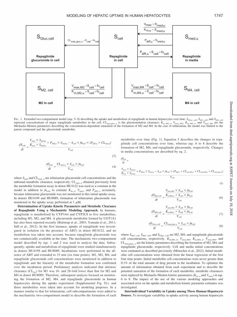
Simultaneous Modeling of Telmisartan Uptake and Metabolism. Inhumans, telmisartan is transformed to a single glucuronide metabolite byUGT1A3 (Stangier et al., 2000; Yamada et al., 2011). Because no specificinhibitor of UGTs is available, modeling of telmisartan uptake and metabolismwas performed simultaneously. The mechanistic two-compartment model wasextended to account for the formation of telmisartan glucuronide over time, asillustrated in Fig. 1. Equation 3 describes the changes in telmisartan cellconcentrations during the uptake experiment, whereas changes in telmisartanglucuronide cell concentrations are defined by eq. 4. Changes in mediaconcentrations over time were defined as shown above in eq. 2.
TABLE 1
Uptake kinetic parameters of seven OATP substrates estimated in human hepatocyte using a mechanistic two-compartment model
Uptake kinetics was measured in cryopreserved human hepatocytes (donor HU4122) plated for 6 h over 2 min at seven concentrations (0.1–300 �M).
Drug Km, u Vmax Pdiff,u fucella fumed CLactive, u
�M pmol � min�1 � 106 cells�1 �l � min�1 � 106 cells�1 �l � min�1 � 106 cells�1
Bosentan 22.5 402 9.74 0.096 0.85 17.9Pitavastatin 1.59 65 13.2 0.053 0.63 40.7Pravastatin 2.25 6.24 0.208 0.963 0.91 2.77Repaglinide 15.6 1236 10.0 0.074 0.95 79.0Rosuvastatin 11.2 104 0.391 0.477 0.93 9.21Telmisartanb 2.03 193 14.1 0.024 0.71 95.2Valsartan 10.4 30 0.064 1 1 2.88
a Data from uptake experiments performed in rat hepatocytes (Ménochet et al., 2012).b Kinetics parameters obtained when telmisartan metabolism was incorporated in the modeling (CLmet � 4.3 �l � min�1 � 106 cells�1).
1746 MENOCHET ET AL.
at ASPE
T Journals on July 10, 2018
dmd.aspetjournals.org
Dow
nloaded from
dScell
dt�
Vmax � Smed,u
Km,u � Smed,u� Pdiff,u � Smed,u � Scell � fucell � �Pdiff,u � CLmet,u
Vcell
(3)
dSmet
dt�
CLmet,u � Scell � fucell
Vcell(4)
where Smet and CLmet, u are telmisartan glucuronide cell concentrations and theunbound metabolic clearance, respectively. CLmet, u obtained previously fromthe metabolite formation assay in donor HU4122 was used as a constant in themodel in addition to fucell to estimate Km, u, Vmax, and Pdiff, u accurately,because telmisartan glucuronide was not monitored in this initial uptake assay.In donors HU4199 and HU8089, formation of telmisartan glucuronide wasmonitored in the uptake assay performed at 1 �M.
Determination of Uptake Kinetic Parameters and Metabolic Clearanceof Repaglinide Using a Mechanistic Modeling Approach. In humans,repaglinide is metabolized by CYP3A4 and CYP2C8 to five metabolites,including M1, M2, and M4. A glucuronide metabolite formed by UGT1A1has also been reported recently (Bidstrup et al., 2003; Yamada et al., 2011;Sall et al., 2012). In the first instance, uptake of repaglinide was investi-gated in isolation (in the presence of ABT) in donor HU4122, and nometabolism was taken into account, because repaglinide glucuronide wasnot commercially available at the time. The mechanistic two-compartmentmodel described by eqs. 1 and 2 was used to analyze the data. Subse-quently, uptake and metabolism of repaglinide were studied simultaneouslyin donors HU4199 and HU8089. Incubations were performed in the ab-sence of ABT and extended to 15 min (six time points). M1, M2, M4, andrepaglinide glucuronide cell concentrations were monitored in addition torepaglinide and the linearity of the metabolite formation was confirmedover the incubation period. Preliminary analysis indicated that intrinsicclearance (CLint) for M1 was 16- and 28-fold lower than that for M2 andM4 in donor HU8089. Therefore, subsequent analysis focused on monitor-ing the formation of M2, M4, and repaglinide glucuronide in humanhepatocytes during the uptake experiment (Supplemental Fig. S1), andthese metabolites were taken into account for modeling purposes. In amanner similar to that for telmisartan, cell subcompartments were added tothe mechanistic two-compartment model to describe the formation of each
metabolite over time (Fig. 1). Equation 5 describes the changes in repa-glinide cell concentrations over time, whereas eqs. 6 to 8 describe theformation of M2, M4, and repaglinide glucuronide, respectively. Changesin media concentrations are described by eq. 2.
dScell
dt�
Vmax � Smed,u
Km,u � Smed,u� Pdiff,u � Smed,u � Scell � fucell � �Pdiff,u � CLmet,M2,u � CLmet,M4,u � CLmet,glue,u
Vcell
(5)
dSM2,cell
dt�
Vmax,M2 � Scell � fucell
Km,M2,u � Scell � fucell
Vcell(6)
dSM4,cell
dt�
Vmax,M4 � Scell � fucell
Km,M4,u � Scell � fucell
Vcell(7)
dSGluc,cell
dt�
CLmet,gluc,u � Scell � fucell
Vcell(8)
where SM2, cell, SM4, cell, and SGluc, cell are M2, M4, and repaglinide glucuronidecell concentrations, respectively. Km,M2, u, Vmax,M2, Km,M4, u, Vmax,M4, andCLmet,gluc, u are the kinetic parameters describing the formation of M2, M4, andrepaglinide glucuronide, respectively. Cell and media initial concentrationswere estimated as described previously (Menochet et al., 2012). Initial metab-olite cell concentrations were obtained from the linear regression of the firstfour time points. Initial metabolite cell concentrations were never greater than0.1% of the total amount of drug present in the incubation. To optimize theamount of information obtained from each experiment and to describe thepotential saturation of the formation of each metabolite, metabolic clearanceswere replaced by Michaelis-Menten kinetic parameters (Km, u and Vmax) in eqs.6 to 8. The impact of the use of the various modeling approaches andassociated error on the uptake and metabolism kinetic parameter estimates wasinvestigated.
Interindividual Variability in Uptake among Three Human HepatocyteDonors. To investigate variability in uptake activity among human hepatocyte
Repaglinide in cell
Repaglinide in media
Repaglinide glucuronide in cell
Scell Smed,uSGluc,cell
SM4, cell
M4 in cell
SM2, cell
M2 in cell
××
+
×
×
××
×+
××
×+
××
FIG. 1. Extended two-compartment model (eqs. 5–8) describing the uptake and metabolism of repaglinide in human hepatocytes over time. SGluc, cell, SM2, cell, and SM4, cell
represent concentrations of major repaglinide metabolites in the cell. CLmet,gluc, u is the glucuronidation clearance; Km, M2, u, Vmax, M2, Km, M4, u, and Vmax, M4 are theMichaelis-Menten parameters describing the concentration-dependent saturation of the formation of M2 and M4. In the case of telmisartan, the model was limited to theparent compound and the glucuronide metabolite.
1747MODELING OF HEPATIC UPTAKE IN HUMAN HEPATOCYTES
at ASPE
T Journals on July 10, 2018
dmd.aspetjournals.org
Dow
nloaded from

donors, uptake of rosuvastatin and repaglinide was measured in three humanhepatocyte donors over a range of concentrations, following mechanisticmodeling data analysis, as described in sections above. For the remainingdrugs in the dataset (pravastatin, valsartan, bosentan, and pitavastatin) uptakewas measured over 2 min in donors HU4199 and HU8089 using a singlesubstrate concentration. On the basis of full kinetic characterization dataobtained for rosuvastatin and repaglinide in three donors, Km, u and Pdiff, u
values were assumed to be constant among donors (Tables 2 and 3). Themechanistic model defined in eq. 1 was therefore used to fit the Vmax using thedata obtained over 2 min at a single substrate concentration, keeping Km, u,Pdiff, u, and fucell constant (as obtained in donor HU4122). The fitting routinewas performed in Matlab using the lsqnonlin function; the differential equa-tions were solved using the ODE45 solver. An analogous method was appliedfor telmisartan, but in this case metabolic clearance was also taken into accountin the fitting routine as shown in eq. 3. Subsequently, CLactive, u for theremaining five drugs could be calculated as the Vmax/Km, u ratio. To verify thevalidity of the assumptions stated above, CLactive, u was calculated for eachdrug in donor HU4122 using the single concentration (1 �M) method and comparedwith the CLactive, u values obtained from the full kinetic characterization.
Statistical Analysis. A Jacobian approach was used in Matlab to estimateS.E. associated with each parameter estimate generated from the mecha-nistic two-compartment model (Landaw and DiStefano, 1984). CVs werecalculated and are expressed as percentages to assess the quality of eachparameter estimate. Goodness of fit was assessed by visual inspection ofthe data and minimum objective function value. Arithmetic means, S.E.s,and CVs associated with each kinetic parameter were calculated for eachdrug when results in the three human hepatocyte donors were available. pvalues were calculated using a Student’s t test, and results were deemedsignificant for p � 0.05.
Collation of Uptake Measurement Data for Estrone-3-Sulfate in Hu-man Hepatocytes from Commercial Supplier. Uptake rate values of estrone-3-sulfate in human cryopreserved hepatocytes were collated from the charac-terization spreadsheets available from BD Gentest (Oxford, UK) betweenSeptember 2008 and September 2011 (http://www.bdbiosciences.com/nvCategory.jsp?action�SELECT&form�formTree_catBean&item�774729).
Uptake was measured at 2 �M over 3 min using 200,000 human hepatocytesin suspension per incubation. By using the uptake rates provided, uptakeclearances were calculated from the ratio of the uptake rate over the substrateconcentration to allow comparison with data obtained in the three humanhepatocyte donors studied here. Demographic information (age and gender) foreach donor available from BD Gentest was also collated, and data were assessedfor any trends. Collated data are summarized in Supplemental Table S1.
Prediction of Hepatic Clearance in Human. In vivo clearance was pre-dicted using the unbound intrinsic clearance (CLint, u) term derived previously(Shitara and Sugiyama, 2006), as described in eq. 9:
CLint,u � �CLactive,u � Pdiff,u �CLmet,u � CLbile,u
CLmet,u � CLbile,u � Pdiff,u(9)
CLactive, u, CLmet, u, and Pdiff, u represent the total unbound uptake clearance,the unbound metabolic clearance, and the passive diffusion clearance; allparameters were obtained experimentally and scaled up to a whole liver usingphysiological scaling factors and are expressed in milliliters per minute perkilogram. Hepatocellularity and liver weight values of 120 � 106 cells/g liverand 21.4 g of liver/kg b.wt., respectively, were used (Brown et al., 2007).CLbile, u is the unbound biliary clearance and was obtained by multiplying thefraction of the dose recovered unchanged in the feces after intravenous ad-ministration by the unbound total plasma clearance. In the case of bosentan,CLmet, u was obtained from the literature (Lave et al., 1996). Details for theindividual drugs are provided in Supplemental Table S2.
Values obtained from in vitro results were compared with the in vivointrinsic clearances estimated from the literature data and are expressed asdescribed in eq. 10:
CLint,h �CLh
fub � �1 �CLh
Qh� (10)
where CLh is the hepatic blood clearance after intravenous administrationcorrected for renal clearance, Qh is hepatic blood flow, and fub is the unboundfraction in the blood. No CLh was available for pitavastatin; therefore, this drugwas omitted from the comparison between predicted and observed hepaticclearances.
Preliminary in vitro-in vivo extrapolation has shown underprediction of theintrinsic hepatic clearance. Therefore, empirical scaling factors required forCLactive, u were calculated for each drug in all three donors. In the first instance,a mean scaling factor per individual donor was used, and the three individualempirical scaling factors were then applied to correct corresponding CLactive, u
in that particular donor. In addition, the average empirical scaling factorobtained for all drugs in all three donors (16 data points in total) was calculatedand applied to correct CLactive, u. The accuracy and bias associated with eachpredictive approach were compared by calculation of the root mean squareerror (rmse) and the geometric mean fold error (gmfe) (Gertz et al., 2010), asdescribed in eqs. 11 and 12, respectively.
rmse � �1
N��predicted � observed2 (11)
gmfe � 10
1N�� log
PredictedObserved� (12)
Comparison of Uptake Data Obtained in Rat and Human Hepatocytes.Uptake data obtained in three human hepatocyte donors were compared
TABLE 2
Uptake and metabolism kinetic parameters of repaglinide estimated in threehuman hepatocyte donors using an extended mechanistic two-compartment model
Uptake kinetics was measured in cryopreserved human hepatocytes plated for 6 h at sevenconcentrations (0.1–300 �M) in the presence (HU4122) or absence (HU4199 and HU8089) of1 mM ABT.
Donor HU4122 HU4199 HU8089 Mean
Km, u, �M 15.6 8.97 13.8 12.8 � 3.4Vmax, pmol � min�1 � 106 cells�1 1236 310 464 670 � 496Pdiff, u, �l � min�1 � 106 cells�1 10.0 16.0 12.0 12.7 � 3.1fucell
a 0.074 0.074 0.074 0.074fumed 0.95 0.94 0.96 0.95 � 0.01CLactive, u, �l � min�1 � 106 cells�1 79.0 34.6 33.6 49.1 � 25.9CLmet, gluc, u, �l � min�1 � 106 cells�1 0.412 0.242 0.327Km, M2, u, �M 41.5 7.40 24.4CLmet, M2, u, �l � min�1 � 106 cells�1 0.950 2.42 1.68Km, M4, u, �M 5.32 8.83 7.07CLmet, M4, u, �l � min�1 � 106 cells�1 0.619 1.29 0.957CLmet,t otal, u, �l � min�1 � 106 cells�1 1.98 3.96 2.97
a Data from uptake experiments performed in rat hepatocytes over an extended incubationtime (Ménochet et al., 2012).
TABLE 3
Uptake kinetic parameters of rosuvastatin estimated in three human hepatocyte donors using a mechanistic two-compartment model
Uptake kinetics was measured in cryopreserved human hepatocytes plated for 6 h over 2 min at seven concentrations (0.1–300 �M).
Donor Km, u Vmax Pdiff, u fumed CLactive, u
�M pmol � min�1 � 106 cells�1 �l � min�1 � 106 cells�1 �l � min�1 � 106 cells�1
HU4122 11.2 104 0.391 0.80 9.21HU4199 9.80 16.6 0.148 1 1.69HU8089 12.0 16.3 0.243 0.96 1.35Mean � S.D. 11.0 � 1.1 45.5 � 50.3 0.261 � 0.123 0.93 � 0.13 4.09 � 4.44
1748 MENOCHET ET AL.
at ASPE
T Journals on July 10, 2018
dmd.aspetjournals.org
Dow
nloaded from

with the results generated previously in rat hepatocytes for the same sevendrugs using a similar experimental and modeling approach (Menochet etal., 2012); data for the distribution coefficient between octanol and waterat pH 7.4 (LogD7.4) were also taken from previous publications. Folddifference in Km, u, CLactive, u, and Pdiff, u were calculated and interspeciesdifferences were considered.
Results
Mechanistic Modeling of Uptake in Donor HU4122. Full uptakekinetics of seven drugs was investigated in human hepatocyte donorHU4122 using a mechanistic modeling approach defined previously.Km, u, Vmax, and Pdiff, u were estimated from the data generated over2-min incubations and are summarized in Table 1. Correspondingkinetic profiles and goodness of fit plots obtained with this model areshown in Supplemental Figs. S2 and S3, respectively. Km, u valuesranged 14-fold, from 1.59 to 22.5 �M for pitavastatin and bosentan,respectively. A greater difference in Vmax among the drugs investi-gated resulted in a 34-fold range in CLactive, u. Pravastatin showed thelowest active uptake clearance (2.77 �l � min�1 � 106 cells�1),whereas telmisartan was the drug most readily taken up by the cellwith CLactive, u of 95.2 �l � min�1 � 106 cells�1. Pdiff, u was the kineticparameter that differed the most among the seven drugs, with a rangeof 222-fold, from 0.064 to 14.1 �l � min�1 � 106 cells�1 for valsartanand telmisartan, respectively.
All media concentrations were corrected for nonspecific binding tothe experimental support; fumed was �0.6 for all drugs investigatedin the current dataset. Nonspecific binding had a minimal influenceon the uptake kinetics of all drugs of interest and most specificallyon rosuvastatin, repaglinide, and pravastatin, which showed fumed
values �0.9. Use of fucell values derived from rat studies allowedmodeling of human hepatocyte uptake data and accurate estimationof all kinetic parameters for the drugs in the current dataset (CV�30%), with the exception of the Km, u and Vmax values forpravastatin (CVs 194 and 36%, respectively). fucell values rangedfrom 0.024 to 1 for telmisartan and valsartan, respectively. Inter-experiment variability of rosuvastatin and estrone-3-sulfate uptakeused as controls was 39 and 40%, respectively (n � 8, donorHU4122).
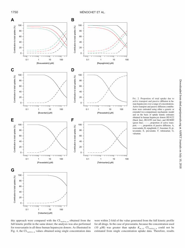
Differing extents of active uptake and passive diffusion were ob-served among the seven drugs investigated, as shown in Fig. 2. Onlyin the case of rosuvastatin, repaglinide, and valsartan was the activeprocess responsible for �50% of the total uptake over the full rangeof concentrations investigated. However, at the lowest concentrationsstudied, the maximal proportion of total uptake due to the activeprocess was �90% for all the drugs investigated. Thus, at low,therapeutically relevant concentrations, the passive permeation is ex-pected to have a limited impact on total uptake into the hepatocytes ofthe drugs investigated. Analogous to rat hepatocytes, a positive linearcorrelation was observed between the logarithm of the passive diffu-sion clearance and LogD7.4 (R2 � 0.886) (Fig. 8C).
Simultaneous Modeling of Uptake and Metabolism of Repaglin-ide. Uptake and metabolism of repaglinide were studied in threehuman hepatocyte donors. The kinetic estimates are summarized inTable 2. In donor HU4122, uptake of repaglinide was investigated inthe presence of 1 mM ABT to inhibit phase I metabolism and allowonly uptake to be characterized in the modeling process. The repa-glinide fucell value of 0.074 was held constant in the mechanisticmodel and is based on values obtained previously in rat hepatocytes.In donors HU4199 and HU8089, the uptake of repaglinide was studiedin the absence of ABT and the formation of three metabolites wasincorporated in the modeling. Repaglinide cell concentrations werefitted with good precision in all cases, as illustrated in the diagnostic
goodness-of-fit plots shown in the Supplemental Fig. S3B. UptakeKm, u of repaglinide was moderate (12.8 � 3.4 �M) and consistentamong the three hepatocyte donors. Pdiff, u was among the highestvalues measured in the current dataset (12.7 � 3.1 �l � min�1 � 106
cells�1), and no significant difference was observed in this parameteracross the donors investigated. Variation in uptake activity resulted inup to 2.4-fold difference between CLactive, u values measured inHU4122 and the additional donors (HU4199 and HU8089) (Fig. 3A).Differences in uptake were reflected in the proportion of total uptakedue to active transport over the range of concentrations. In HU4122,active uptake was the predominant process responsible for total up-take over the range of concentrations studied. In HU4199 andHU8089, contribution of the active process of �70% was observedonly for concentrations �10 �M (Fig. 2).
The kinetics of repaglinide metabolite formation was also investi-gated in donors HU4199 and HU8089. Because the uptake experimentwas performed without ABT, M2, M4, and repaglinide glucuronidewere monitored (Supplemental Fig. S1). Kinetics of repaglinide gluc-uronide formation was best described by a single clearance parameter,whereas formation of M2 and M4 was defined by Michaelis-Mentenparameters used to describe potential saturation of metabolism overthe range of concentrations studied. Clearance due to the formation ofrepaglinide glucuronide was low, contributing 21 and 6% to totalmetabolism in donors HU4199 and HU8089, respectively. Km, u valuefor the M2 pathway was 6-fold greater in HU4199 than in HU8089.However, this resulted in only 2.5-fold greater CLmet,M2, u, due todifferences in metabolic capacity between the two donors. Overall,metabolic clearances in HU4199 and HU8089 were 1.98 and 3.96 �l �min�1 � 106 cells�1, respectively. This was lower than the valueobtained in donor HU4122 when measured in a separate depletion assay(6.61 �l � min�1 � 106 cells�1; data not shown). Uptake clearances were17- and 8.5-fold greater than metabolism for donor HU8089 andHU4199, respectively. Km,M4, u was consistently lower than the uptakeKm, u by on average 2-fold, but the difference in Km,M2, u was less clearbecause of donor variation. CLmet, M2 was greater in comparison with themetabolic clearances of other metabolites formed, consistent with recentstudies in pooled cryopreserved hepatocytes, in which repaglinide me-tabolism was monitored in isolation (Sall et al., 2012).
Interindividual Variability in Uptake and Metabolism Activitybetween Human Hepatocyte Donors. Analysis of uptake kineticdata showed Km to be the most consistent parameter among the threedonors with CVs of 10 and 27% for rosuvastatin and repaglinide,respectively (Tables 2 and 3). Pdiff, u was also similar among the threedonors (CVs 47 and 24% for rosuvastatin and repaglinide, respec-tively), consistent with the reliance of passive permeation on lipophi-licity of the drugs studied. However, for both drugs, uptake activityvaried among the donors, because HU4122 uptake activity was up to2.4- and 6.8-fold greater than activity observed in the two otherdonors for repaglinide and rosuvastatin, respectively (Fig. 3).
To explore the use of uptake estimates obtained at a single lowconcentration in donors HU4199 and HU8089, CLactive, u for pitavas-tatin, valsartan, bosentan, pravastatin, and telmisartan was estimatedusing the uptake rate data generated at 1 �M (10 �M for pravastatin)over 2 min only. Uptake Km and Pdiff were assumed to be consistentamong donors on the basis of full kinetic profiles for repaglinide androsuvastatin. Keeping these parameters constant in the mechanisticmodel allowed fitting of the Vmax and subsequent estimation of theCLactive, u. This approach allowed the delineation of active and pas-sive uptake from a single concentration experiment in a mechanisticmanner, using only a fraction of the number of cells required toinvestigate the full uptake kinetics of a drug. For validation purposes,the same analysis was performed in HU4122, and estimates based on
1749MODELING OF HEPATIC UPTAKE IN HUMAN HEPATOCYTES
at ASPE
T Journals on July 10, 2018
dmd.aspetjournals.org
Dow
nloaded from
this approach were compared with the CLactive, u obtained from thefull kinetic profile in the same donor; the analysis was also performedfor rosuvastatin in all three human hepatocyte donors. As illustrated inFig. 4, the CLactive, u values obtained using single concentration data
were within 2-fold of the value generated from the full kinetic profilefor all drugs. In the case of pravastatin, because the concentration used(10 �M) was greater than uptake Km, u, CLactive, u could not beestimated from single concentration uptake data. Therefore, results
A B
C D
E F
G
[Rosuvastatin] (µM)
0.1 1 10 100
Con
tribu
tion
to to
tal u
ptak
e (%
)
0
20
40
60
80
100
[Repaglinide] (µM)
0.1 1 10 100
Con
tribu
tion
to to
tal u
ptak
e (%
)
0
20
40
60
80
100
[Bosentan] (µM)
0.1 1 10 100
Con
tribu
tion
to to
tal u
ptak
e (%
)
0
20
40
60
80
100
[Pitavastatin] (µM)
0.1 1 10 100
Con
tribu
tion
to to
tal u
ptak
e (%
)
0
20
40
60
80
100
[Pravastatin] (µM)
0.1 1 10 100
Con
tribu
tion
to to
tal u
ptak
e (%
)
0
20
40
60
80
100
[Telmisartan] (µM)
0.1 1 10 100
Con
tribu
tion
to to
tal u
ptak
e (%
)
0
20
40
60
80
100
[Valsartan] (µM)
0.1 1 10 100
Con
tribu
tion
to to
tal u
ptak
e (%
)
0
20
40
60
80
100
FIG. 2. Proportion of total uptake due toactive transport and passive diffusion in hu-man hepatocytes over a range of concentrations.Active transport and passive diffusion contribu-tions were estimated using either a generic orextended two-compartment mechanistic modeland on the basis of uptake kinetic estimatesobtained in human hepatocyte donors HU4122(black line), HU4199 (red line), and HU8089(green line). ——, proportion of active trans-port; – – –, proportion of passive diffusion. A,rosuvastatin; B, repaglinide; C, bosentan; D, pi-tavastatin; E, pravastatin; F, telmisartan; G,valsartan.
1750 MENOCHET ET AL.
at ASPE
T Journals on July 10, 2018
dmd.aspetjournals.org
Dow
nloaded from

presented for this drug are solely based on the full kinetic experimentperformed in donor HU4122. Precision of the estimated uptake clear-ance obtained at the single concentration varied from 22% lower forrosuvastatin in donor HU8089 to 47% higher for bosentan in donorHU4122, but overall no significant bias was observed (gmfe of 1.2).This approach was therefore considered suitable to estimate CLactive, u
in donors HU4199 and HU8089 from single low concentration data.Donor HU4122 showed higher uptake activity than HU4199 and
HU8089 for all drugs investigated, with the exception of bosentan andpitavastatin (Fig. 5). On average, uptake clearance in this donor was2.3- and 2.8-fold greater than that in HU4199 and HU8089, respec-tively. The greatest difference between donors was observed forrosuvastatin. For this drug, uptake clearance in HU4122 was 5.4- and6.8-fold greater than that in HU4199 and HU8089. In contrast, uptakeof telmisartan was consistent among the three donors, with a CV ofonly 7.8%. Overall, HU4199 and HU8089 showed similar levels ofuptake activity. Uptake clearances in these donors were within 30% ofeach other for all drugs, with the exception of pitavastatin and bosen-tan where uptake clearances in HU4199 were 1.9-fold greater thanthose in HU8089. This difference among the three donors was re-flected in the uptake of rosuvastatin when this drug was used as apositive control at 1 �M (Fig. 6A). However, the use of estrone-3-sulfate at 1 �M did not distinguish the three donors in their uptakeactivity, because the differences seen were not statistically significant(p � 0.35) (Fig. 6B). In addition to current data, uptake clearancevalues for estrone-3-sulfate were collated from BD Gentest charac-
terization spreadsheets for 28 individual human hepatocyte donors(details shown in Supplemental Table S1). Clearances ranged from 4to 35.5 �l � min�1 � 106 cells�1 with a mean clearance of 21.4 � 8.5�l � min�1 � 106 cells�1 (CV 40%). No correlation was observedbetween uptake activity and either gender or age for this dataset(Supplemental Fig. S4). Mean CLuptake of estrone-3-sulfate in thecurrent study (19.5 � 7.7 �l � min�1 � 106 cells�1) was comparable tothe mean value obtained from this larger dataset (p � 0.48).
Metabolism of telmisartan was studied in all three donors. Unboundmetabolic clearance of telmisartan in HU4122 was 22-fold lower thanthe active uptake clearance (Table 1). However, metabolic clearanceobtained in this donor was 6-fold greater than the values measured inthe other two donors (4.3 versus 0.7 �l � min�1 � 106 cells�1,respectively). Although this difference could be due to variability inmetabolic capacity between the donors, it is noteworthy that telmis-artan glucuronide was monitored in the entire incubation (cell andmedia) and in the metabolite formation assay performed in donorHU4122, whereas it was only measured in the cell in the uptake assayin donors HU4199 and HU8089, because this metabolite could not bedetected in the media.
Prediction of Hepatic Clearance in Human. Comparison of theobserved and predicted hepatic intrinsic clearances for six of the drugsinvestigated is shown in Fig. 7. The importance of active uptake on
A B
[Repaglinide] (µM)
0 20 40 60 80 100
Tota
l upt
ake
rate
in h
uman
he
pato
cyte
s (p
mol
/min
/106 c
ells
)
0
200
400
600
800
1000
1200
1400
1600
[Rosuvastatin] (µM)
0 100 200 300
Tota
l upt
ake
rate
in h
uman
he
pato
cyte
s (p
mol
/min
/106 c
ells
)
020406080
100120140160180200 FIG. 3. Total uptake kinetic profiles of re-
paglinide (A) and rosuvastatin (B) mea-sured in three batches of cryopreserved hu-man hepatocyte plated for 6 h. Datarepresent the mean of uptake rates mea-sured in duplicate at 2 min in donorHU4122 (F), HU4199 (E) and HU8089( ). Six concentrations were used for repa-glinide (0.1–100 �M) and seven for rosuv-astatin (0.1–300 �M). Lines represent thesimulated total uptake based on uptake ki-netic estimates obtained from the two-com-partment mechanistic model, including me-tabolism in the case of repaglinide.
Observed CLactive,u from full kinetic profile (µL/min/106 cells)
0 20 40 60 80 100
Sin
gle
conc
entra
tion/
Full k
inet
ic
prof
ile C
Lact
ive,
u es
timat
es
00.20.40.60.8
11.21.41.61.8
2
FIG. 4. Comparison between uptake clearances obtained from full kinetic experi-ments in human hepatocytes and the value estimated on the basis of data generatedat 1 �M only using mechanistic model.
BosentanPitavastatin
RepaglinideRosuvastatin
TelmisartanValsartan
0
10
20
30
40
50
60
70
80
90
100
CL up
take
,u (µ
L/m
in/1
06 cel
ls)
FIG. 5. Active uptake clearance of six OATP substrates in human hepatocytedonors generated using a full kinetic approach (F, HU4122) or extrapolated frommeasurements at 1 �M (E, HU4199; , HU8089). Line represents the mean of thethree clearance estimates.
1751MODELING OF HEPATIC UPTAKE IN HUMAN HEPATOCYTES
at ASPE
T Journals on July 10, 2018
dmd.aspetjournals.org
Dow
nloaded from

the prediction of CLint, h was drug-dependent. In the case of prava-statin, rosuvastatin, telmisartan, and valsartan, hepatic clearance wasmainly driven by CLactive, u, because the combined CLmet, u andCLbile, u were much greater than Pdiff, u. For bosentan and repaglinide,CLint, u was a composite of a number of processes, because passivediffusion was greater than metabolic and biliary clearances combined.In vitro-in vivo extrapolation of these data resulted in systematicunderprediction of hepatic intrinsic clearance for all drugs in thedataset, with the exception of valsartan, which was predicted within2-fold of the observed data. The extent of the underprediction wascompound- and donor-dependent, ranging from 5-fold for pravastatinin donor HU4122 to 66-fold for rosuvastatin in donor HU8089.Hence, the empirical scaling factors for CLactive per individual donorwere necessary to recover the observed clearance in vivo and alsoresulted in a large variation (Table 4). For example, clearances indonor HU4122 were corrected by a factor of 6.9 on average, whereasvalues of 25.0 and 25.6 were required in donors HU4199 andHU8089, respectively. Nine of 16 CLint, u values were predictedwithin 2-fold of the observed data (Fig. 7A). The approach involvingempirical scaling factors resulted in low overall bias (2.3), but poorprecision (rmse of 801).
In addition, the whole dataset was considered to accommodate bothdrug and donor variability, resulting in an average empirical scalingfactor of 17.1. Use of this approach resulted in 8 of 16 valuespredicted within 2-fold, improved precision (rmse of 471), and biascomparable to that for the previous scenario (gmfe of 2.52) (Fig. 7B).In all analyses, predicted valsartan CLint, u was more than 5-foldgreater than the observed CLint, h value. In general, use of the averagedataset scaling factor resulted in an overprediction of clearance whendata from donor HU4122 were used (with the exception of bosentan)and underprediction when clearances from donors HU4199 andHU8089 were used.
Comparison between Uptake in Rat and Human Hepatocytes.Uptake kinetic parameters of the seven drugs investigated in thepresent study were estimated previously in rat hepatocytes using amechanistic modeling approach (Menochet et al., 2012). Km, u valueswere on average 4.3-fold greater in rat than in human hepatocytes,mainly because of the higher affinity of pitavastatin and pravastatintoward human transporters (labeled as 1 and 2 in Fig. 8A). Valsartanand bosentan were the only drugs in the dataset showing the oppositetrend with higher affinity in rat hepatocytes. Pitavastatin had thelowest Km, u in human hepatocytes (1.59 �M), whereas telmisartanshowed the highest affinity in rat hepatocytes (3.4 �M). Pravastatinand bosentan showed the lowest affinity toward rat and human trans-porters with Km, u of 37.0 and 22.5 �M in rat and human hepatocytes,respectively. Less than a 2-fold difference was observed betweenKm, u in rat and human hepatocytes for telmisartan, rosuvastatin,valsartan, and bosentan. Overall, the drugs studied covered a similarrange of affinity in both species: 11-fold versus 14-fold in rat andhuman, respectively. Despite the greater affinity observed in humanhepatocytes, uptake clearances were on average 7.1-fold greater in rathepatocytes than in human hepatocytes, due to a large difference inactivity between the two species (Fig. 8B). The greatest differencewas observed for valsartan and rosuvastatin for which CLactive, u
values were 16- and 21-fold greater in rat than in human hepatocytes,respectively. All other drugs had CLactive, u values within 5-fold ofeach other in the two species. The lowest uptake clearance wasobserved for valsartan in human hepatocytes (1.64 �l � min�1 � 106
cells�1 in HU8089) and for pravastatin in rat hepatocytes (12.5 �l �min�1 � 106 cells�1). Telmisartan showed the greatest extent of activeuptake in human hepatocytes with CLactive, u of 95.2 �l � min�1 � 106,whereas active uptake of repaglinide was the greatest in rat hepato-cytes (119 �l � min�1 � 106 cells�1). Therefore, individual CLactive, u
covered a wider range in human than in rat hepatocytes: 58-fold
A B
HU4122 HU4199 HU80890
2
4
6
8
10
12
Rosu
vast
atin
upt
ake
clear
ance
at 1
µM
(µL/
min
/106 c
ells)
HU4122 HU4199 HU80890
5
10
15
20
25
30
35
Estro
ne-3
-sul
fate
upt
ake
clear
ance
at 1
µM
(µ
L/m
in/1
06 cel
ls)
FIG. 6. Uptake clearances of rosuvastatin(A) and estrone-3-sulfate (B) measured as acontrol in human hepatocyte donorsHU4122 (n � 8), HU4199 (n � 3), andHU8089 (n � 4) at 1 �M. ——, representsthe median; �, represents the mean. Thebox represents the 25th and 75th percen-tiles; whiskers are the 5th and 95th percen-tiles.
A B
CLint,h in vivo (mL/min/kg)
1 10 100 1000 10000
Pre
dict
ed C
Lint
,u (m
L/m
in/k
g)
1
10
100
1000
10000
CLint,h in vivo (mL/min/kg)
1 10 100 1000 10000
Pre
dict
ed C
Lint
,u (m
L/m
in/k
g)
1
10
100
1000
10000 FIG. 7. Comparison of predicted and ob-served hepatic clearances for the drugs in-vestigated. Scaling factors were based ei-ther on the donor-specific (A) or empiricalscaling factors obtained using the wholedataset (B). Hepatic clearance was pre-dicted from uptake, metabolic and passivediffusion clearances obtained in three hu-man hepatocyte donors, and biliary clear-ance values reported in the literature. F,bosentan; E, pravastatin; f, repaglinide;�, rosuvastatin; Œ, telmisartan; ‚, valsar-tan. ——, line of unity. – – –, 2-fold differ-ence; - - - - -, 5-fold difference.
1752 MENOCHET ET AL.
at ASPE
T Journals on July 10, 2018
dmd.aspetjournals.org
Dow
nloaded from

versus 8-fold, respectively. As anticipated, interindividual variabilityin uptake clearance was greater in human hepatocytes than in rathepatocytes for all drugs excluding telmisartan.
Unlike active uptake, passive diffusion clearance was consistentbetween the two species, with Pdiff, u in rat being only 1.7-fold greateron average than in human hepatocytes (Fig. 8C). However, Pdiff, u
values obtained for the poorly permeable compounds were muchlower in human than in rat cells, whereas the Pdiff, u values at thehigher end were comparable between the two species (21.4 and 14.1�l � min�1 � 106 cells�1 for telmisartan in rat and human hepatocytes,respectively); Pdiff, u values in human span 222-fold, whereas theyonly covered a 60-fold range in rat hepatocytes. Pdiff, u in humanhepatocytes was positively correlated with LogD7.4, consistent withthe previous analyses in rat hepatocytes. At low concentrations, activetransport was the major process driving total uptake for all drugs inboth species.
A large difference in telmisartan metabolism was observed betweenrat and human hepatocytes; glucuronidation clearance was 32-foldgreater in rat hepatocytes than in human hepatocytes. Metabolic
clearance was only 4-fold lower than active uptake in rat, whereas thedifference between these two processes was up to 71-fold in humanhepatocytes, depending on the individual donor. In contrast, repaglin-ide glucuronidation and overall metabolism data were more consistentbetween the two species in comparison to telmisartan. However,species differences were evident, because the formation of M2 wasincreased by 5-fold between rat and human hepatocytes. In addition,M4 metabolite was not identified as a major metabolite in rat hepa-tocytes, but a clearance of 1.26 �l � min�1 � 106 cells�1 was measuredin human. These differences resulted in a repaglinide total metabolicclearance in humans 4-fold greater than that in rat. Total metabolicclearance was lower than the active uptake in either species; however,this difference was 13-fold on average in human hepatocytes incontrast to 112-fold reported for the rat data (Menochet et al., 2012),probably reflecting the differences in uptake activity between the twospecies.
Discussion
A mechanistic two-compartment model, previously developed inrat hepatocytes was applied here for the analysis of human hepatocytedata, allowing description of the interplay between active uptake,passive diffusion, intracellular binding, and metabolism. Uptake ki-netics of seven OATP substrates was investigated in three humanhepatocyte donors using a standardized experimental method andmodeling approach. The data generated were applied for the predic-tion of in vivo hepatic clearance and comparison with previouslyobtained parameters in rat hepatocytes.
The seven drugs investigated showed a 14-fold range in Km, u
values (Fig. 8A), but because of greater variation in their uptakecapacity, mean CLactive, u ranged 54-fold (Fig. 8B). The CLactive, u
values presented here are generally consistent with literature data.However, higher uptake clearances have been reported for pitavastatinand valsartan (Hirano et al., 2004; Poirier et al., 2009), whereas the
A B
C
Km,u in human hepatocytes (µM)
1 10 100
Km
,u in
rat h
epat
ocyt
es (µ
M)
1
10
100
1
2
3
4
5
6
7
CLactive,u in human hepatocytes (µL/min/106 cells)
1 10 100
CLa
ctiv
e,u i
n ra
t hep
atoc
ytes
(µL/
min
/106 c
ells)
1
10
100
2
5
6
7 31
4
LogD7.4
-2 0 2 4
Log
Pdi
ff,u
-2
-1
0
1
2
Log Pdif f ,u = 0.6316 x LogD7.4 - 0.3143
62
5
4 37
1
FIG. 8. Comparison of the uptake affinity(A), active uptake clearance (B), and pas-sive diffusion clearance (C) of seven OATPsubstrates in rat and human hepatocytes.Km, u, CLactive, u, and Pdiff, u estimates in rathepatocytes were obtained from full kineticexperiments for all drugs. Data representthe mean � S.D. of three experiments.Km, u and Pdiff, u in human hepatocytes wereobtained from full kinetic experiments indonor HU4122, with the exception of rosu-vastatin and repaglinide, for which full ki-netic experiments were conducted in allthree human hepatocyte donors. CLactive, u
in humans is the mean � S.D. of estimatesobtained from the full kinetic experiment indonor HU4122 and extrapolated from ex-periments conducted at 1 �M in donorsHU4199 and HU8089. In A and B, ——,line of unity. – – –, 2-fold difference. In C,——, line of best fit between LogD7.4 andthe passive diffusion clearances generatedin human hepatocytes. F, data generated inhuman hepatocytes; E, data obtained in rathepatocytes. 1, pitavastatin; 2, pravastatin;3, telmisartan; 4, repaglinide; 5, rosuvasta-tin; 6, valsartan; 7, bosentan.
TABLE 4
Empirical scaling factors applied to the active uptake clearances to recover theintrinsic uptake clearance in human livers
DrugScaling Factor
MeanHU4122 HU4199 HU8089
Bosentan 16.1 13.0 24.8 17.9 � 6.1Pravastatin 6.67 ND ND 6.67Repaglinide 4.70 39.2 17.7 20.5 � 17.4Rosuvastatin 9.75 51.9 65.3 42.3 � 29.0Telmisartan 3.23 18.7 17.9 13.3 � 8.7Valsartan 0.740 2.00 2.20 1.64 � 0.79Mean 6.87 � 5.46 25.0 � 20.2 25.6 � 23.7 17.1 � 18.8
N.D., not determined, because CLuptake, u could not be measured accurately at a singleconcentration.
1753MODELING OF HEPATIC UPTAKE IN HUMAN HEPATOCYTES
at ASPE
T Journals on July 10, 2018
dmd.aspetjournals.org
Dow
nloaded from

opposite was observed for pravastatin and telmisartan (Nakai et al.,2001; Ishiguro et al., 2006). Discrepancies observed may be associ-ated with inconsistencies in the uptake experimental design (use ofsingle concentration versus full kinetic profile) and data analysisamong the studies published to date. Uptake activity and passivediffusion were both correlated with lipophilicity, as was the extent ofintracellular binding; however, no correlation could be establishedbetween CLuptake, u and Km, u, LogD7.4, or fucell. Unlike the approachtraditionally used to analyze in vitro uptake data, the mechanistictwo-compartment model provides insight into the effect of the bidi-rectional passive diffusion over a range of concentrations. At lowconcentrations, the passive diffusion had limited contribution to theoverall uptake for all drugs investigated (Fig. 2), which is of relevancebecause the unbound Cmax (after a therapeutic dose) of the drugsstudied ranges from 0.80 nM to 0.52 �M for pitavastatin and valsar-tan, respectively (Sunkara et al., 2007; Deng et al., 2008). However,because these drugs are administered orally, concentrations in thehepatic portal vein could be higher and the contribution of passiveprocesses may differ.
The fucell term in the mechanistic uptake model was based onstudies in rat hepatocytes after an extended incubation time (45–90min) to allow a steady state between intracellular concentration andmedia to be reached. Use of these fucell values allowed a reasonablyprecise estimation of kinetic parameters for all drugs (CV �30%, withthe exception of pravastatin), despite the reduced number of datapoints available for analysis compared with that for rat hepatocytes.This insight into the extent of intracellular binding also permitted thesimultaneous assessment of uptake and metabolism, giving a furtherunderstanding of the interplay between these two processes comparedwith previous studies (Paine et al., 2008; Poirier et al., 2008; Wa-tanabe et al., 2009). In the case of repaglinide, active uptake was onaverage 4.3- and 13-fold greater than passive diffusion and metabolicclearances, respectively. Consistent with the rat data, active uptakehas been identified as the major contributor to repaglinide overallclearance. However, a significant contribution of the passive compo-nent to the total uptake should not be neglected, as supported by onlya 2.4-fold increase in repaglinide area under the curve when coad-ministered with cyclosporine (potent OATP1B1 inhibitor) (Kajosaariet al., 2005; Amundsen et al., 2010). Considering that metabolismrepresents the rate-limiting process for repaglinide hepatic clearance,one may expect that the DDIs observed for this type of drug would bemetabolism driven. However, such interpretation of the current find-ings is inappropriate for inhibitors showing dual effects on uptaketransporters and metabolism, as illustrated in the repaglinide-gemfi-brozil/gemfibrozil glucuronide interaction, leading to a marked in-crease in repaglinide exposure (Niemi et al., 2003). The data analysispresented here provides a mechanistic description of the processesoccurring in the hepatocytes that should improve our ability to quan-titatively predict the risk of complex DDIs involving both uptake andmetabolism.
Analogous to Km, u, passive diffusion clearances showed minimaldonor dependence, although no correlation was evident between Km, u
and Pdiff. Consistency in these parameters across donors allowed theuse of the mechanistic two-compartment model to successfully esti-mate CLactive, u from experiments performed at a single concentration.Although this approach requires prior knowledge of Km, u and Pdiff, u,it permits the investigation of interindividual variability from a re-duced number of human cells, while allowing the mechanistic natureof the data analysis. Although donor HU4122 exhibited the greatestuptake capacity overall, drugs investigated showed different extent ofuptake, depending on the donor used. Rosuvastatin demonstrated thegreatest variability in uptake clearance between donors with a CV of
108% (Table 3), reflecting the variability in uptake Vmax (CV 111%).A similar trend was more evident for drugs with predominant activeuptake in the current dataset. A comparable degree of interindividualvariability has been reported previously (Ishiguro et al., 2006;Yamada et al., 2007). Whereas Yamada et al. (2007) found a variationof 10% in olmesartan uptake clearance among three donors, Ishiguroet al. (2006) found CVs as high as 68% when they investigated theclearance of estradiol-17�-glucuronide in three donors. Emergingabsolute quantification data of uptake transporter expression in anumber of human livers support this interindividual variability (Oht-suki et al., 2012). However, the extent to which this variability intransporter expression in the actual tissue relates to the interindividualvariability in uptake activity in isolated human hepatocytes remains tobe established.
In vitro-in vivo extrapolation resulted in systematic underpredictionof hepatic intrinsic clearances, because in some instances predictedvalues represented �2% of the observed data (rosuvastatin in donorHU8089). Interindividual variability observed in uptake activity re-sulted in drug and donor-specific empirical scaling factors, rangingfrom 6.9 to 25.6 (Table 4). Overall, scaling factors required here weregreater than the values reported previously (3.7–6.5) for rat platedhepatocytes and estimated for individual drugs or small datasets(Poirier et al., 2009; Watanabe et al., 2009; Gardiner and Paine, 2011).This species difference in prediction success is also evident formetabolic clearance based on large datasets (Ito and Houston, 2005).These results also emphasize the difficulty one faces when trying toidentify suitable human hepatocyte donors to investigate the uptake ofNCEs. Considering the large range in individual empirical scalingfactors and compound differences, it is not surprising that the use ofan average factor of 17.1 was not satisfactory for some drugs and inparticular for data obtained in HU4122 donor. These findings empha-size the need to characterize human hepatocytes against a set ofuptake markers to allow prediction of the impact of active uptake onNCE pharmacokinetics in human.
Giacomini et al. (2010) recommended the use of rosuvastatin,pravastatin, or pitavastatin as probes to study DDIs involvingOATP1B1 in vivo. In the present study, we have investigated whetherthese drugs could be used as markers of variability in uptake activityacross different hepatocyte donors. Pravastatin exhibited limited ion-ization when analyzed by LC-MS/MS, and reliable results could notbe generated in all hepatocyte donors. Unlike the general trend ob-served in the dataset, pitavastatin showed greater active uptake indonor HU4199 than in HU4122 (Fig. 5). This could be due to the factthat pitavastatin is almost exclusively transported by OATP1B1 (Hi-rano et al., 2004), and uptake of this compound therefore reflects onlyvariability in a single transporter. Rosuvastatin uptake was a goodmarker of observed interindividual variability in uptake activity re-gardless of whether full kinetics or a single concentration was used. Asignificant difference was observed between donors HU4122 andHU8089 when uptake was measured at 1 �M as a control (p � 0.05)but not between HU4122 and HU4199. Rosuvastatin is transportednot only by OATP1B1 and OATP1B3, but also by Na-taurocholatecotransporting polypeptide (Ho et al., 2006; Kitamura et al., 2008).Therefore, uptake clearance of rosuvastatin might reflect better theoverall uptake capabilities of cells, despite its limited application as aspecific marker of OATP1B1 activity.
The final aim of this study was to investigate differences betweenuptake in rat and human hepatocytes. Km, u values in both specieswere within 2-fold for all drugs studied (Fig. 8A), with the exceptionof pravastatin and pitavastatin, despite the differences in OATPsexpressed (Hagenbuch and Gui, 2008). Likewise, Pdiff was consistentbetween the two species, showing on average less than a 2-fold
1754 MENOCHET ET AL.
at ASPE
T Journals on July 10, 2018
dmd.aspetjournals.org
Dow
nloaded from

difference. The greatest interspecies differences were observed forCLactive, u, because this parameter was on average 7-fold greater in ratthan in human hepatocytes, with rosuvastatin and valsartan exhibitingdifferences of up to 21-fold (Fig. 8B). Although this difference couldbe explained by variation in transporter expression and specificitybetween the two species, it is noteworthy that while rat hepatocyteswere freshly isolated, human cryopreserved hepatocytes were used.Uptake activity has been shown to decrease after cryopreservation andover time in culture (Badolo et al., 2011; Ulvestad et al., 2011).Therefore, although rat hepatocytes can be a useful surrogate forhuman cells in the development of the experimental method, thediscrepancy in transporter expression and activity occurring naturallyor as a result of the isolation/storage techniques confounds directscaling of clearances obtained in rat hepatocytes to human.
In conclusion, a mechanistic two-compartment model was appliedto uptake experiments performed in human hepatocytes to simultane-ously estimate active uptake, passive diffusion, intracellular binding,and metabolism. Although the affinity of the drugs toward the trans-porter proteins remained constant, large variability was observed inuptake activity between the three human hepatocyte donors investi-gated. Uptake activity differed between rat and human hepatocytes;however, information obtained in rat hepatocytes on the extent ofintracellular binding was essential for the modeling of human hepa-tocytes data and for more a informative experimental design of uptakestudies in human hepatocytes with a reduced number of concentrationand time points. Underprediction of hepatic intrinsic clearances em-phasizes the need to account for differences in both transporter ex-pression and activity between hepatocytes and liver tissue and theassociated variability.
Acknowledgments
We acknowledge Dr. David Hallifax and Sue Murby for their assistancewith the LC-MS/MS analysis and Dr. Michael Gertz (all University of Man-chester) for help with the modeling of uptake data.
Authorship Contributions
Participated in research design: Menochet, Kenworthy, Houston, andGaletin.
Conducted experiments: Menochet.Performed data analysis: Menochet and Galetin.Wrote or contributed to the writing of the manuscript: Menochet, Houston,
and Galetin.
References
Amundsen R, Christensen H, Zabihyan B, and Asberg A (2010) Cyclosporine A, but nottacrolimus, shows relevant inhibition of organic anion-transporting protein 1B1-mediatedtransport of atorvastatin. Drug Metab Dispos 38:1499–1504.
Badolo L, Trancart MM, Gustavsson L, and Chesne C (2011) Effect of cryopreservation on theactivity of OATP1B1/3 and OCT1 in isolated human hepatocytes. Chem Biol Interact190:165–170.
Bidstrup TB, Bjørnsdottir I, Sidelmann UG, Thomsen MS, and Hansen KT (2003) CYP2C8 andCYP3A4 are the principal enzymes involved in the human in vitro biotransformation of theinsulin secretagogue repaglinide. Br J Clin Pharmacol 56:305–314.
Bow DA, Perry JL, Miller DS, Pritchard JB, and Brouwer KL (2008) Localization of P-gp(Abcb1) and Mrp2 (Abcc2) in freshly isolated rat hepatocytes. Drug Metab Dispos 36:198–202.
Brown HS, Griffin M, and Houston JB (2007) Evaluation of cryopreserved human hepatocytesas an alternative in vitro system to microsomes for the prediction of metabolic clearance. DrugMetab Dispos 35:293–301.
Chiba M, Ishii Y, and Sugiyama Y (2009) Prediction of hepatic clearance in human from in vitrodata for successful drug development. AAPS J 11:262–276.
De Bruyn T, Ye ZW, Peeters A, Sahi J, Baes M, Augustijns PF, and Annaert PP (2011)Determination of OATP-, NTCP- and OCT-mediated substrate uptake activities in individualand pooled batches of cryopreserved human hepatocytes. Eur J Pharm Sci 43:297–307.
Deng JW, Song IS, Shin HJ, Yeo CW, Cho DY, Shon JH, and Shin JG (2008) The effect ofSLCO1B1*15 on the disposition of pravastatin and pitavastatin is substrate dependent: thecontribution of transporting activity changes by SLCO1B1*15. Pharmacogenet Genomics18:424–433.
Fujino H, Yamada I, Shimada S, Yoneda M, and Kojima J (2003) Metabolic fate of pitavastatin,a new inhibitor of HMG-CoA reductase: human UDP-glucuronosyltransferase enzymes in-volved in lactonization. Xenobiotica 33:27–41.
Gardiner P and Paine SW (2011) The impact of hepatic uptake on the pharmacokinetics oforganic anions. Drug Metab Dispos 39:1930–1938.
Gertz M, Harrison A, Houston JB, and Galetin A (2010) Prediction of human intestinal first-passmetabolism of 25 CYP3A substrates from in vitro clearance and permeability data. DrugMetab Dispos 38:1147–1158.
Giacomini KM, Huang SM, Tweedie DJ, Benet LZ, Brouwer KL, Chu X, Dahlin A, Evers R,Fischer V, Hillgren KM, et al. (2010) Membrane transporters in drug development. Nat RevDrug Discov 9:215–236.
Hagenbuch B and Gui C (2008) Xenobiotic transporters of the human organic anion transportingpolypeptides (OATP) family. Xenobiotica 38:778–801.
Hagenbuch B and Meier PJ (2004) Organic anion transporting polypeptides of the OATP/ SLC21family: phylogenetic classification as OATP/ SLCO superfamily, new nomenclature andmolecular/functional properties. Pflugers Arch 447:653–665.
Hallifax D, Foster JA, and Houston JB (2010) Prediction of human metabolic clearance from invitro systems: retrospective analysis and prospective view. Pharm Res 27:2150–2161.
Hallifax D and Houston JB (2009) Methodological uncertainty in quantitative prediction ofhuman hepatic clearance from in vitro experimental systems. Curr Drug Metab 10:307–321.
Hewitt NJ, Lechon MJ, Houston JB, Hallifax D, Brown HS, Maurel P, Kenna JG, Gustavsson L,Lohmann C, Skonberg C, et al. (2007) Primary hepatocytes: current understanding of theregulation of metabolic enzymes and transporter proteins, and pharmaceutical practice for theuse of hepatocytes in metabolism, enzyme induction, transporter, clearance, and hepatotoxicitystudies. Drug Metab Rev 39:159–234.
Hirano M, Maeda K, Shitara Y, and Sugiyama Y (2004) Contribution of OATP2 (OATP1B1) andOATP8 (OATP1B3) to the hepatic uptake of pitavastatin in humans. J Pharmacol Exp Ther311:139–146.
Ho RH, Tirona RG, Leake BF, Glaeser H, Lee W, Lemke CJ, Wang Y, and Kim RB (2006) Drugand bile acid transporters in rosuvastatin hepatic uptake: function, expression, and pharma-cogenetics. Gastroenterology 130:1793–1806.
Ieiri I, Higuchi S, and Sugiyama Y (2009) Genetic polymorphisms of uptake (OATP1B1, 1B3)and efflux (MRP2, BCRP) transporters: implications for inter-individual differences in thepharmacokinetics and pharmacodynamics of statins and other clinically relevant drugs. ExpertOpin Drug Metab Toxicol 5:703–729.
Ishiguro N, Maeda K, Kishimoto W, Saito A, Harada A, Ebner T, Roth W, Igarashi T, andSugiyama Y (2006) Predominant contribution of OATP1B3 to the hepatic uptake of telmis-artan, an angiotensin II receptor antagonist, in humans. Drug Metab Dispos 34:1109–1115.
Ito K and Houston JB (2005) Prediction of human drug clearance from in vitro and preclinicaldata using physiologically based and empirical approaches. Pharm Res 22:103–112.
Jones HM, Barton HA, Lai Y, Bi YA, Kimoto E, Kempshall S, Tate SC, El-Kattan A, HoustonJB, Galetin A, et al. (2012) Mechanistic pharmacokinetic modelling for the prediction oftransporter-mediated disposition in human from sandwich culture human hepatocyte data.Drug Metab Dispos 40:1007–1017.
Kajosaari LI, Niemi M, Neuvonen M, Laitila J, Neuvonen PJ, and Backman JT (2005) Cyclo-sporine markedly raises the plasma concentrations of repaglinide. Clin Pharmacol Ther78:388–399.
Kitamura S, Maeda K, Wang Y, and Sugiyama Y (2008) Involvement of multiple transporters inthe hepatobiliary transport of rosuvastatin. Drug Metab Dispos 36:2014–2023.
Kotani N, Maeda K, Watanabe T, Hiramatsu M, Gong LK, Bi YA, Takezawa T, Kusuhara H, andSugiyama Y (2011) Culture period-dependent changes in the uptake of transporter substratesin sandwich-cultured rat and human hepatocytes. Drug Metab Dispos 39:1503–1510.
Landaw EM and DiStefano JJ 3rd (1984) Multiexponential, multicompartmental, and noncom-partmental modeling. II. Data analysis and statistical considerations. Am J Physiol 246:R665–R677.
Lave T, Coassolo P, Ubeaud G, Brandt R, Schmitt C, Dupin S, Jaeck D, and Chou RC (1996)Interspecies scaling of bosentan, a new endothelin receptor antagonist and integration of invitro data into allometric scaling. Pharm Res 13:97–101.
Martignoni M, Groothuis GM, and de Kanter R (2006) Species differences between mouse, rat,dog, monkey and human CYP-mediated drug metabolism, inhibition and induction. ExpertOpin Drug Metab Toxicol 2:875–894.
Menochet K, Kenworthy KE, Houston JB, and Galetin A (2012) Simultaneous assessment ofuptake and metabolism in rat hepatocytes: a comprehensive mechanistic model. J PharmacolExp Ther 341:2–15.
Mico BA, Federowicz DA, Ripple MG, and Kerns W (1988) In vivo inhibition of oxidative drugmetabolism by, and acute toxicity of, 1-aminobenzotriazole (ABT). A tool for biochemicaltoxicology. Biochem Pharmacol 37:2515–2519.
Nakai D, Nakagomi R, Furuta Y, Tokui T, Abe T, Ikeda T, and Nishimura K (2001) Humanliver-specific organic anion transporter, LST-1, mediates uptake of pravastatin by humanhepatocytes. J Pharmacol Exp Ther 297:861–867.
Nakakariya M, Shimada T, Irokawa M, Maeda T, and Tamai I (2008) Identification and speciessimilarity of OATP transporters responsible for hepatic uptake of beta-lactam antibiotics. DrugMetab Pharmacokinet 23:347–355.
Niemi M, Backman JT, Kajosaari LI, Leathart JB, Neuvonen M, Daly AK, Eichelbaum M,Kivisto KT, and Neuvonen PJ (2005) Polymorphic organic anion transporting polypeptide 1B1is a major determinant of repaglinide pharmacokinetics. Clin Pharmacol Ther 77:468–478.
Niemi M, Backman JT, Neuvonen M, and Neuvonen PJ (2003) Effects of gemfibrozil, itracona-zole, and their combination on the pharmacokinetics and pharmacodynamics of repaglinide:potentially hazardous interaction between gemfibrozil and repaglinide. Diabetologia 46:347–351.
Ohtsuki S, Schaefer O, Kawakami H, Inoue T, Liehner S, Saito A, Ishiguro N, Kishimoto W,Ludwig-Schwellinger E, Ebner T, et al. (2012) Simultaneous absolute protein quantification oftransporters, cytochromes P450, and UDP-glucuronosyltransferases as a novel approach forthe characterization of individual human liver: comparison with mRNA levels and activities.Drug Metab Dispos 40:83–92.
Paine SW, Parker AJ, Gardiner P, Webborn PJ, and Riley RJ (2008) Prediction of the pharma-cokinetics of atorvastatin, cerivastatin, and indomethacin using kinetic models applied toisolated rat hepatocytes. Drug Metab Dispos 36:1365–1374.
Parker AJ and Houston JB (2008) Rate-limiting steps in hepatic drug clearance: comparison ofhepatocellular uptake and metabolism with microsomal metabolism of saquinavir, nelfinavir,and ritonavir. Drug Metab Dispos 36:1375–1384.
Plise EG, Halladay JS, Cheong J, Sodhi J, and Salphati L (2010) Commonly used inhibitors ofdrug metabolizing enzymes: do they also inhibit drug transporters? Drug Metab Rev 42:139.
1755MODELING OF HEPATIC UPTAKE IN HUMAN HEPATOCYTES
at ASPE
T Journals on July 10, 2018
dmd.aspetjournals.org
Dow
nloaded from
Poirier A, Cascais AC, Funk C, and Lave T (2009) Prediction of pharmacokinetic profile ofvalsartan in humans based on in vitro uptake-transport data. Chem Biodivers 6:1975–1987.
Poirier A, Lave T, Portmann R, Brun ME, Senner F, Kansy M, Grimm HP, and Funk C (2008)Design, data analysis, and simulation of in vitro drug transport kinetic experiments using amechanistic in vitro model. Drug Metab Dispos 36:2434–2444.
Reinoso RF, Telfer BA, Brennan BS, and Rowland M (2001) Uptake of teicoplanin by isolatedrat hepatocytes: comparison with in vivo hepatic distribution. Drug Metab Dispos 29:453–459.
Sall C, Houston JB, and Galetin A (2012) A comprehensive assessment of repaglinide metabolicpathways: impact of choice of in vitro system and relative enzyme contribution to in vitroclearance. Drug Metab Dispos 40:1279–1289.
Shitara Y and Sugiyama Y (2006) Pharmacokinetic and pharmacodynamic alterations of 3-hy-droxy-3-methylglutaryl coenzyme A (HMG-CoA) reductase inhibitors: drug-drug interactionsand interindividual differences in transporter and metabolic enzyme functions. PharmacolTher 112:71–105.
Simonson SG, Raza A, Martin PD, Mitchell PD, Jarcho JA, Brown CD, Windass AS, andSchneck DW (2004) Rosuvastatin pharmacokinetics in heart transplant recipients administeredan antirejection regimen including cyclosporine. Clin Pharmacol Ther 76:167–177.
Soars MG, Grime K, Sproston JL, Webborn PJ, and Riley RJ (2007) Use of hepatocytes to assessthe contribution of hepatic uptake to clearance in vivo. Drug Metab Dispos 35:859–865.
Stangier J, Schmid J, Turck D, Switek H, Verhagen A, Peeters PA, van Marle SP, Tamminga WJ, SollieFA, and Jonkman JH (2000) Absorption, metabolism, and excretion of intravenously and orallyadministered [14C]telmisartan in healthy volunteers. J Clin Pharmacol 40:1312–1322.
Sunkara G, Reynolds CV, Pommier F, Humbert H, Yeh C, and Prasad P (2007) Evaluation of a
pharmacokinetic interaction between valsartan and simvastatin in healthy subjects. Curr MedRes Opin 23:631–640.
Ulvestad M, Bjorquist P, Molden E, Asberg A, and Andersson TB (2011) OATP1B1/1B3activity in plated primary human hepatocytes over time in culture. Biochem Pharmacol82:1219–1226.
Watanabe T, Kusuhara H, Maeda K, Shitara Y, and Sugiyama Y (2009) Physiologically basedpharmacokinetic modeling to predict transporter-mediated clearance and distribution of prav-astatin in humans. J Pharmacol Exp Ther 328:652–662.
Yabe Y, Galetin A, and Houston JB (2011) Kinetic characterization of rat hepatic uptake of 16actively transported drugs. Drug Metab Dispos 39:1808–1814.
Yamada A, Maeda K, Ishiguro N, Tsuda Y, Igarashi T, Ebner T, Roth W, Ikushiro S, andSugiyama Y (2011) The impact of pharmacogenetics of metabolic enzymes and transporterson the pharmacokinetics of telmisartan in healthy volunteers. Pharmacogenet Genomics21:523–530.
Yamada A, Maeda K, Kamiyama E, Sugiyama D, Kondo T, Shiroyanagi Y, Nakazawa H, OkanoT, Adachi M, Schuetz JD, et al. (2007) Multiple human isoforms of drug transporterscontribute to the hepatic and renal transport of olmesartan, a selective antagonist of theangiotensin II AT1-receptor. Drug Metab Dispos 35:2166–2176.
Address correspondence to: Dr. A. Galetin, School of Pharmacy and Phar-maceutical Sciences, University of Manchester, Stopford Building, Oxford Road,Manchester M13 9PT, UK. E-mail: [email protected]
1756 MENOCHET ET AL.
at ASPE
T Journals on July 10, 2018
dmd.aspetjournals.org
Dow
nloaded from