Embed Size (px)
Citation preview

Diversity of fungal pathogens infecting Hordeum L. in Macedonia, symptoms and
morphology
Ilija KAROV 1*
, Sasa MITREV1, Biljana KOVACEVIK
1 and Emilija KOSTADINOVSKA
1
1* University of Goce Delcev, Faculty of Agriculture, Plant Protection Department, Stip -
Republic of Macedonia
Abstract: During April, May and June 2006, 2007, 2008 and 2009 several plant pathogenic
fungi are discovered on barley variety Hordeum vulgare L. Ement. Lam. in Macedonia. The
monitoring confirms the presence of Cochliobolus sativus (Ito & Kurib.) Drechsler ex Dastur,
Blumeria graminis (Erysiphe graminis DC.), Gaeumannomyces graminis var. tritici (Sacc.)
Arx & Oliver, Puccinia hordei Otth., and Ustilago nuda (Jensen) Rostr. The presence of
anamorph Septoria tritici Rob ex Desm., which telemorph is Mycosphaerella graminicola
(Fuckel.) Schroter and anamorph Pseudocercosporella herpotrichoides (Fron) Deighton
which telemorph is Tapesia yallundae Wallwork & Spooner is discovered for the first time in
Macedonia. The most spread fungus is Cochliobolus sativus (Ito & Kurib.) found in eight of
nine monitoring areas.
Key Words: Hordeum vulgare, Cochliobolus sativus, Blumeria graminis, Gaeumannomyces
graminis var. tritici, Puccinia hordei, Ustilago nuda, Tapesia yallundae, Mycosphaerella
graminicolla.
Introduction
The most distributed barley variety in Macedonia is Hordeum vulgare L. Ement. Lam. It is breed
on 55 000 ha with yield of around 3000 kg/ha (Statistical review: Agriculture, 2006, 2007
and 2008). Nine regions producing barley in Macedonia were monitored: Kumanovo, Skopje,
Bitola, Tetovo, Prilep, Stip, Kocani, Sveti Nikole and Probistip, during the period from
January till June 2006, 2007, 2008 and January till April, 2009. The health condition of
barley show that over 10% of entire yield lost is caused by several types of phytopathogenic
fungi, parasiting barley in Macedonia. The presence of Cochliobolus sativus (Ito & Kurib.)
Drechsler ex Dastur, Blumeria graminis (Erysiphe graminis DC.), Gaeumannomyces
graminis var. tritici (Sacc.) Arx & Oliver, Puccinia hordei Otth., and Ustilago nuda (Jensen)
Rostr., is confirmed during this period. The presence of anamorph Septoria tritici Rob ex
Desm., which telomorph is Mycosphaerella graminicolla (Fuckel.) Schroter, and anamorph
Pseudocercosporella herpotrichoides (Fron) Deighton which teleomorph is Tapesia
yallundae Wallwork & Spooner was confirmed for the first time in Macedonia. The aim of
this article is to present the diversity of fungal pathogens, symptoms in the field and
morphology of the microorganisms causing barley diseases in Macedonia.
Materials and methods
Plant material is collected during the field approbations of Hordeum L. in the regions of:
Kumanovo, Skopje, Bitola, Tetovo, Prilep, Stip, Kocani, Sveti Nikole and Probistip in 2006,
2007, 2008 and 2009. Symptoms in the field are photographed and observed under binocular
and microscope, mark OLYMPUS, model XS-402. Facultative fungal parasites are isolate on
nutrient agar PDA (Nelson et al., 1983) and grown on , 7-10 days. For conidia induce,
the pathogen was maintain on zapeck’s Solution Agar (Tuite, 1969), 10 days at temperature
of - Gauemanomuces graminis, is isolated on selective medium SM-GGT3, (Mathre
D.E., 2000);
The identity of fungi is confirmed by the morphology of the pathogen and the use of
identification key (Agrios, third edition, 2007; Compenduum of baley diseases 2008;
Compendium for Crop Production 2003, APS Press);
Pathogenicity is confirmed infecting health barley plants cv “Barun”, spraying the
suspension with 107 CFU on the leaf surface and setting healthy seeds in infected soil;
Resultes
Cochliobolus sativus (Ito & Kurib.) Drechsler ex Dastur the casual agent of spot blotch
and common root rot of barley and wheat, is discovered in the area of: Kumanovo, Skopje,
Bitola, Tetovo, Prilep, Stip, Kocani and Sveti Nikole. This pathogen cause a disease called
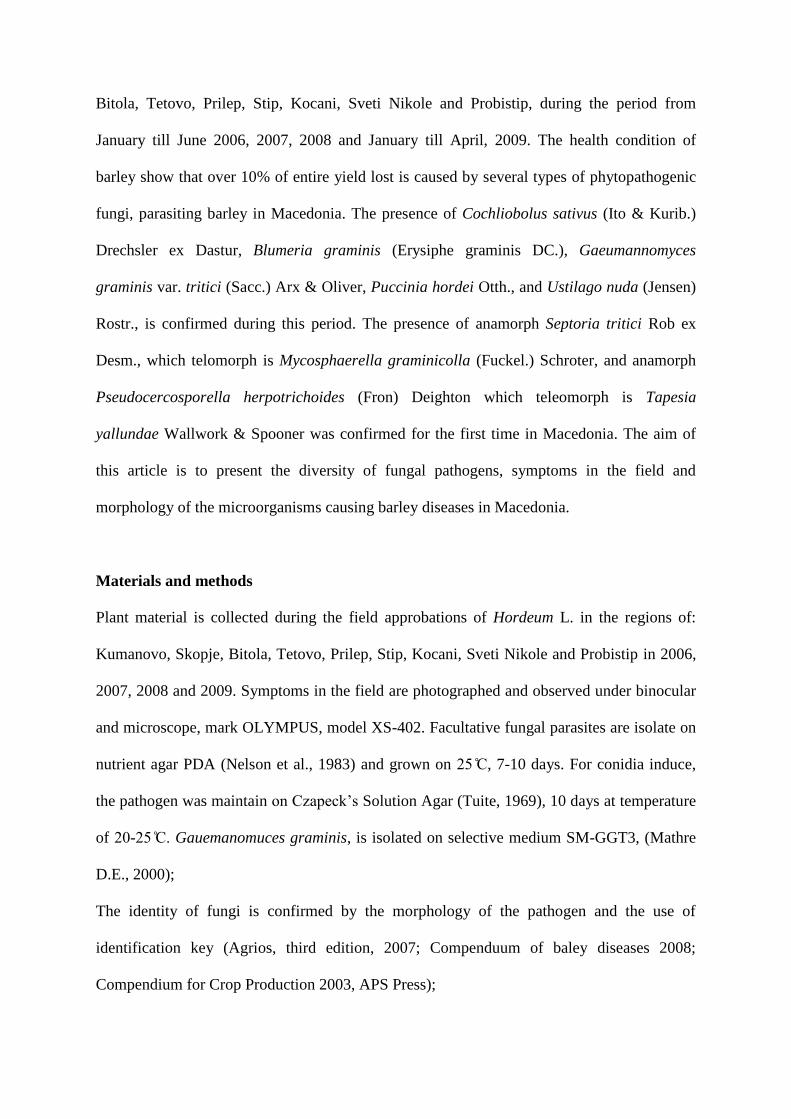
“Foot and root rot of cereals” or “Common root rot”. Primary symptoms, occurred in the
early stage of barley development on coleoptiles, subcrown, primary and secondary roots due
to the presence of conidia inoculum in soil and seed (Agrios, 2005). At the end of the winter
and early in the spring, underground plant parts appeared yellow and not developed enough.
The pathogen from soil inoculum destroy the vascular system, thus the plant is unable to
supply the nutrients and appear yellow and stunted as compared to healthy plants (Fig. 1).
The second infections become from conidia produced on the wheat straw or diseased grass
hosts (Ivanovic M., 1992). Second infections appear on leafs as black to brown flacks which
expand into oval shaped black flack not exceeding 1cm in length (Fig. 2). Some of the plants
recover from the infection (Fig. 3) and mature normally, but the infection progress every year
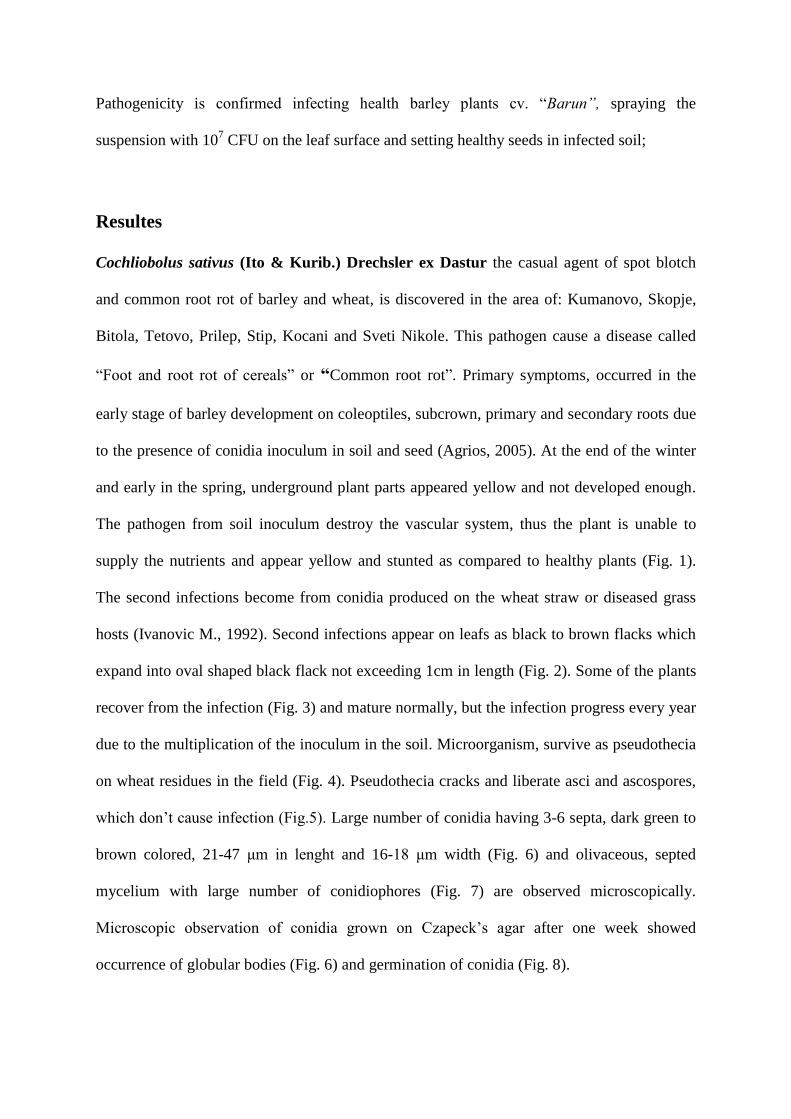
due to the multiplication of the inoculum in the soil. Microorganism, survive as pseudothecia
on wheat residues in the field (Fig. 4). Pseudothecia cracks and liberate asci and ascospores,
which don’t cause infection (Fig ) Large number of conidia having 3-6 septa, dark green to
brown colored, 21-47 μm in lenght and 16-18 μm width (Fig. 6) and olivaceous, septed
mycelium with large number of conidiophores (Fig. 7) are observed microscopically.
Microscopic observation of conidia grown on zapeck’s agar after one week showed
occurrence of globular bodies (Fig. 6) and germination of conidia (Fig. 8).
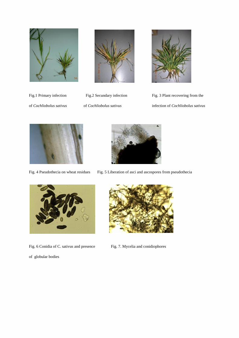
Fig.1 Primary infection Fig.2 Secundary infection Fig. 3 Plant recovering from the
of Cochliobolus sativus of Cochliobolus sativus infection of Cochliobolus sativus
Fig. 4 Pseudothecia on wheat residues Fig. 5 Liberation of asci and ascospores from pseudothecia
Fig. 6 Conidia of C. sativus and presence Fig. 7. Mycelia and conidiophores
of globular bodies
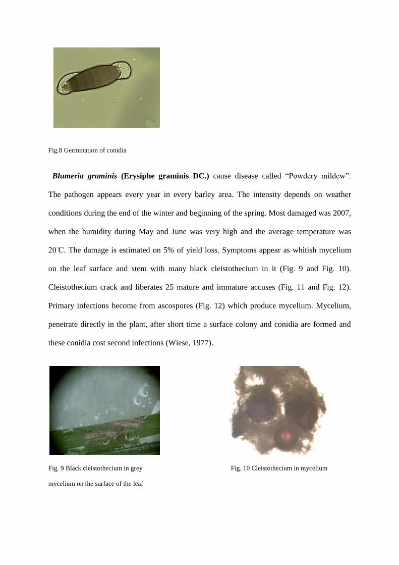
Fig.8 Germination of conidia
Blumeria graminis (Erysiphe graminis DC.) cause disease called “Powdery mildew”
The pathogen appears every year in every barley area. The intensity depends on weather
conditions during the end of the winter and beginning of the spring. Most damaged was 2007,
when the humidity during May and June was very high and the average temperature was
. The damage is estimated on 5% of yield loss. Symptoms appear as whitish mycelium
on the leaf surface and stem with many black cleistothecium in it (Fig. 9 and Fig. 10).
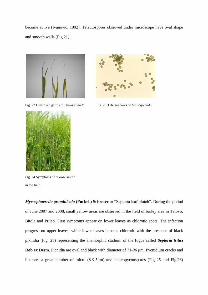
Cleistothecium crack and liberates 25 mature and immature accuses (Fig. 11 and Fig. 12).
Primary infections become from ascospores (Fig. 12) which produce mycelium. Mycelium,
penetrate directly in the plant, after short time a surface colony and conidia are formed and
these conidia cost second infections (Wiese, 1977).
Fig. 9 Black cleistothecium in grey Fig. 10 Cleistothecium in mycelium
mycelium on the surface of the leaf
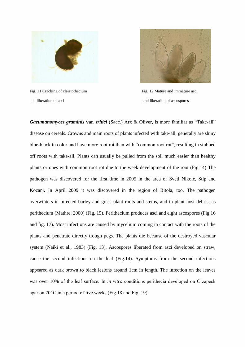
Fig. 11 Cracking of cleistothecium Fig. 12 Mature and immature asci
and liberation of asci and liberation of ascospores
Gaeumanomyces graminis var. tritici (Sacc.) Arx & Oliver, is more familiar as “Take-all”
disease on cereals. Crowns and main roots of plants infected with take-all, generally are shiny
blue-black in color and have more root rot than with “common root rot”, resulting in stubbed
off roots with take-all. Plants can usually be pulled from the soil much easier than healthy
plants or ones with common root rot due to the week development of the root (Fig.14) The
pathogen was discovered for the first time in 2005 in the area of Sveti Nikole, Stip and
Kocani. In April 2009 it was discovered in the region of Bitola, too. The pathogen
overwinters in infected barley and grass plant roots and stems, and in plant host debris, as
perithecium (Mathre, 2000) (Fig. 15). Perithecium produces asci and eight ascospores (Fig.16
and fig. 17). Most infections are caused by mycelium coming in contact with the roots of the
plants and penetrate directly trough pegs. The plants die because of the destroyed vascular
system (Naiki et al., 1983) (Fig. 13). Ascospores liberated from asci developed on straw,
cause the second infections on the leaf (Fig.14). Symptoms from the second infections
appeared as dark brown to black lesions around 1cm in length. The infection on the leaves
was over 10% of the leaf surface. In in vitro conditions perithecia developed on ’zapeck
agar on in a period of five weeks (Fig 18 and Fig 1 )

Fig. 13 Primary infection of G. graminis Fig.14 Secondary infection of G. graminis
Fig. 15 Perithecium of G. graminis on straw Fig. 16 Liberation of asci from perithecium of G. graminis
Fig. 17 Asci and ascospores of G. graminis Fig 18 Perithecia on ’zapeck agar
Fig. 19 Microscopic view of perithecia in
in vitro conditions

Puccinia hordei f.sp. tritici Otth cause the disease called “Stem rust” Symptoms are
observed in the area of Bitola, Skopje, Probistip and Prilep, every year since 2006. Yellow to
orange uredosoruses, elliptical and parallel with the axis, appear on stem and leafs. Over 65%
of the plant surface was covered with these uredosoruses (Fig. 20). The fungus forms five
different types of spores (Kurt et al., 2005). Microscopical observations showed the presence
of uredospores and teleutospores (Fig. 21). Uredospores are one celled, orange and oval with
diameter of 17- 1μm Teleutospores are two-celled, elliptical and slightly sagged between
the cells. The carion of the teleutospores is well diferenced and clearly seen (Fig. 19).
Fig. 20 Uredosoruses of Puccinia hordei Fig. 21 Uredospores and teleutospores
f.sp. tritici on the leaf surface of Puccinia hordei f.sp. tritici
Ustilago nuda ( Jensen ) Rostr. or “Loose smut” is discovered in the area of Kocani and
Bitola in several small parcels where growers used untreated seed from their own production.
This pathogen usually is successfully controlled with seed disinfection (karboksin), otherwice
it can be very destructive. The pathogen completely destroyed the germs, the seed is full with
black powdery smut (Fig. 22 and Fig. 24), consists of teleutospores from the fungus.
Teleutospores are distributed with the wind and spread the infection on large distances
(Cummins et al., 2003). Microscopic observations showed the presence of brown
teleutospores (Fig. 23). Teleutospores are formed from the cells of the mycelium. They are
spread with the wind and infect the healthy flowers and later the seed. The pathogen survive
in form of inactivate mycelium in the seed, until it becomes to germinate, and the mycelium
become active (Ivanovic, 1992). Teleutospores observed under microscope have oval shape
and smooth walls (Fig 21).
Fig. 22 Destroyed germs of Ustilago nuda Fig. 23 Teleutospores of Ustilago nuda
Fig. 24 Symptoms of “Loose smut”
in the field
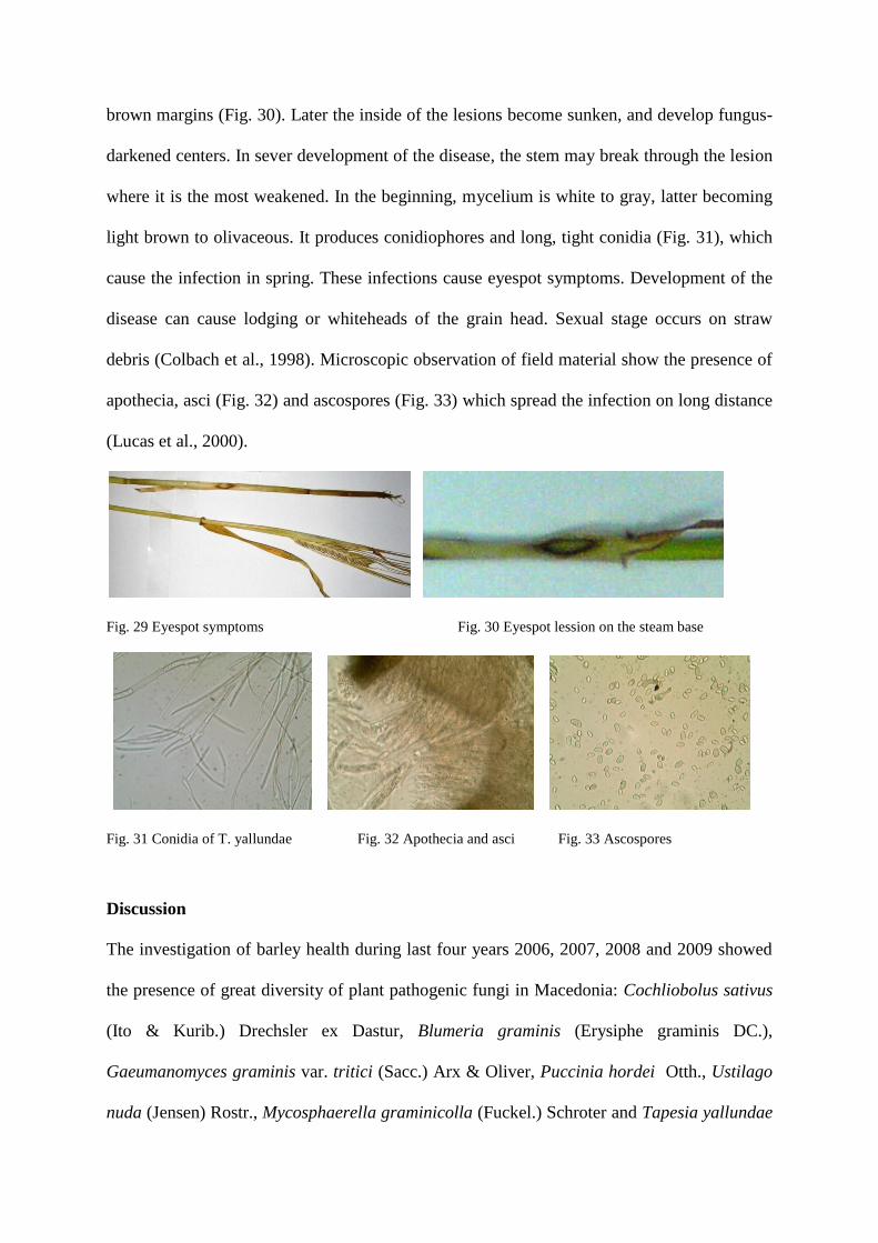
Mycosphaerella graminicola (Fuckel.) Schroter or “Septoria leaf blotch”. During the period
of June 2007 and 2008, small yellow areas are observed in the field of barley area in Tetovo,
Bitola and Prilep. First symptoms appear on lower leaves as chlorotic spots. The infection
progress on upper leaves, while lower leaves become chlorotic with the presence of black
piknidia (Fig. 25) representing the anamorphic stadium of the fugus called Septoria tritici
Rob ex Desm. Picnidia are oval and black with diameter of 71- 6 μm. Pycnidium cracks and
liberates a great number of micro (8- , μm) and macropycnospores (Fig 25 and Fig.26)

which cause the infection. Teleomorphic stadium of the fungus, Mycosphaerella graminicola
is observed in 2008 in the area of Tetovo and Bitola. Perithecia are dark brown to black in
color with diameter of 72- μm (Fig 7). Asci are limpid and have two layers, ascospores
are elliptical and two-celled (Fig. 28).
Fig. 25 Cracking of pycnidia and Fig. 26 Macro and micropicnospores of
Liberation of pycnospores Septoria tritici
Fig. 27 Perithecium of Mycosphaerella Fig. 28 Asci and ascospores
graminicola and liberation of asci
The pathogen overwinters as mycelium and conidia within pycnidia, in infected seed and
diseased plant refuse, left in the field. Seedling infections result in damping off, and provide
inoculums for subsequent infections (Halama, 1996). The infection in the diseased area in
Macedonia is spread from the diseased plant straw left in the field (Fig.27).
Tapesia yallundae Wallwork & Spooner cause disease called “Eyespot”. It is a
teleomorphic stage of the fungus which anamorph is Pseudocercosporella herpotrichoides
(Fron) Deighton. Symptoms appeared as elliptical or “eye” shaped lesions at the stem basis
(Fig. 29 and Fig 30). The eyespot lesions are elliptical, greyish to olivaceous inside with dark
brown margins (Fig. 30). Later the inside of the lesions become sunken, and develop fungus-
darkened centers. In sever development of the disease, the stem may break through the lesion
where it is the most weakened. In the beginning, mycelium is white to gray, latter becoming
light brown to olivaceous. It produces conidiophores and long, tight conidia (Fig. 31), which
cause the infection in spring. These infections cause eyespot symptoms. Development of the
disease can cause lodging or whiteheads of the grain head. Sexual stage occurs on straw
debris (Colbach et al., 1998). Microscopic observation of field material show the presence of
apothecia, asci (Fig. 32) and ascospores (Fig. 33) which spread the infection on long distance
(Lucas et al., 2000).
Fig. 29 Eyespot symptoms Fig. 30 Eyespot lession on the steam base
Fig. 31 Conidia of T. yallundae Fig. 32 Apothecia and asci Fig. 33 Ascospores
Discussion
The investigation of barley health during last four years 2006, 2007, 2008 and 2009 showed
the presence of great diversity of plant pathogenic fungi in Macedonia: Cochliobolus sativus
(Ito & Kurib.) Drechsler ex Dastur, Blumeria graminis (Erysiphe graminis DC.),
Gaeumanomyces graminis var. tritici (Sacc.) Arx & Oliver, Puccinia hordei Otth., Ustilago
nuda (Jensen) Rostr., Mycosphaerella graminicolla (Fuckel.) Schroter and Tapesia yallundae
Wallwork & Spooner. Favourable weather conditions at the end of the winter and beginning
of spring, during these years, induce around 10% of yield loss, in barley production in
Macedonia. Pathogens occurred mostly in areas where barley is produced every year without
crop rotation. The most destructive and intensive was the pathogen Cochliobolus sativus in
the production year 2007/2008 and 2008/2009 on barley, sawed in autumn (October and
November) showing primary infections in December and January of investigated period.
Secondary infections were observed in March and April 2008 and 2009. The most yield loss,
around 20-45% was observed in the region of Kumanovo and Skopje in 2008.
The aim of this study was to make evidence of mycoses appeared on barley in Macedonia,
estimation of yield loss, symptoms and morphology of the pathogen which cause the disease.
During the research, the presence of three different varieties of fungi, their telemorphic and
anamorphic stage, are discovered for the first time in Macedonia: Telemorph Tapesia
yallundae Wallwork & Spooner and anamorph Pseudocercosporella herpotrichoides
(Fron) Deighton, belonging to the class Ascomycetes, order Leotiales, family Dermataceae,
genus Tapesia; Telemorph Mycosphaerella graminicola (Fuckel.) Schroter, anamorph
Septoria tritici Rob ex Desm., belonging to the class Loculoascomycetes, order Dothideales,
family Mycosphaerellaceae, genus Mycosphaerella and Telemorph Cochliobolus sativus
(Ito & Kurib.) Drechsler ex Dastur, which anamorph is Drechslera sorokiniana (Sacc.)
Subram. & Jain., belonging to the class Loculoascomucetes, order Pleosporales, family
Pleosporaseae, genus Cochliobolus.
Republic of Macedonia doesn’t export barley, most of these diseases are introduced in the
country by import of seed material. Considering that most of these diseases are residue born
diseases, crop rotation, on time seedling, soil dreaning and establishment of residue
management is recommended to suppress the infection.
References
Agrios G.N., (2005). Plant Pathology. Fifth edition. USA: Elsevier Academic Press.
Colbach N., Saur L. (1998). Influence of crop management on eyespot development and
infection cycles of winter wheat. European Journal of Plant Pathology, 104 (1): 37-48.
Cummins G.B., Hiratsuka, Y. (2003). Ilustrated genera of rust fungi. Third ed. Minesota,
APS Press.
Halama P. 1996. The occurrence of Mycosphaerella graminicola, telemorph of Septoria
tritici in France. Plant Pathology. 45, 135-138.
Ivanovic M., (1992). Miokoze biljaka. Beograd: Nauka.
Kurt J. Leonard and Les J. Szabo (2005). Stem rust of small grains and grasses caused by
Puccinia graminis. Molecular Plant Pathology 6(2), 99-111.
Lucas J.A., Dyer, P. S. and Murray, T.D. (2000). Pathogenicity, host-specificity, and
population biology of Tapesia spp., causal agents of eyespot disease in cereals. Advances in
Botanical research 33: 226-258.
Mathre D.E. (2000). Take-all Disease on wheat, barley and oats. Plant Health Progress
10.1094/PHP-2000-0623-01-DG.
Naiki T and Cook, R.J. (1983). Factors in loss of pathogenicity in Gaeumannomyces
graminis var. tritici. Phytopathology 73: 1652-1656.
Nelson P.E., Toussoun, T.A., and Marasas, W.F.O., 1983. Fusarium species: An illustrated
Mannual for Identification. Pensylvania State University Press, University Park.
Tuite J. 1969. Plant Pathological Methods. Burgess Publishing Co., Mpls.
Wiese M.V. (1977). Compendium of Wheat Diseases. Minnesota, USA. APS.