Embed Size (px)
Citation preview
DIVERSITY IN ESCHERICHIA COLI O157:H7 BETWEEN HUMAN AND
BOVINE STRAINS
by
JENNIFER ANNE PAGE
B.A., Kansas State University, 2008
A REPORT
submitted in partial fulfillment of the requirements for the degree
MASTER OF SCIENCE
Food Science
KANSAS STATE UNIVERSITY Manhattan, Kansas
2009
Approved by:
Major Professor Dr. Daniel Y.C. Fung
Animal Science and Industry
Abstract
Within the United States, it has been estimated that 60 deaths and 73,000 illnesses are
caused by Escherichia coli O157:H7 infection annually (Gavin et al., 2004). Multiple effects
have been known to occur with the onset of infection from E. coli O157:H7 in which some of
these can become life-threatening. Escherichia coli O157:H7 is defined as a Shiga-toxin-
producing E. coli strain (STEC). This microbial pathogen is a gram-negative bacillus organism
that is motile, non-sorbitol fermenting, and β-glucuronidase negative. The infectious dose of E.
coli O157:H7 can be as low as ten cells (Food and Drug Administration, 2009).
Consumption of contaminated food, mainly undercooked ground beef and non or
incorrectly pasteurized milk, are the primary sources of E. coli O157:H7 infection in human.
Cattle, in particular, are considered chief asymptomatic reservoirs for this pathogen. Carried in
their gut, feces, and milk, cattle carry this Shiga toxin-producing E. coli in ranges from 102 to 105
CFU/g. Although colonized with E. coli O157:H7, cattle and other ruminants show no adverse
side effects from the pathogenic bacteria. There is also a difference in the prevalence of this
pathogen between human and cattle. There has been a low incidence of illness caused by E. coli
O157:H7 in humans when compared to the high prevalence of E. coli 057:H7 found in cattle and
their environment.
It has been discovered, through population genetic analysis, that E. coli O157:H7 and
other O157:H- isolates make up a clone complex. In spite of the clonal nature of E. coli
O157:H7 and other O157:H- isolates, there are significant characteristics showing variability
between the clone complex. These variability aspects can possibly account for the rapid
divergence of E. coli strains including the recently discovered divergence of E. coli O157:H7 in
to two separate lineages. Other possible reasons for a non-linear relationship between cattle
prevalence and human infection include diversity of the Shiga Toxin-Encoding bacteriophage
and receptors in cattle verses human, and finally the difference between the production of Locus
of Enterocyte Effacement (LEE) in both human and cattle lineages.
iii
Table of Contents
List of Figures ................................................................................................................................ iv
List of Tables .................................................................................................................................. v
Acknowledgments.......................................................................................................................... vi
Dedication ..................................................................................................................................... vii
CHAPTER 1 - Introduction of Escherichia coli O157:H7 ............................................................. 1
History and General Knowledge of Escherichia coli O157:H7 ................................................. 1
Disease in Humans and Sources of Infection ............................................................................. 2
Virulence Factors of Escherichia coli O157:H7 ......................................................................... 5
Figures and Tables ...................................................................................................................... 7
CHAPTER 2 - The Presence and Prevalence of Escherichia coli O157:H7 in Cattle ................. 10
CHAPTER 3 - Proposed Reasons for High Prevalence of Escherichia coli O157:H7 in Cattle,
but Low Incidence of Pathogenic Illnesses in Humans from Similar Strains............................... 13
Diversity among Conserved Genomic Regions in Human and Bovine Strains of Escherichia
Coli O157:H7 ............................................................................................................................ 13
Variety among Shiga Toxin Receptors in Human vs. Bovine Strains of Escherichia coli
O157:H7 .................................................................................................................................... 15
The Role of Intimin in Escherichia coli O157:H7 ................................................................... 19
Anti-Terminator Q933 Gene Involvement in Escherichia coli O157:H7 Virulence ................ 21
Locus of Enterocyte Effacement ............................................................................................... 22
Figures and Tables .................................................................................................................... 24
CHAPTER 4 - The Effects of Escherichia coli O157:H7 on the human population: Case Studies
....................................................................................................................................................... 27
Figures and Tables .................................................................................................................... 30
CHAPTER 5 - The Importance of High Escherichia coli O157:H7 Prevalence in Cattle in
Regards to the Relatively low Incidence of Escherichia coli O157:H7-Induced Disease in
Humans ......................................................................................................................................... 31
CHAPTER 6 - Possible Areas of Future Research ....................................................................... 32
REFERENCES ............................................................................................................................. 35
iv
List of Figures
Figure 1.1 Map representing number of E. coli O157:H7 cases during 1990-2006. (Multistate
outbreaks are not included in the numbers shown).. ............................................................... 7
Figure 1.2 Percentages of E. coli O157:H7 outbreaks based on contaminated food sources. ........ 7
Figure 1.3 Possible routes of how E. coli O157:H7 can enter the human population .................... 8
Figure 1.4 (A) General picture of an AB5 toxin molecule. (B) The A subunit of Shiga-Toxin. (C)
The B subunit of Shiga-toxin.. ............................................................................................... 9
Figure 3.1 Depiction of conserved genetic regions through multiple lineage I strains and their
absence in lineage II strains of E. coli O157:H7. ................................................................. 24
Figure 3.2 Model of disease pathway resulting from Shiga-toxin production in E. coli O157:H7
............................................................................................................................................... 25
Figure 3.3 (A) Diagram representing the indirect relationship between the EHEC hemolysin
molecule and disease in humans. (B) Diagram representing the direct relationship between
the EHEC molecule and disease occurance in humans ......................................................... 25
Figure 3.4 PCR products of human verses bovine strains of E. coli O157:H7 and whether or not
they contain Q933 gene. EDL933 (human-origin) does contain the Q933 gene. FAHRP88
(bovine-origin) does not have the Q933 gene………………………………………………..26
Figure 4.1 Dates of illness onset due to Shiga-toxin producing E. coli outbreak, from North
Carolina State Fair, in 2004. ................................................................................................ . 30
v
List of Tables
Table 3.1 Distribution of virulence factors from bovine and human STEC serotypes. The
numbers in the columns represent the number of isolates tested for the specified gene. The
number in parentheses represent of the total category positive for that specific gene. The
serotypes were analyzed for 5 major genes in both bovine and human isolates. Serotypes
used were: O26:H11, O103:H2, O111:H8, O111:H-, O145:H-, and O157:H7…………….24
vi
Acknowledgments
I would like to acknowledge my major professor, Dr. Daniel Y.C. Fung, and master
committee members, Dr. Tiruvoor G. Nagaraja and Dr. Richard Oberst for all of the their time
and guidance in the process of writing this report. I would also like to acknowledge my family
and husband for all of their love and support during my graduate career at Kansas State
University.
vii
Dedication
I would like to dedicate this report to Sean and I’s future daughter. This report topic
became an option only after being informed of my pregnancy. Since then, I have gained more
interest in this field of microbiology and wish to participate in laboratory research within this
area in the future.
1
CHAPTER 1 - Introduction of Escherichia coli O157:H7
History and General Knowledge of Escherichia coli O157:H7 In 1983, a microbial investigation was carried out by state health departments and the
Centers for Disease Control and Prevention (CDC) to determine the cause of two hemorrhagic
colitis outbreaks in 1982 in Michigan and Oregon. Stool specimens were compared to the only
isolate of Escherichia coli O157:H7, at the time, a rare strain of E. coli stored at the CDC in
Atlanta that was recovered in 1975 from a woman with bloody diarrhea. Matching the isolate
from the CDC, it was confirmed that the stool samples contained E. coli O157:H7 (Tarr, 1995).
In 1987, “Shiga-like toxin”, similar to that of Shiga toxin produced by Shigella, was discovered
in E. coli O157:H7 isolates, which helped define its pathogenic ability. These toxic effects
became more publically known after they were involved with the largest US E. coli O157:H7
outbreak in 1993: Jack-in-the-Box outbreak. Increased screening for this pathogen in clinical
laboratories has taken place and the US incidence of infection has risen remarkably since this
outbreak. According to the CDC (CDC, 2009), this lethal pathogen was also responsible for the
following outbreaks: infected ground beef sold at Kroger in 2008, a multi-state outbreak in
ground beef patties in 2006, Taco Bell outbreak in 2006, and a recent outbreak in raw Nestle
cookie dough in 2009. Throughout the decades, E. coli O157:H7 has caused numerous
foodborne outbreaks and a rise in public concern due to its toxigenicity and repulsive effects in
the human. Not only is E. coli O157:H7 now recognized as a common bacterial cause of bloody
diarrhea, but also as the cause of Hemolytic Uremic Syndrome (HUS) in the United States,
Europe and Canada (Besser et al., 1999).
2
Escherichia coli O157:H7 is defined as a Shiga-toxin-producing E. coli strain (STEC)
and it has been suggested that this strain evolved from Enteropathogenic E. coli (EPEC) that
acquired the Shiga toxin gene (Wachsmuth et al., 1997). This pathogen is a Gram-negative
bacillus that is motile, and differs from other E. coli in being non-sorbitol fermenting, and β-
glucuronidase negative. However, E. coli O157 isolates that are non-motile, sorbitol fermenting,
and β-glucuronidase positive have been isolated in Germany and Australia (Kaper and O'Brien,
1998), but are rarely found in the United States. Escherichia coli O157:H7 is a specific isolate
of the bacteria, E. coli. The O refers to the somatic antigen and the H refers to the flagellar
antigen. Part of this strains pathogenic ability derives from the 157st O antigen in combination
with the 7th H antigen and it is these two antigens that are targeted when E. coli O157:H7 is
screened for. Possible signs of E. coli O157:H7 infection include, but are not limited to nausea,
vomiting, slight fever, abdominal pain, HUS, thrombotic thrombocytopenic purpura (TTP), renal
failure, lethargy, seizures, and coma (Su and Brandt, 1995).
Disease in Humans and Sources of Infection In the United States, the most abundant amount of isolates have been recovered from
Washington State, Oregon, Minnesota, and Massachusetts, suggesting that predominance of
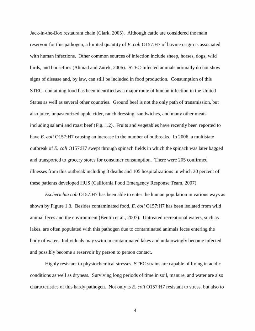
infections are in northern latitudes (Su and Brandt, 1995). According to the Center for Science
in the Public Interest Database (2009), predominance of E. coli O157:H7 infections from 1990-
2006 occurred in the contiguous northern states of North America (Fig. 1.1). Within the United
States, it has been estimated that 60 deaths and 73,000 illnesses are caused by E. coli O157:H7
infection annually (Gavin et al., 2004). Multiple effects have been known to occur with the onset
of infection from E. coli O157:H7 in which some of these can become life-threatening.
Characterized by microangiopathic hemolytic anemia, thrombocytopenia, and renal failure, HUS
3
is a critical syndrome present in nearly 10 percent of children infected with E. coli O157:H7
(Tarr, 1995). HUS occurs predominately in young children and the elder and is the most
common cause of acute renal failure in children. Required dialysis is needed for roughly half of
patients with HUS, while three-quarters require transfusions of erythrocytes or dialysis and
approximately 15 percent of HUS patients suffer from chronic renal failure or death (Tarr et al.,
1989, Robson et al., 1991).
In addition to HUS, central nervous system complications, including blindness, seizures,
and mental retardation, can occur in 30-50 percent of patients infected with E. coli O157:H7
(Cimolai et al., 1992) . The case - fatality rate among nursing home residents can be as high as
36 percent due to, in part, underlying diseases (Su and Brandt, 1995). In a large former nursing
home outbreak with E. coli O157:H7, 17 to 19 residents died due to hemorrhagic colitis,
pneumonia, and HUS (Carter et al., 1987). In non-life-threatening situations, individuals have
been known to experience gastrointestinal complications, dehydration and severe abdominal pain
due to bloody diarrhea. Within 2 to 3 days medical care is usually sought with a majority of
patients recovering within 10 days of the symptom’s onset (Riley et al., 1993).
The infectious dose of E. coli O157:H7 can be as low as ten cells. Consumption of
contaminated food, mainly undercooked ground beef and non or incorrectly pasteurized milk, is
the primary source of E. coli O157:H7 infection. Cattle, in particular, are considered chief
asymptomatic reservoirs for this pathogen. Carried in their gut, feces, and milk, cattle can
contain this Shiga toxin-producing E. coli in ranges from 102 to 105 CFU/g. Ground meat should
be cooked to 165°F in order to ensure that E. coli O157:H7 has been killed (USDA FSIS, 2006).
In 1993, the largest outbreak of E. coli O157:H7 in the United States took place when 732 cases
of illness, including 4 deaths of children, resulted from adulterated hamburgers provided by the
4
Jack-in-the-Box restaurant chain (Clark, 2005). Although cattle are considered the main
reservoir for this pathogen, a limited quantity of E. coli O157:H7 of bovine origin is associated
with human infections. Other common sources of infection include sheep, horses, dogs, wild
birds, and houseflies (Ahmad and Zurek, 2006). STEC-infected animals normally do not show
signs of disease and, by law, can still be included in food production. Consumption of this
STEC- containing food has been identified as a major route of human infection in the United
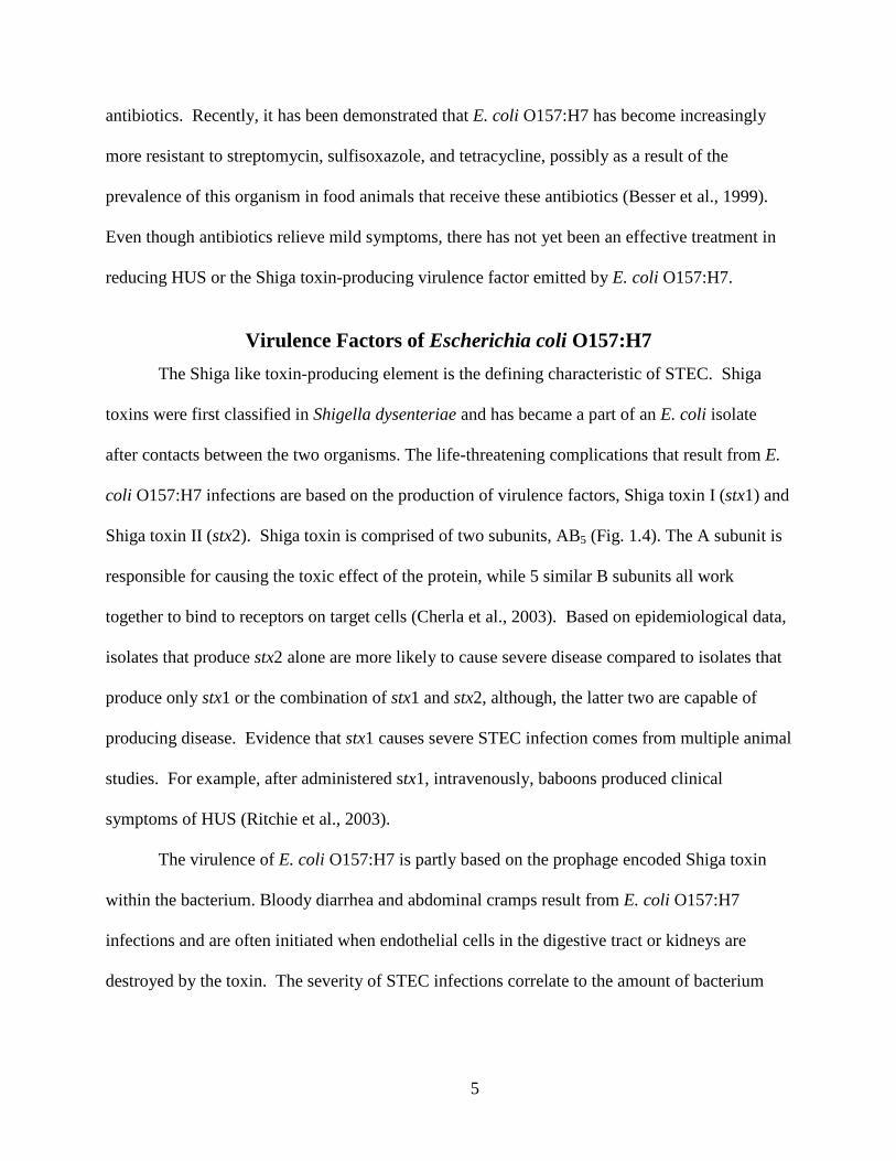
States as well as several other countries. Ground beef is not the only path of transmission, but
also juice, unpasteurized apple cider, ranch dressing, sandwiches, and many other meats
including salami and roast beef (Fig. 1.2). Fruits and vegetables have recently been reported to
have E. coli O157:H7 causing an increase in the number of outbreaks. In 2006, a multistate
outbreak of E. coli O157:H7 swept through spinach fields in which the spinach was later bagged
and transported to grocery stores for consumer consumption. There were 205 confirmed
illnesses from this outbreak including 3 deaths and 105 hospitalizations in which 30 percent of
these patients developed HUS (California Food Emergency Response Team, 2007).
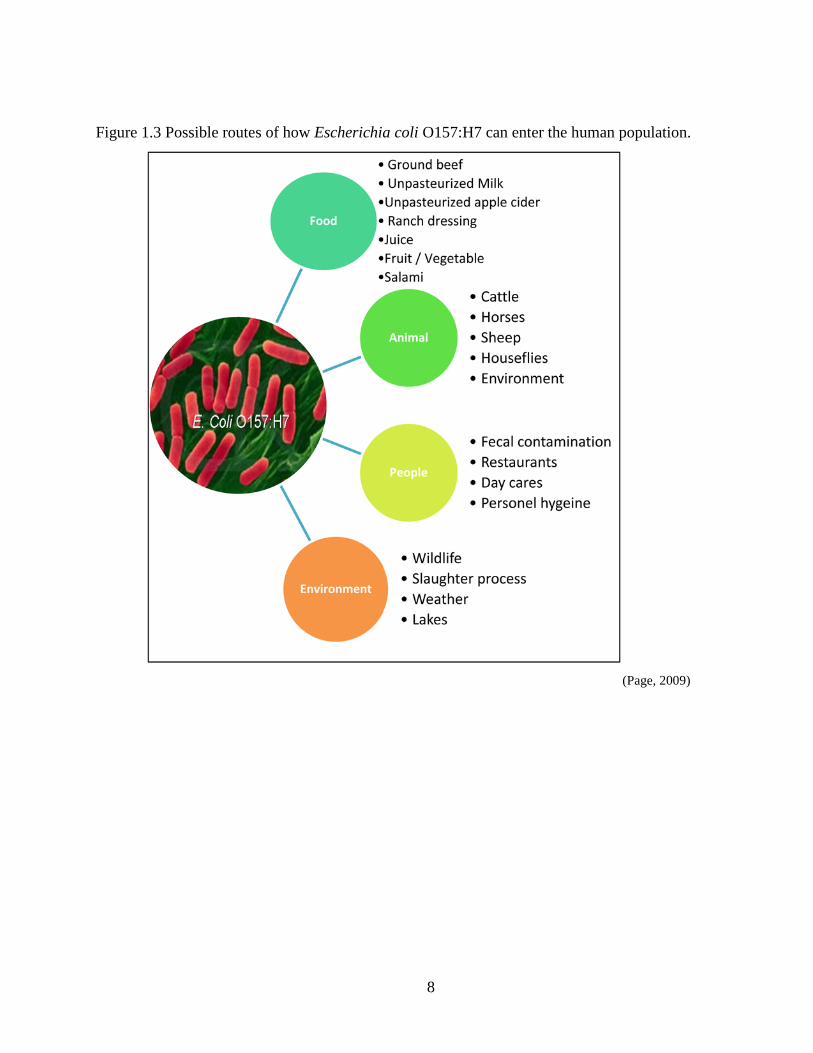
Escherichia coli O157:H7 has been able to enter the human population in various ways as
shown by Figure 1.3. Besides contaminated food, E. coli O157:H7 has been isolated from wild
animal feces and the environment (Beutin et al., 2007). Untreated recreational waters, such as
lakes, are often populated with this pathogen due to contaminated animals feces entering the
body of water. Individuals may swim in contaminated lakes and unknowingly become infected
and possibly become a reservoir by person to person contact.
Highly resistant to physiochemical stresses, STEC strains are capable of living in acidic
conditions as well as dryness. Surviving long periods of time in soil, manure, and water are also
characteristics of this hardy pathogen. Not only is E. coli O157:H7 resistant to stress, but also to
5
antibiotics. Recently, it has been demonstrated that E. coli O157:H7 has become increasingly
more resistant to streptomycin, sulfisoxazole, and tetracycline, possibly as a result of the
prevalence of this organism in food animals that receive these antibiotics (Besser et al., 1999).
Even though antibiotics relieve mild symptoms, there has not yet been an effective treatment in
reducing HUS or the Shiga toxin-producing virulence factor emitted by E. coli O157:H7.
Virulence Factors of Escherichia coli O157:H7 The Shiga like toxin-producing element is the defining characteristic of STEC. Shiga
toxins were first classified in Shigella dysenteriae and has became a part of an E. coli isolate
after contacts between the two organisms. The life-threatening complications that result from E.
coli O157:H7 infections are based on the production of virulence factors, Shiga toxin I (stx1) and
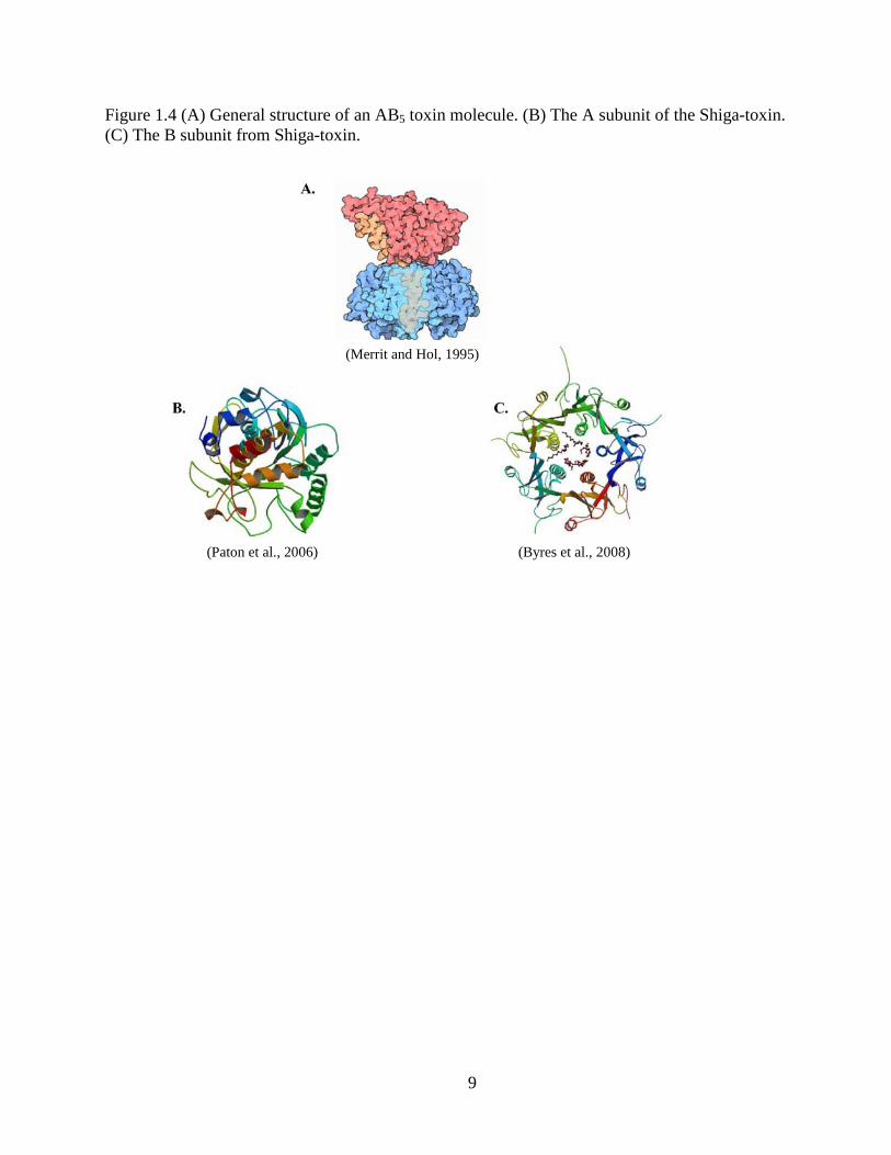
Shiga toxin II (stx2). Shiga toxin is comprised of two subunits, AB5 (Fig. 1.4). The A subunit is
responsible for causing the toxic effect of the protein, while 5 similar B subunits all work
together to bind to receptors on target cells (Cherla et al., 2003). Based on epidemiological data,
isolates that produce stx2 alone are more likely to cause severe disease compared to isolates that
produce only stx1 or the combination of stx1 and stx2, although, the latter two are capable of
producing disease. Evidence that stx1 causes severe STEC infection comes from multiple animal
studies. For example, after administered stx1, intravenously, baboons produced clinical
symptoms of HUS (Ritchie et al., 2003).
The virulence of E. coli O157:H7 is partly based on the prophage encoded Shiga toxin
within the bacterium. Bloody diarrhea and abdominal cramps result from E. coli O157:H7
infections and are often initiated when endothelial cells in the digestive tract or kidneys are
destroyed by the toxin. The severity of STEC infections correlate to the amount of bacterium
6
ingested and the amount of Shiga toxin produced. Unfortunately, low quantities of each can
initiate an E. coli O157:H7 infection in humans.
Shiga toxin is not the only virulent factor in STEC. Intimin is a major concern as this is
the adhesion molecule responsible for attachment of STEC organisms to the epithelial cells of
the intestine, which can cause structural modifications and lesions. Finally, a third virulent
factor in STEC organisms are called Locus for Enterocyte Effacement (LEE), which is a cluster
of genes that encode various virulent factors making the organism pathogenic. One of these
genes is the eae gene which is responsible for encoding the adhesion molecule, intimin. It is
purposed that these three specific virulence factors work together to produce the pathogenic
effects E. coli O157:H7 (Boerlin et al., 1999).
7
Figures and Tables
Figure1.1 Map representing number of Escherichia coli O157:H7 cases during 1990-2006. (Multistate outbreaks are not included in the numbers shown).
Figure 1.2 Percentages of Escherichia coli O157:H7 outbreaks based on contaminated food sources.
(Center of Science in the Public Interest Outbreak Database, 2009).
(Center of Science in the Public Interest outbreak database, 2009)
8
Figure 1.3 Possible routes of how Escherichia coli O157:H7 can enter the human population.
(Page, 2009)
9
Figure 1.4 (A) General structure of an AB5 toxin molecule. (B) The A subunit of the Shiga-toxin. (C) The B subunit from Shiga-toxin.
(Merrit and Hol, 1995)
(Paton et al., 2006) (Byres et al., 2008)
10
CHAPTER 2 - The Presence and Prevalence of Escherichia coli
O157:H7 in Cattle
Escherichia coli O157:H7 was introduced into the cattle population by the means of feed.
Grain- fed cattle seem to have a significantly higher level of E. coli O157:H7 compared to grass-
fed cattle. A grain diet turns a cattle’s rumen into an ideal habitat for E. coli O157:H7 while the
lethal strain of E. coli cannot survive for long periods of time on a grass fed rumen. Not only is
there an increased number of E. coli O157:H7 in rumens of grain-fed cattle, but also in the tons
of manure they produce each year. Approximately a billion tons of contaminated manure is
produced, by agricultural animals, each year and often ends up in locations other than pastures,
causing bacteria to travel to other habitats, animals, and environmental resources. Ultimately,
this brings the pathogen closer to humans and finished food products with a chance of increasing
infection rates in the human population (Pollan, 2006).
Several foodborne illnesses in humans result from the consumption of food products that
are contaminated with pathogenic bacteria. Products can be contaminated via poor water
sources, environmental agents, or other suitable hosts such as animals and insects. Even though
safety policies such as HACCP (Hazard Analysis and Critical Control Points) and GMP’s (Good
Manufacturing Practices) have been put into effect, cattle have been identified as the primary
source of E. coli O157:H7 in the United States. Escherichia coli O157:H7 is transmitted to
human by the cattle in various ways including: contaminated cattle hide, the cattle’s natural
environment, drinking water, shedding of feces, agricultural fairs, and through cross
contamination in the slaughter process. Seasons can have an influential effect on the prevalence
of E. coli O157:H7 isolated from cattle.
11
It has been suggested by Omisakin et al. (2003) that the concentration of E. coli O157:H7
in ruminant feces increases during warmer months of the year, and this fluctuation of bacteria,
during winter to summer months, can range anywhere from 103 to 105 CFU per gram of feces.
The most common range of E. coli O157:H7 being shed, during summer months, is
approximately 103 CFU/g. However, a small proportion of cattle will shed amounts between 103
CFU/g and 105 CFU/g and this specific group is referred to as “super shedders”. This group
causes the most concern as they can contribute the most bacteria to carcasses and meat products
(Omisakin et al., 2003).
The average test shows that E. coli O157:H7 prevalence, in a single herd, can be as high
as 10 percent and in the overall beef cattle population the prevalence can be as high as 28
percent. The prevalence approximations may be slighter lower than actual prevalence data due
to not enough herds tested, not enough animals within the herds tested, tests with low sensitivity,
and seasonal variation during test times (Grauke et al., 2002). Although colonized with E. coli
O157:H7, cattle and other ruminants show no adverse side effects from the pathogenic bacteria.
The Shiga toxin receptor, needed to cause illness, has not been found in cattle and the absence
presents a possible reason for the differing effects between cattle and human (Pruimboom-Brees
et al., 2000).
A study conducted by Cambridge University (Hancock et al., 1994) tested the prevalence
of E. coli O157:H7 in dairy cattle and beef cattle in Washington State. Sixty dairy herds were
tested by taking samples of fecal material along with samples of bulk milk. Twenty-five beef
cattle herds were tested by taking samples from the cattle hide and fecal deposits. It was
concluded that 10 of the 3570 samples in dairy cattle were positive for E. coli O157:H7. Seven
of the 10 positive samples were obtained from calves. The 10 positive samples were from 5 of
12
the 60 herds tested. Ten of the 1412 beef cattle fecal samples contained positive results of E. coli
O157:H7. The 10 positive samples came from 4 of the 25 herds tested. All screening for E. coli
O157:H7 in milk samples returned negative results. It can be suggested that beef cattle have a
higher prevalence of E. coli O157:H7 based on the 0.7% prevalence compared to dairy cattle that
retained 0.28% prevalence. Dairy cattle had E. coli O157:H7 in 8.3% of the herds tested, while
beef cattle had the bacteria in 16% of herds tested (Hancock et al., 1994).
13
CHAPTER 3 - Proposed Reasons for High Prevalence of Escherichia
coli O157:H7 in Cattle, but Low Incidence of Pathogenic Illnesses in
Humans from Similar Strains.
Diversity among Conserved Genomic Regions in Human and Bovine Strains
of Escherichia Coli O157:H7 In the many years given E. coli O157:H7 has been in existence, it has taken thousands of
lives (CDC, 2009), caused thousands of foodborne illnesses and outbreaks (CDC, 2009), and has
drawn a large amount of attention from the public, CDC and other governmental agencies such
as the USDA and FDA(CDC, 2009). Pathogenic effects in the human are not apparent in the
main reservoir, cattle, that serves the human population (Pruimboom-Brees et al., 2000). Not
only is there a difference in the effect between human and cattle when it comes to this pathogen,
but also in the prevalence of this pathogen within the human and cattle populations. There has
been a low incidence of illness caused by E. coli O157:H7 in humans when compared to the high
prevalence of E. coli 0157:H7 found in cattle and their environment. Research, independent
studies, experiments, and analysis of clinical cases have been done in order to determine why the
properties of E. coli O157:H7 differ greatly among humans and cattle (Besser et al., 2006, Kim
et al., 1999, Pruimboom-Brees et al., 2000, Steele et al., 2007.) The genomic difference
between E. coli O157:H7 strains have been a possible answer to why cattle prevalence and
incidence of human illness has not share a linear relationship. Other possible reasons include
diversity of Shiga toxin production and receptors in cattle verses human (Besser et al., 2006), the
difference between the production of Locus of Enterocyte Effacement (LEE) in both human and
cattle lineages (McNally et al., 2001), and the Q933 gene involvement (Ahmad and Zurek,
2006).
14
It has been discovered, through population genetic analysis, that E. coli O157:H7 and
other O157:H- isolates make up a clone complex. In spite of the clonal nature of E. coli
O157:H7 and other O157:H- isolates, significant characteristics showing variability between the
clone complex has been observed through various genomic typing methods, such as pulsed field
gel electrophoresis (PFGE) and octamer-based genome scanning (OBGS). These variability
aspects can possibly account for the rapid divergence of E. coli strains including the recently
discovered divergence of E. coli O157:H7. By the means of OBGS, it has been suggested that E.
coli O157:H7 has diverged in to two separate lineages, lineage I and lineage II. Lineage I has
been known to populate the human species whereas lineage II has appeared more in cattle.
The discovery of two separate lineages was in 1999 and has been followed by a cascade
of research studies supporting its finding (Kim et al., 1999, Steele et al., 2007). Octamer Based
Genome Scanning was the method used to determine the genetic diversity among human and
bovine isolates of E. coli O157:H7. Human and bovine isolates of E. coli O157:H7 were found
to be non-randomly distributed among the two lineages after analysis of OBGS products. The
segregation of isolates has suggested that one of these lineages could be less virulent for humans
or may not have the ability to be transmitted to humans by bovine sources (Kim et al., 1999).
The transmission uncertainty of isolates between human and bovine species could relate to the
high prevalence of E. coli O157:H7 in cattle, but low incidence of infection in humans
comparatively.
Developed recently, was the lineage-specific polymorphism assay (LSPA-6) which is
based on six loci that show bias in their allelic distribution between the two lineages. A study
conducted by Steele et al. (2006) which used LSPA-6 strain and utilized the suppression
subtractive hybridization (SSH) method to identify genomic regions present in E. coli O157:H7
15
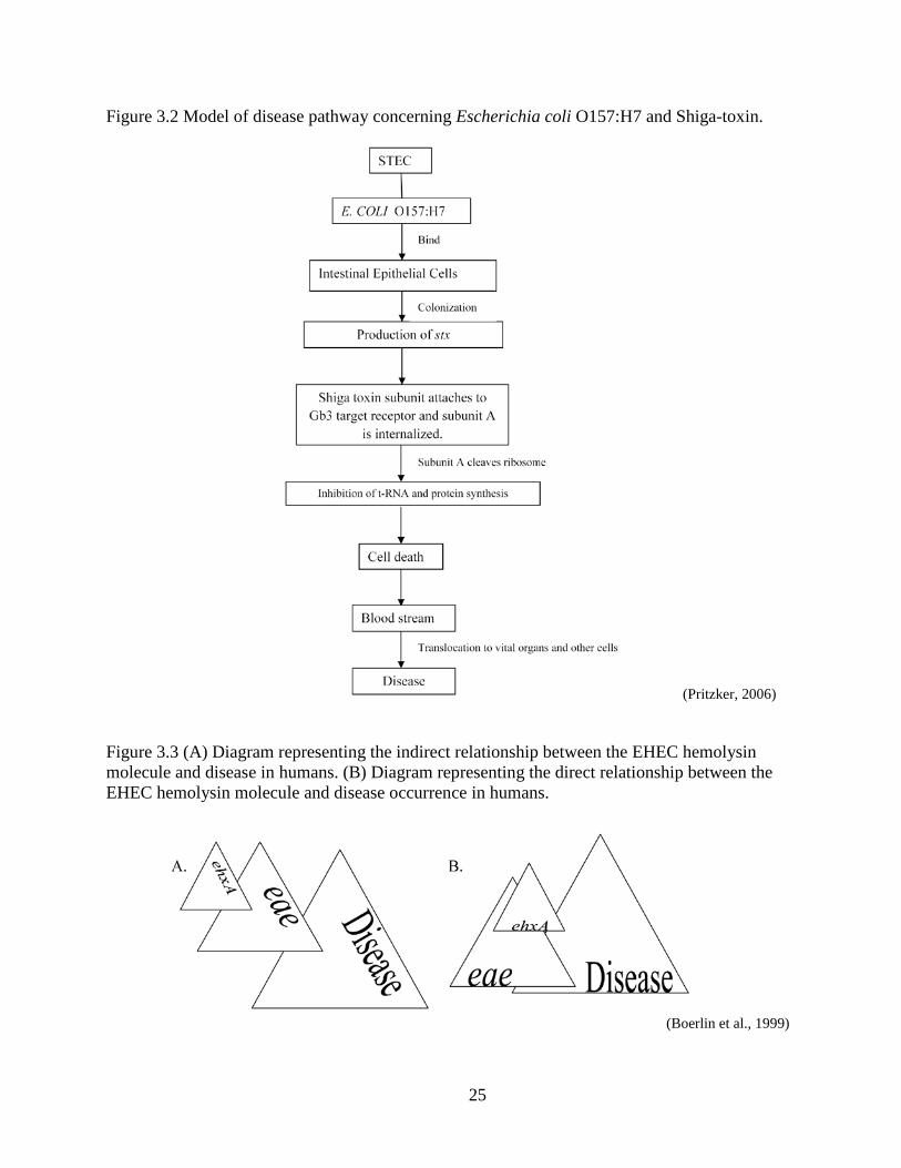
lineage I strains but absent from the lineage II strains. The lineage I strains used in the study
were LSPA-6 111111 and the lineage II strains were LSPA-6 222222. After identifying genomic
regions of difference (RDI), (genomic sequences that were present in the lineage I strain, but
absent in the lineage II strain) the study then identified conserved regions of genomic difference
(CRD). These were candidates that were conserved across multiple lineage I strains and absent
in multiple lineage II strains (Fig. 3.1). After the genomic regions were determined, it was noted
that lineage 1 strains share a set of unique genes that are largely absent in lineage II strains. The
exact contributions of these genes are unknown, but they are thought to be responsible for
expression of virulence factors in lineage I strains (Steele et al., 2007).
Many studies have played a role in trying to determine the cause of the pathogenicity of
E. coli O157:H7 and differences between humans and bovine isolate, while the findings of new
lineages has been a big step.
Variety among Shiga Toxin Receptors in Human vs. Bovine Strains of
Escherichia coli O157:H7 Shiga toxin refers to a family of toxins that is subdivided into two groups, Shiga toxin 1
(stx1) and Shiga toxin 2 (stx2). This toxin is of Shigella dysenteriae bacterium origin and was
first described by Kiyoshi Shiga (Beutin, 2006). Two of the most common sources of Shiga
toxin production has been Shigella dysenteriae and the Shigatoxigenic group of Escherichia coli
(STEC). Shiga toxin is one of the main virulence factors produced in STEC.
The action of Shiga toxin has been associated with HUS and hemorrhagic colitis (HC) in
multiple patients who have tested positive for E. coli O157:H7. Since the toxin is not
recognizable to the human’s immune system, and once the Shiga toxin is released from the
STEC organism, the body’s natural response is to increase permeability of cell barriers so
16
neutrophils and polymorphonuclear leukocytes (PMN) can reach the infection. PMN’s are white
blood cells, varying in nucleus size and shape from each other, that are responsible for attacking
foreign objects in the body (American Heritage® Medical Dictionary, 2007). Using this
opportunity, the toxin breaks through cell walls of the digestive tract, continuing through the
Golgi apparatus to the Endoplasmic Reticulum (ER) and nuclear membrane, and then enters the
blood stream and translocates to other organs such as the central nervous system and kidneys
(Pritzker, 2006). This intracellular method of trafficking is referred to as retrograde transport (see
Fig 3.2).
Shiga toxin is considered a 6-compenent protein structure, AB5. The B subunit is
responsible for attaching to target cell receptors, specifically globotriaosylceramide (Gb3), while
the A subunit is internalized and cleaved into A1 and its other components. A1 travels to the
cytoplasm where it cleaves an amino acid from the 60S ribosomal subunit in the target cell. This
action prevents t-RNA binding and therefore disrupts protein synthesis. With incoherent
synthesis of required proteins, functions of targeted cells seize and cell death occurs. Cell death,
by apoptosis, does not occur in all cell types, but there is sufficient information suggesting that
vascular lesions and tissue damage can result from apoptosis and could be the precursor to severe
disease. The toxin is highly specific for the Gb3 receptor on cell surfaces in order for the B
subunit to attach and the A subunit to enter the cell and for severe disease to result (Pritzker,
2006).
Certain animals, such as cattle, deer and swine do not contain this specific receptor on
some cells which is a potential answer to why cattle have been a reservoir for E. coli O157:H7,
but have not been affected by its pathogenic effects, as in humans. Bovine do however contain
this receptor on their crypt intestinal epithelial cells, but escape cell death by rerouting the toxins
17
to lysosomes instead of the ER where they come into contact with ribosome’s (Cherla et al.,
2003). The bacterium is then shed through bovine feces where it can come into contact with the
human population causing illnesses and outbreaks. A study performed by Pruimboom-brees et
al., (2000) revealed that cattle lack the vascular receptor, Gb3, for E. coli O157:H7 Shiga toxins.
In contrast to humans in which severe disease results from stx production, E. coli O157:H7-
infected cattle are tolerant of the pathogen and remain disease free for most of their lives. On the
other hand, newborn calves are affected by E. coli O157:H7 and suffer from fatal ileocolitis. It is
unknown as to why calves are susceptible to E. coli O157:H7 induced disease, but are tolerant
carriers of the pathogen as adults. Within the study two suggestions were made: Cattle lack
receptors for stx, and newborn calves have receptors for stx, but they disappear with age. The
hypothesis resulted after examination of glycolipid levels in various tissue including kidneys,
ileum, rectum, brainstem and cerebrum of newborn calves. It was determined that high levels of
Gb3 receptors were present in samples from the kidney, brainstem and cerebrum while there was
an undetectable amount in the ileum and rectum. Undetectable amounts of Gb3 were present in
all tissues from adult cattle tissue tested. The research supports the idea that adult cattle do not
contain Gb3 receptors and as a result do not experience E. coli O157:H7 induced disease as do
humans. Calves have given a new insight to Gb3 research and have provided knowledge of how
receptor concentrations differ among species and age (Pruimboom-Brees et al., 2000).
Shiga toxin 2 has been found to be up to 400 times more toxic, in regards to HUS related
illnesses, than Shiga toxin 1. A study was carried out in 2008 that tested for the presence of stx2
in various cells. Mice were given stx2 and no stx1, intraperitoneally, which resulted in weight
loss, paralysis and death. Examination of mice, after death, showed an increase in Ca+ flux
within the cerebral, which suggested that there was an increased neurotransmitter release from
18
neurons caused by the toxin. Since neurons contain a high level of Gb3 receptors, it has been
suggested that neurons are a main target sight for Shiga toxin and its close relative Shiga-like
toxin. Gb3 is more frequently found, for unknown reasons, within renal epithelial cells, and
central nervous system neurons and endothelium which is a possible factor for neurotoxicity
resulting from E. coli O157:H7 illness (Obata et al., 2008).
A previous study conducted (Boerlin et al., 1999) was subject to determine the
associations between virulence factors and STEC disease in humans. Another aim was to
compare bovine and human STEC populations of major serotypes involved in human disease. A
major finding in the study was discovered through multivariate analysis of five virulence factors:
EHEC hemolysin (ehxA), a Protease (espP), intimin encoding gene (eae), stx1 and stx2 encoding
genes. Multivariate analysis is a method of research that involves analysis of more than one
factor at a time instead of focusing on one factor during the study. After investigation of all five
virulence factors, only the eae and stx2 genes had a significant relationship. This supports the
ongoing hypothesis of synergism among the adhesion intimin molecule and Shiga toxin 2
production. The second aim, using univariate analysis, determined that eae and stx2 were
significantly more common in serotypes found in humans when compared to bovine strains. The
opposite was found true for stx1. Shiga toxin 1 was more frequently isolated from serotypes not
found in humans than those associated with humans (Boerlin et al., 1999). This study agrees
with previous studies showing eae and stx2 are more frequent in STEC isolates causing severe
disease and stx1 as being more frequent in some STEC isolates of bovine origin (Table 3.1).
Multiple studies have studied the effects of the individual virulent factors and the specific aspects
of each one. However, the study conducted by Boerlin et al. (1999) presents the idea that there is
19
no single factor responsible for the virulent effects produced by STEC and instead that multiple
factors work together to produce the pathogenic outcome of E. coli O157:H7
Reported by Schmidt et al. (1995), the new genetically analyzed ehxA, has been
associated with severe clinical disease in humans. Boerlin’s previous study (1999) shows a high
prevalence in ehxA and eae in STEC isolates regardless of disease severity. However, the strong
relationship with eae suggests that ehxA is likely to assist in disease as previously suggested by
Schmidt (Boerlin et al., 1999) (Fig 3.3). Shiga toxin has been capable of producing illness in
humans, but not in cattle. This outcome is only partly due to the toxin being released. Besides
the toxin, other virulent factors such as genetic sequences, intimin, LEE, and the Q933 gene all
have a responsibility to the disease-causing component of E. coli O157:H7 and other EHEC
organisms.
Even though genetic sequences, directly, are not virulent they can be the cause of virulent
actions. Some virulence genes, such as stx1, are often regulated by iron concentration. In a
study conducted in 2007, it was determined that E. coli O157:H7 contains a cluster of Open
reading frames (ORF) within a Conserved Region of Difference in Lineage I (CRDI), specific to
lineage I that may represent an iron uptake system. This could be responsible for triggering stx1
gene to continue on with virulent action (Steele et al., 2007).
The Role of Intimin in Escherichia coli O157:H7 Intimin is a vital aspect in relation to both Shiga toxin production and severe disease in
humans. Intimin is an adhesion virulence factor in EHEC E. coli strains. It is found on the cell
surfaces and is considered an attaching and effacing (A/E) protein. It can bind to its receptor,
Tir, which is secreted from bacterial cells into the host cytoplasm of intestinal epithelial cells by
means of Type three Secretion System (TTSS). TTSS is an organelle found mostly in Gram
20
negative bacteria pathogenic bacteria and is used to secret proteins to help the bacteria infect
eukaryotic organisms (Salmond and Reeves, 1993).
A study performed by Cookson and Woodward (1992) tested for the role of intimin, for
adherence, in E. coli O157:H7 strains. To carry out the study, the eae gene was inactivated in
three strains of E. coli O157:H7 from various origins. Adherence of intimin to epithelial cells
showed no intimate adherence, whereas wild type E. coli O157:H7 strains showed a high amount
of adherence. Adherence in wild type E. coli O157:H7 was determined by findings of A/E
lesions and colonies on cells. The study also looked into intimin-independent adherence using
neonatal calf gut explants. The same wild type and mutant strains from the first part were used to
compare adherence in colon tissue and rumen tissue in the neonatal calves. It was found that the
same amount of adherence took place whether the calf was given wild type or the eae-inactivated
version of E. coli O157:H7. The studies’ outcome confirms that intimin does play a strong role
in adherence between E. coli O157:H7 and various cells, but also that there could possibly be
another factor contributing to bacterial adherence causing pathogenic illness (Cookson and
Woodward, 2002).
Variants of the intimin molecule differ between bacterial isolates. The diversity of
intimin subtypes across a diverse collection of E. coli O157:H7 from both bovine and human
origin was examined. The basis of the study was to use Polymerase Chain Reactions (PCR) to
amplify the C-terminal amino acids in the bacteria and then utilize restriction fragment length
polymorphism (RFLP) analysis to differentiate the subtypes. By utilizing this method, three new
subtypes of intimin were found. The origins of the new subtypes were not of importance in the
study, but instead the subtype sequences. The study shows that there are undiscovered intimin
21
receptors and that they could be a reason for the difference between adhesion activity within
pathogenic bacterial strains (Ramachandran et al., 2003).
Anti-Terminator Q933 Gene Involvement in Escherichia coli O157:H7
Virulence Although ruminants appear to be the obvious reservoir for E. coli O157:H7, bovine
serotypes are not frequently isolated from human patients. Part of the reason for this mystery
could be due to the Q933 gene within the E. coli genome. The stx2 gene, which is regulated by
the interaction between the PR’ and Anti-Terminator Q gene, is located upstream of Anti-
Terminator Q. Anti-Terminator Q initiates the late promoter, PR’, and in doing so transcribes the
stx gene furthering Shiga toxin production. In a study by Ahmad and Zureck (2006), it was
proposed that isolates with Q933 produced significantly more Shiga toxin compared to isolates not
containing Q933. The study consisted of 262 environmental strains of E. coli O157:H7 which
were isolated from beef cattle feces and housefly digestive tracts. All strains were screened for
Q933 and Q21 (anti-terminator Q21 of bacteriophage 21). It was found that only 3.4% tested
positive for Q933, while 61.5% tested positive for Q21, and 25.1% carried both Q alleles. Even
though a minimal percent of the tested strains were positive for Q933 alone, it was these strains
that had the highest Shiga toxin production. They produced higher amounts of Shiga toxin
compared to strains that contained Q21 alone or the combination of Q alleles. These conclusions
suggest that Q933 could be an important factor in clinically relevant strains since humans can be
subjected to large amounts of Shiga toxin when infected with E. coli O157:H7 (Ahmad and
Zureck, 2006).
It has been demonstrated that the anti-terminator Q933 is more common in human isolates
than cattle isolates of E. coli O157:H7. It was also demonstrated that geographical regions that
22
have low E. coli O157:H7 illnesses also have a low number of E. coli O157:H7 isolates
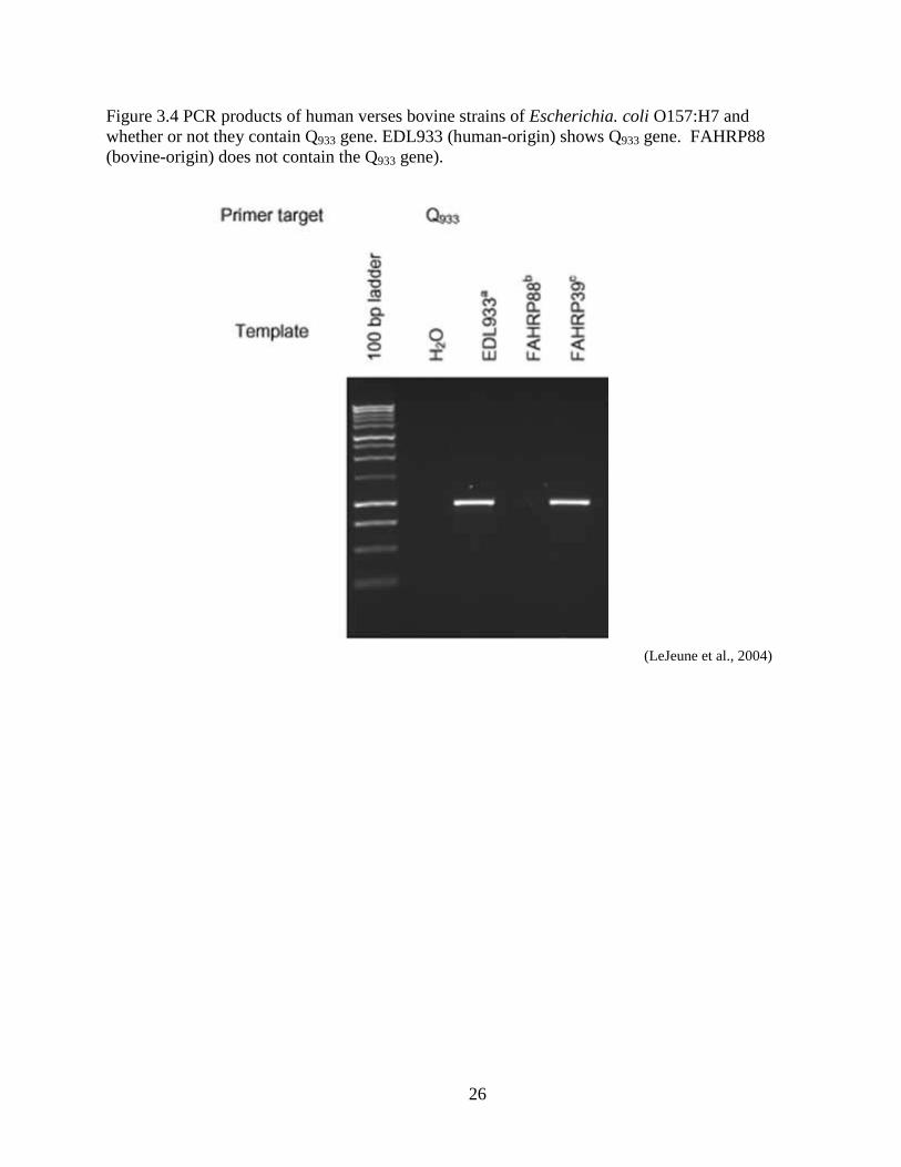
containing Q933 (LeJeune et al., 2004). In the study by LeJeune et al. (2004) 158 E.coli O157:H7
isolates were analyzed for Q933 and Q21. Ninety-one isolates were of bovine origin and 67 were
originally isolated from ill persons. Pulse Field Gel Electrophoresis and Enzyme-linked Immune
Sorbent Assay (ELISA) was done on each strain to gather conclusions. It was determined that the
human strains contained Q933 while the bovine strains contained the Q21 gene. Nine strains of the
158 strains initially tested were known to be of lineage I genotype and seven were of lineage II
genotype. Lineage I E. coli O157:H7 strains are known to be more frequently isolated from
humans than cattle. The study found that the lineage I strains contained the Q933 gene whereas
the lineage II strains, which are more frequent in cattle, contained the Q21 gene and not the Q933
gene (Fig. 3.4). This could help explain why lineage I strains tend to produce Shiga toxin in
higher amounts compared to lineage II strains (LeJeune et al., 2004).
The presence of Q933 is higher in human isolates than cattle isolates, and its presence
corresponds to a higher Shiga toxin production than Q21. This could relate to why cattle can
carry E. coli O157:H7 isolates, but are asymptomatic: Cattle may simply not have E. coli
O157:H7 isolates that contain the Q933 gene.
Locus of Enterocyte Effacement The locus of enterocyte effacement (LEE) is a pathogenicity island, within the EHEC
bacteria, which is clustered with genes that encode multiple virulent factors. Such genes include
ones that encode intimin (eae), intimin’s receptor (Tir), Shiga toxins (stx1 and stx2), EHEC
hemolysin (ehxA), and the LEE effector molecules (espA, espB and espD). Eae and Tir have
played a crucial role in formation of A/E lesions in several cell culture models and are expected
to work simultaneously within bovine and human environments as well. EhxA is also known to
23
contribute to severe disease in humans infected with EHEC (Ritchie et al., 2003). Many studies
have looked into the effects of the individual LEE encoded factors, but have not studied the
combined effects or ratios of LEE factors in both human and bovine populations.
Recent epidemiological data has shown a high prevalence of E. coli O157:H7 in cattle
and their environment, but a relatively low level of infection in humans. A common suggestion
to why this has occurred promotes the pathogenic differences between human and cattle. A
study performed in 2001 has tried to explain the contradicting high E. coli O157:H7 cattle
prevalence and the low human disease incidence from E. coli O157:H7. A range of virulence
determinants, including toxins and adhesions, were screened from human disease and bovine
fecal E. coli O157:H7 strains. Factors were examined in relation to each other. Secreted protein
profiles and actin rearrangement was investigated to note the difference between the human and
bovine strains. Results showed a significant strain and medium dependent variation in espD and
Tir secretion levels. The higher secretion rate of espD in different media suggests different ways
LEE can react between different environments such as the human and bovine atmospheres. Esp
secretion in general correlated to the amount of lesions present during examination of tissue
culture. Strains of human origin produced considerably higher levels of espD than the majority
of strains from bovine origin, which illustrated the human pathogenic potential in which bovine
do not posses. The overall study has suggested that not only do human strains have increased
stx1 and stx2 compared to the bovine population, but also a greater secretion and expression of
other LEE-encoded factors (McNally et al., 2001).
24
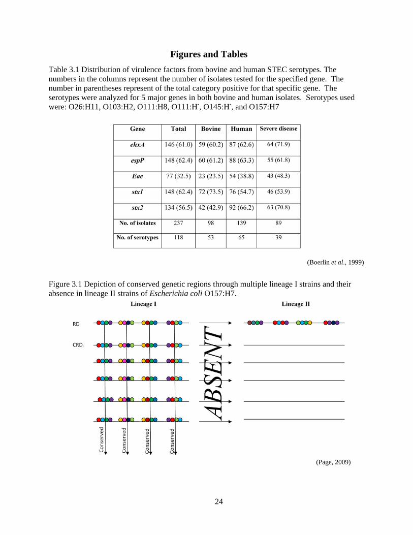
Figures and Tables Table 3.1 Distribution of virulence factors from bovine and human STEC serotypes. The numbers in the columns represent the number of isolates tested for the specified gene. The number in parentheses represent of the total category positive for that specific gene. The serotypes were analyzed for 5 major genes in both bovine and human isolates. Serotypes used were: O26:H11, O103:H2, O111:H8, O111:H-, O145:H-, and O157:H7
(Boerlin et al., 1999)
Figure 3.1 Depiction of conserved genetic regions through multiple lineage I strains and their absence in lineage II strains of Escherichia coli O157:H7.
(Page, 2009)
25
Figure 3.2 Model of disease pathway concerning Escherichia coli O157:H7 and Shiga-toxin.
(Pritzker, 2006)
Figure 3.3 (A) Diagram representing the indirect relationship between the EHEC hemolysin molecule and disease in humans. (B) Diagram representing the direct relationship between the EHEC hemolysin molecule and disease occurrence in humans.
(Boerlin et al., 1999)
26
Figure 3.4 PCR products of human verses bovine strains of Escherichia. coli O157:H7 and whether or not they contain Q933 gene. EDL933 (human-origin) shows Q933 gene. FAHRP88 (bovine-origin) does not contain the Q933 gene).
(LeJeune et al., 2004)
27
CHAPTER 4 - The Effects of Escherichia coli O157:H7 on the
human population: Case Studies
A multistate outbreak of E. coli O157:H7 during December and November of 2000 led
the USDA FSIS to announce a class 1 recall on ground beef. A Green Bay, Wisconsin meat
plant voluntarily recalled 1.1 million pounds of ground beef that had been distributed to 15
different states. The Wisconsin Division of Public Health (WDPH), along with local health
departments, and the Wisconsin State laboratory of Hygiene (WSLH) conducted an in-depth
investigation which involved reported E. coli O157:H7 cases, isolated strains of E. coli O157:H7
and interviews to all suspected cases of E. coli O157:H7. In a press release from the WDPH,
symptoms and sign of infection were made clear to the public and symptomatic persons were
urged to attend their local health department for medical attention and submission of a stool
sample to identify possible E. coli strains. Possible E. coli O157:H7 infections were reported to
the WDPH from local health agencies while all E. coli O157:H7 isolates from specimen were
forwarded to the WSLH for further confirmation using standard methods recommended by the
Centers of Disease Control and Prevention (CDC). Pulse field gel electrophoresis (PFGE) along
with traditional agar methods were utilized to receive the most accurate results. During the
investigation, 74 laboratory- confirmed cases of E. coli O157:H7 were reported from the WDPH.
The patient age ranged from 3 to 84 including six hospitalizations and one case of HUS. After
further investigation of E. coli O157:H7 isolates, it was concluded that there was a total of four
different patient strains of E. coli O157:H7 involved in the outbreak (Proctor et al., 2002). This
study gives us insight as to how many various strains can reside within cattle populations
compared to the number of strains that cause disease in humans. The difference in strains could
28
explain the difference in symptoms among people and the severity of diseases ranging from
diarrhea to HUS.
Escherichia coli O157:H7 has not only been passed from cattle to human by means of
food products. Physical contact between human and cattle and their environment has been a
substantial risk of E. coli infection if proper measures regarding safety and cleanliness were not
considered. In October 2004 one of the largest petting zoo outbreaks of E. coli O157:H7
occurred during the North Carolina State Fair. After three HUS infections were reported in the
state, the North Carolina State Laboratory for Public Health and the US department of
Agriculture conducted an investigation to find the source of the Shiga-toxin-induced HUS
infections. 96 initial environmental samples were taken from fair grounds including: animal
bedding, manure from animal sites, swabs from floors and fans, swabs from apple cider presses,
water samples from fountains, and live flies in animal exhibits. Between October 8 and
November 12, 2004 there were 108 outbreak related cases. 43 of these cases were laboratory-
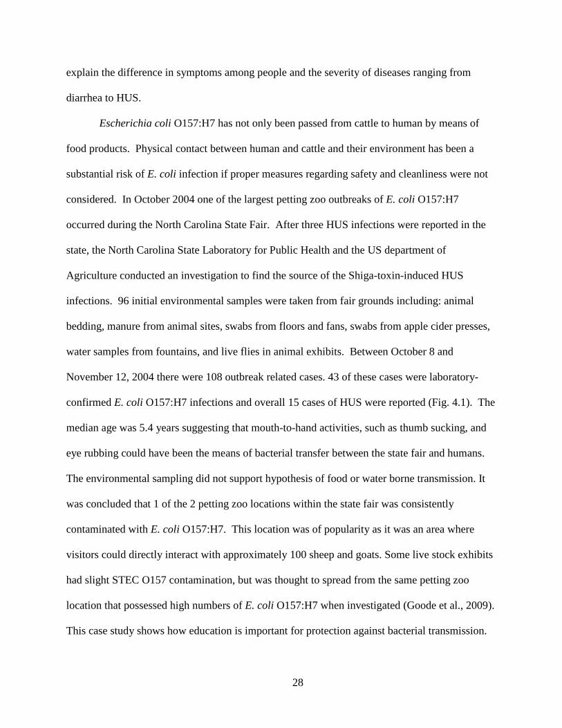
confirmed E. coli O157:H7 infections and overall 15 cases of HUS were reported (Fig. 4.1). The
median age was 5.4 years suggesting that mouth-to-hand activities, such as thumb sucking, and
eye rubbing could have been the means of bacterial transfer between the state fair and humans.
The environmental sampling did not support hypothesis of food or water borne transmission. It
was concluded that 1 of the 2 petting zoo locations within the state fair was consistently
contaminated with E. coli O157:H7. This location was of popularity as it was an area where
visitors could directly interact with approximately 100 sheep and goats. Some live stock exhibits
had slight STEC O157 contamination, but was thought to spread from the same petting zoo
location that possessed high numbers of E. coli O157:H7 when investigated (Goode et al., 2009).
This case study shows how education is important for protection against bacterial transmission.
29
Products such as hand sanitizers have increased awareness and healthy habits, but is not always
utilized which can produce a cascade of person to person transmission.
In 1993, a large E. coli O157:H7 outbreak occurred in California due to undercooked
meat and hamburger patties distributed by the national food chain, Jack-in-the-Box. People
became sick in multiple states, from multiple locations of this chain, including Seattle, Idaho and
Nevada. Overall, four children died due to HUS and over 600 became ill. After investigation it
was discovered that the ground beef patties were actually contaminated with fecal material
causing the severe illnesses in people. It was known as the largest E. coli O157:H7 outbreak in
America up to that time (Department of Defense, 1993). This shows how improper safety
measures can effect food products and put millions of people at risk. Since then, numerous safety
precautions have been made, such as HACCP and GMP’s to help prevent foodborne illnesses
and hinder bacteria from transmission.
30
Figures and Tables
Figure 4.1 Dates of illness onset due to Shiga-toxin producing E. coli outbreak, from North Carolina State Fair, in 2004.
(Goode et al., 2009)
31
CHAPTER 5 - The Importance of High Escherichia coli O157:H7
Prevalence in Cattle in Regards to the Relatively low Incidence of
Escherichia coli O157:H7-Induced Disease in Humans
Multiple studies have supported the idea that E. coli O157:H7 colonizes in high
prevalence within wildlife animals with cattle being the main reservoir. On the other hand,
incidences of disease in humans remain relatively low even with the low infectious dose of the
organism. It is important to understand this relationship to help monitor what strains are capable
of transmission between cattle and human populations. Different E. coli O157:H7 isolates
contain different pathogenic potentials and thus are not always capable of transfer between
species. If the prevalence of various strains is known, then being able to monitor those strains in
humans could give insight to the simplicity of transmission for specific isolates.
There are major differences among Shiga-toxin production, virulent factors, LEE
and genetic sequencing regarding strains of E. coli that effect humans yet do not promote effects
in cattle. This supports the findings of Kim et al. (1999) proposing two different lineages of E.
coli O157:H7. Perhaps the prevalence in cattle includes many isolates of E. coli O157:H7, while
only a few of those are capable of producing pathogenic effects in humans due to the numerous
pathogenic differences among strains.
32
CHAPTER 6 - Possible Areas of Future Research
Genetic analysis of E. coli O157:H7 and its lineages could potentially give more answers
to why the bacteria is so prevalent in cattle yet has such little incidence of infection in human,
comparatively. It was determined that iron uptake could trigger stx1. If this is the case, then
perhaps research concerning bovine and human iron intake could become another area of
interest. E. coli O157:H7 isolates in humans contain less stx1 than bovine strains, so maybe
bovine have a greater influx of iron in general, possibly through feed. Iron uptake could also
have an inverse relationship with stx2. Humans acquire higher amounts of stx2 and less stx1,
suggesting humans do not have the required iron intake to activate the stx1 the same way as the
bovine population. The possible lower concentration of iron in humans could, in return, account
for the increased activation of stx2 that bovine do not have.
Another area of interest for possible research could be Shiga toxin presence in old vs.
young humans. In Pruimboom-Brees study (2000) it was determined that calves had receptors as
do humans whereas adult cattle contained an undetectable amount of receptors. Calves vs. cattle
could be a new insight to age being a factor of E. coli O157:H7-induced disease. Determined,
through multiple studies, HUS is caused by Shiga toxin production and is most common in small
children. Receptor studies in children verses the elder could be an area of growth in research.
Growth in the area of research involving virulence factors should continue as only some
questions have been answered and more questions are arising. Linkages between the multiple
factors and correlating them to disease and various strains, as in bovine and human, can
eliminate bias in future studies. Focus on the effects of the virulent factor combinations instead
of the effects of each separate factor may give more insight to how bovine strains are different
than human strains of E. coli O157:H7. For example, the study done by Boerlin et al. (1999)
33
confirmed that eae and stx2 did not only have a strong correlation to each other, but also
possessed a higher frequency in human serotypes than in non-human serotypes and a higher
frequency in diseased humans compared to non-diseased humans. It can be purposed that the
reaction between the two genes promotes a type of cascade reaction within the immune response
system resulting in disease effects, such as HUS and HC. Since bovine strains do not have the
Gb3 receptor that is vital for stx2 to function, then perhaps reactions with eae are not possible
and therefore omitting the pathogenic affects that are apparent in humans.
Intimin is vital in producing Shiga toxin and disease in humans. Perhaps further research
in intimin subtypes verses E. coli serotypes could give clues as to why E. coli O157:H7 is more
likely to produce disease when compared to other O157:H- isolates. Another benefit to intimin
research would be the ability to characterize E. coli strain serotypes based on the subtype of
intimin it possesses. One last aspect to research in this field is the ability to correlate initmin
subtypes found in bovine verses initmin subtypes found in humans. This could be a potential
factor in why cattle do not adhere Shiga toxin to the ER, but instead push it to lysosomes, which
makes them asymptomatic to E. coli O157:H7 disease.
Locus of Enterocyte Effacement is largely responsible for encoding virulent factors that
produce disease. Future research regarding LEE effector molecules could potentially explain the
difference of virulence between cattle and human reactions to E. coli O157:H7 presence. EspD
is significantly higher in humans along with stx2 and eae. A combination of these three could
explain the difference in pathogenic effects between cattle and human. LEE houses critical
factors involved in E. coli O157:H7-induced disease and could be the potential target for
pathogenic differences between cattle and human populations. Factors that affect LEE induction
34
and the gene-encoding activity could be of interest as well since disease begins at the pathogenic
island.
Attention to more specific biotechnology could help distinguish E. coli O157:H7
strains from one another. This in return could help increase monitoring and surveillance of
different STEC strains as they transfer from cattle to human. Knowledge of strains’ capabilities,
in terms of transmission, could lead to better understanding of the pathogenic qualities that differ
from serotype to serotype.
35
REFERENCES
American Heritage® Medical Dictionary. 2007. The American Heritage Medical Dictionary, 3 ed. Houghton Mifflin Company., Boston, MA.
Ahmad, A. and L. Zurek. 2006. Evaluation of the Anti-Terminator Q933 Gene as a marker for Escherichia coli O157:H7 with high Shiga Toxin Production. Current Microbiology 53:324-328.
Besser, R. E., P. M. Griffin, and L. Slutsker. 1999. Escherichia coli O157:H7 Gasteroenteritis and the Hemolytic Uremic Syndrome: An Emerging Disease. Annual Review of Medine 50:355-367.
Beutin, L. 2006. Emerging enterohaemorrhagic Escherichia coli, causes and effects of the rise of a human pathogen. Journal of Veterinary Medicine, Infectious Diseases and Veterinary Public Health 53:299-305.
Beutin, L., A. Miko, G. Krause, K. Pries, S. Haby, K. Steege and N. Albrecht. 2007. Identification of Human-Pathogenic Strains of Shiga Toxin-Producing Escherichia coli from Food by a Combination of Serotyping and Molecular Typing of Shiga Toxin Genes. Applied and Environmental Microbiology 73:4769-4775.
Boerlin, P., S. McEween, F. Boerlin-Petzold, J. Wilson, R. Johnson and C. Gyles. 1999. Association between Virulence Factors of Shiga Toxin-producing Escherichia coli and Disease in Humans. Journal of Clinical Microbiology 37:497-503.
Byres, E., A. Paton, J. Paton, J. Lofling, D. Smith, M. Wilce, U. Talbot, D. Chong, H. Yu, S. Huang, X. Chen, N. Varki, A. Varki, J. Rossjohn and T. Beddoe. 2008. Crystal structure of the B-subunit of the AB5 toxin from E. coli. Protein Data Bank: 3DWA.
California Food Emergency Response Team. 2007. Investigation of an Escherichia coli O157:H7 Outbreak Associated with Dole Pre-Packaged Spinach. California Department of Health Services Food and Drug Branch. 1-50.
Carter, A., A. Borcayk, J. Carlson, B. Harvey and J. K. Hockin. 1987. A severe outbreak of Escherichia coli O157:H7- associated hemorrhagic colitis in a nursing home. New England Journal of Medicine 316:1496-1500.
Centers for Disease Control and Prevention. 2009. Escherichia coli: Previous outbreaks. National Center for Zoonotic, Vector-Borne, and enteric Diseases. <http://www.cdc.gov/ecoli/>
Center for Science in the Public Interest. 2009. Outlook Alert database, E. coli O157:H7. Online database.
36
<http://www.cspinet.org/foodsafety/outbreak/outbreaks.php?column=pathogen&colval=E.%20coli%20O157:H7>
Cherla, R., S. Lee and V. Tesh. 2003. Shiga toxins and apoptosis. FEMS Microbiology Letters 228:159-166.
Cimolai, N., B. Morrison and J. Carter. 1992. Risk factors for the central nervous system menifestations of gasteroenteritis-associated hemolytic-uremic syndrome. Pediatrics 90:616-621.
Clark, M. 2005. Jack-in-the-Box Escherichia coli Outbreak, About Escherichia coli. Online Post: 2009. Marler Clark LLP, PS, Seattle, Washington.
Cookson, A. and M. Woodward. 2002. The role of intimin in the adherence of enterohaemorrhagic Escherichia coli (EHEC) O157 : H7 to HEp-2 tissue culture cells and to bovine gut explant tissues. International Journal of Medical Microbiology 292:547-553.
Department of Defense. 1993. Case Study: Jack-in-the-Box E. coli crisis. Department of Defense. United States of America.
Gavin, P., L. Peterson, A. Pasquariello, J. Blackburn, M. Hamming, K. Kuo, and R. Thomson. 2004. Evaluation of Performance and Potential Clinical Impact of ProSpecT Shiga Toxin-Producing E. coli in Stool Samples. Journal of Clinical Microbiology 42:1652-1656.
Goode, B., C. O'Reilly, J. Dunn, K. Fullerton, S. Smith, G. J. Keen, L. Durso, M. Davies and S. Montgomery. 2009. Outbreak of Escherichia coli O157. Archives of Pediatrics & Adolescents Medicine 163:42-47.
Grauke, L. J., Kudva, I. Yoon, J. Hunt, C. Williamsand C. Hovde. 2002. Gastrointestinal Tract Location of Escherichia coli. Applied and Environmental microbiology.68:2269–2277.
Hancock, D., T. Besser, M. Kinsel, P. Tarr, D. Rice, and M. Paros. 1994. The Prevalence of Escherichia coli O157:H7 in dairy and beef cattle in Washington State. Epidemiology and Infection 113:199-207.
Kaper, J. and A. O'Brien. 1998. Escherichia coli O157:H7 and other Shiga toxin-producing E. coli strains. American Society for Microbiology. Washington, DC. 1-11.
Kim, J., J. Nietfeldt and K. A. Benson. 1999. Octomer-based genome scanning distinguishes a unique subpopulation of Escherichia coli O157:H7 strains in cattle. Proceedings of the National Academy of Sciences 96:13288-13293.
37
LeJeune, J., Abedon, S., Takemura, K., Christie, N., Sreevastan, S. 2004. Human Escherichia coli O157:H7 Genetic Marker in Isolates of Bovine Origin. Emerging Infectious Diseases. 10: 1482-1485.
McNally, A., A. Roe, S. Simpson, F. Thomson-Carter, E. Hoey, C. Currie, T. Chakraborty, D. Smith, and D. Gally. 2001. Differences in Levels of secreted Locus of Enterocyte Effacement Proteins between Human Disease-Associated and Bovine Escherichia coli O157. Infection and Immunity 69:5107-5114.
Merritt, E. and W. Hol. 1995. AB5 Toxins. Current Opinion in Structural Biology 5:165-171.
Obata, F., K. Tohyama, A. Bonev, G. Kolling, T. Keepers, Gross, LK., Nelson, MT., S. Sato, and T. Obrig. 2008. Shiga toxin 2 affects the central nervous system through receptor globotriaosylceramide localized to neurons. Journal of Infectious Dieseases 198:1398-1406.
Omisakin, F., M. MacRae, I. D. Ogden, and N. J. C. Strachan. 2003. Concentration and
prevalence of Escherichia coli O157 in cattle feces at slaughter. Applied Environmental Microbiology 69:2444-2447.
Ostruff, S. M., D. P. Hopkins, E. G. Sowers and N. K. Strockhine. 1991. Surveillance of Escherichia coli O157 isolation and confirmation. MMWR CDC Surveillance Summary 40:1-6.
Page, J. 2009. Personal works. Master Program. Kansas State University.
Paton, A., Beddoe, T., Thorpe, C., Whisstock, J., Wilce, M., Rossjohn, J., Talbot, U. and Paton, J. 2006. Crystal Structure of the A-subunit of the AB5 Toxin from E. coli. Protein Data Bank: 2IY9. <http://www.rcsb.org/pdb/explore/explore.do?structureId=2IY9>
Pollan, M. 2006. The Vegetable-Industrial Complex, The New York Times. The New York Times, New York City.
Pritzker, F. 2006. Shiga Toxin. Pritzker Olsen Attorneys. <http://www.rcsb.org/pdb/explore/explore.do?structureId=2IY9>
Proctor, M., T. Kurzynski, C. Koschmann, J. Archer and J. Davis. 2002. Four strains of Escherichia coli O157:H7 Isolated from Patients during an Outbreak of Disease Associated with Ground Beef: Importance of Evaluating Multiple Colonies from an Outbreak-Associated Product. Journal of Clinical Microbiology 40:1530-1533.
Pruimboom-Brees, I., T. Morgan, M. Ackermann, E. Nystrom, J. Samual, N. Cornick and H. Moon. 2000. Cattle lack vascular receptors for Escherichia coli O157:H7 Shiga toxins. Proceedings of the National Academy of Sciences 97:10325-10329.
38
Ramachandran, V., K. Brett, M. Hornitzky, M. Dowton, K. Bettelheim, M. Walker and S. Djordjevic. 2003. Distribution of Intimin Subtypes among Escherichia coli Isolates Ruminant and Human Sources. Journal of Clinical Microbiology 41:5022-5032.
Riley, L., R. Remis, S. Helgerson, H. McGee, J. Wells and B. Davis. 1993. Hemorrhagic colitis associated with a rare Escherichia coli serotype. New England Journal of Medicine 308:681-685.
Ritchie, J., C. Thorpe, A. Rogers, and M. Waldor. 2003. Critical Roles for stx2, eae, and tir in Enterohemorrhagic Escherichia coli-induced Diarrhea and Intestinal Inflammation in Infant Rabbits. Infection and Immunity 71:7129-7139.
Robson, W., A. Leung and M, Montgomery. 1991. Causes of death in hemolytic uremic syndrome. Child Nephrology & Urology 11:228-233.
Salmond, P. and G. Reeves. 1993. Membrane traffic wardens and protein secretion in Gram-negative bacteria. Trends in Biochemical Science 18:17-20.
Schmidt, H., Beutin, L., Karch, H. 1995. Molecular Analysis of the Plasmid-Encoded Hemolysin of Escherichia coli O157:H7 strain EDL 933. Infection and Immunity. 63: 1055-1061.
Steele, M., K. Ziebell, Z. Yongxiang, A. Benson, P. Konczy, R. Johnson and V. Gannon. 2007. Indentification of Escherichia coli O157:H7 Genomic Regions Conserved in Strains with a Genotype Associated with Human Infection. Applied and Environmental Microbiology 73:22-31.
Su, C. and L. Brandt. 1995. Escherichia coli O157:H7 Infection in Humans. Annals of Internal Medicine 123:698-714.
Tarr, P. 1995. Escherichia coli O157:H7: Clinical, Diagnostic, and Epidemiological Aspects of Human Infection. Clinical Infectious Diseases 20:1-10.
Tarr, P., M. Neill, J. Allen, C. Siccardi, S. Watkins and R. Hickman. 1989. The increasing incidence of the hemolytic-uremic syndrome in King County, Washington. American Journal of Epidemiology 129:582-586.
United States Department of Agriculture Food Safety and Inspection Service. 2006. Food Safety Education: Is it done yet? <http://www.fsis.usda.gov/Is_It_Done_Yet/Brochure_Text/index.asp>
Wachsmuth, I. K., P. H. Sparling and M. E. Potter. 1997. Enterohemorrhagic Escherichia coli in the United States. FEMS Immunology and Medical Microbiology 18:233-239.


![Isolation and identification of Escherichia coli O157:H7 ... · caused by the consumption of fresh-pressed apple juice [13]. Detection of E. coli O157:H7 in the clinical laboratory](https://img.pdfslide.us/doc/110x75/5e8a9af3f5c74a0ffa56b5f8/isolation-and-identification-of-escherichia-coli-o157h7-caused-by-the-consumption.jpg)