Embed Size (px)
Citation preview
Diverse oligomeric states of CEACAM IgV domainsDaniel A. Bonsora, Sebastian Günthera, Robert Beadenkopfa, Dorothy Beckettb, and Eric J. Sundberga,c,d,1
aInstitute of Human Virology, University of Maryland School of Medicine, Baltimore, MD 21201; bDepartment of Chemistry and Biochemistry, Universityof Maryland, College Park, MD 20742; cDepartment of Medicine, University of Maryland School of Medicine, Baltimore, MD 21201; and dDepartmentof Microbiology and Immunology, University of Maryland School of Medicine, Baltimore, MD 21201
Edited by Barry Honig, Howard Hughes Medical Institute, Columbia University, New York, NY, and approved September 29, 2015 (received for review May14, 2015)
Carcinoembryonic antigen-related cell adhesion molecules (CEACAMs)comprise a large family of cell surface adhesion molecules that bind tothemselves and other family members to carry out numerous cellularfunctions, including proliferation, signaling, differentiation, tumor sup-pression, and survival. They also play diverse and significant roles inimmunity and infection. The formation of CEACAM oligomers iscaused predominantly by interactions between their N-terminal IgVdomains. Although X-ray crystal structures of CEACAM IgV domainhomodimers have been described, how CEACAMs form hetero-dimers or remain monomers is poorly understood. To address thiskey aspect of CEACAM function, we determined the crystal struc-tures of IgV domains that form a homodimeric CEACAM6 complex,monomeric CEACAM8, and a heterodimeric CEACAM6–CEACAM8complex. To confirm and quantify these interactions in solution, weused analytical ultracentrifugation to measure the dimerization con-stants of CEACAM homodimers and isothermal titration calorimetry todetermine the thermodynamic parameters and binding affinities ofCEACAM heterodimers. We found the CEACAM6–CEACAM8 heterodi-meric state to be substantially favored energetically relative tothe CEACAM6 homodimer. Our data provide a molecular basis forthe adoption of the diverse oligomeric states known to exist forCEACAMs and suggest ways in which CEACAM6 and CEACAM8 reg-ulate the biological functions of one another, as well as of additionalCEACAMs with which they interact, both in cis and in trans.
CEACAM | X-ray crystallography | isothermal titration calorimetry |analytical ultracentrifugation
Carcinoembryonic antigen-related cell adhesion molecules(CEACAMs) are Ig-related proteins encoded by 12 genes on
human chromosome 19q13 (1). Expression patterns of eachCEACAM have been observed to be distinct (2). Several CEACAMsare expressed and anchored predominantly on the surfaces ofepithelial, endothelial, lymphocyte, myeloid, and granulocyte cells.Certain CEACAMs, however, are expressed only on one cell typeor tissue, including CEACAM3, CEACAM8, and CEACAM16,which are expressed on phagocytes, granulocytes, and in the innerear, respectively (3–5). With distinct expression patterns and lo-calizations, CEACAMs are typically observed to be involved innumerous and diverse cellular functions, including cell adhesion,proliferation, signaling, differentiation, tumor suppression, andsurvival (6–10). Certain CEACAMs, however, such as CEACAM3and CEACAM16, have specific roles in phagocytosis and hearing,respectively (3, 4). Several pathogenic bacteria, such as Neis-seria meningtidis, Escherichia coli, and Haemophilus influenzause adhesins to interact and anchor themselves to host cell surfacesthrough CEACAM recruitment (11–13). Because CEACAMsare involved in proliferation, tumor suppression, and survival,CEACAM dysregulation is frequently observed in tumor growthand metastasis (8, 14–17). All CEACAMs, except CEACAM16, aretethered to the surfaces of cells through either a single trans-membrane domain or a glycophosphatidylinositol (GPI) anchor attheir C terminus (7). Each CEACAM contains an N-terminal Igvariable-region–like (IgV) domain (6, 7), which is separated fromits C-terminal domain by a variable number of Ig constant region-type 2-like (IgC2-like) domains, ranging in number from zero tosix. One function of CEACAMs is cell adhesion (10). CEACAMs
achieve this primarily through the N-terminal IgV domain, whichcan dimerize either through homo- or heterophillic interactions(18–20). Dimerization of CEACAMs can be either in cis or intrans, with the latter allowing for cell–cell adhesion (21, 22).CEACAM6 and CEACAM8 are both anchored to cells by a
GPI motif, with two interspersing IgC2 domains (6). They arethus unable to directly signal like CEACAM1, which is anchoredby a transmembrane domain and contains a C-terminal cytosolicimmunoreceptor tyrosine-based inhibitory motif (6, 23). CEACAM8is exclusively expressed on granulocytes, whereas CEACAM6 isexpressed on the epithelial cells of the gastrointestinal tract andgranulocytes (24). High levels of CEACAM6 expression are typi-cally observed in several different cancers (17, 25, 26). Studieshave shown that CEACAM6 inhibits anoikis, resistance to apopto-sis in the absence of adhesion to the extracellular matrix, therebypromoting metastasis (27). Although less is known about CEACAM8,mRNA of both CEACAM6 and CEACAM8 are up-regulated inacute lymphoblastic leukemia (28).The homodimerization of CEACAM N-terminal IgV domains,
in particular those of CEACAM1 and CEACAM5, has been de-scribed previously (18, 19). However, the molecular mechanisms bywhich CEACAMs can heterodimerize have yet to be elucidated. Ithas been reported that CEACAM6 can form heterodimers withCEACAM1, CEACAM5, and CEACAM8, whereas CEACAM8can heterodimerize with CEACAM1 and CEACAM6 (20, 29). Likehomodimerization, CEACAM heterodimerization appears to re-quire N-terminal IgV domains (24). In this study, we present X-raycrystallographic and biophysical data showing that, in both crystalsand in solution, CEACAM6 homodimerizes, CEACAM8 is mo-nomeric, and the CEACAM6–CEACAM8 heterodimer representsthe energetically preferred state.
Significance
Carcinoembryonic antigen-related cell adhesion molecules(CEACAMs) are cell surface proteins that regulate cell adhesionand signaling in cancer, infection, and immunity through theirdiverse oligomeric states. Although X-ray crystal structures ofCEACAM homodimers have been described, how they formheterodimers or remain monomers is poorly understood. Herewe present the crystal structures of homodimeric CEACAM6,monomeric CEACAM8, and the heterodimeric CEACAM6–CEACAM8complex. Our crystallographic and biophysical data suggestways in which CEACAM6 and CEACAM8 regulate the biologicalfunctions of one another.
Author contributions: D.A.B., D.B., and E.J.S. designed research; D.A.B. and D.B. per-formed research; D.A.B., S.G., and R.B. contributed new reagents/analytic tools; D.A.B.,D.B., and E.J.S. analyzed data; and D.A.B. and E.J.S. wrote the paper.
The authors declare no conflict of interest.
This article is a PNAS Direct Submission.
Database deposition: The atomic coordinates have been deposited in the Protein DataBank, www.pdb.org [PDB ID codes 4Y8A (CEACAM6), 4Y88 (CEACAM8), 4YIQ (CEACAM6–CEACAM8)].1To whom correspondence should be addressed. Email: [email protected].
This article contains supporting information online at www.pnas.org/lookup/suppl/doi:10.1073/pnas.1509511112/-/DCSupplemental.
www.pnas.org/cgi/doi/10.1073/pnas.1509511112 PNAS | November 3, 2015 | vol. 112 | no. 44 | 13561–13566
BIOPH
YSICSAND
COMPU
TATIONALBIOLO
GY
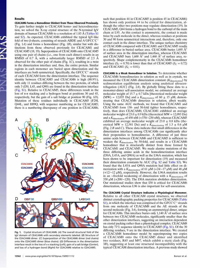
ResultsCEACAM6 Forms a Homodimer Distinct from Those Observed Previously.To gain further insight to CEACAM homo- and heterodimeriza-tion, we solved the X-ray crystal structure of the N-terminal IgVdomain of human CEACAM6 to a resolution of 1.83 Å (Tables S1and S2). As expected, CEACAM6 exhibited the typical IgV-likefold of two β-sheets, consisting of strands ABDE and A′GFCC′C′′(Fig. 1A) and forms a homodimer (Fig. 1B), similar but with dis-tinctions from those observed previously for CEACAM1 andCEACAM5 (18, 19). Superposition of CEACAM6 onto CEACAM5using one pair of chains (i.e., one from each dimer) results in anRMSD of 0.7 Å, with a substantially larger RMSD of 2.5 Åobserved for the other pair of chains (Fig. 1C), resulting in a twistin the dimerization interface and, thus, the entire protein. Similarregions in each monomer are buried upon dimerization and theinterfaces are both symmetrical. Specifically, the GFCC′C′′ strandsof each CEACAM form the dimerization interface. The sequenceidentity between CEACAM5 and CEACAM6 is high (88.9%),with only 11 residues differing between the two proteins, of whichonly 3 (I30, L45, and Q90) are found in the dimerization interface(Fig. S1). Relative to CEACAM5, these differences result in theloss of π-π stacking and a hydrogen bond at positions 30 and 45,respectively, but the gain of a salt bridge at position 90 (Fig. 1D).Mutation of these residues individually in CEACAM5 (F29I,Q44L, and H89Q, with sequence numbering as for CEACAM5;there is a numbering discrepancy of one position in CEACAM6,
such that position 44 in CEACAM5 is position 45 in CEACAM6)has shown only position 44 to be critical for dimerization, al-though the other two positions may regulate dimerization (19). InCEACAM5, Q44 forms a hydrogen bond to the carbonyl of the mainchain at L95. As this contact is asymmetric, the contact is madetwice by each molecule in the dimer, whereas residues at positions30 and 90 form symmetrical interactions and, therefore, only onecontact each in the dimer interface. The unique surface chemistryof CEACAM6 compared with CEACAM1 and CEACAM5 resultsin a difference in buried surface area. CEACAM6 buries 1,695 Å2
of surface area in the dimerization interface, whereas CEACAM1and CEACAM5 bury 1,600 Å2 and 1,460 Å2 of surface area, re-spectively. Shape complementarity in the CEACAM6 homodimerinterface (SC = 0.70) is lower than that of CEACAM5 (SC = 0.72)and CEACAM1 (SC = 0.81).
CEACAM6 Is a Weak Homodimer in Solution. To determine whetherCEACAM6 homodimerizes in solution as well as in crystals, wemeasured the CEACAM6 dimerization constant (Kdimerization) bysedimentation equilibrium analysis using analytical ultracen-trifugation (AUC) (Fig. 2A). By globally fitting these data to amonomer-dimer self-association model, we estimated an averagemolecular weight of 15.7 ± 0.7 kDa [theoretical molecular weight(MW) = 12,035 Da] and a Kdimerization of 60 ± 15 μM, demon-strating that CEACAM6 dimerizes in solution, albeit weakly.Using the same AUC methods, we found that CEACAM1 andCEACAM5 form 130- and 50-fold tighter homodimers, respec-tively, than does CEACAM6; CEACAM1 exhibited an averagemolecular weight of 23.0 ± 0.9 kDa (theoretical MW = 12,093 Da)and a Kdimerization of 450 nM (+370/−230 nM), whereas CEACAM5exhibited an average molecular weight of 23.0 ± 0.8 kDa (the-oretical MW = 12,581 Da) and a Kdimerization of 1.3 ± 0.6 μM(Fig. 2 B and C). These data indicate that small differences in thedimerization interfaces among CEACAMs can significantly altertheir propensities to homodimerize. A difference of just threeamino acids between CEACAM6 and CEACAM5 is sufficient toweaken the Kdimerization by 50-fold, and results in a CEACAM6homodimer that is structurally distinct from those formed byCEACAM1 and CEACAM5. We made alanine mutations of thethree differing amino acids in the homodimerization interface(I30A, L45A, and Q90A) as well as the L96A mutation, which hasbeen shown to be important for dimerization (19) and measuredtheir dimerization constants by AUC (Fig. S2 and Table S3). Wefound that the L45A and Q90A mutation had little effect on di-merization with a Kdimerization of 61 μM (+24/−17 μM) and 48 μM(+12/−12 μM), respectively. However, the L96A mutation resultsin an ∼fivefold weakening of dimerization with a Kdimerization of330 μM (+200/−120). The I30A mutation abolishes dimerization.Our mutational studies show that I30 is critical for CEACAM6dimerization, whereas L96 is also important for self-association.
The CEACAM8 Crystal Structure Indicates a Physiological Monomer.Relative to all other CEACAM crystal structures, we observeddistinct crystallographic packing properties for CEACAM8 (TableS1), in which the interface was comprised of the GFCC′C′′ strandsfrom one molecule of CEACAM8 and the AE strands of thesecond molecule (Fig. 3A), forming an asymmetrical dimer, uniquefor CEACAMs. This interface buries only 1,140 Å2 of surface areabetween two CEACAM8 molecules, significantly smaller than theother dimerization interfaces, suggesting an interaction dependenton crystal packing rather than a physiological complex. CEACAM8has only 71% sequence identity to CEACAM5 (Fig. S1). Of the 30differing residues, 9 are in the dimerization interface. We createda CEACAM8 homodimer model by superimposing two copiesof CEACAM8 onto the CEACAM5 dimer and identifiedtwo residues, R45 and M97, which exhibit a steric clash (Fig.3B), suggesting at least one structural incompatibility with thetypical CEACAM dimerization interface. A previous study showed
Fig. 1. Crystal structure of CEACAM6. (A) The overall structural fold of theIgV domain of CEACAM6 with secondary elements labeled. (B) Structure ofthe CEACAM6 dimer. (C) Superposition of the CEACAM6 dimer (red chains)onto the CEACAM5 dimer (blue chains). (D) Differences in the dimerizationinterface result in the loss of π-π stacking (Left), gain of a salt bridge (Center),and loss of a hydrogen bond (Right) for CEACAM6 relative to CEACAM5.
13562 | www.pnas.org/cgi/doi/10.1073/pnas.1509511112 Bonsor et al.

that the L44R mutation in CEACAM5 (position 45 in CEACAM8)results in a CEACAM5 monomer (19). Although CEACAM8 canhomodimerize, albeit weakly, it is unknown whether the crystalstructure of CEACAM8 represents the measured dimer as seen byAUC or is in fact a monomer. To test this we made two mutations,R45A and L96A, and measured their Kdimerization (Fig. S2). IfCEACAM8 forms a canonical dimer like those observed in theCEACAM1, -5, and -6 structures, in which both R45A and L96Amutations make important dimerization contacts, it therefore wouldbe monomeric as measured by AUC. However, if dimerization ofCEACAM8 forms an asymmetrical dimer as described above,R45A would have no effect on dimerization as it is not buried in thisdimerization interface. We found that both the R45A and L96Amutations resulted in monomeric CEACAM8, demonstrating thatthe observed asymmetrical dimer described above is actually mo-nomeric and that dimeric CEACAM8 most likely forms a canonicalCEACAM dimer similar to those observed for CEACAM1, -5,and -6. We made a further mutation in the CEACAM8 dimerizationinterface, Q90A and also found it to form monomers (Fig. S2).
CEACAM8 Exists Predominately as a Monomer in Solution. To confirmthe monomeric state of CEACAM8 in solution, we performed sedi-mentation equilibrium analysis, by which we estimated a molecu-lar weight of 13.0 ± 0.8 kDa (theoretical MW = 12,192 Da) and a
Kdimerization of 650 μM (+350/−300 μM) for CEACAM8 (Fig. 2D).These data indicate that although CEACAM8 homodimerizationcan occur at the likely nonphysiologically high concentrations that wetested in our AUC experiments, it is most probable that it is found inan exclusively monomeric state. Furthermore, CEACAM8 exhibitsthe weakest homodimerization constant compared with the mea-sured values of other CEACAMs, a 1,300-fold difference comparedwith the strongest, CEACAM1 (Fig. 2E).
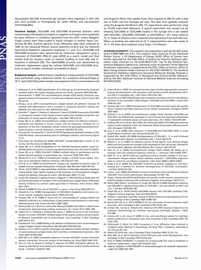
CEACAM6 and CEACAM8 Form a Heterodimer.Cell-based studies haveshown that CEACAM6 and CEACAM8 can form heterodimers(20, 24). With such weak homodimerization constants as wemeasured for CEACAM6 and CEACAM8, a CEACAM6/8heterodimer may represent an energetically more favorable in-teraction, which we tested using isothermal titration calorim-etry (ITC). Upon titrating CEACAM8 into CEACAM6,we observed that this interaction is both enthalpically (ΔH =−4.6 kcal/mol−1) and entropically (−TΔS = −3.2 kcal/mol−1)driven, with a KD of 2.0 ± 0.8 μM (Fig. 4A), indicating that theCEACAM6/8 heterodimer is 30-fold tighter than the CEACAM6homodimer. Several studies have noted that other CEACAMheterodimers can form, including: CEACAM6/1, CEACAM3/5,and CEACAM8/1 (20, 29). Accordingly, we also measured the
Fig. 2. Oligomeric states of homotypic CEACAM preparations. Sedimenta-tion equilibrium analyses (Upper) and residuals of the fits for each curve(Lower) for (A) CEACAM6, (B) CEACAM1, (C) CEACAM5, and (D) CEACAM8.(E) Kdimerization values and SDs for all homotypic CEACAM interactions.
Fig. 3. Crystal structure of CEACAM8. (A) Crystal structure of CEACAM8depicting an asymmetrical dimer due to crystal packing relative to CEACAM5(superimposed onto the CEACAM8 structure shown in gray). (B) An artificialCEACAM8 homodimer modeled by superposition of CEACAM8 onto theCEACAM5 homodimer shows that residues R45 and M97 of CEACAM8 clashin the dimerization interface.
Bonsor et al. PNAS | November 3, 2015 | vol. 112 | no. 44 | 13563
BIOPH
YSICSAND
COMPU
TATIONALBIOLO
GY
affinities of these interactions by ITC but observed no heterodi-mer formation (Fig. S3 A–D). As CEACAMs are highly glycosy-lated proteins and our studies thus far had been conductedwith unglycosylated proteins, we expressed, secreted, and purifiedCEACAM6 and CEACAM8 from HEK293 cells to investigatewhether glycosylation has an effect on the CEACAM6–CEACAM8interaction. Both N-terminal domains are predicted to have threeN-linked glycosylation sites (Fig. S1). A silver-stained SDS/PAGEgel showed that both proteins are glycosylated and polydisperse(Fig. S3E). Titration of glycosylated CEACAM8 into glycosylatedCEACAM6 only results in a twofold reduction in affinity, witha KD of 4.5 ± 0.5 μM, and remains both enthalpically (ΔH =−6.4 kcal/mol−1) and entropically (TΔS = 0.9 kcal/mol−1) driven(Fig. 4B). However, the stoichiometry of the interaction changedfrom 1:1 to 1:0.7, suggesting some inactive species are present.To gain further insight to the CEACAM6/8 heterodimer, we
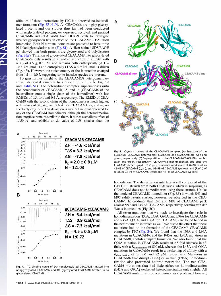
solved its crystal structure to a resolution of 1.85 Å (Fig. 5Aand Table S1). The heterodimer complex superimposes ontothe homodimers of CEACAM1, -5, and -6 (CEACAM6 of theheterodimer onto a single chain of the homodimer) with lowRMSDs of 0.5, 0.4, and 0.4 Å, respectively. The RMSD of CEA-CAM8 with the second chain of the homodimers is much higher,with values of 3.0, 4.6, and 2.6 Å, for CEACAM1, -5, and -6, re-spectively (Fig. 5B). This deviation is greater than that observed forany of the CEACAM homodimers, although the heterodimeriza-tion interface remains similar to them. It buries a smaller surface of1,450 Å2 and exhibits an SC value of 0.58, smaller than the
homodimers. The dimerization interface is still comprised of theGFCC′C′′ strands from both CEACAMs, which is surprising asCEACAM8 does not homodimerize using these strands. Unlikethe modeled CEACAM8 homodimer (Fig. 3B) in which R45 andM97 exhibit steric clashes, however, we observed in the CEA-CAM6/8 heterodimer that R45 and M97 of CEACAM8 packagainst V97 and L45 of CEACAM6, respectively, forming van derWaals interactions (Fig. 5C).All seven mutations that we made to investigate their role in
homodimerization (I30A, L45A, Q90A, and L96A for CEACAM6and R45A, Q90A, and L96A for CEACAM8) are found buried inthe heterodimeric interface as well. We tested the effect that thesemutations had on the formation of the CEACAM6–CEACAM8complex by ITC (Fig. S4). We found that the I30A and L96Amutations in CEACAM6, and the R45A and L96A mutations inCEACAM8, abolish complex formation. We also found that theQ90A mutation in CEACAM8 results in 2.5-fold increase in af-finity with a Kdimerization of 800 nM, whereas the L45A and Q90Amutations in CEACAM6 result in a weakening of affinity with aKdimerization of 12 μM and 22 μM, respectively. Mutations inCEACAM6 that disrupt (I30A) or weaken (L96A) homodime-rization also prevented heterodimerization. The two CEA-CAM6 mutations that had little effect on homodimerization(L45A and Q90A) weakened heterodimerization only slightly. AllCEACAM8 mutations produced monomeric proteins. However,
Fig. 4. ITC binding curve of (A) nonglycosylated CEACAM8 titrated intononglycosylated CEACAM6 and (B) glycosylated CEACAM8 titrated in toglycosylated CEACAM6.
Fig. 5. Crystal structure of the CEACAM6/8 complex. (A) Structure of theCEACAM6–CEACAM8 heterodimer. CEACAM6 and CEACAM8 are cyan andgreen, respectively. (B) Superposition of the CEACAM6–CEACAM8 complex(cyan and green, respectively), CEACAM6 dimer (magenta), and onto theCEACAM5 dimer (gray). (C) 2Fo–Fc composite omit maps of (Left) residues42–48 of CEACAM6 (cyan), and 93–99 of CEACAM8 (yellow), and (Right) ofresidues 93–99 of CEACAM6 (cyan) and 42–48 of CEACAM8 (yellow).
13564 | www.pnas.org/cgi/doi/10.1073/pnas.1509511112 Bonsor et al.

one mutation (Q90A) increases the affinity of heterodimerization,whereas the other two (R45 and L96A) results in no heterodimerformation. Taken together, these data suggest that the chemistriesof both homo- and heterodimerization interfaces are the same.
DiscussionCEACAMs are involved in and regulate diverse cellular func-tions, including cell adhesion and tumor suppression (10, 30). Celladhesion is achieved through dimerization in a trans arrangementvia the N-terminal IgV domains of CEACAMs (21, 22). Thus far,only CEACAM1 and CEACAM5 homodimers have been de-scribed structurally, with CEACAM5 forming homodimers withan affinity of 1 μM and CEACAM1 potentially forming higher-order oligomers (19). In this study, we measured the affinity ofthe homodimerization events for CEACAM1 and CEACAM5as 450 nM and 1.3 μM, respectively. This is much tighter than whatwe observed for CEACAM6, which forms a weak homodimer,and CEACAM8, which is effectively monomeric. These homo-dimerization constants span a range greater than three orders-of-magnitude, despite being modulated by only a few amino aciddifferences in the interface. All of our studies used only CEACAMN-terminal IgV domains. Notably, several studies have suggestedthat the presence of multiple IgC2 domains, each separated byflexible linkers, or the transmembrane motif of CEACAM1, GXXXG,increases the affinity of homophilic binding (21, 31, 32). These in-teractions could potentially strengthen cis engagements. Further-more, the CEACAM1 ectodomain (IgV+3IgC2) in liposomes formclusters of cis dimers (33). Formation of trans homodimers in theseliposomes increases the amount of cis dimer but disrupts the clusters(33), suggesting that the N-terminal domain is flexible enough aroundthe IgC2 domains to allow both cis and trans homodimer interactions.Using ITC, we observed a heterodimeric interaction between
CEACAM6 and CEACAM8 that was energetically favored overany other state of either protein. Glycosylation of CEACAMsoccurs in vivo with at least 35 different glycosylation formsidentified for CEACAM1 (34). Glycosylation of CEACAM6 andCEACAM6 only slightly weakened the interaction; however, wedid observe a change in the stoichiometry. We suspect that cer-tain glycosylation forms of CEACAMs create folded yet in-activate CEACAM molecules that could potentially prevent theformation of the CEACAM6–CEACAM8 interaction and possiblyCEACAM homodimers.Our data suggest that CEACAM6 and CEACAM8 likely function
as regulators of both cis and trans interactions for one another, aswell as potentially for other CEACAMs that bind to either or both ofthem. For example, a granulocyte expressing both CEACAM6 andCEACAM8 would predominately form cis CEACAM6/8 hetero-dimers (Fig. 6A), inhibiting CEACAM6 from engaging in trans in-teractions. As CEACAM8 is expressed exclusively on granulocytesand CEACAM6 is expressed additionally on epithelial cells of thegastrointestinal tract, a trans CEACAM6/8 heterodimer (Fig. 6B)could provide a mechanism by which the immune system can engagethe epithelia.Disruption of the CEACAM6/8 heterodimer has the potential
to increase the pool of free CEACAM6 and CEACAM8 tointerfere with other CEACAM functions. Several studies haveshown that CEACAM6 also forms heterodimers with CEACAM1,CEACAM5, and CEACAM3 (20, 30), whereas CEACAM8 canform a heterodimer with CEACAM1 (35). Although we observedno formation of these other heterodimers by ITC, we cannotdismiss the possibility that these heterodimers do form, but thatthey must do so at concentrations higher than the maximum(35 μM) that we used in our ITC experiments. The presence ofIgC2 domains, or the formation of clusters and therefore an in-crease in the local concentration of these proteins, may make theformation of heterodimers more energetically favorable. Severalsplice variants of CEACAM1 exist, with the long form contain-ing a cytosolic immunoreceptor tyrosine-based inhibitory motif,
allowing CEACAM1 to signal and several short forms that cannotsignal (7, 8). CEACAM6 and CEACAM8 are anchored in themembrane by a GPI moiety and therefore cannot signal in the samemanner as does long-form CEACAM1 (6). Overexpression of theshort form of CEACAM1 interferes with CEACAM1 signaling(36). Assuming that CEACAM6 and CEACAM8 can interact withCEACAM1 at high concentrations on the cell surface, they toohave the potential to disrupt CEACAM1 signaling through for-mation of heterodimers. Furthermore, the CEACAM1/8 in-teraction has been shown to inhibit Toll-like receptor 2-triggeredimmune responses (37). Thus, CEACAM6 could potentially mod-ulate Toll-like receptor-2 inhibition through its recruitment ofCEACAM8.In summary, we present the X-ray crystal structures of homo-
dimeric CEACAM6, monomeric CEACAM8, and heterodimericCEACAM6/8. Coupled with our quantitative biophysical analysesof these CEACAMs with themselves and others, we provide amolecular basis for the diverse oligomeric states of CEACAM IgVdomains that are important for cell adhesion and signaling incancer, infection and immunity.
Materials and MethodsFor details, see SI Materials and Methods.
Protein Production. All nonglycosylated CEACAMN-terminal IgV domains wereexpressed in E. coli and refolded in vitro from inclusion bodies, and werepurified to homogeneity by ion exchange and size-exclusion chromatography.
Fig. 6. Effects of CEACAM6 homodimers, CEACAM8monomers and CEACAM6/8 heterodimers on cis and trans interactions. (A) CEACAM6 and CEACAM8expressed on the same granulocyte energetically favors formation of cisCEACAM6/8 heterodimers. (B) Expression of CEACAM6 and CEACAM8 onepithelial cells and granulocytes, respectively, energetically favors formationof trans CEACAM6/8 heterodimers.
Bonsor et al. PNAS | November 3, 2015 | vol. 112 | no. 44 | 13565
BIOPH
YSICSAND
COMPU
TATIONALBIOLO
GY
Glycosylated CEACAM N-terminal IgV domains were expressed in HEK cellsand were purified to homogeneity by nickel affinity and size-exclusionchromatography.
Structural Analysis. CEACAM6 and CEACAM8 N-terminal domains wereconcentrated individually to 6 mg/mL or together to 9mg/mL and crystallizedby vapor diffusion in conditions optimized from the JCSG+ screen (Qiagen).Single crystals were flash-cooled in liquid nitrogen in mother liquor con-taining 20–30% (vol/vol) glycerol. X-ray diffraction data were collected at100K at the Advanced Photon Source beamline 23-ID-B and the StanfordSynchrotron Radiation Laboratory beamlines 7-1 and 12-2. CEACAM6 andCEACAM8 structures were determined by molecular replacement using amonomer of CEACAM5 (PDB ID code 2QSQ) as a search model and finalmodels built by iterative cycles of manual building in Coot (38) and re-finement in Refmac5 (39). The CEACAM6/8 structure was determined bymolecular replacement using the refined CEACAM6 and CEACAM8 struc-tures as search models; refinement proceeded as above.
Biophysical Analysis. Sedimentation equilibrium measurements of CEACAMswere performed using a Beckman-Coulter XL-I analytical ultracentrifuge at20 °C. Each CEACAM protein sample was prepared at three concentrations and
centrifuged at three rotor speeds. Scans were acquired at 280 nm with a stepsize of 0.001 and five averages per step. The data were globally analyzedusing the program WinNonLin (40). ITC experiments were performed usingan iTC200 instrument (Malvern). A typical experiment was carried out bytitrating CEACAM6 or CEACAM8 loaded in the syringe into a cell loadedwith CEACAM1, CEACAM3, CEACAM5, or CEACAM6 at ∼10:1 molar ratios at25 °C. Heats of dilutions were measured and subtracted from each data set.When no binding was observed at 25 °C measurements were also made at10 °C. All data were analyzed using Origin 7.0 software.
ACKNOWLEDGMENTS. This research was supported in part by the NIH underGrant S10RR15899 (to D.B.). This research used resources of the AdvancedPhoton Source, a US Department of Energy (DOE) Office of Science UserFacility operated for the DOE Office of Science by Argonne National Labo-ratory under Contract no. DE-AC02-06CH11357. Use of the Stanford Syn-chrotron Radiation Lightsource, Stanford Linear Accelerator Center NationalAccelerator Laboratory, is supported by the DOE, Office of Science, Office ofBasic Energy Sciences under Contract no. DE-AC02-76SF00515. The StanfordSynchrotron Radiation Lightsource Structural Molecular Biology Program issupported by the DOE Office of Biological and Environmental Research,and by the NIH, National Institute of General Medical Sciences (includingP41GM103393).
1. Zebhauser R, et al. (2005) Identification of a novel group of evolutionarily conservedmembers within the rapidly diverging murine Cea family. Genomics 86(5):566–580.
2. Hammarström S (1999) The carcinoembryonic antigen (CEA) family: Structures, sug-gested functions and expression in normal and malignant tissues. Semin Cancer Biol9(2):67–81.
3. Zheng J, et al. (2011) Carcinoembryonic antigen-related cell adhesion molecule 16interacts with alpha-tectorin and is mutated in autosomal dominant hearing loss(DFNA4). Proc Natl Acad Sci USA 108(10):4218–4223.
4. Schmitter T, Agerer F, Peterson L, Munzner P, Hauck CR (2004) Granulocyte CEACAM3is a phagocytic receptor of the innate immune system that mediates recognition andelimination of human-specific pathogens. J Exp Med 199(1):35–46.
5. Singer BB, et al. (2002) Carcinoembryonic antigen-related cell adhesion molecule 1expression and signaling in human, mouse, and rat leukocytes: Evidence for re-placement of the short cytoplasmic domain isoform by glycosylphosphatidylinositol-linked proteins in human leukocytes. J Immunol 168(10):5139–5146.
6. Tchoupa AK, Schuhmacher T, Hauck CR (2014) Signaling by epithelial members of theCEACAM family—Mucosal docking sites for pathogenic bacteria. Cell Commun Signal12:27.
7. Gray-Owen SD, Blumberg RS (2006) CEACAM1: Contact-dependent control of im-munity. Nat Rev Immunol 6(6):433–446.
8. Singer BB, et al. (2010) Deregulation of the CEACAM expression pattern causes un-differentiated cell growth in human lung adenocarcinoma cells. PLoS One 5(1):e8747.
9. Singer BB, et al. (2005) CEACAM1 (CD66a) mediates delay of spontaneous and Fasligand-induced apoptosis in granulocytes. Eur J Immunol 35(6):1949–1959.
10. Benchimol S, et al. (1989) Carcinoembryonic antigen, a human tumor marker, func-tions as an intercellular adhesion molecule. Cell 57(2):327–334.
11. Virji M, et al. (2000) Carcinoembryonic antigens are targeted by diverse strains oftypable and non-typable Haemophilus influenzae. Mol Microbiol 36(4):784–795.
12. Hill DJ, Virji M (2003) A novel cell-binding mechanism of Moraxella catarrhalis ubiquitoussurface protein UspA: Specific targeting of the N-domain of carcinoembryonic antigen-related cell adhesion molecules by UspA1. Mol Microbiol 48(1):117–129.
13. Leusch HG, Drzeniek Z, Markos-Pusztai Z, Wagener C (1991) Binding of Escherichia coliand Salmonella strains to members of the carcinoembryonic antigen family: Differentialbinding inhibition by aromatic alpha-glycosides of mannose. Infect Immun 59(6):2051–2057.
14. Obrink B (2008) On the role of CEACAM1 in cancer. Lung Cancer 60(3):309–312.15. Litkouhi B, et al. (2008) Overexpression of CEACAM6 in borderline and invasive mu-
cinous ovarian neoplasms. Gynecol Oncol 109(2):234–239.16. Duxbury MS, et al. (2004) A novel role for carcinoembryonic antigen-related cell
adhesion molecule 6 as a determinant of gemcitabine chemoresistance in pancreaticadenocarcinoma cells. Cancer Res 64(11):3987–3993.
17. Duxbury MS, Ito H, Benoit E, Ashley SW, Whang EE (2004) CEACAM6 is a determinantof pancreatic adenocarcinoma cellular invasiveness. Br J Cancer 91(7):1384–1390.
18. Fedarovich A, Tomberg J, Nicholas RA, Davies C (2006) Structure of the N-terminaldomain of human CEACAM1: Binding target of the opacity proteins during invasionof Neisseria meningitidis and N. gonorrhoeae. Acta Crystallogr D Biol Crystallogr62(Pt 9):971–979.
19. Korotkova N, et al. (2008) Binding of Dr adhesins of Escherichia coli to carcinoembryonicantigen triggers receptor dissociation. Mol Microbiol 67(2):420–434.
20. Oikawa S, et al. (1991) A specific heterotypic cell adhesion activity between membersof carcinoembryonic antigen family, W272 and NCA, is mediated by N-domains. J BiolChem 266(13):7995–8001.
21. Watt SM, et al. (2001) Homophilic adhesion of human CEACAM1 involves N-terminaldomain interactions: Structural analysis of the binding site. Blood 98(5):1469–1479.
22. Zhou H, Fuks A, Alcaraz G, Bolling TJ, Stanners CP (1993) Homophilic adhesion be-tween Ig superfamily carcinoembryonic antigen molecules involves double reciprocalbonds. J Cell Biol 122(4):951–960.
23. Huber M, et al. (1999) The carboxyl-terminal region of biliary glycoprotein controls itstyrosine phosphorylation and association with protein-tyrosine phosphatases SHP-1and SHP-2 in epithelial cells. J Biol Chem 274(1):335–344.
24. Kuroki M, et al. (2001) Identification and comparison of residues critical for cell-adhesionactivities of two neutrophil CD66 antigens, CEACAM6 and CEACAM8. J Leukoc Biol70(4):543–550.
25. Duxbury MS, et al. (2004) Overexpression of CEACAM6 promotes insulin-like growthfactor I-induced pancreatic adenocarcinoma cellular invasiveness. Oncogene 23(34):5834–5842.
26. Schölzel S, et al. (2000) Carcinoembryonic antigen family members CEACAM6 andCEACAM7 are differentially expressed in normal tissues and oppositely deregulatedin hyperplastic colorectal polyps and early adenomas. Am J Pathol 156(2):595–605.
27. Duxbury MS, Ito H, Zinner MJ, Ashley SW, Whang EE (2004) CEACAM6 gene silencingimpairs anoikis resistance and in vivo metastatic ability of pancreatic adenocarcinomacells. Oncogene 23(2):465–473.
28. Lasa A, et al. (2008) High expression of CEACAM6 and CEACAM8 mRNA in acutelymphoblastic leukemias. Ann Hematol 87(3):205–211.
29. Skubitz KM, Skubitz AP (2008) Interdependency of CEACAM-1, -3, -6, and -8 inducedhuman neutrophil adhesion to endothelial cells. J Transl Med 6:78.
30. Oikawa S, et al. (1989) Cell adhesion activity of non-specific cross-reacting antigen(NCA) and carcinoembryonic antigen (CEA) expressed on CHO cell surface: Homophilicand heterophilic adhesion. Biochem Biophys Res Commun 164(1):39–45.
31. Stern N, et al. (2005) Carcinoembryonic antigen (CEA) inhibits NK killing via in-teraction with CEA-related cell adhesion molecule 1. J Immunol 174(11):6692–6701.
32. Patel PC, et al. (2013) Inside-out signaling promotes dynamic changes in the carci-noembryonic antigen-related cellular adhesion molecule 1 (CEACAM1) oligomericstate to control its cell adhesion properties. J Biol Chem 288(41):29654–29669.
33. Klaile E, et al. (2009) The CEACAM1 N-terminal Ig domain mediates cis- and trans-binding and is essential for allosteric rearrangements of CEACAM1 microclusters.J Cell Biol 187(4):553–567.
34. Lucka L, et al. (2005) Identification of Lewis X structures of the cell adhesion moleculeCEACAM1 from human granulocytes. Glycobiology 15(1):87–100.
35. Jiang L, Barclay AN (2010) Identification of leucocyte surface protein interactions byhigh-throughput screening with multivalent reagents. Immunology 129(1):55–61.
36. Müller MM, Klaile E, Vorontsova O, Singer BB, Obrink B (2009) Homophilic adhesionand CEACAM1-S regulate dimerization of CEACAM1-L and recruitment of SHP-2 andc-Src. J Cell Biol 187(4):569–581.
37. Singer BB, et al. (2014) Soluble CEACAM8 interacts with CEACAM1 inhibiting TLR2-triggered immune responses. PLoS One 9(4):e94106.
38. Emsley P, Lohkamp B, Scott WG, Cowtan K (2010) Features and development of Coot.Acta Crystallogr D Biol Crystallogr 66(Pt 4):486–501.
39. Murshudov GN, et al. (2011) REFMAC5 for the refinement of macromolecular crystalstructures. Acta Crystallogr D Biol Crystallogr 67(Pt 4):355–367.
40. Johnson ML, Correia JJ, Yphantis DA, Halvorson HR (1981) Analysis of data from theanalytical ultracentrifuge by nonlinear least-squares techniques. Biophys J 36(3):575–588.
41. Aricescu AR, Lu W, Jones EY (2006) A time- and cost-efficient system for high-levelprotein production in mammalian cells. Acta Crystallogr D Biol Crystallogr 62(Pt 10):1243–1250.
42. Otwinowski Z, Minor W (1997) Processing of X-ray diffraction data collected inoscillation mode. Methods in Enzymology, ed Carter CW Jr. (Academic, Amsterdam),Vol 276, pp 307–326.
43. Kabsch W (2010) Xds. Acta Crystallogr D Biol Crystallogr 66(Pt 2):125–132.44. Winn MD, et al. (2011) Overview of the CCP4 suite and current developments. Acta
Crystallogr D Biol Crystallogr 67(Pt 4):235–242.45. Stein N (2008) CHAINSAW: A program for mutating pdb files used as templates in
molecular replacement. J Appl Cryst 41(3):641–643.46. Vagin A, Teplyakov A (1997) MOLREP: An automated program for molecular replacement.
J Appl Cryst 30(6):1022–1025.
13566 | www.pnas.org/cgi/doi/10.1073/pnas.1509511112 Bonsor et al.