Embed Size (px)
Citation preview

Mol. Cells, Vol. 7, No.1, pp. 13-20
Distribution of Succinic Semialdebyde Reductase in Rat Brain
Jon~ Eun Lee*, Soo Young Choi., Jae Wook Suk., Joung Woo Hong., Byung Kwon Yoo, Eui Yul Choi., Sang Ho Jangl t , Kyung Ah Park and Sung-Woo Ch02 Department of Anatomy, Yonsei University College of Medicine, Seoul 120-752, Korea; IDepartment of Genetic Engineering, College of Natural Sciences, Hallym University, Choonchun 200-702, Korea; 2Department of Biochemistry, College of Medicine, University of Ulsan, Seoul 138-040, Korea
(Received on July 26, 1996)
Succinic semialdehyde reductase (SSR) that catalyzes the reduction of succinic semialdehyde (SSA) to y-hydroxybutyrate (GHB) has been identified as one of the NADPHdependent aldehyde reductases. Reduction of SSA to GHB strongly supports the proposal that GHB biosynthesis may be an important step in the GABA shunt. It is pharmacologically significant in anesthesia, evoking the state of sleep, and an increase in brain dopamine level. Monoclonal antibodies against bovine brain succinic semialdehyde reductase were produced. Using the anti-succinic semialdehyde reductase antibodies, we investigated the distribution of brain succinic semialdehyde reductase in rat brain. The brain tissues were sectioned with a basis on the rat brain atlas of Paxinos and were stained by the immunoperoxidase staining method using monoclonal antibodies. In the section of the frontal lobe, immunoreactive cells were observed in the lateral septal area, the ventral paUidum, which belongs to the substantia innominata. We could observe immunoreactive cells in the reticular thalamic nucleus, which is closely related with 'sleeping', the basal nuclei of Meynert, which is associated with Alzheimer's disease, and hypothalamic nuclei. Immunoreactive cells were also shown in raphe nuclei or the reticular formation of the midbrain, cerebellum, and inferior olivary nuclei of the medulla oblongata. Succinic semialdehyde reductase-immunoreactive cells were distributed extensively in rat brain, especially immunoreactive cells were strongly observed in the areas associated with the limbic system and reticular formation.
y-Aminobutyric acid (GABA) is the major inhibitory neurotransmitter in the mammalian central nervous system, and more is known about its metabolic pathway than that of the amines, peptides and acethylcholine. GABA is mainly produced through enzymatic decarboxylation of glutamic acid by glutamic acid decarboxylase (GAD); degradation of putrescine also yields GABA (Lee and Cho, 1992). The regulation of the metabolic pathways of GABA is complex. The breakdown of GABA occurs through two possible pathways, one of which is through the consecutive action of two enzymes, GABA-transaminase and succinic semialdehyde dehydrogenase, resulting in the formation of succinic semialdehyde (SSA) and succinate (van Bemmelen et al. , 1985). Succinic semialdehyde dehydrogenase produces succinate, which enters the TCA cycle and is converted to glutamate. Glutamate is then recycled to GABA through GAD (GABA shunt) (Doherty and Roth, 1978). There is
* To whom correspondence should be addressed. t Present address: Chungam Biotech Research Center.
another breakdown pathway of GABA in the brain, and this involves the synthesis of y-hydroxybutyrate (GHB) (Doherty et ai. , 1975; Margolis, 1969). GABA transaminase converts GABA to succinic semialdehyde, which is subsequently converted to GHB by succinic semialdehyde reductase (SSR) (Cho et aI. , 1993; Vayer et al. , 1985; 1987). The latter reaction has been the focus of active research, but as of yet, the mechanism of the reduction pathway has not been established, and the research of the reduction of SSA to GHB has not attracted as much interest as that of the transamination of GABA to SSA. However, GHB research is ongoing and one of the results has been the discovery of a binding site that has a high affinity for GHB on cell membranes that form synapses (Benavides et al. , 1982b) and has been studied in the brains of rats and pigs (Hearl and Chur-
The abbreviations used are: GABA, y-aminobutyric acid; GAD, glutamate decarboxylase; GHB, y-hydroxybutyrate; SSA, succinic semialdehyde; SSR, succinic semiadehyde reductase.
© 1997 The Korean Society for Molecular Biology

14 Brain Succinic Semia\dehyde Reductase Mol. Cells
chich, 1985; Rivett et ai. , 1981). The synthesis of GHB is an important step in the GABA shunt.
y-Hydroxybutyrate (GHB) is an endogenous metabolite of the mammalian brain (Roth and Giarman, 1968). It produces CNS depression (sedation), and is thus used as a general anesthetic in humans (Laborit et ai., 1961; Roth and Giarman, 1968). GHB is unevenly distributed in the brain; for example, it is found in the hippocampus, cerebellum and diencephalon (Snead and Morley, 1981). SSR is located in neurons and nerve endings exclusively (Rumigny et ai., 1980). GHB may have a neurophysiological role as a putative neurotransmitter (Benavides et ai., 1982; Maitre and Mandel, 1984). Although the physiological role of GHB is unknown, aldehyde reductase which produces it has been thought to play a scavenging function in the removal of toxic aldehydes (Whittle and Turner, 1981). An enzyme that seems specific for the conversion of succinic semialdehyde to GHB has been purified and characterized in rat, human and bovine brain (Benavides et ai., 1982; Cash et ai., 1979; Cho et ai., 1993; Rumigny et ai. , 1980). This enzyme is a soluble enzyme located in the cytosol of neurons, and has an uneven regional distribution in the brain (Rumigny et ai. , 1981). Several pathways also have been suggested for GHB degradation, such as its conversion into succinic acid and other TCA cycle intermediates (Doherty and Roth, 1978), interconversion into GABA (Cho et ai., 1993; Margolis, 1969; Roth and Giarman, 1969), and breakdown via ~-oxidation (Walkenstein et ai. , 1964).
Some evidence indicates that GHB biosynthesis may be an important step in the GABA shunt, and that GHB may be of pharmacological significance acting as an anesthesia, evoking the state of sleep, and increasing the brain dopamine level (Hearl and Churchich, 1985; Rivett et ai., 1981). Despite many interesting observations, reduction of SSA to GHB has not received due attention, probably because the mechanism by which this reductive pathway operates in vivo is not yet clearly known. NADPH-dependant SSR was purified to apparent homogeneity from bovine brain by several chromatographic procedures (Cho et ai., 1993). Monoclonal antibodies against bovine brain SSR were produced and characterized (Choi et ai., 1995). The mAbs, which specifically recognized SSR on Western blots, were characterized and used as probes for a cross-reactivity study of the brain enzymes from some mammalian and an avian species (Choi et ai. , 1995).
It is with this background that we investigated the localization of SSR in rat brain. These results may clarify the reduction of SSA to GHB, and aid in understanding the physiological role of GHB.
Materials and Methods
Purification of succinic semiaidehyde reductase Succinic semialdehyde reductase from bovine brain
was purified according to the method of Cho pre-
viously described (Cho et ai. , 1993). During purification, enzyme activity was assayed in a solution containing 0.1 M potassium phosphate (PH 7.2), succinic semialdehyde (120 11M) and NADPH (50 11M). The oxidation of NADPH to NADP+ was measured by following the decrease in absorbance at 340 nm by the method of Hearl and Churchich (1985). One unit of enzyme activity was defined as the amount of enzyme required to oxidize 1 I1mol of NADPH per min at 25 ·C. Protein concentration was estimated by the Bradford procedure with bovine serum albumin as a standard (Bradford, 1976).
Production of monocLonai antibodies against succinic semiaidehyde reductase
Monoclonal antibodies against SSR were made according to the method of Choi et al. (1995). Purified enzymes described above were denatured by adding SDS (final concentration 0.1%) and by heat treatment for 1 min at 100 ·C. The denatured protein solution was mixed with an equal volume of complete Freund's adjuvant by sonication and injected into female BALB/c mice (6-8 weeks old). The first injection was followed by three booster injections at 3- to 4-week intervals.
The fusion experiments were performed as follows (Choi and Jeon, 1989; Galfre and Milstein, 1981). Three or four days after final injection, spleen was obtained from the animal and was cut into small pieces. Prepared spleen and Sp2/0-Ag-14 cell suspensions were combined and 1 ml of 50% polyethylene glycol 1500 in incomplete Dulbecco's modified Eagle's medium (DME) was added slowly. The fusion process was stopped by adding incomplete DME after 90 s. Twenty ml of incomplete DME was then added slowly for a period of 10 min. Cells were collected by centrifugation for 1 min at 650 g, suspended carefully in 20 ml of HAT medium (hypoxanthine, aminopterin, and thymidine) by swirling, and centrifuged for 1 min at 650 g. The cells were resuspended in 120 ml of HAT medium and the cell suspension was transferred into each well of 96-well plates.
About two weeks after the fusion , cultured supernatants were collected and first screened by immunodotblot analysis with purified enzyme as an antigen, and then by Western blot analysis. Positive clones, selected by the screening methods, were grown in tissue culture flasks (75 cm2
) and frozen in a liquid nitrogen tank. All positive clones were frozen first and cloned by limiting dilution after thawing.
Immunodot blotting and Western blotting confirmed the monoclonal antibodies to be against the purified enzyme (Towbin et ai. , 1979).
Tissue preparation for light microscope observation The animals were anesthetized by ether and then
perfused through the left ventricle with saline followed by 4% paraformaldehyde (0.1 M phosphate buffered saline, PBS, pH 7.4). The midbrain was re-

Vol. 7 (1997) long Eun Lee et al. 15
moved and placed in a fresh fixative for 4 to 24 h at 4 °c . Tissues were trimmed according to the atlas of Paxinos and Watson (1986). After being dehydrated and cleared, they were embedded in paraplast. Tissues were sectioned at 6 11m thickness, and placed on a slide treated with gelatin. One tissue section was immunohistochemically stained for SSR by using the peroxidase-antiperoxidase (PAP) method of Sternberger (1986), and the other section was given Nissl staining.
Immunoperoxidase staining All tissue sections were immunohistochemically
stained for the SSR monoclonal antibody by using the peroxidase-anti peroxidase (PAP) method of Sternberger (1986). Before staining, tissues were pretreated in 3% hydrogen peroxide solution for 10 min and several rinses in phosphate buffer (PH 7.4, containing 0.1 % Triton X-100). They were incubated in PBS solution containing SSR monoclonal antibody for 72 h at 4 °c, and washed. They also were incubated with goat anti-rabbit gamma globulin (GAR; Chernicon International Inc., Temecula, CA, U.S.A.) diluted 1:50 for 1 h at room temperature. The sections were rinsed several times with the same phosphate buffer and then placed in peroxidase-antiperoxidase (PAP) conjugates (Chemicon International Inc., Temecula, CA, U.S.A.) and diluted 1:100 for 1 h at room temperature. Following several rinses in phosphate buffer, the sections were placed in 0.05% diaminobenzidine (DAB; Sigma Chemical Co., St. Louis, MO, U.S .A.) solution containing 0.01% hydrogen peroxide for 10 min and washed with distilled water. Controls were prepared using incubation solution without primary antibodies. All incubation steps were performed in a humidified chamber.
Tissue slides were made according to a general tissue preparation procedure.
Immunocytochemical staining for electron microscopical preparation
For electron-microscopic observation, the animals were perfused through the left ventricle with saline followed by 3% paraformaldehyde, 3% glutaraldehyde, and 0.1 % picric acid in 0.1 M phosphate buffer (PH 7.4).
The tissue was then sectioned on a vibratome at 50 11m. The free-floating sections were then incubated in 1 % sodium borohydride in 0.1 M phosphate buffer for 30 min followed by a six 5-min rinse in phosphate buffer. The sections were exposed to a graded series of alcohols (10%, 25%, 40%, 25%, 10%) in phosphate buffer for 5 min each to enhance penetration of antibodies while preserving ultrastructural detail (Light et ai., 1983). All tissue sections were immunocytochemically stained for SSR monoclonal antibody by using the peroxidase-antiperoxidase (PAP) method of Sternberger (1986). For electron microscopy, the immunocytochemically-stained tissue
was thoroughly rinsed in a phosphate buffer and placed in 1% osmium tetroxide in 0.1 M phosphate buffer for 30 min. The tissue was then rinsed in a maleate buffer (PH 6.0), stained with uranyl acetate in maleate buffer, rinsed, dehydrated, and embedded with EPON mixture. A selected area was trimmed and mounted on an EPON chuck with a drop of EPON. The thin sections were cut and mounted on the copper grids and viewed without lead citrate staining with a Philips or Hitachi electron microscope.
Results and Discussion
Succinic semialdehyde reductase (SSR) was purified through FPLC Mono-Q chromatography from bovine brain by Cho et al. (1993). We obtained a single protein band on an SDS gel, and monoclonal antibodies against bovine brain succinic semialdehyde reductase were produced. In order to enhance the immunogenicity of the protein, purified enzyme was denatured in the presence of SDS and injected into animals (Choi et al., 1995). From two fusion experiments, 16 positive clones were initially screened by immunodotblot analysis and nine hybridomas out of the 16 clones were finally selected. Since the monoclonal antibodies recognized SSR from several mammalian and an avian species (Choi et al., 1995), cross-reactivity of the mAbs with SSR from rat brain occurred.
y-Hydroxybutyrate (GHB) is an endogenous metabolite of the mammalian brain (Giarman, 1970), and might play a neurophysiological role act as a putative neurotransmitter (Benavides et al., 1982; Maitre and Mandel, 1984). An enzyme that seems specific for conversion of succinic semialdehyde to GHB has recently been isolated and characterized in rat, human and bovine brain (Benavides et al., 1982; Cash et al., 1979; Cho et al., 1993; Rurnigny et at., 1980). Despite the potential significance of SSR, the distribution of SSR has not yet been established.
Snead and Morley (1981) investigated the concentration of GHB in developing and mature rat, monkey, and human brains, together with postmortem GHB concentration. They discovered that there is no big change in the concentration of GHB after death and that GHB is most concentrated in the hypothalamus and cerebral cortex of rats. Also, from the 12 to 14th day after birth there is a significant decrease in the concentration. Furthermore, Vayer et al. (1987), measured the concentration of GHB in the mammalian brain using gas chromatography, and reported high concentrations in the hippocampus, cerebellum, cerebrum of a guinea pig, and especially in the hippocampus of monkeys and the hippocampus, thalamus, and the cerebrum of a rat. Relatively low concentrations were discovered in the cerebral cortex (Snead, 1987). However, most of these results are based on measurement of GHB concentrations in tissue, and there is still no report of the direct observation of

16 Brain Succinic Semialdehyde Reductase Mol. Cells
a b
Figure 1. Distribution of the succinic semialdehyde reductase in a rat brain. a) The section through 0.70 mm from the bregma and 9.70 mm from the interaural line. b) The section through - 1.40 mm from the bregma and 7.60 mm from the interaural line. c) The section through - 3.30 mm from the bregma and 5.70 mm from interaural line. d) The section through - 5.80 mm from the bregma and 3.20 mm from the interaural line of the midbrain level. e) The section through - 7.64 from the bregma and 1.36 mm from the interaural line. f) The section through - 10.04 mm from the bregma and - 1.04 mm from the interaural line. 1-5, cerebellar lobules; 2n, optic nerve; 3V, 3rd ventricle; 3n, oculomotor nerve; 4n, trochlear nerve; 4V, 4th ventricle; 7n, facial nerve; 8vn, vestibulocochlear nerve; aca, anterior commissure; AcbC, accumbens nucleus core; AD, anterodorsal thalamic nucleus; AF, amygdaloid fissure; AH, anterior hypothalamic area; AM, anteromedial thalamic nucleus; Amg, amygdaloid complex area; APT, anterior pretectal nucleus; Aq, cerebral aqueduct; Arc, arcuate nucleus; ATg, anterior tegmental nucleus; A V, anteroventral thalamic nucleus; B, basal nucleus of Mynert; bas, basilar artery; bic, brachium of inf. colliculus; BIC, nucleus of inf. colliculus brachium; CC, corpus callosum; cg, cingulum; CG, central gray; CL, centrolateral thalamic nucleus; CM, central medial thalamic nucleus; CPu, caudate putamen; ctg, central tegmental tract; DA, dorsal hypothalamic area; D3V, dorsal 3rd ventricle; DB, nucleus of diagonal band; DEn, dorsal entopiriform nucleus;
Vol. 7 (1997) long Eun Lee et ai. 17
the distribution of GHB or its synthesizing enzyme in tissue.
In this paper, we investigated the distribution of SSR in rat brain using the anti-succinic semialdehyde reductase antibodies. The brain tissues were sectioned similarly to those seen in the rat brain atlas of Paxinos (1986) and were stained by the immunoperoxidase staining method using monoclonal antibodies. In the section through 0.70 mm from the bregma and 9. 70 mm fro m the interaural line, immunoreactive cells were mainly observed in the lateral septal nuclei (Figs. 1a and 2a), vertical limb of diagonal band nucleus (Fig. 1a), and ventral pallidum (Fig. 1a). These areas belong mainly to the substantia innominata (Perry et ai. , 1984). In the section through - 1.40 mm from the bregma and 7.60 mm from the interaural line (Fig. 1b), immunoreactive cells were shown in caudateputamen of the basal ganglia (Fig. 1b), basal nucleus of Meynert (Fig. 2b), and amygdaloid complex (Fig. 1b). And in the paraventricular thalamic nucleus (Fig. 1b), reticular thalamic nucleus (Fig. 2c) of the thalamus, paraventricular hypothalamic nucleus (Fig. 2e) and supraoptic nucleus (Fig. 2f) of the hypothalamus, immunoreactive cells were also observed. The reticular thalamic nucleus is closely related functionally to the 'sleeping' area (Macchi and Bentivoglio, 1986; Steridae and Llinas, 1988). This area, which receives the afferant fibers from most of the thalamic nucleus, and sends the efferent fibers to the thalamic nucleus, is believed to play an important role in neural conduction in the thalamus (Russchen et ai., 1987; Steridae and Llinas,
Figure 1. Continued.
1988). SSR-immunoreactive cells were found in the basal nucleus of Meynert, which also belongs to substantia innominata. This nucleus is composed of cells containing mainly acetylcholine, and has been shown to undergo significant changes in Alzheimer's disease (Rogers et ai., 1985; Whitehouse et ai., 1982). The immunoreactive cells in the basal nucleus of Meynert were relatively large. In the section through - 3.30 mm from the bregma and 5.70 mm from the interaural line, immunoreactive cells were observed clearly in some basal ganglia and thalamic areas (Fig. 1c), especially the globus pallidus (Fig. 2d), reticular thalamic nucleus, and ventral posterolateral nucleus (Fig. 1c), etc. Immunoreactive cells were also found in the hypothalamic area, raphe nuclei, and reticular formation. In the section through - 5.80 mm from bregma and 3.20 mm from the interaural line of the midbrain level (Fig. 1d), there were some areas that immunostained against SSR monoclonal antibodies, such as dorsal raphe nuclei, median raphe nuclei (Fig. 1d), the rostal linear raphe nuclei (Fig. 3a), red nucleus (Fig. 3a), substantia nigra (Fig. 3b), interpeduncular nucleus (Fig. 1d), etc. These areas are also related to the limbic system, especially the dorsal raphe, median raphe, dorsal and ventral tegmental nucleus, periaqueductal gray matter, and the interpeduncular nucleus, which, combined, constitute the 'midbrain limbic area' (Robertson and Kaitz, 1981). At the level of the inferior colliculus of the midbrain (Fig. Ie), immunoreactive cells were also observed in the doral and median raphe nuclei (Figs. 3c and Ie), anterior tegmental nucleus (Fig. Ie), and reticular for-
OG, dentate gyrus; OMH, dorsomedial hypothalamic nucleus; OpMe, deep mesencephalic nucleus; DR, dorsal raphe nucleus; En, entopiriform nucleus; Ent, enrorhinal cortex; EW, Edinger-Westphal nucleus; f, fornix ; Fl, flocculus; Fr, frontal cortex; GP, globus pallidus; HC, hippocampus; HOB, nucleus of horizontal limb of diagonal band; ic, internal capsule; IC, inferior colliculus; ICj, islands of Calleja; IMO, intermediodorsal thalamic nucleus; IMLF, interstitial nucleus of mlf; JP, interpeduncular nucleus; LC, locus coeruleus; LOOM, laterodorsal thalamic nucleus, dorsomedial; LOVL, laterodorsal thalamic nucleus, ventrolateral; LH, lateral hypothalamic nucleus, II, lateral lemniscus; LP, lateral posterior thalamic nucleus; LPMR, lateral posterior thalamic nucleus, mediorostral ; LSD, lateral septal nucleus, dorsal; LSI, lateral septal nucleus, intermediate; LSV, lateral septal nucleus, ventral ; LV, lateral ventricle; m5, motor root of trigeminal nerve; MA3, medial accessory oculomotor nucleus; mcp, middle cerebellar peduncle; MOC, mediodorsal thalamic nucleus, central; MOM, mediodorsal thalamic nucleus, medial; MOL, mediodorsal thalamic nucleus, lateral, ME, median eminence; Me5, mesencephalic trigeminal nucleus; mfbb, medial forebrain bundle; MG, medial geniculate body; ml, medial lemniscus; mlf, medial longitudinal fasciculus; MnR, median raphe nucleus; Mo5, motor trigeminal nucleus; MPA, medial preoptic area; MS, medial septal nucleus; mt, mammillothalamic tract; MTu, medial tuberal nucleus; NIC, nucleus of inf. colliculus; OC, opt, optic tract; OT, nucleus of optic tract, ox, optic chiasm; P5, peritrigeminal zone; PaAP, paraventricular hypothalamic nucleus; Par, parietal cortex; PC, paracentral thalamic nucleus; PFl, paraflocculus; Pir, piriform nucleus; PLF, posterolateral fissure; Pn, pontine reticular nucleus; PnO, pontine reticular nucleus; Po, posterior thalamic nuclear group; PO, paraolivary nucleus; PPTg, pedunculopotine tegmental nucleus; Pr5 , priciple sensory trigeminal nucleus, PrF, primary fissure ; PRh, perirhinal cortex ; PrS, presubiculum; PT, paratenial thalamic nucleus; PYA, paraventricular thalamic nucleus; PVP, paraventricular thalamic nucleus, posterior, py, pyramidal tract; R, red nucleus; Re, reuniens thalamic nucleus; RF, rhinal fissure; RLi, rostral linear raphe nucleus; Rmg, raphe magnus nucleus; Rt, reticular thalamic nucleus; RtTg, reticulotegmental nucleus; s5, sensory root of trigeminal nerve; SCh, suprachiasmatic nucleus; SC, sup . colliculus; scp, superior cerebellar peduncle; SHi, septo hippocampal nucleus; SI, substantia innominata; sm, stria medullaris thalami; SNC, substantia nigra compacta; SNL, substantia nigra lateral ; SNR, substantia nigra reticular, SO, supraoptic nucleus; Subl, subincertal nucleus; Te, temporal cortex; Tg, tegmental nucleus; tz, trapezoid body; TZ, nucleus of trapezoid body; VC, ventral cochlear nucleus; YEn, ventral entopiriform nucleus; VM, ventromedian thalamic nucleus; VMH, ventromedian hypothalamic nucleus; VP, ventral posterior thalamic nucleus; VPM, ventral posteromedial thalamic nucleus; VPL, ventral posterolateral thalamic nucleus; xscp, decussation of sup. cerebellar peduncle; ZI, zona incerta.

18 Brain Succinic Semialdehyde Reductase Mol. Cells
, , • ,.J ~
• ~ f,~ .' \ .'
"" . " 2(1 ,--
;. . -
Figure 2. SSR-immunoreactive cells in the upper brain of a rat. a) Septal nucleus (Sp) (bar=40 !-lm). b) Basal nucleus of Meynert (B). Some neuron of the reticular thalamic nucleus (RT) are also observed (bar=80 !-lm). c) Reticular thalamic nucleus (RT). There are many immunoreactive cells in each area (bar=52.6 !-lm). d) Globus pallidus (GP). Immunoreactive cells are observed distinctly. The internal capsule (ic) is also shown (bar=105 !-lm). e) Paraventricular hypothalamic nuclei (PY) are observed by the 3rd ventricle (3Y) (bar=80 !-lm). f) Neurons of the supraoptic nucleus (SO) are immunostained against SSR (bar=80 !-lm) .
.. . aq
SN~ OR RLi
.. 3a
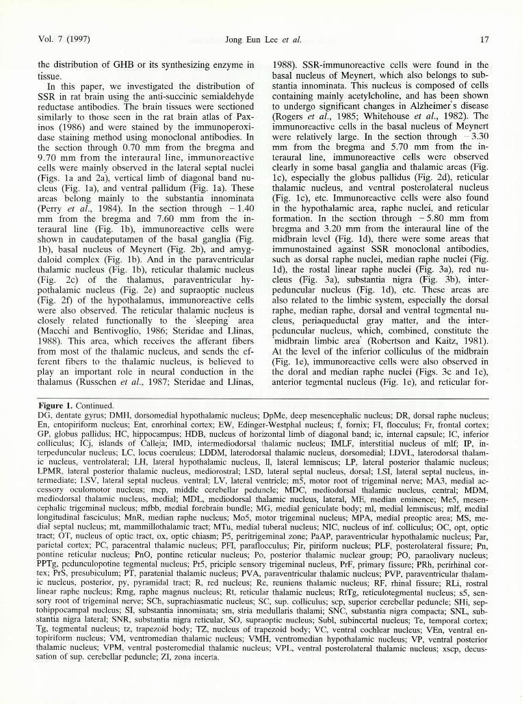
Figure 3. SSR-immunoreactive cells in the brain stem of a rat. a) Immunoreactive cells were observed in a rostral linear raphe nucleus (RLi) and red nucleus (R) (bar=105 !-lm). b) Substantia nigra (SN) (bar=80 !-lm). c) dorsal raphe nucleus (DR). A cerebral queduct (aq) is also shown (bar=105 !-lm). d) Immunoreactive cells are mainly distributed to mesencephalic trigeminal nuclei (Me5), ventral cochlear nuclei (YCA), and pontine reticular nuclei (Pn), and a superior cerebellar peduncle (scp) and trigeminal nerve (s5) are also observed (bar=263 !-lm). e) Inferior olivary nucleus, there are many immunoreactive cells (bar=263 !-lm). f) Cerebellar cortex, many Purkinje cells are immunostained against SSR (bar=105 !-lm).

Vol. 7 (1997) Jong Eun Lee et al. 19
Figure 4. Electron micrograph of SSR-immunoreactive structures in a rat brain. a) Reticular thalamic nucleus. SSR-immunoreactive structures (arrows) are observed around microtubules in the axoplasm of unmyelinated axons (UA) (bar:::1 /-lm). b) Dorsal raphe nucleus. SSR-immunoreactive structures (arrows) are also observed within the mitochondrial matrix (bar:::1 11m).
mation (Fig. I e). This level was sectioned through - 7.64 from bregma and 1.36 mm from the interaural line. In the section through - 10.04 mm from the bregma and - 1.04 mm from the interaural line (Fig. It), immunoreactive cells were found mainly in the mesencephalic trigeminal nucleus (Fig. 3d), ventral cochlear nucleus (Fig. 3d), pontine reticular nucleus (Fig. 3d), and inferior olivary nucleus complex (Fig. 3e) of the brain stem, and were also observed in the dentate nucleus of the cerebellum (Fig. It) and the cerebellar cortex, especially in Purkinje cells (Fig. 3t). These results partly coincided with other biochemical studies (Hechler et al., 1987; 1992; Snead, 1987). Hechler et al. (1987) investigated the regional distribution of high-affinity y-eH]hydroxybutyrate binding sites, which were absent in the caudal parts of the brain (cerebellum, pons, and medulla). According to these results, SSR-immunoreactive cells are widely distributed in the CNS in areas such as the inferior olivary complex, medulla oblongata, and the cerebellum.
Observations through the electron microscope revealed SSR-immunoreactive particles in the axoplasm. The positive structures were found mainly around microtubules in the axoplasm, and within the mitochondrial matrix. In unmyelinated nerve fibers, high electron dense positive materials were also observed around the micro tubules (Fig. 4a) and in the mitochondrial matrix (Fig. 4b). SSR is a hydrophilic enzyme that is present mainly in the cytoplasm of neurons, and has been reported to not be evenly distributed in the brain (Rumigny et ai., 1981). However, SSR-immunoreactive particles were found not only in the axoplasm, but also within the mitochondrial matrix.
Succinic semialdehyde reductase-immunoreactive cells were distributed extensively in rat brain, and were observed particularly in the areas associated with the limbic system and reticular formation. We identified the presence of a specific enzyme associated with the reduction of SSA to GHB in rat
brain by immunoperoxidase staining using mAbs against SSR. These results were associated with the function of y-hydroxybutyrate, which is the production of CNS depression (sedation), and it is used as a general anesthetic for humans.
Acknowledgment
This study was supported by a basic research medical fund, Ministry of Education, Korea for 1995.
References
Benavides, 1., Rumigny, 1. F., Bourguignon, J . J., Cash c., Wermuth, C. G., Mandel, P., Vincendon, G. , and Maitre, M. (1982a) Life Sci. 30, 953-96l.
Benavides, 1. , Rumigny, J. F., Bourguignon, J. J ., Wermuth, C. G., Mandel, P., and Maitre, M. (1982b) 1. Neurochem. 38, 1570-1575.
Bradford, M. M. (1976) Anal. Biochem. 72, 248-254. Cash, C. D. , Maitre, M., and Mandel, P. (1979) 1. Neu
rochem. 33, 1169-1175. Cho, S. W., Song, M. S. , Kim, G. Y , Choi, E. Y, Kang,
W. D., and Choi, S. Y (1993) Eur. 1. Biochem. 211, 757-762.
Choi, E. Y ., and Jeon, K. W. (1989) Exp. Cell Res. 185, 154-165.
Choi, E. Y, Park, S. Y , Jang, S. H., Song, M. S., Cho, S. w., and Choi, S. Y (1995) 1. Neurochem. 64, 371-377.
Doherty, J. D., Snead, O. c., and Roth, R. H. (1975) Anal. Biochem. 65, 268-277.
Doherty, J. D., Hattox, S. E. , Snead, O. c., and Roth, R. H. (1978) 1. Pharmacol. Exp. Ther. 20, 130-139.
Galfre, G., and Milstein, C. (1981) Methods Enzymol. 73, 3-47.
Hearl, W. G., and Churchich, 1. R. (1985) 1. BioI. Chem. 239, 357-36l.
Hechler, V., Weissmann, D., Mach, E., Pujol, J., and Maitre, M. (1987) 1. Neurochem. 49, 1025-1032.
Hechler, V., Goballe, S., and Maitre, M. (1992) Brain Res. 572, 345-348.
Laborit, H., Jouany, J. , Gerard, J. , and Fabiani, P. (1961) Neuropsychopharmacology 2, 490-497.

20 Brain Succinic Semialdehyde Reductase Mol. Cells
Lee, J. E., and Cho, Y. D. (1992) Biochem. Biophys. Res. Commun. 189, 450-454.
Light, A. R., Kavookjian, A. M., and Petrutz, P. (1983) Somatosen. Res. 1, 33-50.
Macchi, G. , and Bentivoglio, M. (1986) in Cerebral Cortex (Jones, E. G. , and Peters, A., eds) Vol. 5, pp. 355-401, Plenum, New York.
Margolis, R. K. (1969) Biochem. Pharmacol. 18, 1243-1246.
Maitre, M., and Mandel, P. (1984) C. R. Seances Acad. Sci. 1lI 298, 341-345.
Pax inos, G., and Watson, C. (1986) The Rat Brain in Stereotaxic Coordinates, 2nd Ed, Academic Press, New York.
Perry, R. H. , Candy, J. M., Perry, E. K., Thompson, J., and Oakley, A. E. (1984) 1. Anal. 138, 713-732.
Rivett, A. 1. , Smith, I. L., and Tipton, K. F. (1981) Biochem. Pharmacal. 30, 741-747.
Robertson, R. T. , and Kaitz, S. S. (1981) 1. Comp. Neurol. 195, 501-525.
Rogers, J. D., Brogan, D., and Mirra, S. S. (1985) Ann. Neurol. 17, 162-170.
Roth, R. H. , and Giarman, N. J. (1968) Biochem. Pharmacol. 17, 735-739.
Roth, R. H. , and Giarman, N. J. (1969) Biochem. Pharmacal. 18, 247-250.
Rumigny, J. F., Maitre, M., Cash, c., and Mandel, P.
(1980) FEBS Lett. 117, 111-116. Rumigny, J. F., Maitre, M. , Cash, C., and Mandel, P.
(1981) 1. Neurochem. 36, 1433-1438. Russchen, F. T. , Amaral, D. G., and Price, J . L. (1987) 1.
Comp. Neural. 256, 175-210. Snead, O. C. (1987) 1. Neurochem. 48, 196-201. Snead, O. c., and Morley, B. J. (1981) Dev. Brain Res. 1,
579-589. Steridae, M., and Llinas, R. (1988) Physiol. Rev. 68, 649-
742. Sternberger, L. A. (1986) Immunocytochemistry, 3rd Ed,
John Wiley, New York. Towbin, H. , Staehelin, T. , and Gordon, J. (1979) Prac.
Natl. A cad. Sci. USA 76, 4350-4354. van Bemmelen, F. J., Schouten, M. J., Fekkes, D., and
Bruinvels, J. (1985) 1. Neurochem. 45, 1471-1474. Vayer, P., Schmitt, M. , Bourguignon, 1. 1. , Mandel, P., and
Maitre, M. (1985) FEBS Lett. 190, 55-60. Vayer, P., Mandel, P., and Maitre, M. (1987) Life Sci. 41,
1547-1557. Walkenstein, S. S., Wiser, R. , Gudmundsen, c., and Kim
mel, H. (1964) Biochim. Biophys. Acta 86, 640-642. Whitehouse, P. 1. , Price, D. L., Struble, R. G. , and Aark, A.
W. (1982) Science 215, 1237-1239. Whittle, S. R. , and Turner, A. J. (1981) Biochim. Biophys.
Acta 657, 94-105.