Embed Size (px)
Citation preview
REVIEW
Distinctive properties of metastasis-initiating cellsToni Celià-Terrassa and Yibin Kang
Department of Molecular Biology, Princeton University, Princeton, New Jersey 08544, USA
Primary tumors are known to constantly shed a largenumber of cancer cells into systemic dissemination, yetonly a tiny fraction of these cells is capable of formingovert metastases. The tremendous rate of attrition duringthe process of metastasis implicates the existence of a rareand unique population of metastasis-initiating cells(MICs). MICs possess advantageous traits that may origi-nate in the primary tumor but continue to evolve duringdissemination and colonization, including cellular plas-ticity, metabolic reprogramming, the ability to enter andexit dormancy, resistance to apoptosis, immune evasion,and co-option of other tumor and stromal cells. Better un-derstanding of the molecular and cellular hallmarks ofMICs will facilitate the development and deployment ofnovel therapeutic strategies.
The majority of cancer deaths is caused by metastasis,when cancer cells manage to escape the primary tumor,survive the treacherous transit through the lymphovascu-lar system, and eventually form secondary tumors in dis-tant organs (Gupta and Massague 2006; Valastyan andWeinberg 2011;Wan et al. 2013). This is a highly challeng-ing process with a tremendous rate of attrition; it is esti-mated that only <0.02% of disseminated tumor cells(DTCs) are able to successfully seed metastases (Luzziet al. 1998; Cameron et al. 2000; Chambers et al. 2002).As a result, although tumor dissemination can occur rela-tively early in cancer progression (Husemann et al. 2008;Massard et al. 2011; Kang and Pantel 2013), sometimeseven at the preneoplastic stage (Rhim et al. 2012), an ex-tended gap time often exists between the formationof the primary tumor and clinicalmanifestations ofmetas-tasis (Yachida et al. 2010;Vanharanta andMassague 2013).Therefore, the capability to initiatemetastatic growth is amajor bottleneckduring cancer progression and representsan ideal window for therapeutic intervention (Fig. 1A).
Metastasis-initiating cells (MICs), by definition, arecancer cells capable of seeding clinically significant meta-static colonies in secondary organs. Like their primary tu-
mor counterparts, the tumor-initiating cells (TICs), MICscan hijack some of the normal stem cell pathways to in-crease cellular plasticity and stemness, which providethem with multiple malignant advantages. However,the MICs must possess additional capabilities that allowthem to survive the metastatic cascade and function asTICs in an organ microenvironment distinctively differ-ent from the primary tumor. These cells form the link be-tween the primary tumor and subsequent metastasis butare exceedingly difficult to identify, track, and character-ize. Even the origin of MICs remains elusive; they mightexist at the primary tumors or emerge during the journeythrough the metastatic cascade (where exposure to ex-treme stress conditions may select for MIC abilities) ormay acquire such capabilities only after arriving at the dis-tant site and engaging the stromal components (Fig. 1B).Such unique challenges in identifying and analyzingMICs demand research tools beyond what are commonlyavailable and used in the study of TICs, such as in vitrotumorsphere assays, in vivo limited dilution tumor initia-tion studies, and analysis using cancer stem cell (CSC)surface markers. In the past few years, new and emergingtechnologies have begun to enable the study of MICs inanimal and clinical models. Genomic sequencing studieshave provided genome-wide comparisons between pri-mary tumors andmatched distantmetastases from cancerpatients and animal models (Campbell et al. 2010;Yachida et al. 2010; McFadden et al. 2014; Gundemet al. 2015). Gene expression analysis at the single-cell lev-el has become a powerful tool to analyze the populationdynamics of tumor cells during metastatic evolution(Lawson et al. 2015). In addition, lineage tracing and bar-code sequencing studies have also been applied to studythe interclonal interactions and population dynamics(Maddipati and Stanger 2015; Wagenblast et al. 2015). Ad-vances in animal modeling of metastasis and detailedanalysis of tumor-intrinsic pathways and tumor–stromalcross-talk further provided unprecedented insights intothe molecular mechanism of metastasis initiation. Someconsensus regarding the hallmarks of MICs has started
[Keywords: cancer metastasis; plasticity; epithelial–mesenchymaltransition; metastasis-initiating cells; metastatic niche]Corresponding author: [email protected] is online at http://www.genesdev.org/cgi/doi/10.1101/gad.277681.116.
© 2016 Celià-Terrassa and Kang This article is distributed exclusively byCold Spring Harbor Laboratory Press for the first six months after the full-issue publication date (see http://genesdev.cshlp.org/site/misc/terms.xhtml). After six months, it is available under a Creative Commons Li-cense (Attribution-NonCommercial 4.0 International), as described athttp://creativecommons.org/licenses/by-nc/4.0/.
892 GENES & DEVELOPMENT 30:892–908 Published by Cold Spring Harbor Laboratory Press; ISSN 0890-9369/16; www.genesdev.org
Cold Spring Harbor Laboratory Press on October 25, 2021 - Published by genesdev.cshlp.orgDownloaded from

to emerge from these studies, including the maintenanceof TIC ability, the flexibility to undergo bidirectional tran-sitions between the epithelial and mesenchymal states,resistance to anoikis and apoptosis, entry into and exitfrom dormancy, evasion of immune system attack, repro-gramming of metabolic activities to adapt to the differentnutrient and oxidative stresses, interclonal cooperations,and the ability to build or take advantage of a supportivestromal niche. Underlying all of these myriad propertiesof MICs is their remarkable cellular plasticity that allowsthem to survive and thrive against all odds. In this review,we summarize the main tumor-intrinsic hallmarks ofMICs and their dynamic interactions with the extrinsicenvironment to manifest their metastasis-forming activi-ties and discuss the possible strategy of targeting MICs incancer therapeutics.
Genomic evolution of MIC traits
Cancer genome sequencing studies have shown that ma-lignant tumors emerge from the sequential accumulationofmutations in driver genes involved in three core cellularprocesses during tumor initiation: cell fate regulation,genome maintenance, and cell survival (Vogelstein et al.2013). These altered processes favor primary tumor in-itiation and may still be essential for MICs to seed metas-tases. However, it was previously unknown whetheradditional driver mutations are needed for metastasis tooccur. Genome sequencing studies have shown high de-grees of similarities among mutations in primary tumorsand metastases (Yachida et al. 2010). The most remark-able finding of these studies is that no consistent metasta-sis-specific mutations have been found other than thosethat are already commonly found in primary tumors(Bozic et al. 2010; Campbell et al. 2010; Yachida et al.2010), suggesting that importantmutations formetastasisare already present in the primary tumor site. These stud-ies frequently reveal a greater enrichment of clonal popu-lations rather than an acquisition of new mutations, asobserved in pancreatic cancer metastasis with amplifica-tions of MYC, RASG13D, and CCDN1 (Campbell et al.2010) and in lobular ER+ breast cancer with ERBB2muta-tions (Shah et al. 2009).A recent study usingwhole-exomesequencing analysis of experimental metastasis models ofmultiple cancer types has shown that metastatic compe-tence arises from the selection of pre-existing mutations,such as RASG13D and BRAFG464V, in heterogeneous popu-lations without the need for additional mutations (Jacobet al. 2015). The selection of these oncogenic pathways fa-vors their prevalence in metastasis, indicating that theyare important contributors to metastatic fitness andthus may be required for MICs. Overall, these findingssuggest that a large number of metastatic properties maybe already forming in the primary tumor through enrich-ment of existing oncogenic mutations that favor metasta-sis initiation.Beyond realignment of genomic mutations, epigenetic
regulationmight be amajor source ofMIC traits, especial-ly in later steps of metastasis. After tumor cells escape theprimary site, the epigenome is subjected to microenviron-mental signal modulation, conferring cellular plasticityand adaptability to new and inhospitable conditions (Sef-tor et al. 2006; Hendrix et al. 2007; Tam and Weinberg2013). Indeed, multiple studies have unveiled evidenceof specific epigenetic pathways involved in themetastaticprogression of different cancer types (Cunha et al. 2014;Gu et al. 2015; Okada et al. 2015; Tang et al. 2015). There-fore, the combination of genetic and epigenetic eventsduring the course of metastasis likely determines the ac-quisition of MIC traits.
Cell fate determinants as regulators of MICs
Adult tissues are hierarchically organized and tightly con-trolled by lineage-specific transcription factors to regulategrowth and differentiation and maintain the homeostasis
Figure 1. Metastasis-initiating cells (MICs) in cancer progres-sion andmetastasis. (A) Schematic depiction of the typical courseof metastatic progression of an early-stage cancer. In many clini-cal cases, tumor dissemination precedes diagnosis of the primarytumor. Surgical debulking and systemic adjuvant treatment elim-inate most of tumor cells at the primary site and throughout thebody. However, a small proportion of DTCs survives the systemictreatment. After a period of dormancy with no clinical sign ofcancer, which could last for months to decades, clinically detect-ablemetastases start to emerge. The subsequent lines of systemictreatment often only temporarily reduce the tumor burden beforemetastatic lesions develop resistance and eventually overwhelmthe patients. The ability to initiatemetastatic outgrowth is there-fore a major bottleneck in cancer progression. (B) Representationof the sequence of events leading to metastasis initiation and ac-quisition ofMICproperties. At the primary tumor site, a tiny frac-tion of long-term self-renewing tumor-initiating cells (TICs) mayrepresent early MICs with driver mutations and high cellularplasticity. During dissemination, the large majority of DTCsdies, except those with strong anoikis resistance. Further attri-tion occurs after DTCs infiltrate distant organs, and MICs needto acquire a series of properties to become fully competent inseeding overt metastases.
Molecular mechanism of metastasis initiation
GENES & DEVELOPMENT 893
Cold Spring Harbor Laboratory Press on October 25, 2021 - Published by genesdev.cshlp.orgDownloaded from

of tissues and organs. During tumorigenesis, themetastat-ic potential of tumors with different cellular origins (adultstem cells, progenitor cells, or differentiated cells) may beshaped by the dominant lineage-specific cell fate reg-ulators expressed in the originating cells. In addition, al-teration or loss of differentiation control may result indedifferentiation, acquisition of stem cell-like activities,and cellular plasticity that facilitate the development ofmetastatic traits (Reya et al. 2001; Ben-Porath et al. 2008).
Accumulating evidence supports the notion that loss ofdifferentiation factors leads to dedifferentiation and acqui-sition of stem cell-like traits that are linked to metastasisinitiation properties (Fig. 2; Cao et al. 2011). Mutation,epigenetic silencing, or reduced expression of luminal dif-ferentiation factors in the mammary gland (GATA3 andELF5) has been shown to promote breast cancer metasta-sis (Kouros-Mehr et al. 2008; Chakrabarti et al. 2012).RARRES3, which is involved in retinoic acid-induced dif-ferentiation signaling, suppresses breast cancer lung me-tastasis initiation by promoting tumor differentiation(Morales et al. 2014). In lung adenocarcinoma, the lossof NKX2-1, a lung lineage-specific transcription factor,increases metastatic seeding (Winslow et al. 2011). In a re-cent follow-up study, NKX2-1 was found to work synerg-istically with other lineage-specific transcription factors(FOXA2 and CDX2) to suppress lung metastasis (Li et al.2015). The simultaneous loss of these three lineage cellfate determinants induces dedifferentiation and stemcell-like properties to promote lungmetastasis. Two otherlung alveolar differentiation transcription factors (GATA6and HOPX) also cooperatively limit the metastatic com-petence of lung adenocarcinoma (Cheung et al. 2013).Similarly, the loss of MITF, a melanocyte differentiationfactor, is sufficient to increase metastasis of melanoma(Cheli et al. 2012).
Opposing the function of lineage-specific differentiationfactors, the increased activity of stem cell factors has been
shown to promote metastasis. For example, the coopera-tion of mammary stem cell (MaSC) transcription factorsSNAI2 and SOX9 induces luminal dedifferentiationtoward a stem cell-like state with metastatic seeding abil-ities (Guo et al. 2012). In a similar fashion, ID1 (inhibitor ofdifferentiation-1) increases breast cancer lung metastasis(Gupta et al. 2007), and the MaSC marker PROCR is alsoreported to be involved in self-renewal and metastasis(Spek and Arruda 2012; Wang et al. 2015a). Interestingly,other factors that support tumor initiation activity seemtoworkonly in themalignant context and arenot involvedin the regulation of normal adult tissue stem cells. For ex-ample, MTDH, an essential factor to support tumorinitiation andmetastasis in breast, prostate, and liver can-cers, is dispensable for embryonic and postnatal develop-ment (Robertson et al. 2014; Wan et al. 2014a,b). Suchfactors will be ideal candidates for therapeutic targetingto prevent metastasis initiation.
Not only are tissue-specific cell fate determinants criti-cal in metastasis initiation, embryonic cell fate regulatorsalso play important roles.With the discovery of the Yama-naka factors—Sox2, Myc, Klf4, Oct4, and others—aspotent reprogramming factors, these genes have also gar-neredmuch attention in cancer research. Eachof these fac-tors has been linked to tumor aggressiveness and poorprognosis (Ben-Porath et al. 2008; Kim et al. 2013; Karetaet al. 2015).MYC is one of themost thoroughly studied on-cogenes (Cole 1986), and KLF4 has also been classified asan oncogene (Leng et al. 2013). Recently, SOX2was shownto maintain self-renewal and survival of CSCs in multipletumor types, including squamous cell carcinoma (Bou-mahdi et al. 2014). In medulloblastoma, SOX2 drives thehierarchical organization of the tumors and promotes re-lapse (Vanner et al. 2014). Interestingly, during embryonicdevelopment, SOX2 specifies cell fate decisions by antag-onizing tissue-specific factors involved in metastasis,such as NKX2-1, CDX2, MITF, and others mentioned
NKX2-1 NKX2-1 FOXA2 FOXA2
ELF5GATA3 GATA3
SOX9SNAI2PROCR+
OKSM OKSMNANOG NANOG
Stem cell sllec erutaMsrotinegorPeussiT
MITF MITF
SOX2NANOG
KLF4OCT4SNAI2SOX9
PROCRMTDH
ID1
MITFFOXA2CDX2
NKX2-1ELF5
GATA3RARRES3
Metastatic dedifferentiated tumor cells
Non-metastatic differentiated tumor cells
BA
Em
bryo
Lung
Ep
ithel
ium
Mam
mar
y gl
and
SOX9
Hai
r Fo
llicl
e
Figure 2. Cell fate determinants in devel-opment and their influence on MICs. (A)Embryonic and adult epithelial cell lineagetranscription factors tightly control self-re-newal and lineage-specific differentiationof normal adult tissue stem cells and em-bryonic stem cells. (OKSM) The Yamanakafactors Oct4, Klf4, Sox2, and Myc. (B) Thesame transcription factors also influencemetastatic behavior of cancer cells and theformation of MICs. Differentiation factorsof normal tissues, such as MITF, GATA3,FOXA2, and others, act as tumor suppres-sors andmetastasis inhibitors. On the otherhand, dedifferentiation factors, such as theYamanaka factors or tissue-specific stemcell factors, drive dedifferentiation, plastic-ity, and metastasis of MICs. Interestingly,these transcription factors constitute acomplex network of reciprocal regulation.For example, SOX2 antagonizes multiple
tissue-specific differentiation factors. Other factors, such asMTDH, exclusively support TIC andMIC activities and have no known func-tion in normal tissue development.
Celià-Terrassa and Kang
894 GENES & DEVELOPMENT
Cold Spring Harbor Laboratory Press on October 25, 2021 - Published by genesdev.cshlp.orgDownloaded from
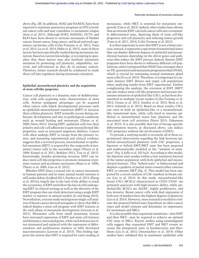
above (Fig. 2B). In addition, SOX2 and NANOG have beenreported to maintain quiescence programs in DTCs/resid-ual cancer cells and may contribute to metastatic relapse(Sosa et al. 2015). Although SOX2, NANOG, OCT4, andKLF4 have been shown to increase metastasis of bladdercancer, breast cancer, lung cancer, and head and neck squ-amous carcinoma cells (Celia-Terrassa et al. 2012; Vairaet al. 2013; Lu et al. 2014; Habu et al. 2015), none of thesefactors has been specifically studied duringmetastasis ini-tiation. Based on current knowledge, it is tempting to spec-ulate that these factors may also facilitate metastaticinitiation by promoting cell plasticity, adaptability, sur-vival, and self-renewal as they do in primary tumors.Therefore, future research should be conducted to studythese cell fate regulators during metastasis initiation.
Epithelial–mesenchymal plasticity and the acquisitionof stem cell-like properties
Cancer cell plasticity is a dynamic state of dedifferentia-tion, with cells acquiring some characteristics of stemcells. Serious malignant advantages can be acquiredwhen cancer cells hijack developmental processes suchas epithelial–mesenchymal transition (EMT) to increasetheir cellular plasticity. EMT normally occurs during em-bryonic development and also in pathological conditionssuch as wound healing and metastasis (Thiery et al.2009; Nieto 2013). During EMT, epithelial cells lose theirpolarity and cell–cell adhesions to gainmesenchymal-likeproperties, such as increased migratory abilities. Cancercells often undergo EMT to escape from the primary tu-mor, and mounting experimental and clinical evidencesuggests that a reversed process, mesenchymal-to-epithe-lial transition (MET), is required for the outgrowth ofmet-astatic tumor cells in the secondary organ (Thiery et al.2009; Korpal et al. 2011; Brabletz 2012; Tsai et al. 2012).Interestingly, besides promoting invasion, EMT can in-duce stem cell-like properties to promote initiation of pri-mary tumors and accelerate metastasis (Mani et al. 2008;Thiery et al. 2009; Guo et al. 2012).Whether EMT plays a crucial role in cancer metastasis
in human patients and in some animal model systems isstill under debate (Ledford 2011; Fischer et al. 2015; Zhenget al. 2015a), largely due to the lack of the ability to trackthe occurrence of EMTand follow the fateof cells undergo-ing EMT in clinical settings as well as the diversity of theEMTprogram that can elude detection using a single EMTmarker or reporter in animal models (Li and Kang 2016).Nevertheless, a recent studyused rigorous single-cell anal-ysis of breast cancer-derived xenografts to show thatMICsindeed display a stem cell program with EMT features atthe early phase of metastasis development (Lawson et al.2015). Metastatic cells from small metastatic lesionshave increased expression of EMT and stem cell featuresanddormancy-associatedgenes,while such features areof-ten attenuated and replaced with the expression of differ-entiation and proliferation markers in fully developedmacrometastases (Lawson et al. 2015). This finding sup-ports the notion that EMT is required for early seeding of
metastasis, while MET is essential for metastatic out-growth (Tsai et al. 2012). Indeed, other studies have shownthat an extremeEMTcan lock cancer cells into a terminal-ly differentiated state, depriving them of stem cell-likeproperties and cell plasticity and reducing tumor growth(Tran et al. 2011, 2014; Celia-Terrassa et al. 2012).It is thus important to note that EMT is not a binary pro-
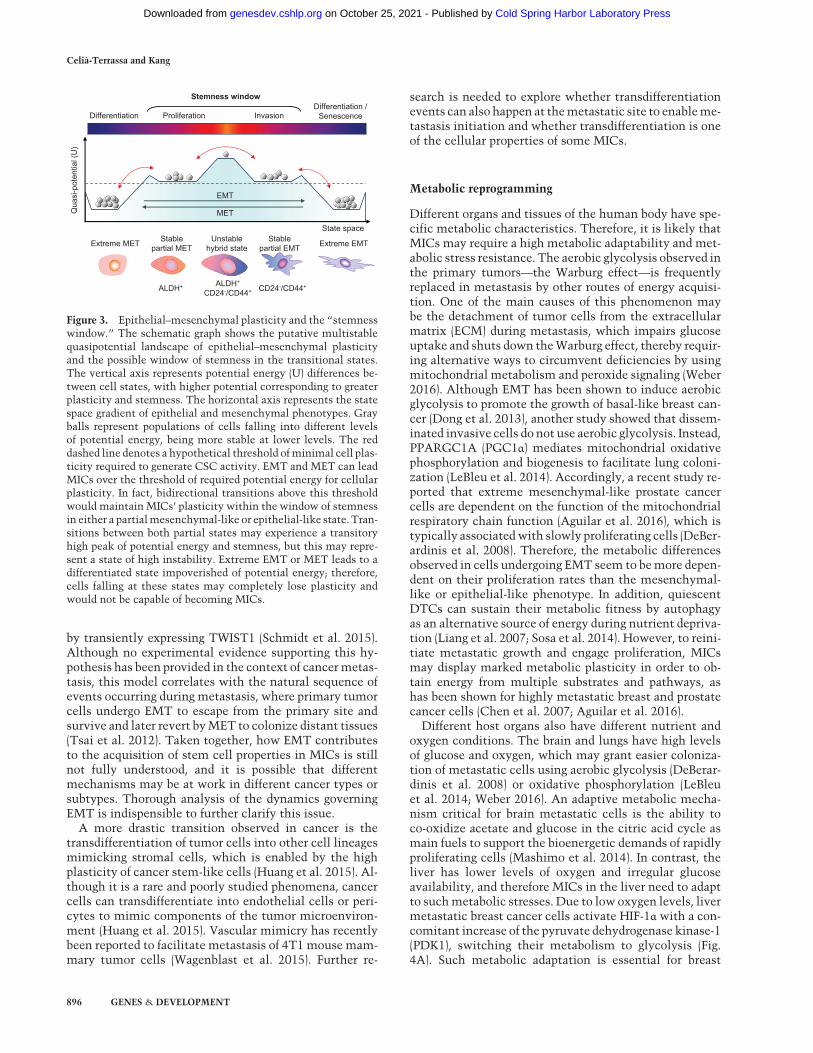
cess; instead, it represents a spectrumof transitional statesthat can display different degrees of epithelial and mesen-chymal features depending on the driver genes and path-ways that induce the EMT process. Indeed, distinct EMTprograms have been shown to influence different cell pop-ulations, and it is proposed that SNAI1has a stronger effectonTICgenerationandmetastasis progression thanSNAI2,which is crucial for sustaining normal mammary glandstemcells (Yeet al. 2015).Therefore, it is important to con-sider distinct EMT drivers and target cell populationswhen analyzing results from EMT experiments. Furthercomplicating the analysis, the reversion of EMT (MET)can also induce stem cell-like properties and increase me-tastasis initiation of epithelial-like CSCs, as has been doc-umented in multiple recent studies (Celia-Terrassa et al.2012; Ocana et al. 2012; Stankic et al. 2013; Beck et al.2015; Schmidt et al. 2015). Based on these results, CSCscan exist in both an epithelial-like or a mesenchymal-like transitional state, while cells fixed at extreme epi-thelial or mesenchymal states lose plasticity and theassociated stem cell activities (Nieto 2013; Oskarssonet al. 2014). It is also possible that loss of lineage-specificdifferentiation factors, as discussed above, can induceCSC properties without the involvement of EMTs.To provide a unifying model to reconcile all of these ex-
perimental observations regarding the link between epi-thelial–mesenchymal plasticity with CSC properties, abipotent or hybrid EMT/MET state has been proposedand mathematically modeled as the “window of stem-ness” (Fig. 3; Jolly et al. 2015a,b). According to this model,the bipotent state resides within a tiny transitory fractionof the tumor population with both epithelial and mesen-chymal features. This “hybrid state” is bidirectional anddisplays a gradient of partial states toward either extremeEMT or extreme MET (Fig. 3). This model has been sup-ported by a recent analysis of CSC markers in breast can-cer (Liu et al. 2014). In this study, mesenchymal-likebreast CSCs (BCSCs) characterized as CD24−CD44+ areprimarily quiescent with high invasive ability, while epi-thelial-like BCSCs are ALDH+, highly proliferative, andless invasive. Breast cancer cells with dual expression ofboth sets of markers have the highest degrees of plasticity(Liu et al. 2014). However,more research is needed to eval-uate the proposed hybrid state hypothesis in other cancertypes and model systems and determine its importancefor metastasis and MICs.It is also possible that sequential transitions—first EMT
and then MET—may be required to achieve an optimalCSC state in MICs. Recent studies using nonmalignantcells suggest that sequential EMT and MET events in-crease the pluripotent state in keratinocytes and fibro-blasts (Liu et al. 2013; Unternaehrer et al. 2014). Otherstudies have validated this in mammary epithelial cells
Molecular mechanism of metastasis initiation
GENES & DEVELOPMENT 895
Cold Spring Harbor Laboratory Press on October 25, 2021 - Published by genesdev.cshlp.orgDownloaded from
by transiently expressing TWIST1 (Schmidt et al. 2015).Although no experimental evidence supporting this hy-pothesis has been provided in the context of cancermetas-tasis, this model correlates with the natural sequence ofevents occurring during metastasis, where primary tumorcells undergo EMT to escape from the primary site andsurvive and later revert byMET to colonize distant tissues(Tsai et al. 2012). Taken together, how EMT contributesto the acquisition of stem cell properties in MICs is stillnot fully understood, and it is possible that differentmechanisms may be at work in different cancer types orsubtypes. Thorough analysis of the dynamics governingEMT is indispensible to further clarify this issue.
A more drastic transition observed in cancer is thetransdifferentiation of tumor cells into other cell lineagesmimicking stromal cells, which is enabled by the highplasticity of cancer stem-like cells (Huang et al. 2015). Al-though it is a rare and poorly studied phenomena, cancercells can transdifferentiate into endothelial cells or peri-cytes to mimic components of the tumor microenviron-ment (Huang et al. 2015). Vascular mimicry has recentlybeen reported to facilitate metastasis of 4T1 mouse mam-mary tumor cells (Wagenblast et al. 2015). Further re-
search is needed to explore whether transdifferentiationevents can also happen at themetastatic site to enableme-tastasis initiation and whether transdifferentiation is oneof the cellular properties of some MICs.
Metabolic reprogramming
Different organs and tissues of the human body have spe-cific metabolic characteristics. Therefore, it is likely thatMICs may require a high metabolic adaptability and met-abolic stress resistance. The aerobic glycolysis observed inthe primary tumors—the Warburg effect—is frequentlyreplaced in metastasis by other routes of energy acquisi-tion. One of the main causes of this phenomenon maybe the detachment of tumor cells from the extracellularmatrix (ECM) during metastasis, which impairs glucoseuptake and shuts down theWarburg effect, thereby requir-ing alternative ways to circumvent deficiencies by usingmitochondrial metabolism and peroxide signaling (Weber2016). Although EMT has been shown to induce aerobicglycolysis to promote the growth of basal-like breast can-cer (Dong et al. 2013), another study showed that dissem-inated invasive cells do not use aerobic glycolysis. Instead,PPARGC1A (PGC1α) mediates mitochondrial oxidativephosphorylation and biogenesis to facilitate lung coloni-zation (LeBleu et al. 2014). Accordingly, a recent study re-ported that extreme mesenchymal-like prostate cancercells are dependent on the function of the mitochondrialrespiratory chain function (Aguilar et al. 2016), which istypically associatedwith slowly proliferating cells (DeBer-ardinis et al. 2008). Therefore, the metabolic differencesobserved in cells undergoing EMT seem to bemore depen-dent on their proliferation rates than the mesenchymal-like or epithelial-like phenotype. In addition, quiescentDTCs can sustain their metabolic fitness by autophagyas an alternative source of energy during nutrient depriva-tion (Liang et al. 2007; Sosa et al. 2014). However, to reini-tiate metastatic growth and engage proliferation, MICsmay display marked metabolic plasticity in order to ob-tain energy from multiple substrates and pathways, ashas been shown for highly metastatic breast and prostatecancer cells (Chen et al. 2007; Aguilar et al. 2016).
Different host organs also have different nutrient andoxygen conditions. The brain and lungs have high levelsof glucose and oxygen, which may grant easier coloniza-tion of metastatic cells using aerobic glycolysis (DeBerar-dinis et al. 2008) or oxidative phosphorylation (LeBleuet al. 2014; Weber 2016). An adaptive metabolic mecha-nism critical for brain metastatic cells is the ability toco-oxidize acetate and glucose in the citric acid cycle asmain fuels to support the bioenergetic demands of rapidlyproliferating cells (Mashimo et al. 2014). In contrast, theliver has lower levels of oxygen and irregular glucoseavailability, and therefore MICs in the liver need to adaptto suchmetabolic stresses. Due to low oxygen levels, livermetastatic breast cancer cells activate HIF-1α with a con-comitant increase of the pyruvate dehydrogenase kinase-1(PDK1), switching their metabolism to glycolysis (Fig.4A). Such metabolic adaptation is essential for breast
EMT
MET
Stemness window
CD24-/CD44+ALDH+
CD24-/CD44+ALDH+
DifferentiationDifferentiation /
SenescenceProliferation Invasion
Qua
si-p
oten
tial (
U)
State spaceUnstable
hybrid stateStable
partial METStable
partial EMT Extreme EMTExtreme MET
Figure 3. Epithelial–mesenchymal plasticity and the “stemnesswindow.” The schematic graph shows the putative multistablequasipotential landscape of epithelial–mesenchymal plasticityand the possible window of stemness in the transitional states.The vertical axis represents potential energy (U) differences be-tween cell states, with higher potential corresponding to greaterplasticity and stemness. The horizontal axis represents the statespace gradient of epithelial and mesenchymal phenotypes. Grayballs represent populations of cells falling into different levelsof potential energy, being more stable at lower levels. The reddashed line denotes a hypothetical threshold ofminimal cell plas-ticity required to generate CSC activity. EMT and MET can leadMICs over the threshold of required potential energy for cellularplasticity. In fact, bidirectional transitions above this thresholdwould maintain MICs’ plasticity within the window of stemnessin either a partialmesenchymal-like or epithelial-like state. Tran-sitions between both partial states may experience a transitoryhigh peak of potential energy and stemness, but this may repre-sent a state of high instability. Extreme EMT or MET leads to adifferentiated state impoverished of potential energy; therefore,cells falling at these states may completely lose plasticity andwould not be capable of becoming MICs.
Celià-Terrassa and Kang
896 GENES & DEVELOPMENT
Cold Spring Harbor Laboratory Press on October 25, 2021 - Published by genesdev.cshlp.orgDownloaded from

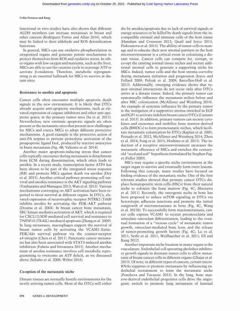
cancer cells to efficiently colonize the liver (Dupuy et al.2015). Another recent study has demonstrated the neces-sity of scavenging energy from the extracellular environ-ment to overcome liver metabolic stress (Loo et al.2015). In this study, colon cancer cells, by down-regulat-ing miR-483 and miR-551, derepress and secrete creatinekinase, brain-type (CKB) into the extracellular space toconvert creatine and ATP into phosphocreatine. Thephosphocreatine is then imported into the MICs to serveas an ATP source for growth functions (Fig. 4A; Looet al. 2015). In metastatic ovarian cells, fatty acids secret-ed from adipocytes are imported by FABP4 inMICs to col-onize the intra-abdominal fat (Nieman et al. 2011). Undernutrient deprivation conditions, mitochondrial HSP90chaperones, including TRAP1, overcomemetabolic stressand promote metastasis by limiting the activation of thenutrient sensor AMPK and preventing autophagy (Cainoet al. 2013).
Exposure to new inhospitable environments and drasticmetabolic reprogramming cause high levels of metabolicstress. Indeed, redox signaling pathways are often up-reg-ulated in metastasis (Pani et al. 2010). For example, itwas recently shown thatmetastaticmelanoma cells adoptdetoxifying mechanisms, such as producing NADPH de-toxifying enzymes of the folate pathway, includingALDH1L2 and MTHFD1, to withstand oxidative stressat the metastatic sites (Piskounova et al. 2015). This isin contrast to previous studies suggesting reactive oxygenspecies (ROS) as prometastatic effectors (Wu 2006; Ishi-kawa et al. 2008; Nishikawa 2008; Porporato et al. 2014).Such contradictory observations may be due to the factthat these earlier studies focused on the action of ROSon the primary tumor site, which may promote cancerprogression by generating genomic instability. ALDH en-zymes arewell-established CSCmarkers in several cancertypes (Ginestier et al. 2007;Medema 2013). Accumulating
Figure 4. Cross-talk betweenMICs and stromal microenvironment and niches at different organs. (A) Metabolic adaptation in the liver.Low oxygen levels in the livermicroenvironment force tumor cells to adapt via HIF-1α/PKD1 induction of glycolyticmetabolism, therebyenabling metastatic colonization. MICs of colon cancer secrete CKB, which phosphorylates extracellular creatine produced by hepato-cytes using extracellular ATP to generate phosphocreatine. Extracellular phosphocreatine is then imported into metastatic cancer cellsby the transporter SLC6A8 to regenerate the ATP as a source of energy for survival and metastatic colonization. (B) Vascular niche andbrain MIC–astrocyte cross-talk. The perivascular niche provides nutrients and oxygen to the infiltrating tumor cells, which secreteanti-PA serpins to protect MICs from astrocyte-derived death signals. Astrocytes also express Jagged1, which activates Notch signalingin MICs to promote self-renewal. Furthermore, astrocytes secrete miR-19a-containing exosomes, which suppress PTEN expressionand activate CCL2-dependent recruitment ofmyeloid cells to promote tumor growth and survival in the brain. (C ) In the bone,MICs com-pete with the hematopoietic stem cells (HSCs) for the HSC niche. Furthermore, osteogenic cells form heterotypic adherens junction withMICs and inducemTOR signaling to promote outgrowth. MICs also use secreted andmembrane-bound VCAM1 to recruit preosteoclasts(pre-Oc) and activate their differentiation to mature osteoclasts (Oc), which in turn promote bone degradation and the formation of the“vicious cycle in bone metastasis.” (D) In the lung, bone marrow-derived cells (BMDCs) facilitate the formation of the premetastaticniche. In addition, the secretion of ECM proteins—tenascin C secreted by tumor cells and periostin (POSTN) secreted by stromal cellssuch as cancer-associated fibroblasts (CAFs)—further establishes the metastatic niche and supports MIC self-renewal by inducing Notchand Wnt signaling, respectively.
Molecular mechanism of metastasis initiation
GENES & DEVELOPMENT 897
Cold Spring Harbor Laboratory Press on October 25, 2021 - Published by genesdev.cshlp.orgDownloaded from
functional in vivo studies have also shown that differentALDH members can increase metastasis in breast andother cancers (Rodriguez-Torres and Allan 2016), whichmay be linked to their aldehyde and ROS detoxificationfunctions.
In general, MICs can use oxidative phosphorylation inoxygenized organs and generate potent mechanisms toprotect themselves from ROS and oxidative stress. In oth-er organs with low oxygen and nutrients, such as the liver,MICs are able to use the creatine cycle to scavenge ATP oractivate β-oxidation. Therefore, metabolic reprogram-ming is an essential hallmark for MICs to survive at dis-tant sites.
Resistance to anoikis and apoptosis
Cancer cells often encounter multiple apoptotic deathsignals in the new environment. It is likely that DTCsalready acquire anti-apoptotic mechanisms, such as ele-vated expression of caspase inhibitors and other anti-apo-ptotic genes, at the primary tumor sites (Su et al. 2015).Nevertheless, new extrinsic apoptotic signals are oftenpresent at themetastatic sites that present newchallengesfor MICs and coerce MICs to adopt different protectivemechanisms. A good example is the protective action ofanti-PA serpins to prevent proteolytic activation of theproapoptotic ligand FasL, produced by reactive astrocytesin brain metastasis (Fig. 4B; Valiente et al. 2014).
Another major apoptosis-inducing stress that cancercells typically encounter duringmetastasis is detachmentfrom ECM during dissemination, which often leads toanoikis. In a recent study, transcription factor ATF4 hasbeen shown to be part of the integrated stress response(ISR) and protects MICs against death via anoikis (Deyet al. 2015). Another critical pathway promoting cell sur-vival and anoikis resistance is the AKT signaling pathway(Vanharanta andMassague 2013;Wan et al. 2013). Variousmechanisms converging on AKT activation have been re-ported to favor survival at different organs. In DTCs, ele-vated expression of neurotrophic receptor NTRK2 (TrkB)inhibits anoikis by activating the PI3K–AKT pathway(Douma et al. 2004). In breast cancer bone metastasis,SRC kinasemediates activation of AKT, which is requiredfor CXCL12/SDF-mediated cell survival and resistance toTNFSF10 (TRAIL)-mediated apoptosis (Zhang et al. 2009).In lung metastasis, macrophages support the survival ofbreast tumor cells by activating the VCAM1–Ezrin–PI3K/Akt survival pathway via the counter-receptorα4-integrin (Chen et al. 2011). Pancreatic cancer metasta-sis has also been associated with STAT3-induced anoikisinhibition (Fofaria and Srivastava 2015). Another mecha-nism of anoikis resistance involves cell metabolic repro-gramming to overcome an ATP deficit, as we discussedabove (Schafer et al. 2009; Weber 2016).
Co-option of the metastatic niche
Distant tissues are normally hostile environments for thenewly arriving tumor cells. Most of the DTCs will either
die by anoikis/apoptosis due to lack of survival signals orenergy resources or be killed by death signals from the in-compatible stromal and immune cells of the host tissue(Hanahan and Coussens 2012; Quail and Joyce 2013;Piskounova et al. 2015). The ability of tumor cells toman-age and re-educate their new stromal partners in the hostmicroenvironment is a critical event in colonizing a dis-tant tissue. Cancer cells can compete for, corrupt, orco-opt the existing normal tissue niches and recruit addi-tional stromal cells to generate a supporting niche forMICs. Indeed, tumor cells and the host stroma coevolveduring metastasis initiation and progression (Joyce andPollard 2009; Polyak et al. 2009; Barcellos-Hoff et al.2013). Additionally, emerging evidence shows that tu-mor–stromal interactions do not occur only after DTCsarrive at a distant tissue. Indeed, the primary tumor cansystemically influence the metastasis milieu before andafter MIC colonization (McAllister and Weinberg 2014).An example of systemic influence by the primary tumoris the instigation of a supportive stroma enriched in EGFand IGF1 to activate indolent breast cancerDTCs (Castanoet al. 2013). In addition, primary tumors can secrete cyto-kines and exosomes and mobilize bone marrow-derivedcells (BMDCs) to form premetastatic niches, which facili-tate metastatic colonization by DTCs (Kaplan et al. 2005;Peinado et al. 2012; McAllister and Weinberg 2014; Zhouet al. 2014; Fong et al. 2015; Hoshino et al. 2015). The in-duction of a receptive microenvironment increases themetastatic efficiency of MICs and enriches the century-old “seed and soil” hypothesis formulated by Stephen Pag-et (Fidler 2003).
MICs may require a specific niche environment at thetarget organ to survive and eventually form overt lesions.Following this concept, many studies have focused onfinding evidence of the metastatic niche. One of the firstrelevant studies showed that prostate cancer DTCs dis-place hematopoietic stem cells (HSCs) from their naturalniche to colonize the bone marrow (Fig. 4C; Shiozawaet al. 2011). Recently, the osteogenic niche in bone hasbeen proposed to induce mTOR signaling in DTCs viaheterotypic adherens junctions and promote the initialoutgrowth of micrometastases in bone (Fig. 4C; Wanget al. 2015b). To successfully form macrometastasis, can-cer cells express VCAM1 to recruit preosteoclasts andstimulate osteoclast differentiation, leading to the even-tual formation of a “vicious cycle” of metastatic tumorgrowth, osteoclast-mediated bone lysis, and the releaseof tumor-promoting growth factors (Fig. 4C; Lu et al.2011; Sethi et al. 2011; Weilbaecher et al. 2011; Ell andKang 2012).
Another important niche location inmany organs is thevasculature. Endothelial cell sprouting abolishes inhibito-ry growth signals in dormant tumor cells to allow metas-tasis of breast cancer cells in different organs (Ghajar et al.2013). Of note, in different types of cancers, certainmicro-RNAs suppress or promote metastasis by influencing en-dothelial recruitment to form the metastatic niche(Pencheva and Tavazoie 2013). In the lung, bone mar-row-derived endothelial progenitor cells drive the angio-genic switch to promote lung metastasis of luminal
Celià-Terrassa and Kang
898 GENES & DEVELOPMENT
Cold Spring Harbor Laboratory Press on October 25, 2021 - Published by genesdev.cshlp.orgDownloaded from

breast cancer cells (Gao et al. 2008). In the brain, vascularco-option of breast cancer cells through L1CAM-mediatedadhesion facilitates MIC access to nutrients and oxygen,while tumor-derived anti-PA serpin protected MICsfrom FasL death signals from astrocytes (Fig. 4B; Valienteet al. 2014). MICs can also generate their own niche bybuilding a supportive ECM in distant organs. For example,breast cancer cells secrete tenascin C, an ECM protein, inlungs to a stimulate stemness and favor metastasis (Fig.4D; Oskarsson et al. 2011).In order to cultivate a supporting “soil” at secondary or-
gan sites, tumor cells can also activate other nonnichecells, such as fibroblasts in metastatic sites, and turnthem into cancer-associated fibroblasts (CAFs) with me-tastasis-promoting functions (Kalluri and Zeisberg 2006),such as producing ECM niche components periostin(POSTN) and tenascin C (Fig. 4D; O’Connell et al. 2011;Malanchi et al. 2012). In addition, TGF-β released from co-lorectal cancer cells stimulated CAFs to secrete IL-11,which feeds back to tumor cells to activate STAT3 signal-ing, favoring the survival of metastatic cells in the liver(Calon et al. 2012). In the brain stroma, reactive astrocytesalso mediate important cross-talks with MICs to enhancetheir proliferation, survival, and metastasis (Kodack et al.2015). Astrocytes promote stem cell-like traits to breastcancer cells by activating Notch signaling in the brain(Fig. 4B; Xing et al. 2013). A recent study demonstratedhow PTEN expression is suppressed in MICs by the inter-action with astrocytes. In this study, astrocyte-derivedexosomes transfer the PTEN targeting miR-19a to theMICs. PTEN repression increases NFκB-dependent CCL2secretion and recruitment ofmyeloid cells to promote sur-vival and growth ofMICs in the brain (Fig. 4C; Zhang et al.2015).Amain threat toMICs is the immunecells present at the
new organ sites. The immune system is believed to pre-vent the formation of >80% of all primary tumors (Hana-han and Weinberg 2011). Even if DTCs have successfullyevaded the immune system at the primary tumor site,they are likely to encounter new, hostile immune cellswith the ability to recognize and kill them in the circula-tion and at metastatic sites. Indeed, the plasticity ofMICs to readily fluctuate between EMT–MET statesmight facilitate the immune evasion during metastasis.EMT transcription factors have been shown to haveimmunosuppressive functions. SNAI1 induces CD4+
CD25− Treg immune-suppressive cells and impairs den-dritic cell activity (Kudo-Saito et al. 2009). Moreover,ZEB1 repression of miR-200s up-regulates its target PD-L1, a known immune checkpoint regulator ofCD8+T cells(Chen et al. 2014). The secretionofTGF-β fromtumor cellscan repress the production of cytolytic and proapoptoticfactors by CD8+ CTLs (Thomas and Massague 2005).Therefore, mesenchymal-like DTCs, which often have el-evated expression of TGF-β, may escape attack by CTLsupon arrival in distant tissues. In contrast, BMP4, anothermember of the TGF-β family, functions as a metastasissuppressor in breast cancer by blockingG-CSF-induced ex-pansion ofmyeloid-derived suppressor cells (MDSCs) (Caoet al. 2014). As another mechanism to compromise the
function of innate immune cells during metastasis, mela-nomacells express FcγRIIb that negatively regulates B-cellrecognition and humoral immunity to promote liver me-tastasis (Cohen-Solal et al. 2010).Alternatively, some immune cells can be subverted by
DTCs to promote their metastatic growth. For example,activated M2 macrophages can promote metastatic colo-nization of different cancers by supporting growth,survival, and vascularization while impairing immunoge-nicity (Qian andPollard 2010;Quail and Joyce 2013). In thelungs, breast cancer cells can interactwithmacrophages toactivate the PI3K–AKT pathway and protect the cancercells from apoptotic signals (Chen et al. 2011). In fact, ab-lation of macrophage activation by blocking CSF-1R orCCR2 is a promising strategy to preventmacrophage insti-gation of metastasis outgrowth (Quail and Joyce 2013).Taken together, MICs have evolved multiple mecha-
nisms to turn a potentially hostile environment in a sec-ondary organ into a supportive niche. This can beachieved by releasing systemic growth and survival sig-nals from the primary tumor to foster a premetastaticniche, competing for existing normal stem cell niches,and engaging and converting the stromal cells to thwartdeath signals and immune attack.
Exit from dormancy
Metastatic dormancy is a frequent occurrence in manycancer types, with distant relapse occurring many yearsafter the successful treatment of an early-stage primarytumor and initial complete remission. Dormant DTCshave been definedwith threemain features: growth arrest,survival, and therapy resistance (Ghajar 2015). Further-more, their entry into dormancy and reactivation notonly is triggered by intrinsic programs but is also depen-dent on specialized microenvironmental niches, extrinsicsignals, and immune effects (Giancotti 2013; Quail andJoyce 2013; Sosa et al. 2014).Due to technical limitations, it is impractical to follow
a single cell for years and witness its awakening from dor-mancy to initiatemetastatic outgrowth, especially in clin-ical settings. Consequently, little has been known abouthow dormant cells escape growth arrest to initiate metas-tasis. Some studies propose different mechanisms for dif-ferent organ-specific metastases (Sosa et al. 2014). In bonemetastasis, elevated expression of VCAM1 induced by in-flammatory pathways in tumor cells promotes the transi-tion from indolent micrometastasis to overt metastasis(Lu et al. 2011). In lung metastasis, BMP signaling fromthe parenchyma restrains breast DTCs from exiting a dor-mant state by repressing self-renewal and inducing differ-entiation (Gao et al. 2012). Production of BMP inhibitors,such as Coco, by tumor cells can release them from laten-cy, prevent differentiation, and promotemetastasis initia-tion. Thus, the ability of dormantDTCs to overcome suchanti-growth signals is what turns them into active MICs.Other signals from the stromal niche can also induce thereactivation of growth and self-renewal pathways, such asERK, Wnt, and Notch (Giancotti 2013). For example, an
Molecular mechanism of metastasis initiation
GENES & DEVELOPMENT 899
Cold Spring Harbor Laboratory Press on October 25, 2021 - Published by genesdev.cshlp.orgDownloaded from
ECM component of the metastatic niche, tenascin C, canactivate Notch and β-catenin signaling (Oskarsson et al.2011), while POSTN can present Wnt ligands to activateWNT/TCF signaling (Fig. 4D;Malanchi et al. 2012).More-over, the perivascular niche can reactivate metastaticgrowth of dormant DTCs by endothelial sprouting andsecretion of POSTNandTGF-β (Ghajar et al. 2013). There-fore, MICs can overcome dormancy by activating self-renewal and stem cell-related pathways, such as Wnt,Notch, and TGF-β.
We discussed above how EMT or MET can generatestem cell properties in cancer cells and how mesenchy-mal-like cancer cells are less proliferative than epitheli-al-like cancer cells (Brabletz 2012; Liu et al. 2014).According to paradigm, it has been proposed, but not yetproven, thatmesenchymal-like TICs remain in a dormantstate upon arrival in a distant organ and need to undergoMET in order to reactivate and initiate metastasis (Gian-cotti 2013). In this scenario, both processes of EMT andMET would be critical for metastasis: EMT for enteringdormancy, promoting survival, and drug resistance andMET as the mechanism to reactivate proliferation andself-renewal to initiatemetastasis. This could also explainthe pathological observation that metastases display epi-thelial traits rather thanmesenchymal characters (Chafferet al. 2007; Korpal et al. 2011; Tsai et al. 2012; Chui 2013).
Drug resistance of MICs
A close correlation between metastasis and treatment re-sistance is frequently observed. Metastatic tumors are in-variably more chemoresistant than primary tumors, asevidenced by the marked decrease of chemotherapyresponse rate in metastatic settings as compared withneoadjuvant settings (Gonzalez-Angulo et al. 2007). Con-versely, poor response to neoadjuvant chemotherapy of-ten correlates with earlier metastatic recurrence andshorter survival, indicating that chemoresistant tumorsare prone to metastasize (Gonzalez-Angulo et al. 2007).Therefore, the generation of MIC properties may be phe-notypically linked to enhanced drug resistance capacities.MICs enriched with CSC-like features may benefit fromresistant mechanisms of CSCs, such as a stronger DNAdamage response (Wang 2015), elevated expression of ef-flux drug pumps (Schinkel et al. 1994; Zhou et al. 2001;Dean et al. 2005), and ALDH detoxifying enzymes (Hon-oki et al. 2010; Rausch et al. 2010). Therefore, inhibitorsof pathways involved in CSC regulation, such as antibod-ies againstNOTCH, FZD, IL6R, and other relevant signal-ing pathway receptors, may also have a therapeuticimpact on MICs (Brooks et al. 2015).
Importantly, EMT induction is well known to increasechemoresistance (Thiery et al. 2009; Yu et al. 2013; Zhenget al. 2015b) and recently has been shown to induce che-moresistance in lung metastases using an EMT lineagetracing system in breast cancer (Fischer et al. 2015). Thesestudies help explain why conventional treatments likegemcitabine or cyclophosphamide usually do not affectmesenchymal-like cells. Therefore, the existence of dor-
mant mesenchymal-like clones at a distant site could re-sist many conventional treatments (Giancotti 2013;Kang and Pantel 2013) and require novel therapeutic strat-egies targeting EMT-related pathways and features. Forexample, tumor cells undergoing EMT become resistantto EGFR inhibitors due to the activation of AXL kinase,which may be blocked with specific kinase inhibitors(Zhang et al. 2012). The inhibition of PKCα and FRA1can suppress tumor initiation by mesenchymal-likeCSCs and is therefore a potential target for mesenchy-mal-like MICs (Tam et al. 2013). However, dormant can-cer cells can also escape existing cancer treatmentsbecause of their quiescent status or niche protection(Braun et al. 2000; Naumov et al. 2003). Therefore, dor-mancy-specific treatment strategies should be designedto target the dormant cells (Sosa et al. 2014; Ghajar2015). Furthermore, other MIC-associated features, suchas metabolic reprogramming and activation of survivalpathways, are additional candidates for developing newtreatment options (Holohan et al. 2013; Loo et al. 2015).
Besides these MIC-intrinsic properties, tumor-associat-ed stroma has also been found to severely increase resis-tance to traditional cancer therapies (Gilbert andHemann 2010; Sun et al. 2012). In metastasis, primary tu-mor-associated endothelial cells produce TNFα to in-crease CXCL1/2 in cancer cells. These attract myeloidcells at the metastatic site, which produces S100A8/9 tofeed back to metastatic cells and stimulate increased che-moresistance (Acharyya et al. 2012). The tumor–stromalniche interactions discussed earlier provide additional op-portunities to disrupt a prosurvival niche for MICs andsensitize them to anti-cancer agents (Wan et al. 2013).
MIC heterogeneity: clonal or polyclonal metastasis
Primary tumors are heterogeneous masses of cells con-taining multiple subclones that are genetically and epige-netically different (Marusyk et al. 2012). Primary tumorsare considered to arise from single TICs capable of bothself-renewing and producing heterogeneity (HanahanandWeinberg 2011; Greaves andMaley 2012). Inmetasta-sis, the classical view also considers a single tumor cell asthe origin of metastases, based on chromosomal analysis(Talmadge et al. 1982). However, circulating tumor cells(CTCs) have been found to be genetically and phenotypi-cally heterogeneous (Stoecklein et al. 2008; Kang and Pan-tel 2013; Yu et al. 2013), raising the possibility ofpolyclonal seeding and metastases. Until now, little wasknown about the clonal population dynamics throughoutthe different steps of metastasis leading into the forma-tion of overt metastases. However, recent studies usinglineage tracing, barcode sequencing, and whole-genomesequencing are shedding light on this question and havedemonstrated a mostly polyclonal nature of metastasis(Fig. 5; McFadden et al. 2014; Gundem et al. 2015; Maddi-pati and Stanger 2015; Wagenblast et al. 2015).
Mutation analysis between the primary tumor and themetastatic lesions indicated polyclonal metastatic spreadin the lymph nodes but not in the liver (McFadden et al.
Celià-Terrassa and Kang
900 GENES & DEVELOPMENT
Cold Spring Harbor Laboratory Press on October 25, 2021 - Published by genesdev.cshlp.orgDownloaded from
2014). Another whole-genome sequencing study analyzed51 tumors of 10 prostate cancer patients, including prima-ry tumors and multiple metastases in the same patients,and revealed the coexistence of multiple clones in theme-tastases, including those from metastasis-to-metastasisspreads (Gundem et al. 2015). Molecular barcoding offersanother effective method to track clonal populations inexperimental animal models of metastasis, and this ap-proach has recently been used to analyze metastasis het-erogeneity generated by the 4T1 mouse mammarytumor cell line (Wagenblast et al. 2015). In this study,orthotopic injection of barcoded cells generated metasta-ses composed of multiple, different clones in various tis-sues, although it cannot be ruled out that independentmetastatic nodules in the same organ might be seededmonoclonally.Lineage tracing using fluorescence markers is another
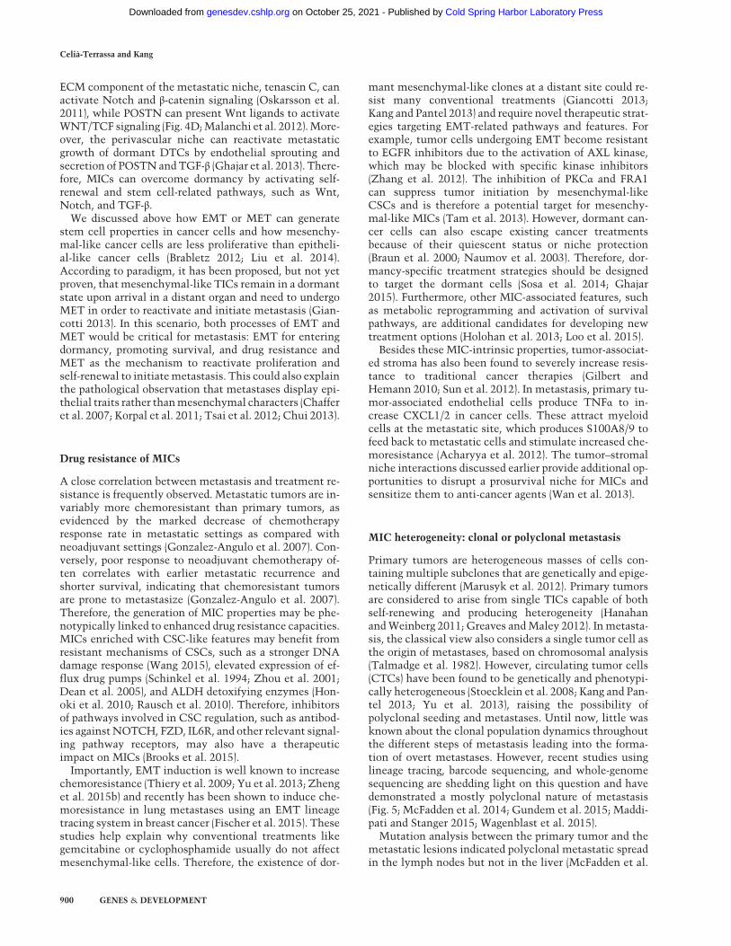
robust method to study polyclonal metastases in animalmodels. Combining the multicolor “confetti” mousemodel for multiclonal tracking with the K-rasLSL.G12D/+;p53R172H/+; PdxCre (KPC) mousemodel of pancreatic duc-tal adenocarcinoma (PDAC), a high frequency of polyclon-al metastasis was revealed, including 11%–14% in thelungs and liver and 80% in the peritoneum and dia-phragm. Interestingly, during the metastatic outgrowthto overt lesions, the clonal diversity usually decreases,leading to formation of monoclonal or polyclonal expan-sions that appear to depend on themetastatic site (Maddi-pati and Stanger 2015). Taken together, emergingevidence suggests that MICs are heterogeneous, and dif-ferent clones are often involved in seeding and formingovert metastasis. Depending on the interaction of MICclones and the conditions of the host organ, themetastaticoutgrowth can remain polyclonal or become monoclonal(Fig. 5A). Additionally, the demonstration of polyclonalmetastasis suggests the contribution of different hetero-typic interactions among different tumor clonal subpopu-lations to initiate metastasis.
Clonal cooperation
The importance of tumor heterogeneity in cancer evolu-tion has led to the idea that tumors may function asecosystems of interactive populations within an environ-ment. Consequently, several studies have started to focuson the ecological cooperation or competitive interactionsbetween tumor populations (Merlo et al. 2006; Moreno2008; Neelakantan et al. 2015; Tabassum and Polyak2015). In primary breast tumors, recent research usingmouse models has characterized the polyclonal origin ofcertain tumor types and the interclonal cooperation be-tween multiple subclones (Cleary et al. 2014). Small sub-populations can drive the growth of other non-cell-autonomous clones through paracrine interaction andmi-croenvironment modulation (Marusyk et al. 2014). In themetastatic context, this phenomenon was first reportedby coinjecting nonmetastatic cells with metastatic cellsto increase the metastasis of the former (Miller 1983). Co-operation can be promoted by endocrine and exosome sig-naling between different clones (Martorana et al. 1998;Neelakantan et al. 2015). ECM proteins, such as SPARC,also serve as messengers of cooperation to enhance inva-sion and metastasis (Fig. 5B; Mateo et al. 2014). In addi-tion, heterotypic interactions among EMT and non-EMTcells have also been demonstrated to increase metastasisprogression of hamster cheek pouch carcinoma cells(Tsuji et al. 2008) aswell as in xenograftmodels of prostatecancer metastasis (Celia-Terrassa et al. 2012). In the latterstudy, both clonal populations seeded distant organs, al-though only the non-EMT clonal population—enrichedin epithelial-like CSCs—expanded to overt metastases.Interestingly, the different clones presented complemen-tary essential properties for metastasis, invasion in themesenchymal-like clones and self-renewal/proliferationin the epithelial-like clones. As a result, the combinationof clones enhanced metastasis, including new organsnever colonized by either clone alone. This kind of
Monoclonal Phenotypicallyheterogenic monoclonal
Polyclonal
BA
Polyclonal seeding
Induced invasiveness
Opportunistic MICs
Circulating clusters of DTCs
Clone A Clone B
Polyclonal micrometastasis
Macrometastasis
Mesenchymal-like trailblazers
Figure 5. Clonal cooperation in metastasis. (A) Representation of different macrometastasis outputs from initial polyclonal dissemina-tion and seeding. Polyclonal seeding andmicrometastasis may develop polyclonal (left) or monoclonal (middle) macrometastasis depend-ing on the clonal and tumor–stroma interaction dynamics in the target organ. (Right) In addition,metastasis heterogeneity can result fromthe generation of multiple phenotypes from a single metastatic clone. (B, left) Mesenchymal-like secretory cells can induce invasive phe-notypes in epithelial-like TICs through secreted factors such as SPARC, facilitating their escape from the primary site. (Middle) In addi-tion, noninvasive TICs/MICs can opportunistically follow trailblazer invasive cells to escape from the primary site or extravasate andinfiltrate a distant tissue. (Right) Polyclonal seeding of a distant organ as a result of clonal cooperation.
Molecular mechanism of metastasis initiation
GENES & DEVELOPMENT 901
Cold Spring Harbor Laboratory Press on October 25, 2021 - Published by genesdev.cshlp.orgDownloaded from

cooperation has been further characterized in other mod-els proposing a leading invasive cell followed by “opportu-nistic” cells (Fig. 5B; Chapman et al. 2014; Westcott et al.2015).
This synergy is in agreement with the observation thatCTC clusters have dramatically increased metastatic po-tential compared with single CTCs (Aceto et al. 2014)and the polyclonal nature of metastases (Gundem et al.2015; Maddipati and Stanger 2015). Therefore, MICsmight be “opportunistic” cells, which benefit from the es-tablishment of heterotypic interactions with other DTCswith complementary abilities that can be exploited byMICs (Fig. 5B).
The polyclonal origin of metastases and cooperativeinteractions between different clonal populations in me-tastasis may be highly relevant to the understanding ofthe so-called polygenic drug resistance (Holohan et al.2013). Heterogeneous polyclonal metastases confer agreat diversity of drug resistance. Furthermore, hierarchi-cally organized populations are reported to stochasticallytransition between phenotypic states to balance cancercell populations in any direction, even from non-stem-like cells to stem-like cells (Gupta et al. 2011). Such ahigh degree of plasticity ofMICs, combinedwith the poly-clonal nature of metastasis, presents a major challenge forconventional therapy.
Concluding remarks and perspectives
In this review, we enumerate a compendium of relevanthallmarks of MICs. Many of these hallmarks are repre-sented within the same MIC population; however, itshould be kept inmind that not all of them need to be pos-sessed by the same tumor cell to initiate metastasis. Agreat degree of diversity, including the requirement of dif-ferent assortments of MIC hallmarks, exists among MICsin different cancer types and subtypes and in differentmetastatic organ sites. Furthermore,many of theMIC fea-tures are required only at specific windows of metastaticprogression. Importantly, the discovery of polyclonal me-tastasis introduces an additional layer of variability, as dif-ferent clonal populations may cooperate to collectivelyseed metastasis, and even non-MIC populations can pro-vide important functions to complementMICs in enhanc-ing their metastatic competency. Therefore, the ability toform a metastatic lesion may not be the privilege of a spe-cific tumor cell population with the requisite molecularand functional hallmarks ofMICs. Instead,metastasis ini-tiation may be the culmination of a highly fluid processinvolving multiple iterations of transitional cellularstates, dynamic interactions between clonal tumor popu-lations, and both short-distance and long-range interac-tions between tumor cells and the host organs. Despiteall of these ambiguities in defining MICs, a central coreproperty of MICs is their cellular plasticity, which under-lies almost all other MIC hallmarks (Fig. 6). This mostfundamental hallmark of MICs may therefore representa potential Achilles’ heel of cancer that can be exploitedin developing new treatments. Future research should ad-
dress these key questions: Is cellular plasticity really cru-cial for metastatic initiation, and how can we targetcellular plasticity? Cellular plasticity targeting treat-ments are unlikely to be cytotoxic when applied as singleagents in treatments and therefore are likely to fail stan-dard clinical trials that often rely on reduced tumorburden as an indication of effectiveness. Instead, suchtreatmentmay demonstrate efficacy onlywhen combinedwith other treatments to induce stress on potential MICsand in adjuvant settings. These will be major hurdles toadvance cellular plasticity targeting treatment throughthe traditional drug development and clinical trial pipe-lines. Nevertheless, as technical innovations continue tobring about major breakthroughs in the study of MICs inclinical settings and experimental models, our newly de-veloped insights into themysterious process ofmetastasisinitiation will undoubtedly lead to improved preventionand treatment of metastatic diseases.
Acknowledgments
We thank members of our laboratories for helpful discussions,and, in particular, H.A. Smith and D. Liu for critical reading ofthe manuscript. We also apologize to the many investigatorswhose important studies could not be cited directly here owingto space limitations. The work was supported by a SusanG. Komen Fellowship to T.C.-T. (PDF15332075), and grantsfrom the Brewster Foundation, the Breast Cancer Research Foun-dation, the Department of Defense (BC123187), and the NationalInstitutes of Health (R01CA141062) to Y.K.
References
Aceto N, Bardia A, Miyamoto DT, Donaldson MC, Wittner BS,Spencer JA, YuM, Pely A, EngstromA, ZhuH, et al. 2014. Cir-culating tumor cell clusters are oligoclonal precursors ofbreast cancer metastasis. Cell 158: 1110–1122.
Proliferation
Self-renewal
Drug resistance
Anoikis/apoptosis resistance
Dormancyexit
Stromalco-option
ClonalCooperation
Immuneevasion
Cellular plasticity
Metabolicadaptation
Figure 6. Cellular plasticity as the core characteristic of MICs.This diagram lists nine characteristics commonly observed inMICs. These abilities are often enabled by cancer cell plasticity,represented as the core of MIC properties. Not all of these proper-tiesmayneed to be acquired simultaneously byMICs to growme-tastases. Instead, multiple different combinations may influencethe emergence of MICs in the affected organs.
Celià-Terrassa and Kang
902 GENES & DEVELOPMENT
Cold Spring Harbor Laboratory Press on October 25, 2021 - Published by genesdev.cshlp.orgDownloaded from

Acharyya S, Oskarsson T, Vanharanta S, Malladi S, Kim J, MorrisPG, Manova-Todorova K, Leversha M, Hogg N, Seshan VE,et al. 2012. A CXCL1 paracrine network links cancer chemo-resistance and metastasis. Cell 150: 165–178.
Aguilar E,Marin deMas I, Zodda E,Marin S,Morrish F, SelivanovV,Meca-Cortés O,DelowarH, PonsM, Izquierdo I, et al. 2016.Metabolic reprogramming and dependencies associated withepithelial cancer stem cells independent of the epithelial–mesenchymal transition program. Stem Cells doi: 10.1002/stem.2286.
Barcellos-Hoff MH, Lyden D, Wang TC. 2013. The evolution ofthe cancer niche during multistage carcinogenesis. Nat RevCancer 13: 511–518.
Beck B, LapougeG, Rorive S, Drogat B, Desaedelaere K, DelafailleS, Dubois C, Salmon I, Willekens K, Marine JC, et al. 2015.Different levels of Twist1 regulate skin tumor initiation,stemness, and progression. Cell Stem Cell 16: 67–79.
Ben-Porath I, Thomson MW, Carey VJ, Ge R, Bell GW, Regev A,Weinberg RA. 2008. An embryonic stem cell-like gene expres-sion signature in poorly differentiated aggressive human tu-mors. Nat Genet 40: 499–507.
Boumahdi S, DriessensG, LapougeG,Rorive S,NassarD, LeMer-cier M, Delatte B, Caauwe A, Lenglez S, Nkusi E, et al. 2014.SOX2 controls tumour initiation and cancer stem-cell func-tions in squamous-cell carcinoma. Nature 511: 246–250.
Bozic I, Antal T, Ohtsuki H, Carter H, KimD, Chen S, Karchin R,Kinzler KW,Vogelstein B,NowakMA. 2010. Accumulation ofdriver and passenger mutations during tumor progression.Proc Natl Acad Sci 107: 18545–18550.
Brabletz T. 2012. To differentiate or not—routes towards metas-tasis. Nat Rev Cancer 12: 425–436.
Braun S, Kentenich C, Janni W, Hepp F, de Waal J, Willgeroth F,Sommer H, Pantel K. 2000. Lack of effect of adjuvant chemo-therapy on the elimination of single dormant tumor cells inbone marrow of high-risk breast cancer patients. J Clin Oncol18: 80–86.
Brooks MD, Burness ML, Wicha MS. 2015. Therapeutic implica-tions of cellular heterogeneity and plasticity in breast cancer.Cell Stem Cell 17: 260–271.
Caino MC, Chae YC, Vaira V, Ferrero S, Nosotti M, Martin NM,Weeraratna A, O’Connell M, JerniganD, Fatatis A, et al. 2013.Metabolic stress regulates cytoskeletal dynamics and metas-tasis of cancer cells. J Clin Invest 123: 2907–2920.
Calon A, Espinet E, Palomo-Ponce S, Tauriello DV, Iglesias M,Cespedes MV, Sevillano M, Nadal C, Jung P, Zhang XH,et al. 2012. Dependency of colorectal cancer on a TGF-β-driv-en program in stromal cells for metastasis initiation. CancerCell 22: 571–584.
Cameron MD, Schmidt EE, Kerkvliet N, Nadkarni KV, MorrisVL, Groom AC, Chambers AF, MacDonald IC. 2000. Tempo-ral progression of metastasis in lung: cell survival, dormancy,and location dependence of metastatic inefficiency. CancerRes 60: 2541–2546.
Campbell PJ, Yachida S, Mudie LJ, Stephens PJ, Pleasance ED,Stebbings LA, Morsberger LA, Latimer C, McLaren S, LinML, et al. 2010. The patterns and dynamics of genomic insta-bility inmetastatic pancreatic cancer.Nature 467: 1109–1113.
Cao PD, Cheung WK, Nguyen DX. 2011. Cell lineage specifica-tion in tumor progression and metastasis. Discov Med 12:329–340.
Cao Y, Slaney CY, Bidwell BN, Parker BS, Johnstone CN, RautelaJ, Eckhardt BL, Anderson RL. 2014. BMP4 inhibits breast can-cermetastasis by blockingmyeloid-derived suppressor cell ac-tivity. Cancer Res 74: 5091–5102.
Castano Z, Marsh T, Tadipatri R, Kuznetsov HS, Al-Shahrour F,Paktinat M, Greene-Colozzi A, Nilsson B, Richardson AL,McAllister SS. 2013. Stromal EGF and IGF-I together modu-late plasticity of disseminated triple-negative breast tumors.Cancer Discov 3: 922–935.
Celia-Terrassa T, Meca-Cortes O, Mateo F, de Paz AM, Rubio N,Arnal-Estape A, Ell BJ, Bermudo R, Diaz A, Guerra-RebolloM,et al. 2012. Epithelial–mesenchymal transition can suppressmajor attributes of human epithelial tumor-initiating cells. JClin Invest 122: 1849–1868.
Chaffer CL, Thompson EW,Williams ED. 2007. Mesenchymal toepithelial transition in development and disease. Cells Tis-sues Organs 185: 7–19.
Chakrabarti R, Hwang J, Andres Blanco M, Wei Y, Lukacisin M,Romano RA, Smalley K, Liu S, Yang Q, Ibrahim T, et al.2012. Elf5 inhibits the epithelial–mesenchymal transition inmammary gland development and breast cancer metastasisby transcriptionally repressing Snail2. Nat Cell Biol 14:1212–1222.
Chambers AF, Groom AC, MacDonald IC. 2002. Disseminationand growth of cancer cells inmetastatic sites.Nat RevCancer2: 563–572.
Chapman A, Fernandez del Ama L, Ferguson J, Kamarashev J,Wellbrock C, Hurlstone A. 2014. Heterogeneous tumor sub-populations cooperate to drive invasion. Cell Rep 8: 688–695.
Cheli Y, Giuliano S, Fenouille N, Allegra M, Hofman V, HofmanP, Bahadoran P, Lacour JP, Tartare-Deckert S, Bertolotto C,et al. 2012. Hypoxia and MITF control metastatic behaviourin mouse and human melanoma cells. Oncogene 31:2461–2470.
Chen EI, Hewel J, Krueger JS, TirabyC,WeberMR, Kralli A, Beck-er K, Yates JR III, Felding-Habermann B. 2007. Adaptation ofenergy metabolism in breast cancer brain metastases. CancerRes 67: 1472–1486.
Chen Q, Zhang XH, Massague J. 2011. Macrophage binding to re-ceptor VCAM-1 transmits survival signals in breast cancercells that invade the lungs. Cancer Cell 20: 538–549.
Chen L, GibbonsDL, Goswami S, CortezMA,AhnYH, Byers LA,Zhang X, Yi X, Dwyer D, Lin W, et al. 2014. Metastasis is reg-ulated via microRNA-200/ZEB1 axis control of tumour cellPD-L1 expression and intratumoral immunosuppression.Nat Commun 5: 5241.
Cheung WK, Zhao M, Liu Z, Stevens LE, Cao PD, Fang JE, West-brook TF, Nguyen DX. 2013. Control of alveolar differentia-tion by the lineage transcription factors GATA6 and HOPXinhibits lung adenocarcinoma metastasis. Cancer Cell 23:725–738.
Chui MH. 2013. Insights into cancer metastasis from a clinico-pathologic perspective: epithelial–mesenchymal transition isnot a necessary step. Int J Cancer 132: 1487–1495.
Cleary AS, Leonard TL, Gestl SA, Gunther EJ. 2014. Tumour cellheterogeneity maintained by cooperating subclones in Wnt-driven mammary cancers. Nature 508: 113–117.
Cohen-Solal JF, Cassard L, Fournier EM, Loncar SM, FridmanWH, Sautes-Fridman C. 2010. Metastatic melanomas expressinhibitory low affinity Fcγ receptor and escape humoral im-munity. Dermatol Res Pract 2010: 657406.
ColeMD. 1986. Themyc oncogene: its role in transformation anddifferentiation. Annu Rev Genet 20: 361–384.
Cunha S, Lin YC, Goossen EA, DeVette CI, Albertella MR,Thomson S,MulvihillMJ,WelmAL. 2014. The RON receptortyrosine kinase promotes metastasis by triggering MBD4-de-pendent DNA methylation reprogramming. Cell Rep 6:141–154.
Molecular mechanism of metastasis initiation
GENES & DEVELOPMENT 903
Cold Spring Harbor Laboratory Press on October 25, 2021 - Published by genesdev.cshlp.orgDownloaded from
DeanM, Fojo T, Bates S. 2005. Tumour stem cells and drug resis-tance. Nat Rev Cancer 5: 275–284.
DeBerardinis RJ, Lum JJ, Hatzivassiliou G, Thompson CB. 2008.The biology of cancer: metabolic reprogramming fuels cellgrowth and proliferation. Cell Metab 7: 11–20.
Dey S, Sayers CM, Verginadis II, Lehman SL, Cheng Y, CernigliaGJ, Tuttle SW, Feldman MD, Zhang PJ, Fuchs SY, et al. 2015.ATF4-dependent induction of heme oxygenase 1 preventsanoikis and promotes metastasis. J Clin Invest 125:2592–2608.
Dong C, Yuan T,WuY,WangY, Fan TW,Miriyala S, Lin Y, Yao J,Shi J, Kang T, et al. 2013. Loss of FBP1 by Snail-mediated re-pression provides metabolic advantages in basal-like breastcancer. Cancer Cell 23: 316–331.
DoumaS, VanLaarT, Zevenhoven J,MeuwissenR, VanGarderenE, Peeper DS. 2004. Suppression of anoikis and induction ofmetastasis by the neurotrophic receptor TrkB. Nature 430:1034–1039.
Dupuy F, Tabaries S, Andrzejewski S, Dong Z, Blagih J, AnnisMG, Omeroglu A, Gao D, Leung S, Amir E, et al. 2015.PDK1-dependent metabolic reprogramming dictates meta-static potential in breast cancer. Cell Metab 22: 577–589.
Ell B, Kang Y. 2012. SnapShot: bone metastasis. Cell 151: 690–690.e1.
Fidler IJ. 2003. The pathogenesis of cancer metastasis: the ‘seedand soil’ hypothesis revisited. Nat Rev Cancer 3: 453–458.
Fischer KR, Durrans A, Lee S, Sheng J, Li F, Wong ST, Choi H, ElRayes T, Ryu S, Troeger J, et al. 2015. Epithelial-to-mesenchy-mal transition is not required for lung metastasis but contrib-utes to chemoresistance. Nature 527: 472–476.
Fofaria NM, Srivastava SK. 2015. STAT3 induces anoikis resis-tance, promotes cell invasion andmetastatic potential in pan-creatic cancer cells. Carcinogenesis 36: 142–150.
Fong MY, Zhou W, Liu L, Alontaga AY, Chandra M, Ashby J,Chow A, O’Connor ST, Li S, Chin AR, et al. 2015. Breast-can-cer-secreted miR-122 reprograms glucose metabolism in pre-metastatic niche to promote metastasis. Nat Cell Biol 17:183–194.
Gao D, Nolan DJ, Mellick AS, Bambino K, McDonnell K, MittalV. 2008. Endothelial progenitor cells control the angiogenicswitch in mouse lung metastasis. Science 319: 195–198.
Gao H, Chakraborty G, Lee-Lim AP, Mo Q, Decker M, Vonica A,Shen R, Brogi E, Brivanlou AH, Giancotti FG. 2012. The BMPinhibitor Coco reactivates breast cancer cells at lung meta-static sites. Cell 150: 764–779.
Ghajar CM. 2015. Metastasis prevention by targeting the dor-mant niche. Nat Rev Cancer 15: 238–247.
Ghajar CM, Peinado H, Mori H, Matei IR, Evason KJ, Brazier H,Almeida D, Koller A, Hajjar KA, Stainier DY, et al. 2013.The perivascular niche regulates breast tumour dormancy.Nat Cell Biol 15: 807–817.
Giancotti FG. 2013. Mechanisms governing metastatic dor-mancy and reactivation. Cell 155: 750–764.
Gilbert LA, Hemann MT. 2010. DNA damage-mediated induc-tion of a chemoresistant niche. Cell 143: 355–366.
Ginestier C, Hur MH, Charafe-Jauffret E, Monville F, Dutcher J,Brown M, Jacquemier J, Viens P, Kleer CG, Liu S, et al.2007. ALDH1 is a marker of normal and malignant humanmammary stem cells and a predictor of poor clinical outcome.Cell Stem Cell 1: 555–567.
Gonzalez-AnguloAM,Morales-Vasquez F, HortobagyiGN. 2007.Overview of resistance to systemic therapy in patients withbreast cancer. Adv Exp Med Biol 608: 1–22.
Greaves M, Maley CC. 2012. Clonal evolution in cancer. Nature481: 306–313.
Gu L, Frommel SC, Oakes CC, Simon R, Grupp K, Gerig CY, BarD, Robinson MD, Baer C, Weiss M, et al. 2015. BAZ2A (TIP5)is involved in epigenetic alterations in prostate cancer and itsoverexpression predicts disease recurrence. Nat Genet 47:22–30.
Gundem G, Van Loo P, Kremeyer B, Alexandrov LB, Tubio JM,Papaemmanuil E, Brewer DS, Kallio HM, Hognas G, AnnalaM, et al. 2015. The evolutionary history of lethal metastaticprostate cancer. Nature 520: 353–357.
Guo W, Keckesova Z, Donaher JL, Shibue T, Tischler V, Rein-hardt F, Itzkovitz S, Noske A, Zurrer-Hardi U, Bell G, et al.2012. Slug and Sox9 cooperatively determine the mammarystem cell state. Cell 148: 1015–1028.
GuptaGP,Massague J. 2006.Cancermetastasis: building a frame-work. Cell 127: 679–695.
Gupta GP, Perk J, Acharyya S, de Candia P, Mittal V, Todorova-Manova K, Gerald WL, Brogi E, Benezra R, Massague J. 2007.ID genes mediate tumor reinitiation during breast cancerlung metastasis. Proc Natl Acad Sci 104: 19506–19511.
Gupta PB, Fillmore CM, Jiang G, Shapira SD, Tao K, KuperwasserC, Lander ES. 2011. Stochastic state transitions give rise tophenotypic equilibrium in populations of cancer cells. Cell146: 633–644.
Habu N, Imanishi Y, Kameyama K, Shimoda M, Tokumaru Y,Sakamoto K, Fujii R, Shigetomi S, Otsuka K, Sato Y, et al.2015. Expression of Oct3/4 and Nanog in the head and necksquamous carcinoma cells and its clinical implications for de-layed neck metastasis in stage I/II oral tongue squamous cellcarcinoma. BMC Cancer 15: 730.
Hanahan D, Coussens LM. 2012. Accessories to the crime: func-tions of cells recruited to the tumor microenvironment. Can-cer Cell 21: 309–322.
Hanahan D, Weinberg RA. 2011. Hallmarks of cancer: the nextgeneration. Cell 144: 646–674.
Hendrix MJ, Seftor EA, Seftor RE, Kasemeier-Kulesa J, KulesaPM, Postovit LM. 2007. Reprogramming metastatic tumourcells with embryonic microenvironments. Nat Rev Cancer7: 246–255.
HolohanC, Van Schaeybroeck S, Longley DB, Johnston PG. 2013.Cancer drug resistance: an evolving paradigm. Nat Rev Can-cer 13: 714–726.
Honoki K, Fujii H, Kubo A, Kido A, Mori T, Tanaka Y, TsujiuchiT. 2010. Possible involvement of stem-like populations withelevatedALDH1 in sarcomas for chemotherapeutic drug resis-tance. Oncol Rep 24: 501–505.
Hoshino A, Costa-Silva B, Shen TL, Rodrigues G, Hashimoto A,Tesic Mark M, Molina H, Kohsaka S, Di Giannatale A, CederS, et al. 2015. Tumour exosome integrins determine organo-tropic metastasis. Nature 527: 329–335.
Huang Z, Wu T, Liu AY, Ouyang G. 2015. Differentiation andtransdifferentiation potentials of cancer stem cells. Oncotar-get 6: 39550–39563.
HusemannY, Geigl JB, Schubert F,Musiani P,MeyerM, BurghartE, Forni G, Eils R, FehmT, Riethmuller G, et al. 2008. System-ic spread is an early step in breast cancer. Cancer Cell 13:58–68.
Ishikawa K, Takenaga K, Akimoto M, Koshikawa N, YamaguchiA, Imanishi H, Nakada K, Honma Y, Hayashi J. 2008. ROS-generatingmitochondrial DNAmutations can regulate tumorcell metastasis. Science 320: 661–664.
Jacob LS, Vanharanta S, ObenaufAC, PirunM,VialeA, SocciND,Massague J. 2015.Metastatic competence can emergewith se-lection of preexisting oncogenic alleles without a need of newmutations. Cancer Res 75: 3713–3719.
Celià-Terrassa and Kang
904 GENES & DEVELOPMENT
Cold Spring Harbor Laboratory Press on October 25, 2021 - Published by genesdev.cshlp.orgDownloaded from

JollyMK, BoaretoM, Huang B, Jia D, LuM, Ben-Jacob E, OnuchicJN, Levine H. 2015a. Implications of the hybrid epithelial/mesenchymal phenotype in metastasis. Front Oncol 5: 155.
Jolly MK, Jia D, Boareto M, Mani SA, Pienta KJ, Ben-Jacob E, Le-vine H. 2015b. Coupling the modules of EMT and stemness: atunable ‘stemness window’ model. Oncotarget 6:25161–25174.
Joyce JA, Pollard JW. 2009.Microenvironmental regulation ofme-tastasis. Nat Rev Cancer 9: 239–252.
Kalluri R, Zeisberg M. 2006. Fibroblasts in cancer. Nat Rev Can-cer 6: 392–401.
Kang Y, Pantel K. 2013. Tumor cell dissemination: emerging bio-logical insights from animalmodels and cancer patients.Can-cer Cell 23: 573–581.
KaplanRN,RibaRD, Zacharoulis S, BramleyAH, Vincent L, Cos-ta C, MacDonald DD, Jin DK, Shido K, Kerns SA, et al. 2005.VEGFR1-positive haematopoietic bone marrow progenitorsinitiate the pre-metastatic niche. Nature 438: 820–827.
Kareta MS, Gorges LL, Hafeez S, Benayoun BA, Marro S, ZmoosAF, Cecchini MJ, Spacek D, Batista LF, O’Brien M, et al.2015. Inhibition of pluripotency networks by the Rb tumorsuppressor restricts reprogramming and tumorigenesis. CellStem Cell 16: 39–50.
Kim J, Hoffman JP, Alpaugh RK, Rhim AD, Reichert M, StangerBZ, Furth EE, Sepulveda AR, Yuan CX, Won KJ, et al. 2013.An iPSC line from human pancreatic ductal adenocarcinomaundergoes early to invasive stages of pancreatic cancer pro-gression. Cell Rep 3: 2088–2099.
Kodack DP, Askoxylakis V, Ferraro GB, Fukumura D, Jain RK.2015. Emerging strategies for treating brain metastases frombreast cancer. Cancer Cell 27: 163–175.
KorpalM, Ell BJ, Buffa FM, IbrahimT, BlancoMA, Celia-TerrassaT, Mercatali L, Khan Z, Goodarzi H, Hua Y, et al. 2011. Directtargeting of Sec23a by miR-200s influences cancer cell secre-tome and promotes metastatic colonization. Nat Med 17:1101–1108.
Kouros-Mehr H, Bechis SK, Slorach EM, Littlepage LE, EgebladM, Ewald AJ, Pai SY, Ho IC, Werb Z. 2008. GATA-3 links tu-mor differentiation and dissemination in a luminal breast can-cer model. Cancer Cell 13: 141–152.
Kudo-Saito C, ShirakoH, Takeuchi T, KawakamiY. 2009. Cancermetastasis is accelerated through immunosuppression duringSnail-induced EMT of cancer cells. Cancer Cell 15: 195–206.
Lawson DA, Bhakta NR, Kessenbrock K, Prummel KD, Yu Y,Takai K, Zhou A, Eyob H, Balakrishnan S, Wang CY, et al.2015. Single-cell analysis reveals a stem-cell program in hu-man metastatic breast cancer cells. Nature 526: 131–135.
LeBleu VS, O’Connell JT, Gonzalez Herrera KN,WikmanH, Pan-tel K, Haigis MC, de Carvalho FM, Damascena A, DomingosChinen LT, Rocha RM, et al. 2014. PGC-1α mediates mito-chondrial biogenesis and oxidative phosphorylation in cancercells to promotemetastasis.NatCell Biol 16: 992–1003, 1001–1015.
Ledford H. 2011. Cancer theory faces doubts. Nature 472: 273.Leng Z, Tao K, Xia Q, Tan J, Yue Z, Chen J, Xi H, Li J, Zheng H.
2013. Kruppel-like factor 4 acts as an oncogene in colon cancerstem cell-enriched spheroid cells. PLoS One 8: e56082.
LiW, KangY. 2016. Probing the fifty shades of EMT inmetastasis.Trends Cancer 2: 65–67.
Li CM, Gocheva V, Oudin MJ, Bhutkar A, Wang SY, Date SR, NgSR, Whittaker CA, Bronson RT, Snyder EL, et al. 2015. Foxa2and Cdx2 cooperate with Nkx2-1 to inhibit lung adenocarci-noma metastasis. Genes Dev 29: 1850–1862.
Liang J, Shao SH, Xu ZX, Hennessy B, Ding Z, LarreaM, Kondo S,Dumont DJ, Gutterman JU, Walker CL, et al. 2007. The ener-
gy sensing LKB1–AMPK pathway regulates p27(kip1) phos-phorylation mediating the decision to enter autophagy orapoptosis. Nat Cell Biol 9: 218–224.
Liu X, SunH, Qi J, Wang L, He S, Liu J, Feng C, ChenC, LiW, GuoY, et al. 2013. Sequential introduction of reprogramming fac-tors reveals a time-sensitive requirement for individual fac-tors and a sequential EMT–MET mechanism for optimalreprogramming. Nat Cell Biol 15: 829–838.
Liu S, Cong Y, Wang D, Sun Y, Deng L, Liu Y, Martin-Trevino R,Shang L,McDermott SP, LandisMD, et al. 2014. Breast cancerstem cells transition between epithelial and mesenchymalstates reflective of their normal counterparts. Stem Cell Re-ports 2: 78–91.
Loo JM, Scherl A, Nguyen A, Man FY, Weinberg E, Zeng Z, SaltzL, Paty PB, Tavazoie SF. 2015. Extracellular metabolic ener-getics can promote cancer progression. Cell 160: 393–406.
Lu X, Mu E, Wei Y, Riethdorf S, Yang Q, Yuan M, Yan J, Hua Y,Tiede BJ, Lu X, et al. 2011. VCAM-1 promotes osteolytic ex-pansion of indolent bone micrometastasis of breast cancerby engaging α4β1-positive osteoclast progenitors. CancerCell 20: 701–714.
Lu X, Mazur SJ, Lin T, Appella E, Xu Y. 2014. The pluripotencyfactor nanog promotes breast cancer tumorigenesis and me-tastasis. Oncogene 33: 2655–2664.
Luzzi KJ, MacDonald IC, Schmidt EE, Kerkvliet N, Morris VL,Chambers AF, Groom AC. 1998. Multistep nature of meta-static inefficiency: dormancy of solitary cells after successfulextravasation and limited survival of early micrometastases.Am J Pathol 153: 865–873.
Maddipati R, Stanger BZ. 2015. Pancreatic cancermetastases har-bor evidence of polyclonality. Cancer Discov 5: 1086–1097.
Malanchi I, Santamaria-Martinez A, Susanto E, Peng H, Lehr HA,Delaloye JF, Huelsken J. 2012. Interactions between cancerstemcells and their niche governmetastatic colonization.Na-ture 481: 85–89.
Mani SA, Guo W, Liao MJ, Eaton EN, Ayyanan A, Zhou AY,Brooks M, Reinhard F, Zhang CC, Shipitsin M, et al. 2008.The epithelial–mesenchymal transition generates cells withproperties of stem cells. Cell 133: 704–715.
Martorana AM, Zheng G, Crowe TC, O’Grady RL, Lyons JG.1998. Epithelial cells up-regulate matrix metalloproteinasesin cells within the same mammary carcinoma that have un-dergone an epithelial–mesenchymal transition. Cancer Res58: 4970–4979.
Marusyk A, Almendro V, Polyak K. 2012. Intra-tumour heteroge-neity: a looking glass for cancer?Nat Rev Cancer 12: 323–334.
Marusyk A, Tabassum DP, Altrock PM, Almendro V, Michor F,Polyak K. 2014. Non-cell-autonomous driving of tumourgrowth supports sub-clonal heterogeneity.Nature 514: 54–58.
Mashimo T, Pichumani K, Vemireddy V, Hatanpaa KJ, Singh DK,Sirasanagandla S,Nannepaga S, Piccirillo SG, Kovacs Z, FoongC, et al. 2014. Acetate is a bioenergetic substrate for humanglioblastoma and brain metastases. Cell 159: 1603–1614.
Massard C, Loriot Y, Fizazi K. 2011. Carcinomas of an unknownprimary origin—diagnosis and treatment.Nat RevClinOncol8: 701–710.
Mateo F, Meca-Cortes O, Celia-Terrassa T, Fernandez Y, AbasoloI, Sanchez-Cid L, BermudoR, Sagasta A, Rodriguez-CarunchioL, Pons M, et al. 2014. SPARC mediates metastatic coopera-tion between CSC and non-CSC prostate cancer cell subpopu-lations. Mol Cancer 13: 237.
McAllister SS, Weinberg RA. 2014. The tumour-induced system-ic environment as a critical regulator of cancer progressionand metastasis. Nat Cell Biol 16: 717–727.
Molecular mechanism of metastasis initiation
GENES & DEVELOPMENT 905
Cold Spring Harbor Laboratory Press on October 25, 2021 - Published by genesdev.cshlp.orgDownloaded from
McFaddenDG, Papagiannakopoulos T, Taylor-Weiner A, StewartC, Carter SL, Cibulskis K, Bhutkar A, McKenna A, Dooley A,VernonA, et al. 2014. Genetic and clonal dissection ofmurinesmall cell lung carcinoma progression by genome sequencing.Cell 156: 1298–1311.
Medema JP. 2013. Cancer stem cells: the challenges ahead. NatCell Biol 15: 338–344.
Merlo LM, Pepper JW, Reid BJ,Maley CC. 2006. Cancer as an evo-lutionary and ecological process.Nat Rev Cancer 6: 924–935.
Miller FR. 1983. Tumor subpopulation interactions in metasta-sis. Invasion Metastasis 3: 234–242.
MoralesM, Arenas EJ, Urosevic J, GuiuM, Fernandez E, Planet E,Fenwick RB, Fernandez-Ruiz S, Salvatella X, Reverter D, et al.2014. RARRES3 suppresses breast cancer lung metastasis byregulating adhesion and differentiation. EMBO Mol Med 6:865–881.
Moreno E. 2008. Is cell competition relevant to cancer? Nat RevCancer 8: 141–147.
NaumovGN, Townson JL, MacDonald IC,Wilson SM, BramwellVH, GroomAC, Chambers AF. 2003. Ineffectiveness of doxor-ubicin treatment on solitary dormant mammary carcinomacells or late-developing metastases. Breast Cancer Res Treat82: 199–206.
NeelakantanD, DrasinDJ, FordHL. 2015. Intratumoral heteroge-neity: clonal cooperation in epithelial-to-mesenchymal tran-sition and metastasis. Cell Adh Migr 9: 265–276.
Nieman KM, Kenny HA, Penicka CV, Ladanyi A, Buell-GutbrodR, Zillhardt MR, Romero IL, Carey MS, Mills GB, Hotamisli-gil GS, et al. 2011. Adipocytes promote ovarian cancer metas-tasis and provide energy for rapid tumor growth. Nat Med 17:1498–1503.
Nieto MA. 2013. Epithelial plasticity: a common theme in em-bryonic and cancer cells. Science 342: 1234850.
Nishikawa M. 2008. Reactive oxygen species in tumor metasta-sis. Cancer Lett 266: 53–59.
Ocana OH, Corcoles R, Fabra A, Moreno-Bueno G, Acloque H,Vega S, Barrallo-Gimeno A, Cano A, Nieto MA. 2012. Meta-static colonization requires the repression of the epithelial–mesenchymal transition inducer Prrx1. Cancer Cell 22:709–724.
O’Connell JT, SugimotoH,CookeVG,MacDonald BA,MehtaAI,LeBleu VS, Dewar R, Rocha RM, Brentani RR, Resnick MB,et al. 2011. VEGF-A and tenascin-C produced by S100A4+ stro-mal cells are important for metastatic colonization. Proc NatlAcad Sci 108: 16002–16007.
Okada T, Sinha S, Esposito I, Schiavon G, Lopez-Lago MA, Su W,Pratilas CA,AbeleC,Hernandez JM,OharaM, et al. 2015. TheRho GTPase Rnd1 suppresses mammary tumorigenesis andEMT by restraining Ras–MAPK signalling. Nat Cell Biol 17:81–94.
Oskarsson T, Acharyya S, Zhang XH, Vanharanta S, Tavazoie SF,Morris PG, Downey RJ, Manova-Todorova K, Brogi E, Mas-sague J. 2011. Breast cancer cells produce tenascin C as a met-astatic niche component to colonize the lungs. Nat Med 17:867–874.
Oskarsson T, Batlle E, Massague J. 2014. Metastatic stem cells:sources, niches, and vital pathways. Cell Stem Cell 14:306–321.
Pani G, Galeotti T, Chiarugi P. 2010. Metastasis: cancer cell’s es-cape from oxidative stress. Cancer Metastasis Rev 29:351–378.
Peinado H, Aleckovic M, Lavotshkin S, Matei I, Costa-Silva B,Moreno-Bueno G, Hergueta-Redondo M, Williams C, Gar-cia-Santos G, Ghajar C, et al. 2012. Melanoma exosomes edu-
cate bone marrow progenitor cells toward a pro-metastaticphenotype through MET. Nat Med 18: 883–891.
Pencheva N, Tavazoie SF. 2013. Control of metastatic progres-sion by microRNA regulatory networks. Nat Cell Biol 15:546–554.
Piskounova E, AgathocleousM, MurphyMM, Hu Z, HuddlestunSE, Zhao Z, Leitch AM, Johnson TM, DeBerardinis RJ, Morri-son SJ. 2015. Oxidative stress inhibits distant metastasis byhuman melanoma cells. Nature 527: 186–191.
Polyak K, Haviv I, Campbell IG. 2009. Co-evolution of tumorcells and their microenvironment. Trends Genet 25: 30–38.
Porporato PE, PayenVL, Perez-Escuredo J, De Saedeleer CJ, Danh-ier P, Copetti T, Dhup S, Tardy M, Vazeille T, Bouzin C, et al.2014. A mitochondrial switch promotes tumor metastasis.Cell Rep 8: 754–766.
Qian BZ, Pollard JW. 2010.Macrophage diversity enhances tumorprogression and metastasis. Cell 141: 39–51.
Quail DF, Joyce JA. 2013. Microenvironmental regulation of tu-mor progression and metastasis. Nat Med 19: 1423–1437.
RauschV, Liu L, Kallifatidis G, Baumann B,Mattern J, Gladkich J,Wirth T, Schemmer P, BuchlerMW,ZollerM, et al. 2010. Syn-ergistic activity of sorafenib and sulforaphane abolishes pan-creatic cancer stem cell characteristics. Cancer Res 70:5004–5013.
Reya T, Morrison SJ, Clarke MF, Weissman IL. 2001. Stem cells,cancer, and cancer stem cells. Nature 414: 105–111.
RhimAD,Mirek ET, Aiello NM,Maitra A, Bailey JM,McAllisterF, Reichert M, Beatty GL, Rustgi AK, Vonderheide RH, et al.2012. EMT and dissemination precede pancreatic tumor for-mation. Cell 148: 349–361.
Robertson CL, Srivastava J, Siddiq A, Gredler R, Emdad L, Rajase-karanD, AkielM, Shen XN, Guo C, Giashuddin S, et al. 2014.Genetic deletion of AEG-1 prevents hepatocarcinogenesis.Cancer Res 74: 6184–6193.
Rodriguez-TorresM, Allan AL. 2016. Aldehyde dehydrogenase asa marker and functional mediator of metastasis in solid tu-mors. Clin Exp Metastasis 33: 97–113.
Schafer ZT, Grassian AR, Song L, Jiang Z, Gerhart-Hines Z, IrieHY, Gao S, Puigserver P, Brugge JS. 2009. Antioxidant and on-cogene rescue ofmetabolic defects caused by loss of matrix at-tachment. Nature 461: 109–113.
Schinkel AH, Smit JJ, van Tellingen O, Beijnen JH, Wagenaar E,van Deemter L,Mol CA, van der ValkMA, Robanus-MaandagEC, te Riele HP, et al. 1994. Disruption of themousemdr1a P-glycoprotein gene leads to a deficiency in the blood-brain bar-rier and to increased sensitivity to drugs. Cell 77: 491–502.
Schmidt JM, Panzilius E, Bartsch HS, Irmler M, Beckers J, Kari V,Linnemann JR, Dragoi D, Hirschi B, Kloos UJ, et al. 2015.Stem-cell-like properties and epithelial plasticity arise as sta-ble traits after transient Twist1 activation. Cell Rep 10:131–139.
Seftor EA, Meltzer PS, Kirschmann DA, Margaryan NV, SeftorRE, HendrixMJ. 2006. The epigenetic reprogramming of poor-ly aggressive melanoma cells by a metastatic microenviron-ment. J Cell Mol Med 10: 174–196.
Sethi N, Dai X, Winter CG, Kang Y. 2011. Tumor-derived JAG-GED1 promotes osteolytic bone metastasis of breast cancerby engaging notch signaling in bone cells. Cancer Cell 19:192–205.
Shah SP, Morin RD, Khattra J, Prentice L, Pugh T, Burleigh A,Delaney A, Gelmon K, Guliany R, Senz J, et al. 2009. Muta-tional evolution in a lobular breast tumour profiled at singlenucleotide resolution. Nature 461: 809–813.
ShiozawaY, Pedersen EA,HavensAM, JungY,MishraA, Joseph J,Kim JK, Patel LR, Ying C, Ziegler AM, et al. 2011. Human
Celià-Terrassa and Kang
906 GENES & DEVELOPMENT
Cold Spring Harbor Laboratory Press on October 25, 2021 - Published by genesdev.cshlp.orgDownloaded from
prostate cancermetastases target the hematopoietic stem cellniche to establish footholds in mouse bone marrow. J Clin In-vest 121: 1298–1312.
Sosa MS, Bragado P, Aguirre-Ghiso JA. 2014. Mechanisms of dis-seminated cancer cell dormancy: an awakening field.Nat RevCancer 14: 611–622.
SosaMS, Parikh F,MaiaAG, EstradaY, BoschA, Bragado P, EkpinE, George A, Zheng Y, Lam HM, et al. 2015. NR2F1 controlstumour cell dormancy via SOX9- and RARβ-driven quies-cence programmes. Nat Commun 6: 6170.
Spek CA, Arruda VR. 2012. The protein C pathway in cancer me-tastasis. Thromb Res 129: S80–S84.
Stankic M, Pavlovic S, Chin Y, Brogi E, Padua D, Norton L, Mas-sague J, Benezra R. 2013. TGF-β–Id1 signaling opposes Twist1and promotes metastatic colonization via a mesenchymal-to-epithelial transition. Cell Rep 5: 1228–1242.
Stoecklein NH, Hosch SB, Bezler M, Stern F, Hartmann CH, VayC, Siegmund A, Scheunemann P, Schurr P, Knoefel WT, et al.2008. Direct genetic analysis of single disseminated cancercells for prediction of outcome and therapy selection in esoph-ageal cancer. Cancer Cell 13: 441–453.
Su Z, Yang Z, Xu Y, Chen Y, Yu Q. 2015. Apoptosis, autophagy,necroptosis, and cancer metastasis. Mol Cancer 14: 48.
SunY,Campisi J, HiganoC, BeerTM, Porter P, Coleman I, True L,Nelson PS. 2012. Treatment-induced damage to the tumormicroenvironment promotes prostate cancer therapy resis-tance through WNT16B. Nat Med 18: 1359–1368.
Tabassum DP, Polyak K. 2015. Tumorigenesis: it takes a village.Nat Rev Cancer 15: 473–483.
Talmadge JE, Wolman SR, Fidler IJ. 1982. Evidence for the clonalorigin of spontaneous metastases. Science 217: 361–363.
TamWL,Weinberg RA. 2013. The epigenetics of epithelial–mes-enchymal plasticity in cancer. Nat Med 19: 1438–1449.
TamWL, Lu H, Buikhuisen J, Soh BS, Lim E, Reinhardt F, Wu ZJ,Krall JA, Bierie B, Guo W, et al. 2013. Protein kinase Cα is acentral signaling node and therapeutic target for breast cancerstem cells. Cancer Cell 24: 347–364.
Tang YN,DingWQ,GuoXJ, YuanXW,WangDM, Song JG. 2015.Epigenetic regulation of Smad2 and Smad3 by profilin-2 pro-motes lung cancer growth and metastasis. Nat Commun 6:8230.
Thiery JP, Acloque H, Huang RY, Nieto MA. 2009. Epithelial–mesenchymal transitions in development and disease. Cell139: 871–890.
ThomasDA,Massague J. 2005. TGF-β directly targets cytotoxicTcell functions during tumor evasion of immune surveillance.Cancer Cell 8: 369–380.
TranDD,CorsaCA, BiswasH,Aft RL, LongmoreGD. 2011. Tem-poral and spatial cooperation of Snail1 and Twist1 during ep-ithelial–mesenchymal transition predicts for human breastcancer recurrence. Mol Cancer Res 9: 1644–1657.
Tran HD, Luitel K, Kim M, Zhang K, Longmore GD, Tran DD.2014. Transient SNAIL1 expression is necessary for metastat-ic competence in breast cancer. Cancer Res 74: 6330–6340.
Tsai JH, Donaher JL, Murphy DA, Chau S, Yang J. 2012. Spatio-temporal regulation of epithelial–mesenchymal transition isessential for squamous cell carcinoma metastasis. CancerCell 22: 725–736.
Tsuji T, Ibaragi S, Shima K, Hu MG, Katsurano M, Sasaki A, HuGF. 2008. Epithelial–mesenchymal transition induced bygrowth suppressor p12CDK2–AP1 promotes tumor cell localinvasion but suppresses distant colony growth. Cancer Res68: 10377–10386.
Unternaehrer JJ, Zhao R, KimK, CesanaM, Powers JT, Ratanasir-intrawoot S, Onder T, Shibue T, Weinberg RA, Daley GQ.
2014. The epithelial–mesenchymal transition factor SNAILparadoxically enhances reprogramming. Stem Cell Rep 3:691–698.
Vaira V, Faversani A, Martin NM, Garlick DS, Ferrero S, NosottiM, Kissil JL, Bosari S, AltieriDC. 2013. Regulation of lung can-cer metastasis by Klf4–Numb-like signaling. Cancer Res 73:2695–2705.
Valastyan S, Weinberg RA. 2011. Tumor metastasis: molecularinsights and evolving paradigms. Cell 147: 275–292.
ValienteM,Obenauf AC, Jin X, ChenQ, ZhangXH, LeeDJ, ChaftJE, KrisMG,Huse JT, Brogi E, et al. 2014. Serpins promote can-cer cell survival and vascular co-option in brain metastasis.Cell 156: 1002–1016.
Vanharanta S,Massague J. 2013.Origins ofmetastatic traits.Can-cer Cell 24: 410–421.
Vanner RJ, RemkeM, Gallo M, Selvadurai HJ, Coutinho F, Lee L,KushidaM, Head R, Morrissy S, Zhu X, et al. 2014. Quiescentsox2+ cells drive hierarchical growth and relapse in sonichedgehog subgroup medulloblastoma. Cancer Cell 26: 33–47.
Vogelstein B, PapadopoulosN,VelculescuVE, Zhou S,Diaz LA Jr,Kinzler KW. 2013. Cancer genome landscapes. Science 339:1546–1558.
Wagenblast E, Soto M, Gutierrez-Angel S, Hartl CA, Gable AL,Maceli AR, Erard N, Williams AM, Kim SY, Dickopf S, et al.2015. A model of breast cancer heterogeneity reveals vascularmimicry as a driver of metastasis. Nature 520: 358–362.
Wan L, Pantel K, Kang Y. 2013. Tumor metastasis: moving newbiological insights into the clinic. Nat Med 19: 1450–1464.
Wan L, Hu G, Wei Y, Yuan M, Bronson RT, Yang Q, Siddiqui J,Pienta KJ, Kang Y. 2014a. Genetic ablation of metadherin in-hibits autochthonous prostate cancer progression and metas-tasis. Cancer Res 74: 5336–5347.
Wan L, LuX, Yuan S,WeiY,Guo F, ShenM,YuanM,ChakrabartiR, Hua Y, Smith HA, et al. 2014b. MTDH–SND1 interactionis crucial for expansion and activity of tumor-initiating cellsin diverse oncogene- and carcinogen-induced mammary tu-mors. Cancer Cell 26: 92–105.
WangQE. 2015. DNA damage responses in cancer stem cells: im-plications for cancer therapeutic strategies. World J BiolChem 6: 57–64.
Wang D, Cai C, Dong X, Yu QC, Zhang XO, Yang L, Zeng YA.2015a. Identification of multipotent mammary stem cells byprotein C receptor expression. Nature 517: 81–84.
Wang H, Yu C, Gao X, Welte T, Muscarella AM, Tian L, Zhao H,Zhao Z, Du S, Tao J, et al. 2015b. The osteogenic niche pro-motes early-stage bone colonization of disseminated breastcancer cells. Cancer Cell 27: 193–210.
Weber GF. 2016. Metabolism in cancer metastasis. Int J Cancer138: 2061–2066.
Weilbaecher KN, Guise TA,McCauley LK. 2011. Cancer to bone:a fatal attraction. Nat Rev Cancer 11: 411–425.
Westcott JM, Prechtl AM,Maine EA, Dang TT, EsparzaMA, SunH, Zhou Y, Xie Y, Pearson GW. 2015. An epigenetically dis-tinct breast cancer cell subpopulation promotes collective in-vasion. J Clin Invest 125: 1927–1943.
Winslow MM, Dayton TL, Verhaak RG, Kim-Kiselak C, SnyderEL, Feldser DM, Hubbard DD, DuPage MJ, Whittaker CA,Hoersch S, et al. 2011. Suppression of lung adenocarcinomaprogression by Nkx2-1. Nature 473: 101–104.
Wu WS. 2006. The signaling mechanism of ROS in tumor pro-gression. Cancer Metastasis Rev 25: 695–705.
Xing F, Kobayashi A, Okuda H, Watabe M, Pai SK, Pandey PR,Hirota S, Wilber A,Mo YY,Moore BE, et al. 2013. Reactive as-trocytes promote themetastatic growth of breast cancer stem-
Molecular mechanism of metastasis initiation
GENES & DEVELOPMENT 907
Cold Spring Harbor Laboratory Press on October 25, 2021 - Published by genesdev.cshlp.orgDownloaded from
like cells by activating Notch signalling in brain. EMBO MolMed 5: 384–396.
Yachida S, Jones S, Bozic I, Antal T, Leary R, Fu B, KamiyamaM,Hruban RH, Eshleman JR, Nowak MA, et al. 2010. Distantmetastasis occurs late during the genetic evolution of pancre-atic cancer. Nature 467: 1114–1117.
Ye X, Tam WL, Shibue T, Kaygusuz Y, Reinhardt F, Ng Eaton E,Weinberg RA. 2015. Distinct EMT programs control normalmammary stem cells and tumour-initiating cells. Nature525: 256–260.
YuM, Bardia A, Wittner BS, Stott SL, Smas ME, Ting DT, IsakoffSJ, Ciciliano JC, Wells MN, Shah AM, et al. 2013. Circulatingbreast tumor cells exhibit dynamic changes in epithelial andmesenchymal composition. Science 339: 580–584.
Zhang XH, Wang Q, Gerald W, Hudis CA, Norton L, Smid M,Foekens JA, Massague J. 2009. Latent bone metastasis inbreast cancer tied to Src-dependent survival signals. CancerCell 16: 67–78.
Zhang Z, Lee JC, Lin L, Olivas V, Au V, LaFramboise T, Abdel-Rahman M, Wang X, Levine AD, Rho JK, et al. 2012. Activa-tion of the AXL kinase causes resistance to EGFR-targetedtherapy in lung cancer. Nat Genet 44: 852–860.
Zhang L, Zhang S, Yao J, Lowery FJ, Zhang Q, HuangWC, Li P, LiM, Wang X, Zhang C, et al. 2015. Microenvironment-inducedPTEN loss by exosomal microRNA primes brain metastasisoutgrowth. Nature 527: 100–104.
Zheng X, Carstens JL, Kim J, ScheibleM, Kaye J, Sugimoto H,WuCC, LeBleu VS, Kalluri R. 2015a. Epithelial-to-mesenchymaltransition is dispensable for metastasis but induces chemore-sistance in pancreatic cancer. Nature 527: 525–530.
Zheng X, Carstens JL, Kim J, ScheibleM, Kaye J, Sugimoto H,WuCC, LeBleu VS, Kalluri R. 2015b. Epithelial-to-mesenchymaltransition is dispensable for metastasis but induces chemore-sistance in pancreatic cancer. Nature 527: 525–530.
Zhou S, Schuetz JD, Bunting KD, Colapietro AM, Sampath J,Morris JJ, Lagutina I, Grosveld GC, Osawa M, Nakauchi H,et al. 2001. The ABC transporter Bcrp1/ABCG2 is expressedin a wide variety of stem cells and is a molecular determinantof the side-population phenotype. Nat Med 7: 1028–1034.
ZhouW, Fong MY, Min Y, Somlo G, Liu L, Palomares MR, Yu Y,Chow A, O’Connor ST, Chin AR, et al. 2014. Cancer-secretedmiR-105 destroys vascular endothelial barriers to promotemetastasis. Cancer Cell 25: 501–515.
Celià-Terrassa and Kang
908 GENES & DEVELOPMENT
Cold Spring Harbor Laboratory Press on October 25, 2021 - Published by genesdev.cshlp.orgDownloaded from

10.1101/gad.277681.116Access the most recent version at doi: 30:2016, Genes Dev.
Toni Celià-Terrassa and Yibin Kang Distinctive properties of metastasis-initiating cells
References
http://genesdev.cshlp.org/content/30/8/892.full.html#ref-list-1
This article cites 188 articles, 28 of which can be accessed free at:
License
Commons Creative
.http://creativecommons.org/licenses/by-nc/4.0/at Creative Commons License (Attribution-NonCommercial 4.0 International), as described
). After six months, it is available under ahttp://genesdev.cshlp.org/site/misc/terms.xhtmlsix months after the full-issue publication date (see This article is distributed exclusively by Cold Spring Harbor Laboratory Press for the first
ServiceEmail Alerting
click here.right corner of the article or
Receive free email alerts when new articles cite this article - sign up in the box at the top
© 2016 Celià-Terrassa and Kang; Published by Cold Spring Harbor Laboratory Press
Cold Spring Harbor Laboratory Press on October 25, 2021 - Published by genesdev.cshlp.orgDownloaded from