Embed Size (px)
Citation preview
fmicb-10-01971 August 27, 2019 Time: 12:8 # 1
ORIGINAL RESEARCHpublished: 27 August 2019
doi: 10.3389/fmicb.2019.01971
Edited by:Ludmila Chistoserdova,
University of Washington,United States
Reviewed by:Karsten Becker,
University of Münster, GermanyVasvi Chaudhry,
University of Tübingen, Germany
*Correspondence:Maria Miragaia
Specialty section:This article was submitted to
Evolutionary and GenomicMicrobiology,
a section of the journalFrontiers in Microbiology
Received: 02 April 2019Accepted: 12 August 2019Published: 27 August 2019
Citation:Espadinha D, Sobral RG,
Mendes CI, Méric G, Sheppard SK,Carriço JA, de Lencastre H and
Miragaia M (2019) Distinct Phenotypicand Genomic Signatures UnderlieContrasting Pathogenic Potential
of Staphylococcus epidermidis ClonalLineages. Front. Microbiol. 10:1971.
doi: 10.3389/fmicb.2019.01971
Distinct Phenotypic and GenomicSignatures Underlie ContrastingPathogenic Potential ofStaphylococcus epidermidis ClonalLineagesDiana Espadinha1,2, Rita G. Sobral3, Catarina Inês Mendes4, Guillaume Méric5,Samuel K. Sheppard5,6, João A. Carriço4, Hermínia de Lencastre2,7 and Maria Miragaia1*
1 Laboratory of Bacterial Evolution and Molecular Epidemiology, Instituto de Tecnologia Química e Biológica António Xavier,Universidade Nova de Lisboa, Oeiras, Portugal, 2 Laboratory of Molecular Genetics, Instituto de Tecnologia Química eBiológica António Xavier, Universidade Nova de Lisboa, Oeiras, Portugal, 3 Laboratory of Molecular Microbiology of BacterialPathogens, UCIBIO/REQUIMTE, Faculdade de Ciências e Tecnologia, Universidade Nova de Lisboa, Costa de Caparica,Portugal, 4 Molecular Microbiology and Infection Unit, Instituto de Medicina Molecular, Faculdade de Medicina de Lisboa,Universidade de Lisboa, Lisbon, Portugal, 5 The Milner Centre for Evolution, University of Bath, Bath, United Kingdom,6 MRC CLIMB Consortium, Bath, United Kingdom, 7 Laboratory of Microbiology and Infectious Diseases, The RockefellerUniversity, New York, NY, United States
Background: Staphylococcus epidermidis is a common skin commensal that hasemerged as a pathogen in hospitals, mainly related to medical devices-associatedinfections. Noteworthy, infection rates by S. epidermidis have the tendency to risesteeply in next decades together with medical devices use and immunocompromizedpopulation growth. Staphylococcus epidermidis population structure includes twomajor clonal lineages (A/C and B) that present contrasting pathogenic potentials.To address this distinction and explore the basis of increased pathogenicity ofA/C lineage, we performed a detailed comparative analysis using phylogenetic andintegrated pangenome-wide-association study (panGWAS) approaches and comparedthe lineages’s phenotypes in in vitro conditions mimicking carriage and infection.
Results: Each S. epidermidis lineage had distinct phenotypic signatures in skin andinfection conditions and differed in genomic content. Combination of phenotypic andgenotypic data revealed that both lineages were well adapted to skin environmentalcues. However, they appear to occupy different skin niches, perform distinct biologicalfunctions in the skin and use different mechanisms to complete the same function:lineage B strains showed evidence of specialization to survival in microaerobic and lipidrich environment, characteristic of hair follicle and sebaceous glands; lineage A/C strainsshowed evidence for adaption to diverse osmotic and pH conditions, potentially allowingthem to occupy a broader and more superficial skin niche. In infection conditions, A/Cstrains had an advantage, having the potential to bind blood-associated host matrixproteins, form biofilms at blood pH, resist antibiotics and macrophage acidity and toproduce proteases. These features were observed to be rare in the lineage B strains.PanGWAS analysis produced a catalog of putative S. epidermidis virulence factors andidentified an epidemiological molecular marker for the more pathogenic lineage.
Frontiers in Microbiology | www.frontiersin.org 1 August 2019 | Volume 10 | Article 1971
fmicb-10-01971 August 27, 2019 Time: 12:8 # 2
Espadinha et al. S. epidermidis Clonal Lineages Pathogenicity
Conclusion: The prevalence of A/C lineage in infection is probably related to ahigher metabolic and genomic versatility that allows rapid adaptation during transitionfrom a commensal to a pathogenic lifestyle. The putative virulence and phenotypicfactors associated to A/C lineage constitute a reliable framework for future studies onS. epidermidis pathogenesis and the finding of an epidemiological marker for the morepathogenic lineage is an asset for the management of S. epidermidis infections.
Keywords: S. epidermidis, pan genome, GWAS, clonal lineages, pathogen, commensal
BACKGROUND
Staphylococcus epidermidis is one of the most abundantcommensal bacteria of healthy human skin and mucosa. Thisorganism has emerged in recent decades as an importantopportunistic pathogen, being the main cause of nosocomialinfections associated to indwelling medical devices such asperipheral or central intravenous catheters (CVCs) (Otto, 2009).Infections by S. epidermidis usually occur due to a breach in theskin barrier resulting from the insertion of the medical devices,allowing S. epidermidis to penetrate the host tissues.
The progression to infection after skin penetration dependson the ability of S. epidermidis to rapidly change from acommensal to a pathogenic state. As skin commensals,S. epidermidis survive and grow under nutrient limitation,at a low temperature (<37◦C) and pH (∼4.5–6.4) (Schmid-Wendtner and Korting, 2006) and at diverse osmotic pressuresresulting from the production/evaporation of sweat andfluctuations in environmental humidity (Wilson, 2005).Furthermore, they have to cope with cell desquamation, andoxidative stress resulting from UV exposure (Kammeyer andLuiten, 2015). Once in the bloodstream, upon skin barrierbreach, the environmental landscape changes dramaticallyand S. epidermidis suddenly face a nutrient-rich and alkalineenvironment with a higher temperature, pro-inflammatorymolecules and reactive oxygen species (ROS) generated byimmune cells (Akira et al., 2006; Weiss and Schaible, 2015) andeventual antibiotic pressure (Ciofu et al., 2017). However, thefactors of S. epidermidis that contribute for the transition fromhealth to disease state are not completely understood.
One of the factors thought to be crucial for transition fromskin to blood is the formation of biofilms on the surface ofmedical devices, which can be composed of a mesh of proteins,exopolysaccharides and extracellular DNA (Qin et al., 2007;Otto, 2009). These biofilms can confer protection against thehost immune system (Vuong et al., 2004) and resistance toantibiotics (Farber et al., 1990; Khardori et al., 1995; Singhet al., 2010), making infections extremely difficult to treat (Otto,2009). Other mechanisms that have been shown to be importantfor S. epidermidis pathogenicity include the ability to evadehuman innate immunity, namely through processes involved inresistance to antimicrobial peptides (AMP) (Cheung et al., 2010).Besides the ica operon, which is directly involved in biofilmformation (Heilmann et al., 1996) and the insertion sequenceIS256, shown to modulate biofilm formation and antibioticresistance (Kozitskaya et al., 2004), other virulence factors
have been proposed. These include the primary attachment tohost extracellular matrices and intercellular aggregation, toxins,proteases and lipases, all suggested to be implicated in invasivepotential (Otto, 2012).
Whole genome sequence analysis (Conlan et al., 2012; Mericet al., 2015) has shown that the S. epidermidis populationwas composed of two main phylogenetic clusters: lineage A/C,containing most of the isolates from colonization and infectionand lineage B, comprising mainly colonization isolates. In spiteof their clinical relevance, the factors associated to the successof A/C strains, both as colonizers and pathogens, are notfully understood.
Here we hypothesize that the success of A/C strains ascolonizers and as pathogens is associated with an increasedability to adapt to environmental constraints imposed by bothcolonization and infection states. To address this, we analyzed thegenomes of representative isolates from A/C and B clusters andcharacterized their phenotypic traits in conditions that mimic thecolonization and infection scenarios.
RESULTS AND DISCUSSION
S. epidermidis Population Structure IsComposed of Two Clusters WithDifferent Pathogenic PotentialIn previous studies we have characterized a collection of 1714S. epidermidis from different isolation dates, geographic andclinical origins (Miragaia et al., 2007; Rolo et al., 2012). Based onmultilocus sequence typing (MLST) data we selected a collectionof 83 isolates that represent the diversity of strains in terms ofgenetic backgrounds (71 sequence types, between 1996 and 2001).To understand how isolates of different origins were related, wegenerated a pan genome based on the annotated genomes from82 S. epidermidis strains under study (one genome was excludedfrom the genomic analysis due to possible contamination, seeMaterials and Methods) and compared the genomic content ofthe strains according to their isolation source. The pangenomeobtained had a total size of 5.0 Mbp with 31.3% of GC content,encoding a total of 6682 genes. From these, approximately onethird (n = 1653) comprised the core genome (present in allstrains in this study), with the remainder (5029) comprisingthe accessory genome. Gene accumulation curves that plot pangenome size as a function of the number of genomes sequenced,suggest an open pan genome that is characteristic of populations
Frontiers in Microbiology | www.frontiersin.org 2 August 2019 | Volume 10 | Article 1971
fmicb-10-01971 August 27, 2019 Time: 12:8 # 3
Espadinha et al. S. epidermidis Clonal Lineages Pathogenicity
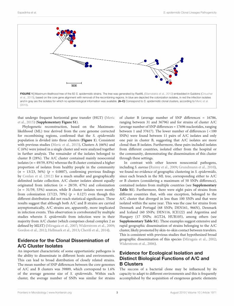
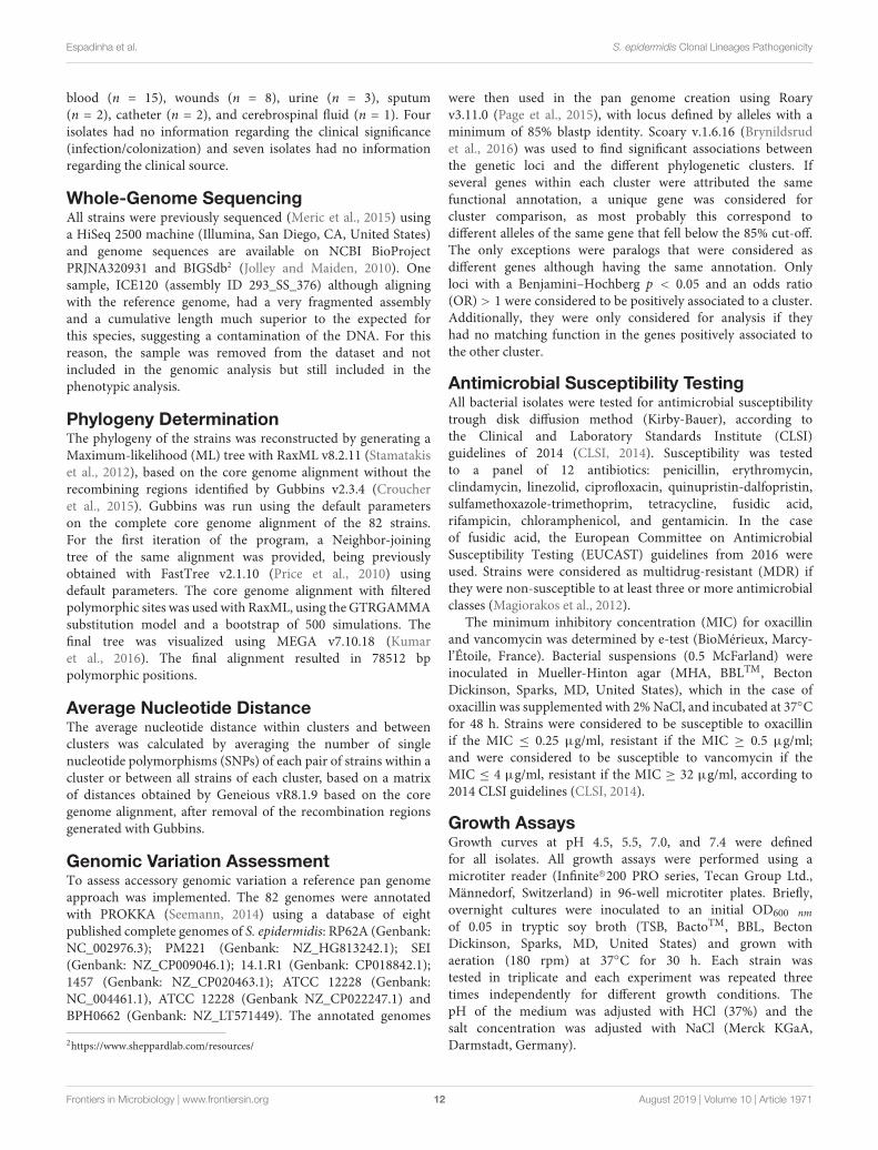
FIGURE 1 | Maximum-likelihood tree of the 82 S. epidermidis strains. The tree was generated by RaxML (Stamatakis et al., 2012) embedded in Gubbins (Croucheret al., 2015), based on the core gene alignment with removal of the recombining regions. In blue are depicted the colonization isolates, in red the infection isolatesand in gray are the isolates for which no epidemiological information was available. (A–C) Correspond to S. epidermidis clonal clusters, according to Meric et al.(2015).
that undergo frequent horizontal gene transfer (HGT) (Mericet al., 2015) (Supplementary Figure S1).
Phylogenetic reconstruction, based on the Maximum-likelihood (ML) tree derived from the core genome correctedfor recombining regions, confirmed that the S. epidermidispopulation is divided into three clusters (Figure 1). Consistentwith previous studies (Meric et al., 2015), Clusters A (66%) andC (6%) were joined in a single cluster and were analyzed togetherin further analysis. The remainder of the isolates belonged tocluster B (28%). The A/C cluster contained mainly nosocomialisolates (n = 49/59, 83%) whereas the B cluster contained a higherproportion of isolates from healthy people in the community(n = 13/23, 56%) (p = 0.0007), confirming previous findingsby Conlan et al. (2012) for a much smaller and geographicallydelimited isolate collection. A/C cluster isolates almost equallyoriginated from infection (n = 28/59, 47%) and colonization(n = 31/59, 53%) sources, while B cluster isolates were mostlyfrom colonization (17/23, 70%) (p = 0.127) even though thisdifferent distribution did not reach statistical significance. Theseresults suggest that although both A/C and B strains are carriedasymptomatically, A/C strains are, apparently, more implicatedin infection events. This observation is corroborated by multiplestudies wherein S. epidermidis from infection were in theirmajority from A/C cluster (which comprises clonal complex 2 asdefined by MLST) (Miragaia et al., 2007; Widerstrom et al., 2009;Gordon et al., 2012; Hellmark et al., 2013; Cherifi et al., 2014).
Evidence for the Clonal Dissemination ofA/C Cluster IsolatesAn important characteristic of some opportunistic pathogens isthe ability to disseminate in different hosts and environments.This can lead to broad distribution of closely related strains.The mean number of SNP differences between the core genomesof A/C and B clusters was 39889, which correspond to 1.6%of the average genome size of S. epidermidis. Within eachcluster, the average number of SNPs was similar for strains
of cluster B (average number of SNP differences = 16786,ranging between 31 and 36796) and for strains of cluster A/C(average number of SNP differences = 17696 nucleotides, rangingbetween 1 and 37617). The lower number of differences (<100SNPs) were found between 11 pairs of A/C isolates and onlyone pair in cluster B, suggesting that A/C isolates are moreclonal than B isolates. Furthermore, these pairs included isolatesfrom different countries, isolated either from the hospital orthe community, demonstrating the dissemination of this clusterthrough these settings.
In contrast with other known nosocomial pathogens,including S. aureus (Ruimy et al., 2009; Grundmann et al., 2010),we found no evidence of geographic clustering in S. epidermidis,since each branch in the ML tree, corresponding either to A/Cor B clusters (considering a maximum of 50 SNPs difference),contained isolates from multiple countries (see SupplementaryTable S1). Furthermore, there were eight pairs of strains fromdifferent countries that, with one exception, belonged to theA/C cluster that diverged in less than 100 SNPs and that wereisolated within the same year. This was the case for strains fromDenmark and Portugal (68 SNPs; DEN161, 966N), Denmarkand Iceland (60 SNPs; DEN116, ICE122) and Argentina andHungary (17 SNPs; AGT24, HUR105), among others (seeSupplementary Table S1). These examples suggest extensive andrapid geographic dissemination of strains belonging to the A/Ccluster, likely promoted by skin-to-skin contact between travelers.This is consistent with previous studies that hypothesized broadgeographic dissemination of this species (Miragaia et al., 2002;Widerstrom et al., 2006).
Evidence for Ecological Isolation andDistinct Biological Functions of A/C andB ClustersThe success of a bacterial clone may be influenced by itscapacity to adapt to different environments and this is frequentlyaccomplished by the acquisition of exogenous genetic elements
Frontiers in Microbiology | www.frontiersin.org 3 August 2019 | Volume 10 | Article 1971
fmicb-10-01971 August 27, 2019 Time: 12:8 # 4
Espadinha et al. S. epidermidis Clonal Lineages Pathogenicity
(Uhlemann et al., 2012; Walther et al., 2018), which will composethe accessory genome. To assess differences in gene content of thetwo S. epidermidis clusters, we analyzed and quantified accessorygenome variation. The matrix for presence/absence of all thegenes in the genomes is represented in Supplementary TableS2. The majority of A/C and B pan genomes was composedof accessory genes (A/C: 74%, 3723/5029; B: 67%, 3381/5029).Among this accessory genome, 1648 genes were exclusive to A/Ccluster and these genes varied in frequency from 93% to 2% and1306 genes were exclusive to cluster B and ranged from 100 to 4%,suggesting high genome plasticity and cluster-specific functions.The number of accessory genes shared between strains of thesame cluster (3723 in A/C and 3381 genes in B) is 1.8–1.6 higherthan the number of genes shared between the two clusters (2075genes). This difference could be due to the existence of a barrier togenetic transfer between them. However, in contrast to S. aureus,this apparently low frequency of genetic transfer is not evidentlyassociated with the presence of Restriction/Modification (R-M)systems (Waldron and Lindsay, 2006; Corvaglia et al., 2010)and/or the clustered regularly interspaced short palindromicrepeats (CRISPR) (Marraffini and Sontheimer, 2008). Therewas no clear association of a hsdR/hsdM system to any of theclusters and cas and csm genes were equally distributed amongstrains of the two clusters [A/C: 12% and B: 17% (p > 0.05)](Supplementary Table S2). An alternative explanation for thelower level of genetic exchange between the clusters, in thisapparently recombinogenic species (Miragaia et al., 2007; Mericet al., 2015), might be the existence of tropism of each ofthese lineages to specific skin niches (Grice et al., 2009; Ohet al., 2014) that could have lead to some ecological isolation ofthe two genetic clusters. Actually, skin microbiota compositionwas previously shown to vary according to the environmentalcharacteristics of the skin ecological niche sampled (Costelloet al., 2009; Grice et al., 2009). To test the hypothesis ofecological isolation we have examined the seven MLST genes,which constitute a good representation of the core genes, eitherin terms of genome distribution and genetic diversity (Gomeset al., 2005; Miragaia et al., 2007). Data showed an apparentallelic segregation, where aroE gene presented no common allelesbetween the clusters and the remaining genes (arcC, gtr, mutS,pyrR, tpi, yqiL), comprising between 11 and 20 alleles, had amaximum of two alleles shared by the two clusters.
Furthermore, we looked for genes that were significantlyassociated to each cluster using a pan genome wide-associationapproach, that scores genes for associations with specificepidemiological features. In particular, 166 genes were identifiedto have a strong positive association (Benjamini–Hochbergp < 0.05) with cluster A/C (see Supplementary Table S3)and 244 genes with a strong association with cluster B (seeSupplementary Table S4). Tables 1, 2 include the genes amongthese, which have functions that were either present exclusively inone of the clusters or that were present in both but in significantlydifferent frequencies. Strains of the A/C cluster were enrichedfor genes involved in processes such as biofilm formation andadhesion to host matrix proteins, proteolysis, resistance toantibiotics and adaptation to low pH. Conversely, strains fromcluster B were enriched for genes involved in the detoxification
of formate and formaldehyde, oxidative stress response, hostinteraction through type VII secretion system, lipid metabolismand cell wall biosynthesis. This is consistent with S. epidermidisclusters A/C and B performing distinct biological functions andpossibly occupying different skin niches.
S. epidermidis of A/C and B ClustersHave Adapted to Skin EnvironmentConditions Using Different StrategiesSkin is the first barrier of the human body against externalaggressions and contains molecules that derive from themetabolism of skin cells and our microbiota, but also fromthe environment (Wilson, 2005). On the skin, S. epidermidishave to deal with nutrient limitation, harsh and variableenvironmental conditions and mechanic stresses. Perspirationis one of the central contributors for the composition ofskin milieu and a major physiological function that assistsin thermoregulation, skin surface hydration and immunedefense. Perspiration is composed mainly of water, butcontains several other metabolites that contribute for itsmultiple functions, such as antimicrobial peptides (AMP),immunoglobulins, natural moisturizing factors (lactate,urea, electrolytes, amino acids), vitamins, metals and salts(Wilson, 2005).
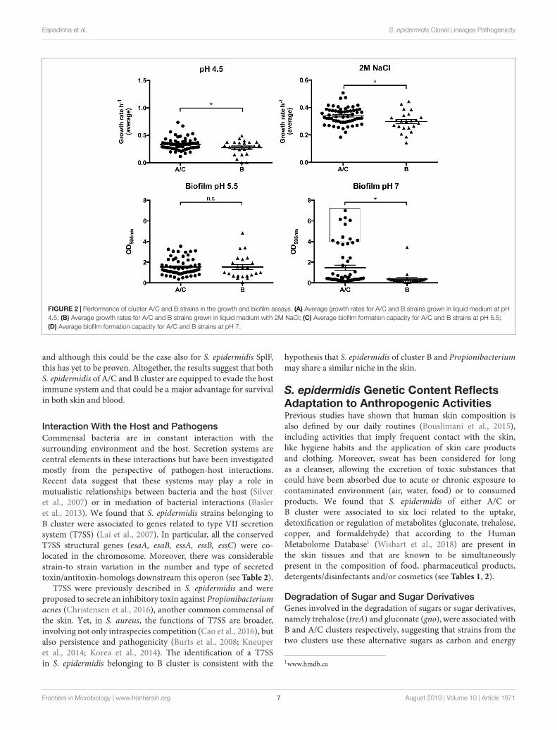
To understand if the S. epidermidis clusters differ in theirability to adapt to skin we have compared their ability to grow andproduce biofilm in conditions that mimic the skin environment(acidic pH and increased salt concentrations). Moreover, welooked in more detail for the genes that were associated by thepan genomic wide-association approach to each lineage and thatcould provide an advantage in the skin environmental conditions.
Growth and Biofilm Formation at Acidic pHStaphylococcus epidermidis belonging to A/C cluster appear tohave a distinctive ability to adapt to the two acidic pH valuestested (pH 4.5 and pH 5.5), as strains from the A/C clusterreached, in average, higher cell densities in stationary phase(∼12% higher in both cases) and presented either a greater(pH4.5: µA/C = 0.335; µB = 0.274, p = 0.043) or similar averagegrowth rate (pH 5.5: µA/C = 0.626; µB = 0.594, p = 0.273)when compared to strains from the B cluster (see Figure 2Aand Table 3). Additionally, strains belonging to the A/C clusterwere associated to a gene involved in the arginine deiminasemetabolism (arcD, an arginine/ornithine antiporter) (see Table 1)usually carried by the arginine catabolic mobile element (ACME).Although a genomic copy of arcD gene with a similar functionis known to be ubiquitous in the S. epidermidis genome, thisextra copy together with the remaining arc operon within ACMEwas previously suggested to be important for pH homeostasis inS. epidermidis and to contribute to additional tolerance to low-pHin community-associated Staphylococcus aureus USA300 clone(Thurlow et al., 2013; Lindgren et al., 2014). Other functionsassociated to A/C cluster strains that can eventually contributefor the observed increased growth rate is the flavin-containingoxidoreductase (YwqN), the YdjH sugar kinase; and the hexose-6-phosphate:phosphate antiporter (UhpT) (Park et al., 2015),
Frontiers in Microbiology | www.frontiersin.org 4 August 2019 | Volume 10 | Article 1971
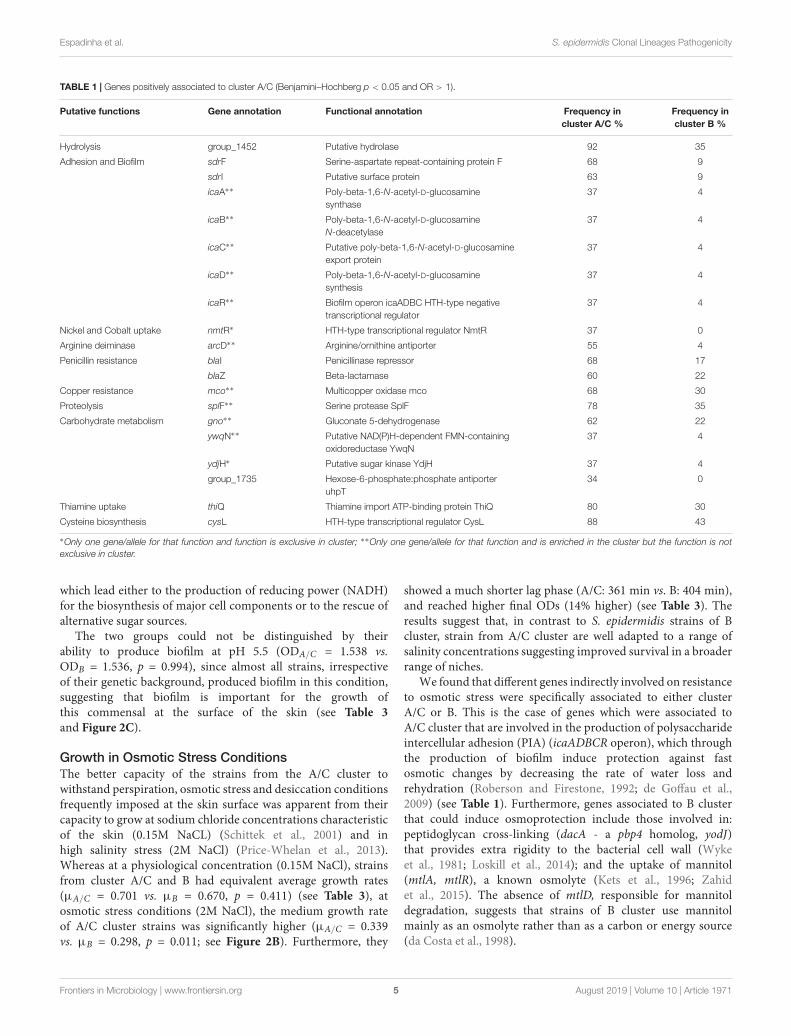
fmicb-10-01971 August 27, 2019 Time: 12:8 # 5
Espadinha et al. S. epidermidis Clonal Lineages Pathogenicity
TABLE 1 | Genes positively associated to cluster A/C (Benjamini–Hochberg p < 0.05 and OR > 1).
Putative functions Gene annotation Functional annotation Frequency incluster A/C %
Frequency incluster B %
Hydrolysis group_1452 Putative hydrolase 92 35
Adhesion and Biofilm sdrF Serine-aspartate repeat-containing protein F 68 9
sdrI Putative surface protein 63 9
icaA∗∗ Poly-beta-1,6-N-acetyl-D-glucosaminesynthase
37 4
icaB∗∗ Poly-beta-1,6-N-acetyl-D-glucosamineN-deacetylase
37 4
icaC∗∗ Putative poly-beta-1,6-N-acetyl-D-glucosamineexport protein
37 4
icaD∗∗ Poly-beta-1,6-N-acetyl-D-glucosaminesynthesis
37 4
icaR∗∗ Biofilm operon icaADBC HTH-type negativetranscriptional regulator
37 4
Nickel and Cobalt uptake nmtR∗ HTH-type transcriptional regulator NmtR 37 0
Arginine deiminase arcD∗∗ Arginine/ornithine antiporter 55 4
Penicillin resistance blaI Penicillinase repressor 68 17
blaZ Beta-lactamase 60 22
Copper resistance mco∗∗ Multicopper oxidase mco 68 30
Proteolysis splF∗∗ Serine protease SplF 78 35
Carbohydrate metabolism gno∗∗ Gluconate 5-dehydrogenase 62 22
ywqN∗∗ Putative NAD(P)H-dependent FMN-containingoxidoreductase YwqN
37 4
ydjH∗ Putative sugar kinase YdjH 37 4
group_1735 Hexose-6-phosphate:phosphate antiporteruhpT
34 0
Thiamine uptake thiQ Thiamine import ATP-binding protein ThiQ 80 30
Cysteine biosynthesis cysL HTH-type transcriptional regulator CysL 88 43
∗Only one gene/allele for that function and function is exclusive in cluster; ∗∗Only one gene/allele for that function and is enriched in the cluster but the function is notexclusive in cluster.
which lead either to the production of reducing power (NADH)for the biosynthesis of major cell components or to the rescue ofalternative sugar sources.
The two groups could not be distinguished by theirability to produce biofilm at pH 5.5 (ODA/C = 1.538 vs.ODB = 1.536, p = 0.994), since almost all strains, irrespectiveof their genetic background, produced biofilm in this condition,suggesting that biofilm is important for the growth ofthis commensal at the surface of the skin (see Table 3and Figure 2C).
Growth in Osmotic Stress ConditionsThe better capacity of the strains from the A/C cluster towithstand perspiration, osmotic stress and desiccation conditionsfrequently imposed at the skin surface was apparent from theircapacity to grow at sodium chloride concentrations characteristicof the skin (0.15M NaCL) (Schittek et al., 2001) and inhigh salinity stress (2M NaCl) (Price-Whelan et al., 2013).Whereas at a physiological concentration (0.15M NaCl), strainsfrom cluster A/C and B had equivalent average growth rates(µA/C = 0.701 vs. µB = 0.670, p = 0.411) (see Table 3), atosmotic stress conditions (2M NaCl), the medium growth rateof A/C cluster strains was significantly higher (µA/C = 0.339vs. µB = 0.298, p = 0.011; see Figure 2B). Furthermore, they
showed a much shorter lag phase (A/C: 361 min vs. B: 404 min),and reached higher final ODs (14% higher) (see Table 3). Theresults suggest that, in contrast to S. epidermidis strains of Bcluster, strain from A/C cluster are well adapted to a range ofsalinity concentrations suggesting improved survival in a broaderrange of niches.
We found that different genes indirectly involved on resistanceto osmotic stress were specifically associated to either clusterA/C or B. This is the case of genes which were associated toA/C cluster that are involved in the production of polysaccharideintercellular adhesion (PIA) (icaADBCR operon), which throughthe production of biofilm induce protection against fastosmotic changes by decreasing the rate of water loss andrehydration (Roberson and Firestone, 1992; de Goffau et al.,2009) (see Table 1). Furthermore, genes associated to B clusterthat could induce osmoprotection include those involved in:peptidoglycan cross-linking (dacA - a pbp4 homolog, yodJ)that provides extra rigidity to the bacterial cell wall (Wykeet al., 1981; Loskill et al., 2014); and the uptake of mannitol(mtlA, mtlR), a known osmolyte (Kets et al., 1996; Zahidet al., 2015). The absence of mtlD, responsible for mannitoldegradation, suggests that strains of B cluster use mannitolmainly as an osmolyte rather than as a carbon or energy source(da Costa et al., 1998).
Frontiers in Microbiology | www.frontiersin.org 5 August 2019 | Volume 10 | Article 1971
fmicb-10-01971 August 27, 2019 Time: 12:8 # 6
Espadinha et al. S. epidermidis Clonal Lineages Pathogenicity
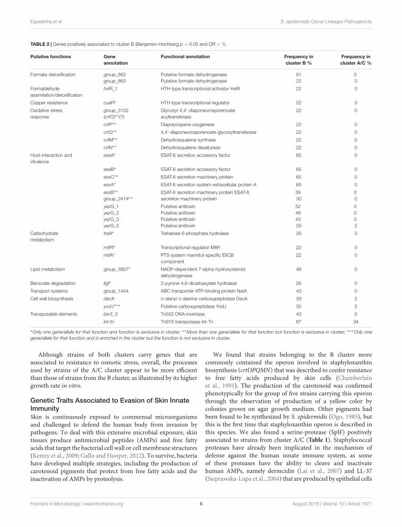
TABLE 2 | Genes positively associated to cluster B (Benjamini–Hochberg p < 0.05 and OR > 1).
Putative functions Geneannotation
Functional annotation Frequency incluster B %
Frequency incluster A/C %
Formate detoxification group_862group_863
Putative formate dehydrogenasePutative formate dehydrogenase
9122
00
Formaldehydeassimilation/detoxification
hxlR_1 HTH-type transcriptional activator HxlR 22 0
Copper resistance cueR∗ HTH-type transcriptional regulator 22 0
Oxidative stressresponse
group_3122(crtO)∗∗(?)
Glycosyl-4,4′-diaponeurosporenoateacyltransferase
22 0
crtP∗∗ Diapolycopene oxygenase 22 0
crtQ∗∗ 4,4′-diaponeurosporenoate glycosyltransferase 22 0
crtM∗∗ Dehydrosqualene synthase 22 0
crtN∗∗ Dehydrosqualene desaturase 22 0
Host-interaction andVirulence
esaA∗ ESAT-6 secretion accessory factor 65 0
esaB∗ ESAT-6 secretion accessory factor 65 0
essC∗∗ ESAT-6 secretion machinery protein 65 0
esxA∗ ESAT-6 secretion system extracellular protein A 65 0
essB∗∗
group_2414∗∗ESAT-6 secretion machinery protein ESAT-6secretion machinery protein
3930
00
yezG_1yezG_2yezG_3yezG_5
Putative antitoxinPutative antitoxinPutative antitoxinPutative antitoxin
52484326
0002
Carbohydratemetabolism
treA∗ Trehalose-6-phosphate hydrolase 26 0
mtlR∗ Transcriptional regulator MtlR 22 0
mtlA∗ PTS system mannitol-specific EIICBcomponent
22 0
Lipid metabolism group_3907∗ NADP-dependent 7-alpha-hydroxysteroiddehydrogenase
48 0
Benzoate degradation ligI∗ 2-pyrone-4,6-dicarbaxylate hydrolase 26 0
Transport systems group_1444 ABC transporter ATP-binding protein NatA 43 0
Cell wall biosynthesis dacA D-alanyl-D-alanine carboxypeptidase DacA 39 2
yodJ∗∗∗ Putative carboxypeptidase YodJ 35 2
Transposable elements bin3_3 Tn552 DNA-invertase 43 0
int-tn Tn916 transposase Int-Tn 87 34
∗Only one gene/allele for that function and function is exclusive in cluster; ∗∗More than one gene/allele for that function but function is exclusive in cluster; ∗∗∗Only onegene/allele for that function and is enriched in the cluster but the function is not exclusive in cluster.
Although strains of both clusters carry genes that areassociated to resistance to osmotic stress, overall, the processesused by strains of the A/C cluster appear to be more efficientthan those of strains from the B cluster, as illustrated by its highergrowth rate in vitro.
Genetic Traits Associated to Evasion of Skin InnateImmunitySkin is continuously exposed to commensal microorganismsand challenged to defend the human body from invasion bypathogens. To deal with this extensive microbial exposure, skintissues produce antimicrobial peptides (AMPs) and free fattyacids that target the bacterial cell wall or cell membrane structures(Kenny et al., 2009; Gallo and Hooper, 2012). To survive, bacteriahave developed multiple strategies, including the production ofcarotenoid pigments that protect from free fatty acids and theinactivation of AMPs by proteolysis.
We found that strains belonging to the B cluster morecommonly contained the operon involved in staphyloxanthinbiosynthesis (crtOPQMN) that was described to confer resistanceto free fatty acids produced by skin cells (Chamberlainet al., 1991). The production of the carotenoid was confirmedphenotypically for the group of five strains carrying this operonthrough the observation of production of a yellow color bycolonies grown on agar growth medium. Other pigments hadbeen found to be synthesized by S. epidermidis (Ogo, 1985), butthis is the first time that staphyloxanthin operon is described inthis species. We also found a serine-protease (SplF) positivelyassociated to strains from cluster A/C (Table 1). Staphylococcalproteases have already been implicated in the mechanism ofdefense against the human innate immune system, as someof these proteases have the ability to cleave and inactivatehuman AMPs, namely dermcidin (Lai et al., 2007) and LL-37(Sieprawska-Lupa et al., 2004) that are produced by epithelial cells
Frontiers in Microbiology | www.frontiersin.org 6 August 2019 | Volume 10 | Article 1971
fmicb-10-01971 August 27, 2019 Time: 12:8 # 7
Espadinha et al. S. epidermidis Clonal Lineages Pathogenicity
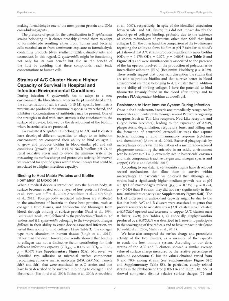
FIGURE 2 | Performance of cluster A/C and B strains in the growth and biofilm assays. (A) Average growth rates for A/C and B strains grown in liquid medium at pH4.5; (B) Average growth rates for A/C and B strains grown in liquid medium with 2M NaCl; (C) Average biofilm formation capacity for A/C and B strains at pH 5.5;(D) Average biofilm formation capacity for A/C and B strains at pH 7.
and although this could be the case also for S. epidermidis SplF,this has yet to be proven. Altogether, the results suggest that bothS. epidermidis of A/C and B cluster are equipped to evade the hostimmune system and that could be a major advantage for survivalin both skin and blood.
Interaction With the Host and PathogensCommensal bacteria are in constant interaction with thesurrounding environment and the host. Secretion systems arecentral elements in these interactions but have been investigatedmostly from the perspective of pathogen-host interactions.Recent data suggest that these systems may play a role inmutualistic relationships between bacteria and the host (Silveret al., 2007) or in mediation of bacterial interactions (Basleret al., 2013). We found that S. epidermidis strains belonging toB cluster were associated to genes related to type VII secretionsystem (T7SS) (Lai et al., 2007). In particular, all the conservedT7SS structural genes (esaA, esaB, essA, essB, essC) were co-located in the chromosome. Moreover, there was considerablestrain-to strain variation in the number and type of secretedtoxin/antitoxin-homologs downstream this operon (see Table 2).
T7SS were previously described in S. epidermidis and wereproposed to secrete an inhibitory toxin against Propionibacteriumacnes (Christensen et al., 2016), another common commensal ofthe skin. Yet, in S. aureus, the functions of T7SS are broader,involving not only intraspecies competition (Cao et al., 2016), butalso persistence and pathogenicity (Burts et al., 2008; Kneuperet al., 2014; Korea et al., 2014). The identification of a T7SSin S. epidermidis belonging to B cluster is consistent with the
hypothesis that S. epidermidis of cluster B and Propionibacteriummay share a similar niche in the skin.
S. epidermidis Genetic Content ReflectsAdaptation to Anthropogenic ActivitiesPrevious studies have shown that human skin composition isalso defined by our daily routines (Bouslimani et al., 2015),including activities that imply frequent contact with the skin,like hygiene habits and the application of skin care productsand clothing. Moreover, sweat has been considered for longas a cleanser, allowing the excretion of toxic substances thatcould have been absorbed due to acute or chronic exposure tocontaminated environment (air, water, food) or to consumedproducts. We found that S. epidermidis of either A/C orB cluster were associated to six loci related to the uptake,detoxification or regulation of metabolites (gluconate, trehalose,copper, and formaldehyde) that according to the HumanMetabolome Database1 (Wishart et al., 2018) are present inthe skin tissues and that are known to be simultaneouslypresent in the composition of food, pharmaceutical products,detergents/disinfectants and/or cosmetics (see Tables 1, 2).
Degradation of Sugar and Sugar DerivativesGenes involved in the degradation of sugars or sugar derivatives,namely trehalose (treA) and gluconate (gno), were associated withB and A/C clusters respectively, suggesting that strains from thetwo clusters use these alternative sugars as carbon and energy
1www.hmdb.ca
Frontiers in Microbiology | www.frontiersin.org 7 August 2019 | Volume 10 | Article 1971
fmicb-10-01971 August 27, 2019 Time: 12:8 # 8
Espadinha et al. S. epidermidis Clonal Lineages Pathogenicity
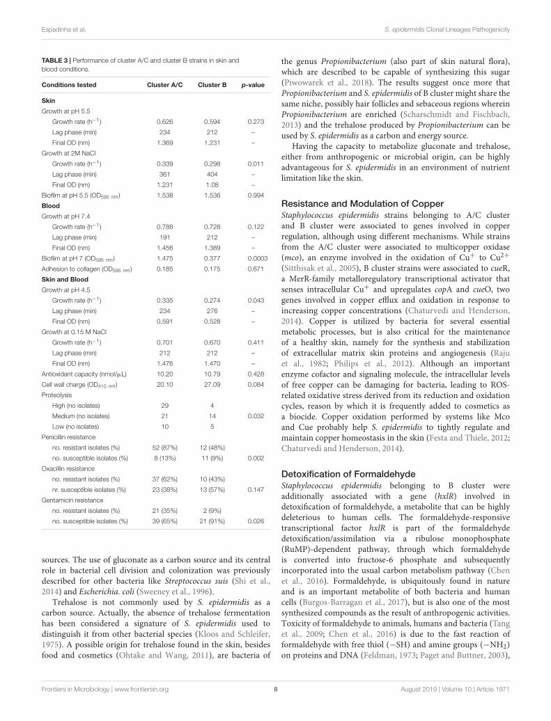
TABLE 3 | Performance of cluster A/C and cluster B strains in skin andblood conditions.
Conditions tested Cluster A/C Cluster B p-value
Skin
Growth at pH 5.5
Growth rate (h−1) 0.626 0.594 0.273
Lag phase (min) 234 212 –
Final OD (nm) 1.369 1.231 –
Growth at 2M NaCl
Growth rate (h−1) 0.339 0.298 0.011
Lag phase (min) 361 404 –
Final OD (nm) 1.231 1.08 –
Biofilm at pH 5.5 (OD595 nm) 1.538 1.536 0.994
Blood
Growth at pH 7.4
Growth rate (h−1) 0.788 0.728 0.122
Lag phase (min) 191 212 –
Final OD (nm) 1.456 1.389 –
Biofilm at pH 7 (OD595 nm) 1.475 0.377 0.0003
Adhesion to collagen (OD595 nm) 0.185 0.175 0.671
Skin and Blood
Growth at pH 4.5
Growth rate (h−1) 0.335 0.274 0.043
Lag phase (min) 234 276 –
Final OD (nm) 0.591 0.528 –
Growth at 0.15 M NaCl
Growth rate (h−1) 0.701 0.670 0.411
Lag phase (min) 212 212 –
Final OD (nm) 1.476 1.470 –
Antioxidant capacity (nmol/µL) 10.20 10.79 0.428
Cell wall charge (OD410 nm) 20.10 27.09 0.084
Proteolysis
High (no isolates) 29 4
Medium (no isolates) 21 14 0.032
Low (no isolates) 10 5
Penicillin resistance
no. resistant isolates (%) 52 (87%) 12 (48%)
no. susceptible isolates (%) 8 (13%) 11 (9%) 0.002
Oxacillin resistance
no. resistant isolates (%) 37 (62%) 10 (43%)
nr. susceptible isolates (%) 23 (38%) 13 (57%) 0.147
Gentamicin resistance
no. resistant isolates (%) 21 (35%) 2 (9%)
no. susceptible isolates (%) 39 (65%) 21 (91%) 0.026
sources. The use of gluconate as a carbon source and its centralrole in bacterial cell division and colonization was previouslydescribed for other bacteria like Streptococcus suis (Shi et al.,2014) and Escherichia. coli (Sweeney et al., 1996).
Trehalose is not commonly used by S. epidermidis as acarbon source. Actually, the absence of trehalose fermentationhas been considered a signature of S. epidermidis used todistinguish it from other bacterial species (Kloos and Schleifer,1975). A possible origin for trehalose found in the skin, besidesfood and cosmetics (Ohtake and Wang, 2011), are bacteria of
the genus Propionibacterium (also part of skin natural flora),which are described to be capable of synthesizing this sugar(Piwowarek et al., 2018). The results suggest once more thatPropionibacterium and S. epidermidis of B cluster might share thesame niche, possibly hair follicles and sebaceous regions whereinPropionibacterium are enriched (Scharschmidt and Fischbach,2013) and the trehalose produced by Propionibacterium can beused by S. epidermidis as a carbon and energy source.
Having the capacity to metabolize gluconate and trehalose,either from anthropogenic or microbial origin, can be highlyadvantageous for S. epidermidis in an environment of nutrientlimitation like the skin.
Resistance and Modulation of CopperStaphylococcus epidermidis strains belonging to A/C clusterand B cluster were associated to genes involved in copperregulation, although using different mechanisms. While strainsfrom the A/C cluster were associated to multicopper oxidase(mco), an enzyme involved in the oxidation of Cu+ to Cu2+
(Sitthisak et al., 2005), B cluster strains were associated to cueR,a MerR-family metalloregulatory transcriptional activator thatsenses intracellular Cu+ and upregulates copA and cueO, twogenes involved in copper efflux and oxidation in response toincreasing copper concentrations (Chaturvedi and Henderson,2014). Copper is utilized by bacteria for several essentialmetabolic processes, but is also critical for the maintenanceof a healthy skin, namely for the synthesis and stabilizationof extracellular matrix skin proteins and angiogenesis (Rajuet al., 1982; Philips et al., 2012). Although an importantenzyme cofactor and signaling molecule, the intracellular levelsof free copper can be damaging for bacteria, leading to ROS-related oxidative stress derived from its reduction and oxidationcycles, reason by which it is frequently added to cosmetics asa biocide. Copper oxidation performed by systems like Mcoand Cue probably help S. epidermidis to tightly regulate andmaintain copper homeostasis in the skin (Festa and Thiele, 2012;Chaturvedi and Henderson, 2014).
Detoxification of FormaldehydeStaphylococcus epidermidis belonging to B cluster wereadditionally associated with a gene (hxlR) involved indetoxification of formaldehyde, a metabolite that can be highlydeleterious to human cells. The formaldehyde-responsivetranscriptional factor hxlR is part of the formaldehydedetoxification/assimilation via a ribulose monophosphate(RuMP)-dependent pathway, through which formaldehydeis converted into fructose-6 phosphate and subsequentlyincorporated into the usual carbon metabolism pathway (Chenet al., 2016). Formaldehyde, is ubiquitously found in natureand is an important metabolite of both bacteria and humancells (Burgos-Barragan et al., 2017), but is also one of the mostsynthesized compounds as the result of anthropogenic activities.Toxicity of formaldehyde to animals, humans and bacteria (Tanget al., 2009; Chen et al., 2016) is due to the fast reaction offormaldehyde with free thiol (−SH) and amine groups (−NH2)on proteins and DNA (Feldman, 1973; Paget and Buttner, 2003),
Frontiers in Microbiology | www.frontiersin.org 8 August 2019 | Volume 10 | Article 1971
fmicb-10-01971 August 27, 2019 Time: 12:8 # 9
Espadinha et al. S. epidermidis Clonal Lineages Pathogenicity
making formaldehyde one of the most potent protein and DNAcross-linking agents.
The presence of genes for the detoxification in S. epidermidisstrains belonging to B cluster probably allowed them to adaptto formaldehyde resulting either from human and bacterialcells metabolism or from continuous exposure to formaldehydecontaining products (dyes, synthetic textiles, disinfectants, andcosmetics). In this regard, S. epidermidis might be functioningnot only for its own benefit but also in the benefit ofthe host by avoiding that these compounds reach toxicconcentrations to human cells.
Strains of A/C Cluster Have a HigherCapacity of Survival in Hospital andInfection Environmental ConditionsDuring infection S. epidermidis has to adapt to a newenvironment, the bloodstream, wherein the pH is stabilized at 7.4,the concentration of salt is steady (0.15 M), specific host matrixproteins are produced, the immune response is exacerbated, andinhibitory concentrations of antibiotics may be present. One ofthe strategies to deal with such stresses is the attachment to thesurface of a device, followed by the development of the biofilm,where bacterial cells are protected.
To evaluate if S. epidermidis belonging to A/C and B clustershave developed different capacities to adapt to an infectionenvironment, we compared their ability to bind collagen I,to grow and produce biofilm in blood-similar pH and saltconditions (growth: pH 7.4; 0.15 M NaCl; biofilm: pH 7), toresist oxidative stress and to evade the immune system (bymeasuring the surface charge and proteolytic activity). Moreover,we searched for specific genes within these lineages that could beassociated to a higher infectious capacity.
Binding to Host Matrix Proteins and BiofilmFormation at Blood pHWhen a medical device is introduced into the human body, itssurface becomes coated with a layer of host proteins (Vaudauxet al., 1995; von Eiff et al., 2002; Arrecubieta et al., 2007; Singhet al., 2012). Foreign-body associated infections are attributedto the attachment of bacteria to these host proteins, such ascollagen I from tissues, and fibronectin and fibrinogen fromblood, through binding of surface proteins (Patti et al., 1994;Foster and Hook, 1998) followed by the production of biofilm. Tounderstand if S. epidermidis belonging to the two genetic lineagesdiffered in their ability to cause device-associated infection, wetested their ability to bind collagen I (see Table 3), the collagentype more abundant in human tissues (Singh et al., 2012),rather than the skin. However, our results showed that bindingto collagen was not a distinctive factor contributing for theirdifferent infectious capacity (ODA/C = 0.185 vs. ODB = 0.175,p = 0.067) (see Supplementary Figure S2A). Moreover, weidentified two adhesins or microbial surface componentsrecognizing adhesive matrix molecules (MSCRAMMs), namelySdrF and SdrI, that were associated to A/C strains and thathave been described to be involved in binding to collagen I andfibronectin (Hartford et al., 2001; Sakinc et al., 2005; Arrecubieta
et al., 2007), respectively. In spite of the identified associationbetween SdrF and A/C cluster, this did not impact directly thephenotype of collagen binding, probably due to the existenceof known redundancy of proteins other than SdrF that bindcollagen I. On the other hand, the comparison of the two lineagesregarding the ability to form biofilm at pH 7 (similar to blood’spH) showed that A/C strains produced significantly more biofilm(ODA/C = 1.475; ODB = 0.377, p = 0.0003) (see Table 3 andFigure 2D) and were simultaneously associated to the presenceof the ica operon, involved in the production of polysaccharideintercellular adhesion (PIA) (Benjamini–Hochberg p = 0.029).These results suggest that upon skin disruption the strains thatare able to produce biofilm and that survive better in bloodenvironment are those belonging to A/C cluster that in additionto the ability of binding collagen I have the potential to bindfibronectin (mainly found in the blood after injury) and toproduce PIA-dependent biofilms at blood’s pH.
Resistance to Host Immune System During InfectionOnce in the bloodstream, bacteria are immediately recognized bymonocytes and neutrophils through several Pattern recognizingreceptors (such as Toll-Like receptors, Nod-Like receptors andC-type lectin receptors), leading to the expression of AMPs,phagocytosis, degranulation, respiratory burst and killing andthe formation of neutrophil extracellular traps that capturebacteria inducing a rapid inflammatory response (cytokinesand chemokines) (Akira et al., 2006). Bacterial phagocytosis bymacrophages occurs via the formation of a membrane-enclosedphagosome containing the microbe in an acidic environment(can be as low as pH 4.5), extremely rich in bactericidal enzymesand toxic compounds (reactive oxygen and nitrogen species andcopper) (Weiss and Schaible, 2015).
According to our data, S. epidermidis strains have developedseveral mechanisms that allow them to survive withinmacrophages. In particular, we observed that although A/Cstrains had a significantly higher medium growth rate at pH4.5 (pH of macrophages milieu) (µA/C = 0.335; µB = 0.274,p = 0.043) than B strains, they did not vary significantly in theirtotal antioxidant capacity (see Supplementary Figure S2B). Thelack of difference in antioxidant capacity might be due to thefact that both A/C and B clusters were associated to genes thatprovide resistance to oxidative stress (A/C cluster: mco; B cluster:crtOPQMN operon) and tolerance to copper (A/C cluster: mco;B cluster: cueR) (see Tables 1, 2). Especially, staphyloxanthinproduced by crtOPQMN was described in S. aureus to participatein the scavenging of free radicals and to have impact in virulence(Clauditz et al., 2006; Mishra et al., 2011).
We have also compared the surface charge and proteolyticactivity of the two clusters, as a measure of the capacityto evade the host immune system. According to our data,strains of the A/C and B clusters showed a similar averagevalue of surface charge measured by the relative percentage ofunbound cytochrome C, but the values obtained varied from0 and 78% among strains (see Supplementary Figure S2Cand Supplementary Table S5). In particular, closely relatedstrains in the phylogenetic tree (DEN116 and ICE21, 101 SNPs)showed completely distinct relative surface charges (72 and
Frontiers in Microbiology | www.frontiersin.org 9 August 2019 | Volume 10 | Article 1971
fmicb-10-01971 August 27, 2019 Time: 12:8 # 10
Espadinha et al. S. epidermidis Clonal Lineages Pathogenicity
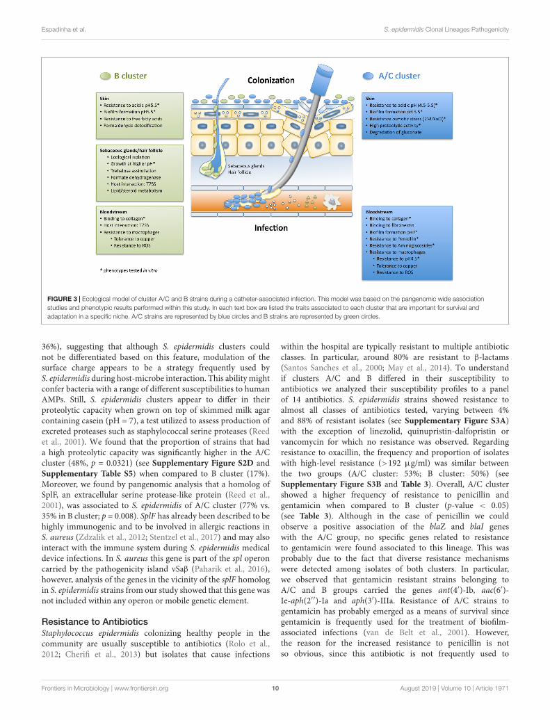
FIGURE 3 | Ecological model of cluster A/C and B strains during a catheter-associated infection. This model was based on the pangenomic wide associationstudies and phenotypic results performed within this study. In each text box are listed the traits associated to each cluster that are important for survival andadaptation in a specific niche. A/C strains are represented by blue circles and B strains are represented by green circles.
36%), suggesting that although S. epidermidis clusters couldnot be differentiated based on this feature, modulation of thesurface charge appears to be a strategy frequently used byS. epidermidis during host-microbe interaction. This ability mightconfer bacteria with a range of different susceptibilities to humanAMPs. Still, S. epidermidis clusters appear to differ in theirproteolytic capacity when grown on top of skimmed milk agarcontaining casein (pH = 7), a test utilized to assess production ofexcreted proteases such as staphylococcal serine proteases (Reedet al., 2001). We found that the proportion of strains that hada high proteolytic capacity was significantly higher in the A/Ccluster (48%, p = 0.0321) (see Supplementary Figure S2D andSupplementary Table S5) when compared to B cluster (17%).Moreover, we found by pangenomic analysis that a homolog ofSplF, an extracellular serine protease-like protein (Reed et al.,2001), was associated to S. epidermidis of A/C cluster (77% vs.35% in B cluster; p = 0.008). SplF has already been described to behighly immunogenic and to be involved in allergic reactions inS. aureus (Zdzalik et al., 2012; Stentzel et al., 2017) and may alsointeract with the immune system during S. epidermidis medicaldevice infections. In S. aureus this gene is part of the spl operoncarried by the pathogenicity island νSaβ (Paharik et al., 2016),however, analysis of the genes in the vicinity of the splF homologin S. epidermidis strains from our study showed that this gene wasnot included within any operon or mobile genetic element.
Resistance to AntibioticsStaphylococcus epidermidis colonizing healthy people in thecommunity are usually susceptible to antibiotics (Rolo et al.,2012; Cherifi et al., 2013) but isolates that cause infections
within the hospital are typically resistant to multiple antibioticclasses. In particular, around 80% are resistant to β-lactams(Santos Sanches et al., 2000; May et al., 2014). To understandif clusters A/C and B differed in their susceptibility toantibiotics we analyzed their susceptibility profiles to a panelof 14 antibiotics. S. epidermidis strains showed resistance toalmost all classes of antibiotics tested, varying between 4%and 88% of resistant isolates (see Supplementary Figure S3A)with the exception of linezolid, quinupristin-dalfopristin orvancomycin for which no resistance was observed. Regardingresistance to oxacillin, the frequency and proportion of isolateswith high-level resistance (>192 µg/ml) was similar betweenthe two groups (A/C cluster: 53%; B cluster: 50%) (seeSupplementary Figure S3B and Table 3). Overall, A/C clustershowed a higher frequency of resistance to penicillin andgentamicin when compared to B cluster (p-value < 0.05)(see Table 3). Although in the case of penicillin we couldobserve a positive association of the blaZ and blaI geneswith the A/C group, no specific genes related to resistanceto gentamicin were found associated to this lineage. This wasprobably due to the fact that diverse resistance mechanismswere detected among isolates of both clusters. In particular,we observed that gentamicin resistant strains belonging toA/C and B groups carried the genes ant(4′)-Ib, aac(6′)-Ie-aph(2′′)-Ia and aph(3′)-IIIa. Resistance of A/C strains togentamicin has probably emerged as a means of survival sincegentamicin is frequently used for the treatment of biofilm-associated infections (van de Belt et al., 2001). However,the reason for the increased resistance to penicillin is notso obvious, since this antibiotic is not frequently used to
Frontiers in Microbiology | www.frontiersin.org 10 August 2019 | Volume 10 | Article 1971
fmicb-10-01971 August 27, 2019 Time: 12:8 # 11
Espadinha et al. S. epidermidis Clonal Lineages Pathogenicity
treat device-related infections. A possibility is that blaZregulators are maintained associated to A/C strains becausethey are important for SCCmec acquisition by S. epidermidisas was previously described for S. aureus (Katayama et al.,2003). Actually, all strains that were resistant to methicillincontained blaZ.
Identification of Epidemiological Markersfor S. epidermidis A/C and B ClustersThe results from this study highlighted the phenotypic andgenotypic differences between the A/C and B clusters andprovided several lines of evidence supporting the higherpathogenic potential of A/C strains. Thus, the finding of goodepidemiological markers for these two clusters would assist ina better prognosis of the infection and would improve clinicaldecision-making (namely more appropriate treatment approach)and, ultimately, patient outcomes.
To identify good epidemiological markers for the A/C and Bclusters we looked for genes that were associated to the clustersand had frequencies above 90% (see Supplementary Table S3and Table 2). We found two genes meeting these criteria: agene encoding a hypothetical protein with a putative DUF1641domain with a formate dehydrogenase activity associated tocluster B; and one gene encoding for a hydrolase (yxeP) ofunknown function associated to cluster A/C. In this latter,there were only three isolates belonging to B cluster thatcontained yxeP. These three strains cluster together in thetree, with two strains being more closely related (70 SNPs)than the other (∼2500 SNPs of difference), all collected fromnasal colonization in Portugal from draftees sharing the sameenvironment, suggesting that the acquisition of yxeP by strainsof this lineage might be a rare genetic event.
Our results confirmed the presence of a putative formatedehydrogenase gene (group_862) as highly specific of strainsbelonging to B cluster, as previously suggested (Conlan et al.,2012). Formate dehydrogenase was described to be importantfor formate detoxification and NADH/H + production duringanaerobic fermentative growth in E. coli (Knappe and Sawers,1990; Sawers, 2005), but in S. aureus this enzyme is only expressedin a microaerobic environment (Leibig et al., 2011). It is thustempting to speculate that fdh might enable cluster B strains tosurvive in specific microaerobic niches in the skin, as hair folliclesand sebaceous glands, like Propionibacterium spp.
CONCLUSION
Staphylococcus epidermidis is the main colonizer of thehuman skin but also one the most important opportunisticpathogens associated to medical device-related infections thatoccur when the skin barrier is disrupted and the host isimmunocompromised.
In this study we have compared the two unique clustersthat compose S. epidermidis population (A/C and B) regardingtheir genomic content, using genome-wide association studies,and regarding their phenotypic performance in conditions thatmimic colonization and infection, which allowed us to create
a portrait of each cluster that sheds some light into theirecology and adaptation to commensalism or pathogenicity(see Figure 3). Herein we have shown that strains fromcluster A/C and cluster B differ extensively in both theircore and accessory genomes and are associated to differentgenes enrolled in biological functions that were frequentlyrelated to their phenotypic performance in the skin and bloodconditions, suggesting that the two clusters have been ecologicallyisolated. Strains from cluster A/C grew faster or reachedhigher final cell densities in all skin acidic pH (4.5–5.5) andsalt concentrations tested (0.15 M, 2 M), whereas B clusterstrains were less fit under acidic and osmotic stress. Moreover,B strains were specifically associated to functions related toresistance to free fatty acids, lipid metabolism, microaerobicenvironment and interspecies interaction (Propionibacteriumspp.). In infection conditions, however, A/C strains weresuperior in their ability to resist antibiotics, to grow inmacrophage milieu pH conditions (pH 4.5) and to formbiofilm at blood pH (∼pH 7). Moreover, they were associatedto functions related to binding to host-matrix proteins.Overall, B strains seem to have adapted to survive in amicroaerophilic and lipid rich environment such as hair folliclesand sebaceous glands; whereas A/C strains probably occupy amore superficial and broader niche in the skin as they werebetter adapted to the changing osmotic and pH conditions ofthe skin surface.
Herein we have also produced a population and genome-widebased catalog of genes associated to A/C cluster and identifiedenvironmental conditions that favor the growth of A/C strainsthat could serve as the basis for future studies on S. epidermidispathogenesis and eventually for the development of precisionantimicrobial strategies. Additionally, the finding of a geneticmarker for the more pathogenic genetic lineage can improvesignificantly the prognosis and management of medical-devicerelated infections caused by this bacterium.
MATERIALS AND METHODS
Bacterial IsolatesThe bacterial collection comprised a total of 83 S. epidermidisisolates, collected from different countries (Argentina, n = 1;Bulgaria, n = 2; Cape Verde, n = 4; Colombia, n = 1; Denmark,n = 14; Greece, n = 3; Hungary, n = 2; Iceland, n = 13;Italy, n = 2; Japan, n = 2; Mexico, n = 5; Portugal, n = 31;Taiwan, n = 2; and Uruguay, n = 1) between 1996 and 2001and representative of the population found in the hospital(n = 60) and community settings (n = 23). These isolates wereselected to represent all the diversity in sequence types previouslyfound among a temporal and geographically diverse collectionof nosocomial and community-associated S. epidermidis isolates(Miragaia et al., 2007; Rolo et al., 2012). Isolates were classifiedas being from colonization (n = 45) or infection (n = 34)according to defined clinical parameters. Colonization isolateswere recovered from several products including nasal swabs(n = 35), wounds (n = 6), sputum (n = 1), urine (n = 1)and blood (n = 1). Infection products were recovered from
Frontiers in Microbiology | www.frontiersin.org 11 August 2019 | Volume 10 | Article 1971
fmicb-10-01971 August 27, 2019 Time: 12:8 # 12
Espadinha et al. S. epidermidis Clonal Lineages Pathogenicity
blood (n = 15), wounds (n = 8), urine (n = 3), sputum(n = 2), catheter (n = 2), and cerebrospinal fluid (n = 1). Fourisolates had no information regarding the clinical significance(infection/colonization) and seven isolates had no informationregarding the clinical source.
Whole-Genome SequencingAll strains were previously sequenced (Meric et al., 2015) usinga HiSeq 2500 machine (Illumina, San Diego, CA, United States)and genome sequences are available on NCBI BioProjectPRJNA320931 and BIGSdb2 (Jolley and Maiden, 2010). Onesample, ICE120 (assembly ID 293_SS_376) although aligningwith the reference genome, had a very fragmented assemblyand a cumulative length much superior to the expected forthis species, suggesting a contamination of the DNA. For thisreason, the sample was removed from the dataset and notincluded in the genomic analysis but still included in thephenotypic analysis.
Phylogeny DeterminationThe phylogeny of the strains was reconstructed by generating aMaximum-likelihood (ML) tree with RaxML v8.2.11 (Stamatakiset al., 2012), based on the core genome alignment without therecombining regions identified by Gubbins v2.3.4 (Croucheret al., 2015). Gubbins was run using the default parameterson the complete core genome alignment of the 82 strains.For the first iteration of the program, a Neighbor-joiningtree of the same alignment was provided, being previouslyobtained with FastTree v2.1.10 (Price et al., 2010) usingdefault parameters. The core genome alignment with filteredpolymorphic sites was used with RaxML, using the GTRGAMMAsubstitution model and a bootstrap of 500 simulations. Thefinal tree was visualized using MEGA v7.10.18 (Kumaret al., 2016). The final alignment resulted in 78512 bppolymorphic positions.
Average Nucleotide DistanceThe average nucleotide distance within clusters and betweenclusters was calculated by averaging the number of singlenucleotide polymorphisms (SNPs) of each pair of strains within acluster or between all strains of each cluster, based on a matrixof distances obtained by Geneious vR8.1.9 based on the coregenome alignment, after removal of the recombination regionsgenerated with Gubbins.
Genomic Variation AssessmentTo assess accessory genomic variation a reference pan genomeapproach was implemented. The 82 genomes were annotatedwith PROKKA (Seemann, 2014) using a database of eightpublished complete genomes of S. epidermidis: RP62A (Genbank:NC_002976.3); PM221 (Genbank: NZ_HG813242.1); SEI(Genbank: NZ_CP009046.1); 14.1.R1 (Genbank: CP018842.1);1457 (Genbank: NZ_CP020463.1); ATCC 12228 (Genbank:NC_004461.1), ATCC 12228 (Genbank NZ_CP022247.1) andBPH0662 (Genbank: NZ_LT571449). The annotated genomes
2https://www.sheppardlab.com/resources/
were then used in the pan genome creation using Roaryv3.11.0 (Page et al., 2015), with locus defined by alleles with aminimum of 85% blastp identity. Scoary v.1.6.16 (Brynildsrudet al., 2016) was used to find significant associations betweenthe genetic loci and the different phylogenetic clusters. Ifseveral genes within each cluster were attributed the samefunctional annotation, a unique gene was considered forcluster comparison, as most probably this correspond todifferent alleles of the same gene that fell below the 85% cut-off.The only exceptions were paralogs that were considered asdifferent genes although having the same annotation. Onlyloci with a Benjamini–Hochberg p < 0.05 and an odds ratio(OR) > 1 were considered to be positively associated to a cluster.Additionally, they were only considered for analysis if theyhad no matching function in the genes positively associated tothe other cluster.
Antimicrobial Susceptibility TestingAll bacterial isolates were tested for antimicrobial susceptibilitytrough disk diffusion method (Kirby-Bauer), according tothe Clinical and Laboratory Standards Institute (CLSI)guidelines of 2014 (CLSI, 2014). Susceptibility was testedto a panel of 12 antibiotics: penicillin, erythromycin,clindamycin, linezolid, ciprofloxacin, quinupristin-dalfopristin,sulfamethoxazole-trimethoprim, tetracycline, fusidic acid,rifampicin, chloramphenicol, and gentamicin. In the caseof fusidic acid, the European Committee on AntimicrobialSusceptibility Testing (EUCAST) guidelines from 2016 wereused. Strains were considered as multidrug-resistant (MDR) ifthey were non-susceptible to at least three or more antimicrobialclasses (Magiorakos et al., 2012).
The minimum inhibitory concentration (MIC) for oxacillinand vancomycin was determined by e-test (BioMérieux, Marcy-l’Étoile, France). Bacterial suspensions (0.5 McFarland) wereinoculated in Mueller-Hinton agar (MHA, BBLTM, BectonDickinson, Sparks, MD, United States), which in the case ofoxacillin was supplemented with 2% NaCl, and incubated at 37◦Cfor 48 h. Strains were considered to be susceptible to oxacillinif the MIC ≤ 0.25 µg/ml, resistant if the MIC ≥ 0.5 µg/ml;and were considered to be susceptible to vancomycin if theMIC ≤ 4 µg/ml, resistant if the MIC ≥ 32 µg/ml, according to2014 CLSI guidelines (CLSI, 2014).
Growth AssaysGrowth curves at pH 4.5, 5.5, 7.0, and 7.4 were definedfor all isolates. All growth assays were performed using amicrotiter reader (Infinite R©200 PRO series, Tecan Group Ltd.,Männedorf, Switzerland) in 96-well microtiter plates. Briefly,overnight cultures were inoculated to an initial OD600 nmof 0.05 in tryptic soy broth (TSB, BactoTM, BBL, BectonDickinson, Sparks, MD, United States) and grown withaeration (180 rpm) at 37◦C for 30 h. Each strain wastested in triplicate and each experiment was repeated threetimes independently for different growth conditions. ThepH of the medium was adjusted with HCl (37%) and thesalt concentration was adjusted with NaCl (Merck KGaA,Darmstadt, Germany).
Frontiers in Microbiology | www.frontiersin.org 12 August 2019 | Volume 10 | Article 1971
fmicb-10-01971 August 27, 2019 Time: 12:8 # 13
Espadinha et al. S. epidermidis Clonal Lineages Pathogenicity
Biofilm AssaysThe biofilm formation was tested at pH 5.5 and pH 7and assays were performed on 96-well microtiter plates(Corning R©96 Well TC-Treated flat bottom, Sigma-Aldrich,St. Louis, MO, United States). Overnight cultures wereinoculated to an initial OD600 nm of 0.05 in TSB andgrown in static conditions with no aeration at 37◦C for24 h. After incubation, the contents of the wells werewashed with water, heat fixed at 60◦C, stained with 0.06%crystal violet and resuspended in acetic acid (30%). Theoptical density was measured at 595 nm in a microtiterplate reader (Infinite R©200 PRO series, Tecan Group Ltd.,Männedorf, Switzerland). Each strain was tested in triplicateand each experiment was repeated three times independently.S. epidermidis strains RP62A and ATCC12228 were used aspositive and negative controls of biofilm formation (Christensenet al., 1995), respectively.
Collagen AssaysStaphylococcus epidermidis adhesion to collagen was assayedby adapting the protocol described by Bowden et al. (2002).Briefly, early log-phase S. epidermidis cultures (OD600 between0.3 and 0.7) were harvested and centrifuged (5000 g for 5 min).Cells were washed, resuspended in PBS, adjusted to a finalOD600 of 1 and inoculated in 96-well microtiter plates coatedwith collagen I (CorningTM BioCoatTM Collagen I 96-wellClear Flat Bottom TC-treated Microplate, VWR, Radnor, PA,United States) for 2 h at room temperature (RT). After gentlewashes with PBS, adherent cells were fixed with 25% (v/v)aqueous formaldehyde and incubated at RT for 30 min. Theplates were washed gently with PBS, stained with 0.5% crystalviolet for 5 min, washed again and read on a microtiter platereader (Infinite R©200 PRO series, Tecan Group Ltd., Männedorf,Switzerland) at 595 nm.
Antioxidant Capacity AssayOvernight cultures were adjusted to 0.1 (OD650) in TSBand let grow to 0.7 (OD650), incubated with or withoutH2O2 (30%, VWR, Radnor, PA, United States) for 20 minand harvested by pelleting. Cells were washed twice,resuspended in with 1x PBS and lysed with 2.5 µl lysozyme(20 mg/ml, Sigma-Aldrich, St. Louis, MO, United States),5 µL lysostaphin (10 mg/ml, Ambi products LLC, Lawrence,NY, United States), and 3 µl RNAase (10 mg/ml, Sigma-Aldrich, St. Louis, MO, United States) for 2 h and the lysatewas harvested after centrifugation at 4◦C and 5000 g for10 min. Antioxidant capacity of the samples was testedusing the Total Antioxidant Capacity Assay kit (Sigma-Aldrich, St. Louis, MO, United States), following themanufacturer’s instructions.
Surface Charge AssayThe protocol used to assay the surface charge was adaptedfrom Peschel et al. (1999). Bacterial cultures were grownovernight and harvested by centrifugation (5 min, 5000 g).Cells were washed twice with MOPS buffer (20 mM, pH
7) and resuspended in the same buffer to 15 (OD600).Cell suspensions and cytochrome c (1 mg/ml) weremixed in equal volumes and incubated for 10 min at RT(protected from light and with continuous soft agitation).Cells were centrifuged (13,000 rpm, 3 min) and unboundcytochrome c was measured by absorbance (OD410). Thehigher the absorbance value, the more positively charged isthe cell envelope.
Proteolysis AssayOvernight cultures were grown to 0.7 (OD600) in TSB andwere spotted onto 10% skimmed milk agar (Sigma-Aldrich, St.Louis, MO, United States). Plates were incubated at 37◦C for24–48 h and lysis halos were measured from the border of thespotted bacterial growth until the border of the halo. The strainswere categorized into three percentiles (high, medium, and low)depending on the size of the halos produced.
Phenotypic Assays Statistical AnalysisThe statistical analysis was performed using the softwareGraphPad Prism 6. The significance of the differences betweencluster A/C and cluster B regarding growth rates, biofilmproduction, collagen adhesion, cell wall net charge andantioxidant capacity was assessed using the two-tailed non-parametric Student’s t-test. The significance of differenceregarding proteolysis and antimicrobial resistance was analyzedusing Chi-square test. A p-value < 0.05 was consideredstatistically significant.
DATA AVAILABILITY
Genome sequences used in this study are availableon NCBI BioProject PRJNA320391 and BIGSdb(https://www.sheppardlab.com/resources/).
ETHICS STATEMENT
Isolates from colonization in the hospital in Portugal wereall obtained from nasal screening of patients from Hospitalda Força Aérea (Lisbon, Portugal). Collection of theseisolates was performed with approval from the local medicalEthics Committee (“Comissão de Ética para a Sáude daForça Aérea,” Hospital da Força Aérea, Lisboa, Portugal)and Infection Control Committee ("Comissão de HigieneHospitalar do Hospital da Força Aérea," Hospital da ForçaAérea, Lisboa, Portugal) and written informed consentwas not required, according to Ethics Committee-approvedguidelines. Isolates from colonization in the community wereobtained from screening of draftees attending Centro deFormação da Ota (Lisbon, Portugal), which was performedwith written informed consent and approval from all thenecessary military authorities. Nosocomial strains fromother countries were collected as part of the hospitalroutine diagnostic testing. The strains were de-identifiedand analyzed anonymously and the strains, not a human,
Frontiers in Microbiology | www.frontiersin.org 13 August 2019 | Volume 10 | Article 1971
fmicb-10-01971 August 27, 2019 Time: 12:8 # 14
Espadinha et al. S. epidermidis Clonal Lineages Pathogenicity
were studied. Ethical approval and informed consent werethus not required.
AUTHOR CONTRIBUTIONS
DE cultured the isolates and performed the phenotypicexperiments. DE and MM carried out the data analysisand interpretation, and wrote the manuscript. CM and JCwere involved in the data analysis and manuscript revision.GM and SS performed the sequencing of the isolates andreviewed the manuscript. RS and HL helped in the writing andrevision of the manuscript. All authors read and approved thefinal manuscript.
FUNDING
DE and CM were supported by Ph.D. grants PD/BD/52206/2013and SFRH/BD/129483/2017, respectively, from the Fundaçãopara a Ciência e Tecnologia (FCT). This work waspartially supported by project PTDC/FIS-NAN/0117/2014,project PTDC/CVT-CVT/29510/2017, project PTDC/BIA-MIC/31645/2017, and project EXPOSE - SAICT-POL/23222/2016 from FCT; Projects LISBOA-01-0145-FEDER-007660 (Microbiologia Molecular, Estrutural e Celular) andUID/Multi/04378/2019) funded by FEDER funds throughCOMPETE2020 - Programa Operacional Competitividade eInternacionalização (POCI); by ONEIDA project (LISBOA-01-0145-FEDER- 016417) co-funded by FEEI - “Fundos EuropeusEstruturais e de Investimento” from “Programa OperacionalRegional Lisboa2020” and by national funds through FCT;
Operacional Competitividade e Internacionalização, ProgramaOperacional Regional de Lisboa (FEDER) and Fundação para aCiência e a Tecnologia.
SUPPLEMENTARY MATERIAL
The Supplementary Material for this article can be foundonline at: https://www.frontiersin.org/articles/10.3389/fmicb.2019.01971/full#supplementary-material
FIGURE S1 | R-plot depicting the open pan genome of S. epidermidis.
FIGURE S2 | Performance of strains from cluster A/C and B in the collagenbinding assay (A), antioxidant capacity assay (B), surface charge assay (C), andproteolytic capacity assay (D).
FIGURE S3 | Antimicrobial susceptibility testing of A/C and B cluster strains. (A)Antimicrobial susceptibility profile of cluster A/C and B strains by disk diffusionassay; (B) Minimum inhibitory concentration for oxacillin of cluster A/C and BMRSE strains by e-test. Legend: penicillin (PEN), oxacillin (OXA), erythromycin(ERY), clindamicin (DA), cloramphenicol (C), rifampicin (RIF), linezolid (LZD),ciprofloxacin (CIP), quinopristin-dalfopristin (QD), trimethoprim-sulphamethoxazole(SXT), tetracyclin (TET), fusidic acid (FD), gentamicin (GEN), andvancomycin (VAN).
TABLE S1 | Nucleotide distance matrix generated in Geneious based on the coregenome alignment without the recombination regions.
TABLE S2 | List of genes composing the pan genome generated by Roary.
TABLE S3 | List of genes positively associated to cluster A/C(Benjamini–Hochberg p < 0.05 and OR > 1).
TABLE S4 | List of genes positively associated to cluster B (Benjamini–Hochbergp < 0.05 and OR > 1).
TABLE S5 | Summary of the epidemiological and phenotypic characteristics ofcluster A/C and cluster B strains.
REFERENCESAkira, S., Uematsu, S., and Takeuchi, O. (2006). Pathogen recognition
and innate immunity. Cell 124, 783–801 doi: 10.1016/j.cell.2006.02.015
Arrecubieta, C., Lee, M. H., Macey, A., Foster, T. J., and Lowy, F. D. (2007). SdrF, aStaphylococcus epidermidis surface protein, binds type I collagen. J. Biol. Chem.282, 18767–18776. doi: 10.1074/jbc.m610940200
Basler, M., Ho, B. T., and Mekalanos, J. J. (2013). Tit-for-tat: type VI secretionsystem counterattack during bacterial cell-cell interactions. Cell 152, 884–894.doi: 10.1016/j.cell.2013.01.042
Bouslimani, A., Porto, C., Rath, C. M., Wang, M., Guo, Y., Gonzalez, A., et al.(2015). Molecular cartography of the human skin surface in 3D. Proc. Nat. Acad.Sci. U. S. A. 112, E2120–E2199.
Bowden, M. G., Visai, L., Longshaw, C. M., Holland, K. T., Speziale, P., andHook, M. (2002). Is the GehD lipase from Staphylococcus epidermidis acollagen binding adhesin? J. Biol. Chem. 277, 43017–43023. doi: 10.1074/jbc.m207921200
Brynildsrud, O., Bohlin, J., Scheffer, L., and Eldholm, V. (2016). Rapid scoring ofgenes in microbial pan-genome-wide association studies with Scoary. GenomeBiol. 17:238.
Burgos-Barragan, G., Wit, N., Meiser, J., Dingler, F. A., Pietzke, M., Mulderrig,L., et al. (2017). Mammals divert endogenous genotoxic formaldehydeinto one-carbon metabolism. Nature 548, 549–554. doi: 10.1038/nature23481
Burts, M. L., DeDent, A. C., and Missiakas, D. M. (2008). EsaC substrate for theESAT-6 secretion pathway and its role in persistent infections of Staphylococcusaureus. Mol. Microbiol. 69, 736–746. doi: 10.1111/j.1365-2958.2008.06324.x
Cao, Z., Casabona, M. G., Kneuper, H., Chalmers, J. D., and Palmer, T. (2016). Thetype VII secretion system of Staphylococcus aureus secretes a nuclease toxin thattargets competitor bacteria. Nat. Microbiol. 2:16183.
Chamberlain, N. R., Mehrtens, B. G., Xiong, Z., Kapral, F. A., Boardman, J. L.,and Rearick, J. I. (1991). Correlation of carotenoid production, decreasedmembrane fluidity, and resistance to oleic acid killing in Staphylococcus aureus18Z. Infect. Immun. 59, 4332–4337.
Chaturvedi, K. S., and Henderson, J. P. (2014). Pathogenic adaptations to host-derived antibacterial copper. Front. Cell. Infect. Microbiol. 4:3. doi: 10.3389/fcimb.2014.00003.
Chen, N. H., Djoko, K. Y., Veyrier, F. J., and McEwan, A. G. (2016). Formaldehydestress responses in bacterial pathogens. Front. Microbiol. 7:257. doi: 10.3389/fmicb.2016.00257.
Cherifi, S., Byl, B., Deplano, A., Nagant, C., Nonhoff, C., Denis, O., et al.(2014). Genetic characteristics and antimicrobial resistance of Staphylococcusepidermidis isolates from patients with catheter-related bloodstream infectionsand from colonized healthcare workers in a Belgian hospital. Annals of ClinicalMicrobiology And Antimicrobials. 13:20 doi: 10.1186/1476-0711-13-20
Cherifi, S., Byl, B., Deplano, A., Nonhoff, C., Denis, O., and Hallin, M. (2013).Comparative epidemiology of Staphylococcus epidermidis isolates from patientswith catheter-related bacteremia and from healthy volunteers. J. Clin. Microbiol.51, 1541–1547. doi: 10.1128/jcm.03378-12
Cheung, G. Y., Rigby, K., Wang, R., Queck, S. Y., Braughton, K. R., Whitney, A. R.,et al. (2010). Staphylococcus epidermidis strategies to avoid killing by humanneutrophils. Plos Pathog. 6:e1001133. doi: 10.1371/journal.ppat.1001133.
Christensen, G. D., Baldassarri, L., and Simpson, W. A. (1995). Methods forstudying microbial colonization of plastics. Methods Enzymol. 253, 477–500.doi: 10.1016/s0076-6879(95)53040-1
Frontiers in Microbiology | www.frontiersin.org 14 August 2019 | Volume 10 | Article 1971
fmicb-10-01971 August 27, 2019 Time: 12:8 # 15
Espadinha et al. S. epidermidis Clonal Lineages Pathogenicity
Christensen, G. J., Scholz, C. F., Enghild, J., Rohde, H., Kilian, M., Thurmer,A., et al. (2016). Antagonism between Staphylococcus epidermidis andPropionibacterium acnes and its genomic basis. BMC Genomics 17:152. doi:10.1186/s12864-016-2489-5.
Ciofu, O., Rojo-Molinero, E., Macia, M. D., and Oliver, A. (2017). Antibiotictreatment of biofilm infections. APMIS 125, 304–19.
Clauditz, A., Resch, A., Wieland, K. P., Peschel, A., and Gotz, F. (2006).Staphyloxanthin plays a role in the fitness of Staphylococcus aureus and itsability to cope with oxidative stress. Infect. Immun. 74, 4950–4953. doi: 10.1128/iai.00204-06
CLSI, (2014). Performance Standards for Antimicrobial Susceptibility Testing,Twenty-Fourth Informational Supplement, M100-S24. Wayne, PA: Clinical andLaboratory Standards Institute.
Conlan, S., Mijares, L. A., NISC Comparative Sequencing Program., Becker, J.,Blakesley, R. W., Bouffard, G. G., et al. (2012). Staphylococcus epidermidis pan-genome sequence analysis reveals diversity of skin commensal and hospitalinfection-associated isolates. Genome Biol. 13:R64.
Corvaglia, A. R., Francois, P., Hernandez, D., Perron, K., Linder, P., and Schrenzel,J. (2010). A type III-like restriction endonuclease functions as a major barrierto horizontal gene transfer in clinical Staphylococcus aureus strains. Proc. Nat.Acad. Sci. U.S.A. 107, 11954–11958. doi: 10.1073/pnas.1000489107
Costello, E. K., Lauber, C. L., Hamady, M., Fierer, N., Gordon, J. I., and Knight, R.(2009). Bacterial community variation in human body habitats across space andtime. Science 326, 1694–1697. doi: 10.1126/science.1177486
Croucher, N. J., Page, A. J., Connor, T. R., Delaney, A. J., Keane, J. A., Bentley,S. D., et al. (2015). Rapid phylogenetic analysis of large samples of recombinantbacterial whole genome sequences using Gubbins. Nucleic Acids Res. 43:e15doi: 10.1093/nar/gku1196
da Costa, M. S., Santos, H., and Galinski, E. A. (1998). An overview of the roleand diversity of compatible solutes in Bacteria and Archaea. Adv. Biochem. Eng.Biotechnol. 61, 117–153. doi: 10.1007/bfb0102291
de Goffau, M. C., Yang, X., van Dijl, J. M., and Harmsen, H. J. (2009). Bacterialpleomorphism and competition in a relative humidity gradient. EnvironmentalMicrobiology. 11, 809–822. doi: 10.1111/j.1462-2920.2008.01802.x
Farber, B. F., Kaplan, M. H., and Clogston, A. G. (1990). Staphylococcusepidermidis extracted slime inhibits the antimicrobial action of glycopeptideantibiotics. J. Infect. Dis. 161, 37–40. doi: 10.1093/infdis/161.1.37
Feldman, M. Y. (1973). Reactions of nucleic acids and nucleoproteins withformaldehyde. Prog. Nucleic Acid Res. Mol. Biol. 13:1–49. doi: 10.1016/s0079-6603(08)60099-9
Festa, R. A., and Thiele, D. J. (2012). Copper at the front line of the host-pathogenbattle. PLoS Pathog. 8:e1002887. doi: 10.1371/journal.ppat.1002887.
Foster, T. J., and Hook, M. (1998). Surface protein adhesins of Staphylococcusaureus. Trends Microbiol. 6, 484–488. doi: 10.1016/s0966-842x(98)01400-0
Gallo, R. L., and Hooper, L. V. (2012). Epithelial antimicrobial defence of the skinand intestine. Nat. Rev. Immunol. 12, 503–516. doi: 10.1038/nri3228
Gomes, A. R., Vinga, S., Zavolan, M., and de Lencastre, H. (2005). Analysis of thegenetic variability of virulence-related loci in epidemic clones of methicillin-resistant Staphylococcus aureus. Antimicrob. Agents Chemother. 49, 366–379.doi: 10.1128/aac.49.1.366-379.2005
Gordon, R. J., Miragaia, M., Weinberg, A. D., Lee, C. J., Rolo, J., Giacalone, J. C.,et al. (2012). Staphylococcus epidermidis colonization is highly clonal across UScardiac centers. J. Infect. Dis. 205, 1391–1398. doi: 10.1093/infdis/jis218
Grice, E. A., Kong, H. H., Conlan, S., Deming, C. B., Davis, J., Young, A. C., et al.(2009). Topographical and temporal diversity of the human skin microbiome.Science 324, 1190–1192. doi: 10.1126/science.1171700
Grundmann, H., Aanensen, D. M., van den Wijngaard, C. C., Spratt, B. G.,Harmsen, D., and Friedrich, A. W. (2010). Geographic distribution ofStaphylococcus aureus causing invasive infections in Europe: a molecular-epidemiological analysis. PLoS Med. 7:e1000215. doi: 10.1371/journal.pmed.1000215.
Hartford, O., O’Brien, L., Schofield, K., Wells, J., and Foster, T. J. (2001). The Fbe(SdrG) protein of Staphylococcus epidermidis HB promotes bacterial adherenceto fibrinogen. Microbiology 147(Pt 9), 2545–2552. doi: 10.1099/00221287-147-9-2545
Heilmann, C., Schweitzer, O., Gerke, C., Vanittanakom, N., Mack, D., and Gotz,F. (1996). Molecular basis of intercellular adhesion in the biofilm-formingStaphylococcus epidermidis. Mole. Microbiol. 20, 1083–10891.
Hellmark, B., Soderquist, B., Unemo, M., and Nilsdotter-Augustinsson, A. (2013).Comparison of Staphylococcus epidermidis isolated from prosthetic jointinfections and commensal isolates in regard to antibiotic susceptibility, agrtype, biofilm production, and epidemiology. Int. J. Med. Microbiol. 303, 32–39.doi: 10.1016/j.ijmm.2012.11.001
Jolley, K. A., and Maiden, M. C. (2010). BIGSdb: scalable analysis of bacterialgenome variation at the population level. BMC Bioinfom. 11:595. doi: 10.1186/1471-2105-11-595.
Kammeyer, A., and Luiten, R. M. (2015). Oxidation events and skin aging. AgeingRes. Rev. 21, 16–29. doi: 10.1016/j.arr.2015.01.001
Katayama, Y., Zhang, H. Z., Hong, D., and Chambers, H. F. (2003). Jumping thebarrier to beta-lactam resistance in Staphylococcus aureus. J. Bacteriol. 185,5465–5472. doi: 10.1128/jb.185.18.5465-5472.2003
Kenny, J. G., Ward, D., Josefsson, E., Jonsson, I. M., Hinds, J., Rees, H. H., et al.(2009). The Staphylococcus aureus response to unsaturated long chain freefatty acids: survival mechanisms and virulence implications. PLoS One 4:e4344.doi: 10.1371/journal.pone.0004344.
Kets, E. P., Galinski, E. A., de Wit, M., de Bont, J. A., and Heipieper, H. J.(1996). Mannitol, a novel bacterial compatible solute in Pseudomonas putidaS12. J. Bacteriol. 178, 6665–6670. doi: 10.1128/jb.178.23.6665-6670.1996
Khardori, N., Yassien, M., and Wilson, K. (1995). Tolerance of Staphylococcusepidermidis grown from indwelling vascular catheters to antimicrobial agents.J. Ind. Microbiol. 15, 148–51.
Kloos, W. E., and Schleifer, K. H. (1975). Isolation and characterizationof staphylococci from human skin II. descriptions of four new species:staphylococcus warneri, staphylococcus capitis, staphylococcus hominis, andstaphylococcus simulans. Int. J. Syst. Evol. Microbiol. 25, 62–79. doi: 10.1099/00207713-25-1-62
Knappe, J., and Sawers, G. (1990). A radical-chemical route to acetyl-CoA: theanaerobically induced pyruvate formate-lyase system of Escherichia coli. FEMSMicrobiol. Rev. 6, 383–398. doi: 10.1016/0378-1097(90)90689-n
Kneuper, H., Cao, Z. P., Twomey, K. B., Zoltner, M., Jager, F., Cargill, J. S.,et al. (2014). Heterogeneity in ess transcriptional organization and variablecontribution of the Ess/Type VII protein secretion system to virulenceacross closely related Staphylocccus aureus strains. Mol. Microbiol. 93,928–493.
Korea, C. G., Balsamo, G., Pezzicoli, A., Merakou, C., Tavarini, S., Bagnoli, F.,et al. (2014). Staphylococcal Esx proteins modulate apoptosis and release ofintracellular Staphylococcus aureus during infection in epithelial cells. Infect.Immun. 82, 4144–4153. doi: 10.1128/iai.01576-14
Kozitskaya, S., Cho, S. H., Dietrich, K., Marre, R., Naber, K., and Ziebuhr, W.(2004). The bacterial insertion sequence element IS256 occurs preferentiallyin nosocomial Staphylococcus epidermidis isolates: association with biofilmformation and resistance to aminoglycosides. Infect. Immun. 72, 1210–1215.doi: 10.1128/iai.72.2.1210-1215.2004
Kumar, S., Stecher, G, and Tamura, K. (2016). MEGA7: molecular evolutionarygenetics analysis version 7.0 for bigger datasets. Mol. Biol. Evol. 33, 1870–1874.doi: 10.1093/molbev/msw054
Lai, Y., Villaruz, A. E., Li, M., Cha, D. J., Sturdevant, D. E., and Otto, M. (2007). Thehuman anionic antimicrobial peptide dermcidin induces proteolytic defencemechanisms in staphylococci. Mol. Microbiol. 63, 497–506. doi: 10.1111/j.1365-2958.2006.05540.x
Leibig, M., Liebeke, M., Mader, D., Lalk, M., Peschel, A., and Gotz, F. (2011).Pyruvate formate lyase acts as a formate supplier for metabolic processes duringanaerobiosis in Staphylococcus aureus. J. Bacteriol. 193, 952–962. doi: 10.1128/jb.01161-10
Lindgren, J. K., Thomas, V. C., Olson, M. E., Chaudhari, S. S., Nuxoll, A. S.,Schaeffer, C. R., et al. (2014). Arginine deiminase in Staphylococcus epidermidisfunctions to augment biofilm maturation through pH homeostasis. J. Bacteriol.196, 2277–2289. doi: 10.1128/jb.00051-14
Loskill, P., Pereira, P. M., Jung, P., Bischoff, M., Herrmann, M., Pinho, M. G.,et al. (2014). Reduction of the peptidoglycan crosslinking causes a decrease instiffness of the Staphylococcus aureus cell envelope. Biophys. J. 107, 1082–1089.doi: 10.1016/j.bpj.2014.07.029
Magiorakos, A. P., Srinivasan, A., Carey, R. B., Carmeli, Y., Falagas, M. E.,Giske, C. G., et al. (2012). Multidrug-resistant, extensively drug-resistantand pandrug-resistant bacteria: an international expert proposal for interimstandard definitions for acquired resistance. Clin. Microbiol. Infect. 18, 268–81.
Frontiers in Microbiology | www.frontiersin.org 15 August 2019 | Volume 10 | Article 1971
fmicb-10-01971 August 27, 2019 Time: 12:8 # 16
Espadinha et al. S. epidermidis Clonal Lineages Pathogenicity
Marraffini, L. A., and Sontheimer, E. J. (2008). CRISPR interference limitshorizontal gene transfer in staphylococci by targeting DNA. Science 322, 1843–1845. doi: 10.1126/science.1165771
May, L., Klein, E. Y., Rothman, R. E., and Laxminarayan, R. (2014). Trends inantibiotic resistance in coagulase-negative staphylococci in the United States,1999 to 2012. Antimicrob. Agents Chemother. 58, 1404–1409. doi: 10.1128/aac.01908-13
Meric, G., Miragaia, M., de Been, M., Yahara, K., Pascoe, B., Mageiros, L., et al.(2015). Ecological overlap and horizontal gene transfer in Staphylococcus aureusand Staphylococcus epidermidis. Genome Biol. Evol. 7, 1313–2138.
Miragaia, M., Couto, I., Pereira, S. F., Kristinsson, K. G., Westh, H., Jarlovm, J. O.,et al. (2002). Molecular characterization of methicillin-resistant Staphylococcusepidermidis clones: evidence of geographic dissemination. J. Clin. Microbiol. 40,430–438. doi: 10.1128/jcm.40.2.430-438.2002
Miragaia, M., Thomas, J. C., Couto, I., Enright, M. C., and de Lencastre, H. (2007).Inferring a population structure for Staphylococcus epidermidis from multilocussequence typing data. J. Bacteriol. 189, 2540–2552. doi: 10.1128/jb.01484-06
Mishra, N. N., Liu, G. Y., Yeaman, M. R., Nast, C. C., Proctor, R. A., McKinnell, J.,et al. (2011). Carotenoid-related alteration of cell membrane fluidity impactsStaphylococcus aureus susceptibility to host defense peptides. Antimicrob.Agents Chemother. 55, 526–531. doi: 10.1128/aac.00680-10
Ogo, K. (1985). Properties of pigments produced by S. epidermidis. J. OkayamaMed. Assoc. 97, 749–759. doi: 10.4044/joma1947.97.7-8_749
Oh, J., Byrd, A. L., Deming, C., Conlan, S., NISC Comparative SequencingProgram, Kong, H. H., et al. (2014). Biogeography and individuality shapefunction in the human skin metagenome. Nature 514, 59–64. doi: 10.1038/nature13786
Ohtake, S., and Wang, Y. J. (2011). Trehalose: current use and future applications.J. Pharm. Sci. 100, 2020–2053. doi: 10.1002/jps.22458
Otto, M. (2009). Staphylococcus epidermidis–the “accidental” pathogen. Nat. Rev.Microbiol. 7, 555–567. doi: 10.1038/nrmicro2182
Otto, M. (2012). Molecular basis of Staphylococcus epidermidis infections. Semin.Immunopathol. 34, 201–214. doi: 10.1007/s00281-011-0296-2
Page, A. J., Cummins, C. A., Hunt, M., Wong, V. K., Reuter, S., Holden, M. T., et al.(2015). Roary: rapid large-scale prokaryote pan genome analysis. Bioinformatics31, 3691–3693. doi: 10.1093/bioinformatics/btv421
Paget, M. S., and Buttner, M. J. (2003). Thiol-based regulatory switches. Annu. Rev.Genetics 37, 91–121. doi: 10.1146/annurev.genet.37.110801.142538
Paharik, A. E., Salgado-Pabon, W., Meyerholz, D. K., White, M. J., Schlievert, P. M.,and Horswill, A. R. (2016). The spl serine proteases modulate Staphylococcusaureus protein production and virulence in a rabbit model of pneumonia.mSphere 1:e00208-16.
Park, J. Y., Kim, J. W., Moon, B. Y., Lee, J., Fortin, Y. J., Austin, F. W., et al. (2015).Characterization of a novel two-component regulatory system, HptRS, theregulator for the hexose phosphate transport system in Staphylococcus aureus.Infect. Immun. 83, 1620–1628. doi: 10.1128/iai.03109-14
Patti, J. M., Allen, B. L., McGavin, M. J., and Hook, M. (1994). MSCRAMM-mediated adherence of microorganisms to host tissues. Annual Review ofMicrobiology.;48:585–617. doi: 10.1146/annurev.micro.48.1.585
Peschel, A., Otto, M., Jack, R. W., Kalbacher, H., Jung, G., and Gotz, F. (1999).Inactivation of the dlt operon in Staphylococcus aureus confers sensitivity todefensins, protegrins, and other antimicrobial peptides. J. Biol. Chem. 274,8405–5410.
Philips, N., Samuel, P., Parakandi, H., Gopal, S., Siomyk, H., Ministro, A., et al.(2012). Beneficial regulation of fibrillar collagens, heat shock protein-47, elastinfiber components, transforming growth factor-beta1, vascular endothelialgrowth factor and oxidative stress effects by copper in dermal fibroblasts.Connect. Tissue Res. 53, 373–378. doi: 10.3109/03008207.2012.665970
Piwowarek, K., Lipinska, E., Hac-Szymanczuk, E., Kieliszek, M., and Scibisz, I.(2018). Propionibacterium spp.-source of propionic acid, vitamin b12, andother metabolites important for the industry. Appl. Microbiol. Biotechnol. 102,515–538. doi: 10.1007/s00253-017-8616-7
Price, M. N., Dehal, P. S., and Arkin, A. P. (2010). FastTree 2–approximatelymaximum-likelihood trees for large alignments. Plos One. 5:e9490. doi: 10.1371/journal.pone.0009490.
Price-Whelan, A., Poon, C. K., Benson, M. A., Eidem, T. T., Roux, C. M., Boyd, JM,et al. (2013). Transcriptional profiling of Staphylococcus aureus during growth
in 2 M NaCl leads to clarification of physiological roles for Kdp and Ktr K+uptake systems. mBio 4:e00407-13.
Qin, Z., Ou, Y., Yang, L., Zhu, Y., Tolker-Nielsen, T., Molin, S., et al. (2007).Role of autolysin-mediated DNA release in biofilm formation of Staphylococcusepidermidis. Microbiology 153(Pt 7), 2083–2092. doi: 10.1099/mic.0.2007/006031-0
Raju, K. S., Alessandri, G., Ziche, M., and Gullino, P. M. (1982). Ceruloplasmin,copper ions, and angiogenesis. J. Nat. Cancer Inst. 69, 1183–1188.
Reed, S. B., Wesson, C. A., Liou, L. E., Trumble, W. R., Schlievert, P. M., Bohach,G. A., et al. (2001). Molecular characterization of a novel Staphylococcus aureusserine protease operon. Infect. Immun. 69, 1521–1527. doi: 10.1128/iai.69.3.1521-1527.2001
Roberson, E. B., and Firestone, M. K. (1992). Relationship between Desiccationand exopolysaccharide production in a soil Pseudomonas sp. Appl. Environ.Microbiol. 58, 1284–1291.
Rolo, J., de Lencastre, H., and Miragaia, M. (2012). Strategies of adaptation ofStaphylococcus epidermidis to hospital and community: amplification anddiversification of SCCmec. J. Antimicrob. Chemother. 67, 1333–1341. doi: 10.1093/jac/dks068
Ruimy, R., Armand-Lefevre, L., Barbier, F., Ruppe, E., Cocojaru, R., Mesli, Y.,et al. (2009). Comparisons between geographically diverse samples of carriedStaphylococcus aureus. J. Bacteriol. 191, 5577–5583. doi: 10.1128/jb.00493-09
Sakinc, T., Woznowski, M., Ebsen, M., and Gatermann, S. G. (2005). surface-associated protein of Staphylococcus saprophyticus is a lipase. Infect. Immun. 73,6419–6428. doi: 10.1128/iai.73.10.6419-6428.2005
Santos Sanches, I., Mato, R., de Lencastre, H., Tomasz, A., CEM/NETCollaborators and the International Collaborators, (2000). Patterns ofmultidrug resistance among methicillin-resistant hospital isolates of coagulase-positive and coagulase-negative staphylococci collected in the internationalmulticenter study RESIST in 1997 and 1998. Microb. Drug Res. 6, 199–211.doi: 10.1089/mdr.2000.6.199
Sawers, R. G. (2005). Formate and its role in hydrogen production in Escherichiacoli. Biochem. Soc. Trans. 33(Pt 1), 42–46. doi: 10.1042/bst0330042
Scharschmidt, T. C., and Fischbach, M. A. (2013). What lives on our skin: ecology,genomics and therapeutic opportunities of the skin microbiome. Drug Discov.Today Dis. Mech. 10, e83–e89. doi: 10.1016/j.ddmec.2012.12.003
Schittek, B, Hipfel, R, Sauer, B, Bauer, J, Kalbacher, H, Stevanovic, S, et al. (2001).Dermcidin: a novel human antibiotic peptide secreted by sweat glands. Nat.Immunol. 2, 1133–1137. doi: 10.1038/ni732
Schmid-Wendtner, M. H., and Korting, H. C. (2006). The pH of the skin surfaceand its impact on the barrier function. Skin Pharmacol. Physiol. 19, 296–302.doi: 10.1159/000094670
Seemann, T. (2014). Prokka: rapid prokaryotic genome annotation. Bioinformatic30, 2068–2069. doi: 10.1093/bioinformatics/btu153
Shi, Z., Xuan, C., Han, H., Cheng, X., Wang, J., Feng, Y., et al. (2014). Gluconate 5-dehydrogenase (Ga5DH) participates in Streptococcus suis cell division. ProteinCell 5, 761–769. doi: 10.1007/s13238-014-0074-8
Sieprawska-Lupa, M., Mydel, P., Krawczyk, K., Wojcik, K., Puklo, M., Lupa,B., et al. (2004). Degradation of human antimicrobial peptide LL-37 byStaphylococcus aureus-derived proteinases. Antimicrob. Agents Chemother. 48,4673–4679. doi: 10.1128/aac.48.12.4673-4679.2004
Silver, A. C., Kikuchi, Y., Fadl, A. A., Sha, J., Chopra, A. K., and Graf, J. (2007).Interaction between innate immune cells and a bacterial type III secretionsystem in mutualistic and pathogenic associations. Proc. Nat. Acad. Sci. U.S.A.104, 9481–6948.
Singh, B., Fleury, C., Jalalvand, F., and Riesbeck, K. (2012). Human pathogensutilize host extracellular matrix proteins laminin and collagen for adhesion andinvasion of the host. FEMS Microbiol. Rev. 36, 1122–1180. doi: 10.1111/j.1574-6976.2012.00340.x
Singh, R., Ray, P., Das, A., and Sharma, M. (2010). Penetration ofantibiotics through Staphylococcus aureus and Staphylococcus epidermidisbiofilms. J. Antimicrob. Chemother. 65, 1955–1958. doi: 10.1093/jac/dkq257
Sitthisak, S., Howieson, K., Amezola, C., and Jayaswal, R. K. (2005).Characterization of a multicopper oxidase gene from Staphylococcusaureus. Applied and Environmental Microbiology. 71, 5650–5653.doi: 10.1128/aem.71.9.5650-5653.2005
Frontiers in Microbiology | www.frontiersin.org 16 August 2019 | Volume 10 | Article 1971
fmicb-10-01971 August 27, 2019 Time: 12:8 # 17
Espadinha et al. S. epidermidis Clonal Lineages Pathogenicity
Stamatakis, A., Aberer, A. J., Goll, C., Smith, S. A., Berger, S. A., and Izquierdo-Carrasco, F. (2012). RAxML-Light: a tool for computing terabyte phylogenies.Bioinformatics 28, 2064–2066. doi: 10.1093/bioinformatics/bts309
Stentzel, S., Teufelberger, A., Nordengrun, M., Kolata, J., Schmidt, F., vanCrombruggen, K., et al. (2017). Staphylococcal serine protease-like proteins arepacemakers of allergic airway reactions to Staphylococcus aureus. J. Allergy Clin.Immunol. 139:492-500.e8.
Sweeney, N. J., Klemm, P., McCormick, B. A., Moller-Nielsen, E., Utley, M.,Schembri, M. A., et al. (1996). The Escherichia coli K-12 gntP gene allows E. colif-18 to occupy a distinct nutritional niche in the streptomycin-treated mouselarge intestine. Infect. Immun. 64, 3497–3503.
Tang, X., Bai, Y., Duong, A., Smith, M. T., Li, L., and Zhang, L. (2009).Formaldehyde in China: production, consumption, exposure levels, and healtheffects. Environ. Int. 35, 1210–1224. doi: 10.1016/j.envint.2009.06.002
Thurlow, L. R., Joshi, G. S., Clark, J. R., Spontak, J. S., Neely, C. J., Maile, R.,et al. (2013). Functional modularity of the arginine catabolic mobile elementcontributes to the success of USA300 methicillin-resistant Staphylococcusaureus. Cell Host Microbe 13, 100–107. doi: 10.1016/j.chom.2012.11.012
Uhlemann, A. C., Porcella, S. F., Trivedi, S., Sullivan, SB., Hafer, C., Kennedy,AD., et al. (2012). Identification of a highly transmissible animal-independentStaphylococcus aureus ST398 clone with distinct genomic and cell adhesionproperties. mBio 3:e00027-12.
van de Belt, H., Neut, D., Schenk, W., van Horn, J. R., van der Mei, H. C.,and Busscher, HJ. (2001). Infection of orthopedic implants and the use ofantibiotic-loaded bone cements. Rev. Acta Orthop. Scand. 72, 557–751.
Vaudaux, P. E., Francois, P., Proctor, R. A., McDevitt, D., Foster, T. J., Albrecht,R. M., et al. (1995). Use of adhesion-defective mutants of Staphylococcus aureusto define the role of specific plasma proteins in promoting bacterial adhesion tocanine arteriovenous shunts. Infect. Immun. 63, 585–590.
von Eiff, C., Peters, G., and Heilmann, C. (2002). Pathogenesis of infections due tocoagulase-negative staphylococci. Lancet Infect. Dis. 2, 677–685.
Vuong, C., Voyich, J. M., Fischer, E. R., Braughton, K. R., Whitney, A. R.,DeLeo, F. R., et al. (2004). Polysaccharide intercellular adhesin (PIA) protectsStaphylococcus epidermidis against major components of the human innateimmune system. Cell. Microbiol. 6, 269–275. doi: 10.1046/j.1462-5822.2004.00367.x
Waldron, D. E., and Lindsay, J. A. (2006). Sau1: a novel lineage-specific typeI restriction-modification system that blocks horizontal gene transfer intoStaphylococcus aureus and between S. Aureus isolates of different lineages.J. Bacteriol. 188, 5578–85.
Walther, B., Klein, K. S., Barton, A. K., Semmler, T., Huber, C., Merle, R., et al.(2018). Equine methicillin-resistant sequence type 398 Staphylococcus aureus(MRSA) harbor mobile genetic elements promoting host adaptation. Front.Microbiol. 9:2516. doi: 10.3389/fmicb.2018.02516.
Weiss, G., and Schaible, U. E. (2015). Macrophage defense mechanisms againstintracellular bacteria. Immunol. Rev. 264, 182–203. doi: 10.1111/imr.12266
Widerstrom, M., Monsen, T., Karlsson, C., and Wistrom, J. (2006). Molecularepidemiology of meticillin-resistant coagulase-negative staphylococci in aSwedish county hospital: evidence of intra- and interhospital clonal spread.J. Hosp. Infect. 64, 177–83
Widerstrom, M., Monsen, T., Karlsson, C., Edebro, H., Johansson, A., andWistrom, J. (2009). Clonality among multidrug-resistant hospital-associatedStaphylococcus epidermidis in northern Europe. Scand. J. Infect. Dis. 41,642–649. doi: 10.1080/00365540903146987
Wilson, M. (2005). Microbial Inhabitants of Humans: Their Ecology and Role inHealth and Disease. Cambridge: Cambridge University Press
Wishart, D. S., Feunang, Y. D., Marcu, A., Guo, A. C., Liang, K., Vazquez-Fresno,R., et al. (2018). HMDB 4.0: the human metabolome database for 2018. NucleicAcids Res. 46, D608–D617.
Wyke, A. W., Ward, J. B., Hayes, M. V., and Curtis, N. A. (1981). A role in vivofor penicillin-binding protein-4 of Staphylococcus aureus. Eur. J. Biochem. 119,389–933.
Zahid, N., Schweiger, P., Galinski, E., and Deppenmeier, U. (2015). Identificationof mannitol as compatible solute in Gluconobacter oxydans. Appl. Microbiol.Biotechnol. 99, 5511–5521. doi: 10.1007/s00253-015-6626-x
Zdzalik, M., Karim, AY, Wolski, K, Buda, P, Wojcik, K, Brueggemann, S, et al.(2012). Prevalence of genes encoding extracellular proteases in Staphylococcusaureus - important targets triggering immune response in vivo. FEMS Immunol.Med. Microbiol. 66, 220–229. doi: 10.1111/j.1574-695x.2012.01005.x
Conflict of Interest Statement: The authors declare that the research wasconducted in the absence of any commercial or financial relationships that couldbe construed as a potential conflict of interest.
Copyright © 2019 Espadinha, Sobral, Mendes, Méric, Sheppard, Carriço, deLencastre and Miragaia. This is an open-access article distributed under the termsof the Creative Commons Attribution License (CC BY). The use, distribution orreproduction in other forums is permitted, provided the original author(s) and thecopyright owner(s) are credited and that the original publication in this journalis cited, in accordance with accepted academic practice. No use, distribution orreproduction is permitted which does not comply with these terms.
Frontiers in Microbiology | www.frontiersin.org 17 August 2019 | Volume 10 | Article 1971