Embed Size (px)
Citation preview

11/7/2016
1
FRONTIERS IN
UPPER EXTREMITY SURGERY
BRANDON EARP, MD
BRIGHAM AND WOMEN’S HOSPITAL
BOSTON, MA
NOV EMBER 5 , 2 016
Distal Radius Fractures
Disclosures
I have stock ownership in Johnson and Johnson and Pfizer.
Background
The radius is the most common bone in the arm to sustain a fracture.
2.5% of all ED visits
Bimodal age distribution: 5-24yo (athletic/high energy injuries) and elderly “fragility” fractures
25% of pediatric fractures and 18% of “elderly” fractures
Increasing incidence of osteoporosis-related fractures as our population ages

11/7/2016
2
Skeletal Anatomy
Distal radius begins at the metaphyseal flare
Articular surface is biconcave and triangular with apex at styloid, base at DRUJ
Two facets: Scaphoid and Lunate, with sigmoid notch to accommodate ulna.
What do we look for radiographically?
Ulnar variance Radial inclination Dorsal inclination or tilt Articular step-off Associated injuries Ulna fracture
Ulnar styloid Ulnar neck
Scaphoid fracture SL/LT ligament injury DRUJ disruption TFCC injury Proximal injury of forearm/elbow
What do we look for radiographically?
Ulnar variance Radial inclination Dorsal inclination or tilt Articular step-off Associated injuries Ulna fracture
Ulnar styloid Ulnar neck
Scaphoid fracture SL/LT ligament injury DRUJ disruption TFCC injury Proximal injury of forearm/elbow

11/7/2016
3
What do we look for radiographically?
Ulnar variance Radial inclination Dorsal inclination or tilt Articular step-off Associated injuries Ulna fracture
Ulnar styloid Ulnar neck
Scaphoid fracture SL/LT ligament injury DRUJ disruption TFCC injury Proximal injury of forearm/elbow
What do we look for radiographically?
Ulnar variance Radial inclination Dorsal inclination or tilt Articular step-off Associated injuries Ulna fracture
Ulnar styloid Ulnar neck
Scaphoid fracture SL/LT ligament injury DRUJ disruption TFCC injury Proximal injury of forearm/elbow
What do we look for radiographically?
Ulnar variance Radial inclination Dorsal inclination or tilt Articular step-off Dorsal Comminution Associated injuries Ulna fracture
Ulnar styloid Ulnar neck
Scaphoid fracture SL/LT ligament injury DRUJ disruption TFCC injury Proximal injury of forearm/elbow

11/7/2016
4
Adjunct imaging
CT- understand fracture pattern, assess for other fractures
MRI – will commonly show TFCC injury- not commonly indicated to treat this acutely. Assess other soft tissue injury or occult fracture
Indications for surgical treatment- AAOS
Dorsal tilt >10 degrees Intra-articular stepoff >2mm Ulnar positive variance >2mm
But… Depends on:
Initial displacementComminutionProgressive loss of reductionPatient factors
AGE
Important to have a discussion in regard to treatment options
Especially the elderly patients
If appropriate, get family involved in the decision making process

11/7/2016
5
Good functional result despite malunion
Types of Fractures
Classification: Type I bending/metaphyseal

11/7/2016
6
Classification: Type II shearing/articular
Classification: Type III compression/articular
Classification: Type IV avulsion/fx dislocation
11/7/2016
7
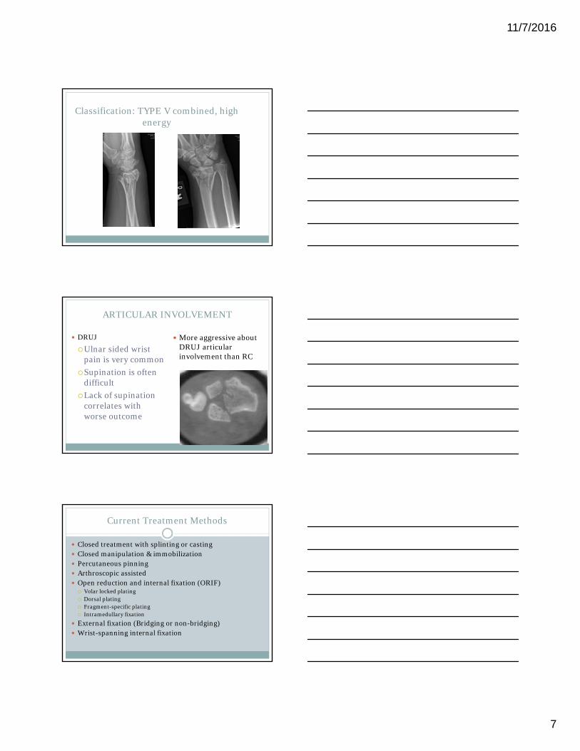
Classification: TYPE V combined, high energy
ARTICULAR INVOLVEMENT
DRUJ
Ulnar sided wrist pain is very common
Supination is often difficult
Lack of supination correlates with worse outcome
More aggressive about DRUJ articular involvement than RC
Current Treatment Methods
Closed treatment with splinting or casting Closed manipulation & immobilization Percutaneous pinning Arthroscopic assisted Open reduction and internal fixation (ORIF) Volar locked plating Dorsal plating Fragment-specific plating Intramedullary fixation
External fixation (Bridging or non-bridging) Wrist-spanning internal fixation
11/7/2016
8
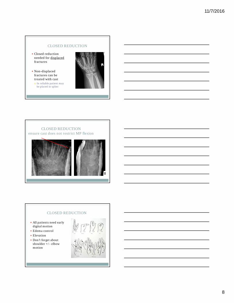
CLOSED REDUCTION
Closed reduction needed for displacedfractures
Non-displaced fractures can be treated with cast In reliable patient may
be placed in splint
CLOSED REDUCTIONensure cast does not restrict MP flexion
CLOSED REDUCTION
All patients need early digital motion
Edema control
Elevation
Don’t forget about shoulder +/- elbow motion

11/7/2016
9
Closed Reduction, Percutaneous Pinning (CRPP)
Can be used for many fracture patternsShearing injuries that can not be easily platedSevere soft tissue traumaThere is risk of loss of reductionPin-track infectionNeeds cast or splint
Intramedullary Nail
Alternative to k-wire fixation
Better for extra-articular fractures
Need good metaphyseal bone (1.5-2 cm)
*Radial sensory nerve irritation
More stable than k-wires alone
Similar stability to volar plates
Can start immediate ROM
Avoid open volar soft tissue envelope
Less OR time?
IM Nail

11/7/2016
10
External Fixation
Much less commonly used
Bridging or Non-Bridging
Problems include over-distraction, hand stiffness
Use mainly for severe soft tissue injury
Temporary fix until able to perform formal ORIF
External Fixation
Combined Dorsal/Volar Approach

11/7/2016
11
Bridge Plate (Wrist Spanning)
Severe articular comminution
Internal “ex fix”
Span zone of injury until fracture consolidation
Requires second procedure
Wrist stiffness is usually not a problem
Need to avoid tendon irritation and adhesions
May be combined with C-wires
Associated Conditions
CARPAL INJURIES

11/7/2016
12
ULNA STYLOID FRACTURES
Ulnar sided wrist pain is common in post operative period
Presence of ulnar styloid fracture should create concern for DRUJ instability
Incidence 3 to 37%
Need to asses normal side
Increased motion compared to contra-lateral side
Fluoroscopic evaluation if in doubt
Usually treated non-op even if DR is treated.
ULNA STYLOID FRACTURES
DISTAL ULNA FRACTURES
Carpal Tunnel
Acute CTSDevelops typically
over hours after injury
Progressive
Painful
Median nerve contusionCan be present
at time of injuryImproves with
reductionIs not
progressive

11/7/2016
13
Cases
Case 1: shortening, comminution, stepoff
Very shortenedIntraarticular step-off on lateral with dorsal comminution. Expect significant “crush” of cancellous bone due to diaphysis compressing into the distal segments
Case 1
Bone loss in metaphysisSupported by the locking rafting screws

11/7/2016
14
Case 2-stepoff, sigmoid notch involvement
Case 2
Case 2

11/7/2016
15
Case 3- stepoff, volar comminution
Case 3
Case 4 – volar shear
11/7/2016
16
Case 4
Case 4
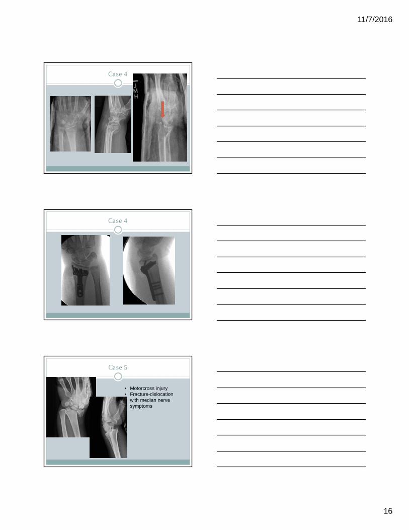
Case 5
• Motorcross injury• Fracture-dislocation
with median nerve symptoms
11/7/2016
17
CTR, BR REL, ORIF
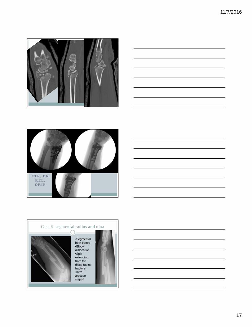
Case 6- segmental radius and ulna
•Segmental both bones•Elbow dislocation•Split extending from the distal radius fracture•Intra-articular stepoff

11/7/2016
18
Case 6
•Wrist spanning plate, not a wrist fusion plate•Acts as an internal ex fix
Case 7- external fixator
•Allows traction –mostly longitudinal, although can address other planes•Ligamentotaxis – see midcarpal joint•sigmoid notch, radiocarpal joint
Case 7- revised to ORIF

11/7/2016
19
Case 8- Distal radius with SL injury
•SL >3mm•SL angle on lateral >60deg
Case 8
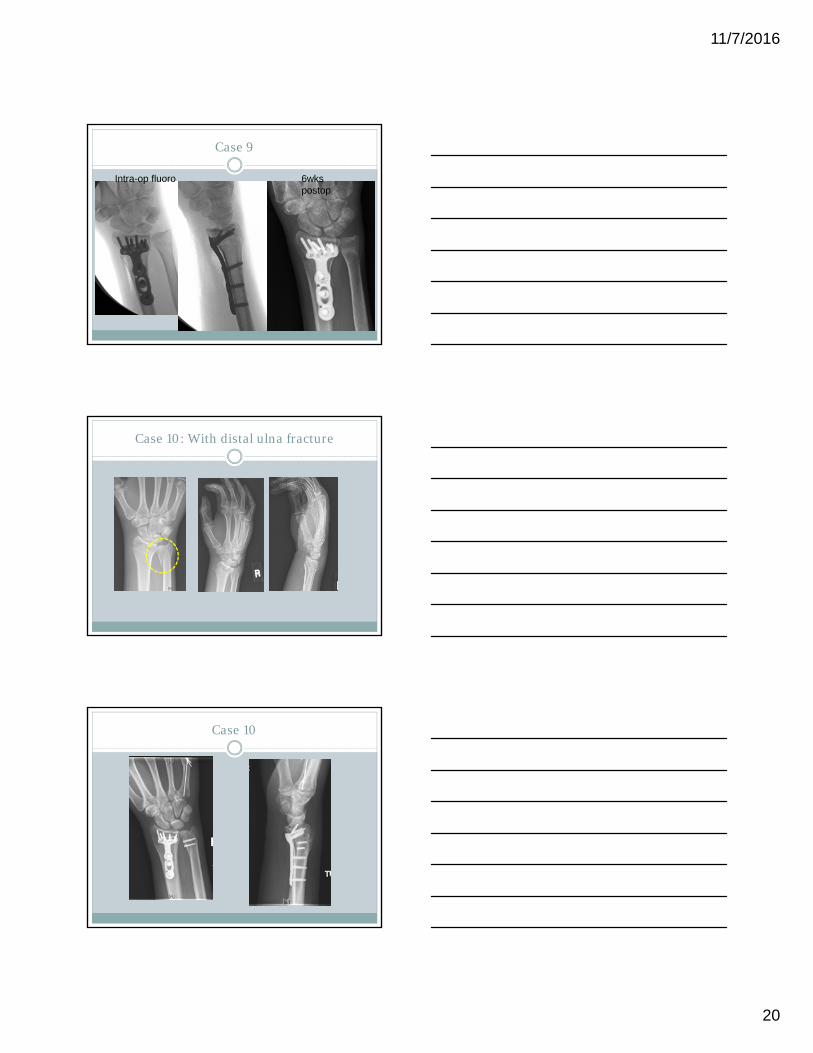
Case 9-Distal radius with ulnar neck fracture
11/7/2016
20
Case 9
Intra-op fluoro 6wks postop
Case 10: With distal ulna fracture
Case 10

11/7/2016
21
Case 11: DRUJ disruption
Case 11
Tendon Irritation
Can have flexor and extensor tendon irritationPts will generally complain of
pain with motion after fracture has healed
FPL common on flexorEDC common for extensorHWR prior to rupture
11/7/2016
22
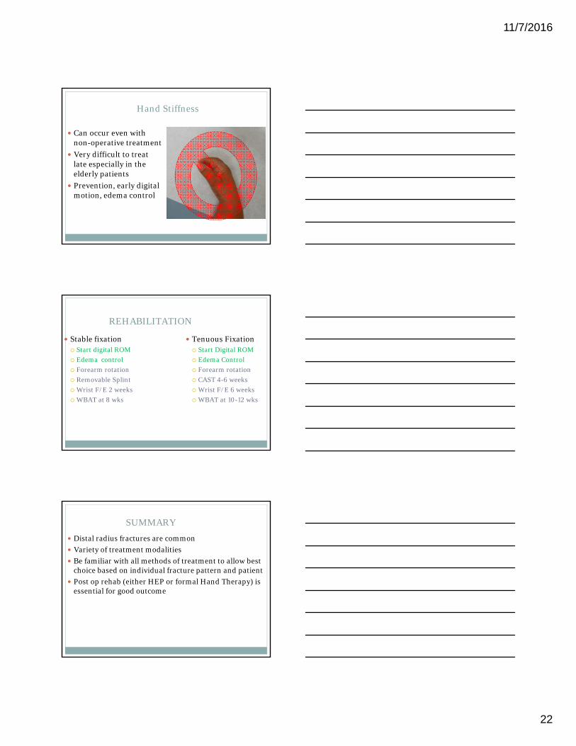
Hand Stiffness
Can occur even with non-operative treatment
Very difficult to treat late especially in the elderly patients
Prevention, early digital motion, edema control
REHABILITATION
Stable fixation Start digital ROM
Edema control
Forearm rotation
Removable Splint
Wrist F/E 2 weeks
WBAT at 8 wks
Tenuous Fixation Start Digital ROM
Edema Control
Forearm rotation
CAST 4-6 weeks
Wrist F/E 6 weeks
WBAT at 10-12 wks
SUMMARY
Distal radius fractures are common
Variety of treatment modalities
Be familiar with all methods of treatment to allow best choice based on individual fracture pattern and patient
Post op rehab (either HEP or formal Hand Therapy) is essential for good outcome

11/7/2016
23
Thank you