Embed Size (px)
Citation preview

1
DISSERTATION
TITLE OF STUDY:
INTEREUKIN 6 LEVELS IN ADENOTONSILLAR HYPERPLASIA AND CHRONIC
RECURRENT TONSILLITIS
Principal researcher: DR. ELAINE AGONO YUKO
M. Med ENT HEAD AND NECK SURGERY
REG: H58/72205/08
Supervisors: DR. MAJOR (RTD) GONTIER C.S.
CONSULTANT PATHOLOGIST, SENIOR LECTURER,
DEPARTMENT OF PATHOLOGY, UNIVERSITY OF
NAIROBI.
DR. OBURRA H.O
ASSOCIATE PROFESSOR,
OTORHINOLARYNGOLOGY, HEAD AND NECK
SURGERY, DEPARTMENT OF SURGERY
UNIVERSITY OF NAIROBI
A study submitted in part fulfilment of the requirements for the degree of Master of Medicine in
Ear, Nose and Throat- Head and Neck Surgery, at the University Of Nairobi

- 1 -
DECLARATION
This is my original work and has not been presented for a degree in any other university.
Signed____________________ Date__________________
Dr. Elaine Agono Yuko
This thesis will be supervised by:
Professor Oburra H.O
Associate Professor, Otorhinolaryngology, Head and Neck Surgery, Department Of Surgery
University Of Nairobi
Signed____________________ Date____________________
DR. MAJOR (RTD) GONTIER C.S.
Consultant Pathologist, Senior Lecturer, Department Of Pathology, University Of Nairobi.
Signed___________________ Date____________________

- 2 -
ACNOWLEDMENTS
I thank and acknowledge the many people who assisted me in this work: My supervisors
Professor H.O Obura and Dr Gontier for their patience, mentorship and guidance; Isaiah
Anyango and Simon Ogolla of KAVI for assisting me in processing of the specimens and
interpretation of the data; Mr Joseph Kamungu, Sr. Emily Changuli and Sr. Salome Warugongo,
nurses of the ENT satellite theatre; Dr Meera Patel and Dr Gachambi Mwangi who ran the race
with me; The Late Dr James Achola for his counsel and support and lastly but never least my
Father Engineer David Yuko for holding my hand unconditionally.
T o those whose contributions I have neglected to note, thank you.
I dedicate this work to Joyce Yuko, my mum.

- 3 -
TABLE OF CONTENTS:
1. Acronyms and abbreviations ……………………………………………………..….4
2. Abstract ………………………………………………………………….…………..5
3. Introduction …………………….……………………………………...….…..…… 7
4. Anatomy …….………. ………….……………………………………….….....…...9
5. Cytokines…………………………..…………………………….……….……..….11
6. Interleukin 6………………………….……………………….………….…….……15
7. Literature review…………………………….………………….………..…..……….16
8. Hypothesis…………………………………………………………….……….…….. 19
9. Justification of study…………………………………..…………….…….…..……...19
10. Aims and objectives ………………………………………….…….………..….……20
11. Materials and methods……………………………….…….………….………..….….22
12. Data analysis……………………………………………………….…………………..22
13. Ethical consideration…………………………………………………………..……….23
14. Analysis............................................................................................................................23
15. Results.............................................................................................................................24
16. Discussion..........................................................................................................................32
17. Conclusion.........................................................................................................................34
18. Recommendations..............................................................................................................34

- 4 -
19. Appendix 1- Patient information form………….…………………...........................34
20. Appendix 2- Consent form ……………..….....………..............................................40
21. Appendix 3-History and examination profoma……………………….…....................39
22. Appendix 4- Implementation Timetable and budget………………………………….43
23. Appendix 4- Interleukin 6 assay……………………………………………………….45
24. References……………………………………………………………………………50

- 5 -
ACRONYMS AND ABBREVIATIONS:
AH ADENOID HYPERTROPHY
ATH ADENOTONSILLAR HYPERPLASIA
ATS ADENOTONSILLAR SURGERY
CRP C REACTIVE PROTEIN
CRT CHRONIC RECURRENT TONSILLITIS
ELISA ENZYME LINKED IMMUNOSORBENT ASSAY
ENT EAR NOSE AND THROAT
GCSF GRANULOCYTE COLONY STIMULATING FACTOR
HIV HUMAN IMMUNODEFICIENCY VIRUS
IL INTERLEUKIN
INF INTERFERON
KNH KENYATTA NATIONAL HOSPITAL
OSA OBSTRUCTIVE SLEEP APNEA
ORL HNS OTORHINOLARYNGOLOGY HEAD AND NECK SURGERY
PHA-P PHYTO-HEMOAGGLUTINATION P IMMNUNOGEN
SPSS STATISTICAL PACKAGE FOR SOCIAL SCIENCES (SPSS)
TNF-α TUMOR NECROTIC FACTOR ALFA
TNF-β TUMOR NECROTIC FACTOR BETA
WBC WHITE BLOOD CELLS

- 6 -
INTERLEUKIN 6 LEVELS IN ADENOTONSILLAR HYPERPLASIA AND CHRONIC
RECURRENT TONSILLITIS
ABSTRACT
BACKGROUND
The primary etiology of adenotonsillar hyperplasia and chronic recurrent tonsillitis is
inflammation which is mediated by cytokines such as Interleukin-6 which is both a pro and anti-
inflammatory agent. In this study the IL-6 levels in adenotonsillar tissue of patients with AH,
ATH and CRT were measured.
OBJECTIVE
To measure the levels of interleukin 6 in adenotonsillar tissue in adenotonsillar hyperplasia and
chronic recurrent tonsillitis.
STUDY DESIGN
Prospective cross-sectional study
MATERIALS AND METHODS
83 patients undergoing adenotonsillectomy for ATH and CRT were recruited and IL-6 assays
carried out and correlated with the use of medications such as antihistamines, topical steroids and
antibiotics prior to surgery.
DATA ANALYSIS
Data was entered into preformatted sheets and analyzed with the SPSS17.0. Means, percentages
and statistical significance were calculated. Independent t-test and Analysis of variance
(ANOVA) with LSD Post Hoc multiple comparisons were used.
RESULTS

- 7 -
The male to female ratio was 53.01:46.99. Patients with ATH and AH were72.3% with grade 3
and 4 tonsils as the majority at 66.2%. The highest level of IL-6 was 1.029 while the lowest was
0.104 with a mean of 0.4347. Patients who had tonsillectomies had higher IL- 6 levels compared
to those who had adenoidectomies with a mean difference which was significant (p<0.001). The
mean difference in IL6 levels of patients who were on antihistamines versus those who were not
was not significant (p= 0.444). The mean difference of IL-6 of patients on topical steroids and
those who were not was significant (p<0.001)
CONCLUSION
IL- 6 levels were more elevated in patients with both CRT and ATH than in patients with AH
only. Thus elevated IL-6 levels may be a mark of disease chronicity
The use of topical nasal steroids leads to reduced IL6 levels thus are an effective medical
treatment for AH. The use of oral antihistamines did not significantly affect IL6 levels.

- 8 -
INTRODUCTION:
Adenoidectomy and tonsillectomy are the most commonly performed procedures in
Otorhinolaryngology, Head and Neck Surgery (ORL-HNS) practice in the world and the
worldwide picture mirrors the situation in the ORL HNS practice in Kenya.1, 2. The primary
indications for adenotonsillectomy are chronic infection and upper airway obstruction which
becomes more pronounced during sleep when the oropharyngeal musculature is relaxed.3, 4.
There may be significant advantage in pursuing medical therapy for ATH and CRT as this may
limit the surgical risks and complications especially for poor surgical candidates and reduce the
psychological and financial burden to the patients and their families.
Ongoing research on adenotonsillar disease include the role of atopy, tonsillar tissue as a
reservoir for Human Immunodefiency Virus (HIV), the topical use of Leukotriene inhibitors and
topical steroids on tonsil tissue and the role of biofilm forming bacteria.5,6 ATH and CRT are
both sequelae of inflammation. Recent studies indicate that local inflammation and mucosal
immunity function independently from the systemic response. Inflammation is primarily
mediated by cytokines which are peptides involved in regulation of both cellular and humoral
immune response by relaying information between cell signalling molecules. Cytokine levels
may differ in ATH and CRT.7
Adenoids and tonsils are most immunologically active from four to ten years of age with
involution by adolescence, with continued growth of the skull base growth plates adenoids and
tonsils are rarely obstructive after this period. The exact mechanisms underlying follicular
lymphoid proliferation and hyperplasia remain poorly understood.8
Previously it has been widely assumed that tonsillar and adenoidal tissue enlarge at a rate faster
than the bony structure of the nasopharynx during early childhood thus reducing the airway
diameter. Recent studies in normal children showed that the adenotonsillar growth is
proportionate to the somatic growth of the airway, and that any deviation would be abnormal.9
Research indicates that localized inflammation in the nasopharyngeal area is involved in the
pathophysiology of upper airway obstruction and subsequently obstructive sleep apnoea in
children. Assessment of adenotonsillar tissues from children with obstructive sleep apnoea

- 9 -
(OSA) has shown markedly increased inflammatory cell proliferation especially T-cell
lymphocytes and increased expression of pro-inflammatory cytokines and other inflammatory
mediators, such as TNF-α, IL-6 and IL-1α, when compared with adenotonsillar tissues surgically
removed in the treatment of children with CRT.10 It is postulated that in OSA, respiratory viruses
and recurrent vibration of the upper airway from snoring will promote localized inflammation
with subsequent mucosal swelling and over-expression of inflammatory cytokines.11 Studies
examining exhaled breath condensate and induced sputum in children with OSAS have revealed
the upregulation of localized inflammatory processes in upper airway tissues.12
- 10 -
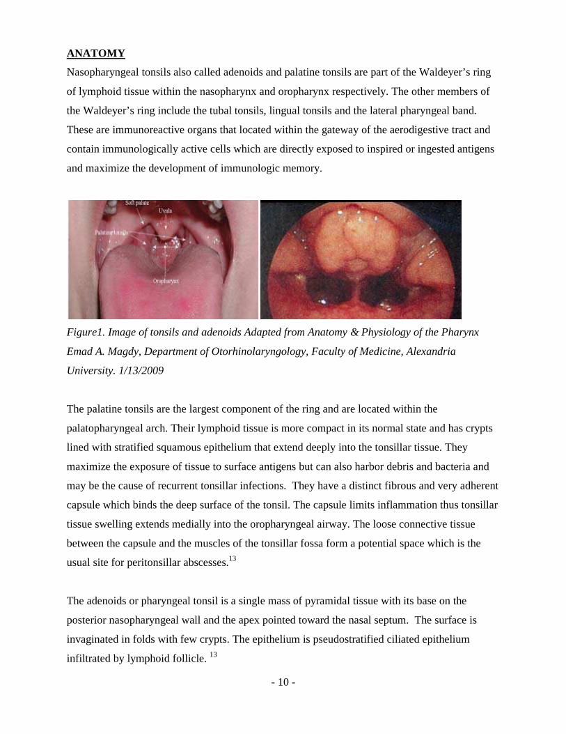
ANATOMY
Nasopharyngeal tonsils also called adenoids and palatine tonsils are part of the Waldeyer’s ring
of lymphoid tissue within the nasopharynx and oropharynx respectively. The other members of
the Waldeyer’s ring include the tubal tonsils, lingual tonsils and the lateral pharyngeal band.
These are immunoreactive organs that located within the gateway of the aerodigestive tract and
contain immunologically active cells which are directly exposed to inspired or ingested antigens
and maximize the development of immunologic memory.
Figure1. Image of tonsils and adenoids Adapted from Anatomy & Physiology of the Pharynx
Emad A. Magdy, Department of Otorhinolaryngology, Faculty of Medicine, Alexandria
University. 1/13/2009
The palatine tonsils are the largest component of the ring and are located within the
palatopharyngeal arch. Their lymphoid tissue is more compact in its normal state and has crypts
lined with stratified squamous epithelium that extend deeply into the tonsillar tissue. They
maximize the exposure of tissue to surface antigens but can also harbor debris and bacteria and
may be the cause of recurrent tonsillar infections. They have a distinct fibrous and very adherent
capsule which binds the deep surface of the tonsil. The capsule limits inflammation thus tonsillar
tissue swelling extends medially into the oropharyngeal airway. The loose connective tissue
between the capsule and the muscles of the tonsillar fossa form a potential space which is the
usual site for peritonsillar abscesses.13
The adenoids or pharyngeal tonsil is a single mass of pyramidal tissue with its base on the
posterior nasopharyngeal wall and the apex pointed toward the nasal septum. The surface is
invaginated in folds with few crypts. The epithelium is pseudostratified ciliated epithelium
infiltrated by lymphoid follicle. 13

- 11 -
TONSIL SIZE AND POSITION
The size and the position of the tonsils significantly affect the diagnosis and symptomatology of
upper airway obstruction secondary to tonsillar hyperplasia. Tonsils may be bi-lobed with
extension into the hypopharynx, or more rarely into the nasopharynx. Inferior extension is
associated with a history of obstruction and relatively normal appearing tonsils.Brodsky
classification is a useful determinant for tonsillectomy and is a grading system of tonsillar size
that is expressed as the estimated percentage of patent oropharyngeal airway between the
palatine tonsils.14
Figure 2: Brodsky classification of tonsillar size, Adapted from Lawrence Elikan, M.D. Seckin
Ulualp, M.D.University of Texas Medical Branch Department of Otolaryngology. Special
situations in management of tonsil and adenoid disorders, January 11, 2006
FUNCTION AND IMMUNOLOGY
The immunologic structure of the tonsils and adenoids is divided into four compartments from
the surface in; reticular crypt epithelium, extra follicular area, mantle zone of the lymphoid
follicle, and the germinal centre of the lymphoid follicle. Without afferent lymphatics the
lymphoid nodules in these structures are exposed to antigen only in the crypts of the palatine
tonsils and the folds of the adenoids. Membrane cells and antigen presenting cells are involved in
transporting the antigen through the epithelial layer via transcytosis and presenting them to T-
helper cells. This stimulates the B cells in the germinal zone of the lymphoid follicle to
differentiate and produce antibodies. Palatine tonsils and adenoids are involved in the

- 12 -
production of mostly secretory IgA, which is transported to the surface and provides mucosal
immune protection and assists in preventing bacteria and viruses from adhering to pharyngeal
mucosa15
CYTOKINES
Cytokines are soluble intercellular signalling molecules that act via specific receptors to regulate
host cell function. Cytokines can act in an autocrine, paracrine, or endocrine fashion via lymph
or plasma and have local and systemic actions. Different cell types may secrete the same
cytokine while a single cytokine can exhibit pleiotropy thus acts on several different cell types.
Redundancy implies that similar functions can be stimulated by different cytokines. They are
often produced in a cascade and can act synergistically or antagonistically.
Cytokines regulate immunity, inflammation, and hematopoiesis. They are produced in response
to an immune stimulus and are very potent biological molecules at very low concentrations and
generally though not exclusively act over short distances and short time spans. They bind to
specific membrane receptors, which signal the cell via second messengers to alter gene
expression. Responses to cytokines include increasing or decreasing expression of membrane
proteins (including cytokine receptors), proliferation, and secretion of effector molecules. There
are six major types of cytokines chemokines, colony-stimulating factors, interferons,
interleukins, transforming growth factors, and tumour necrosis factors (TNF). Cytokines may
also be classified by the cell of production such as lymphokines by lymphocytes and monokines
by monocytes. Chemokines have chemotactic activities while interleukins are produced by
leukocytes.16
CYTOKINE ACTIVITIES
Cytokines are made by many cell populations, but the predominant producers are T helper
lymphocyes (Th) and macrophages. The largest group of cytokines is that which stimulates
immune cell proliferation and differentiation. This includes Interleukin 1 (IL-1), which activates
T cells; IL-2 which stimulates proliferation of antigen-activated T and B cells; IL-4, IL-5, and
IL-6, which stimulate proliferation and differentiation of B cells, Interferon gamma (IFNg),
which activates macrophages and IL-3, IL-7 and Granulocyte Monocyte Colony-Stimulating
Factor (GM-CSF), which stimulate hematopoiesis. IFN alpha and IFN beta inhibit virus

- 13 -
replication in infected cells, while IFN gamma stimulates antigen-presenting cell MHC
expression.16
TH1 AND TH2 BALANCE, REGULATION, AND INVOLVEMENT IN DISEASE
T lymphocytes are a major source of cytokines and they bear antigen-specific receptors on their
cell surface to allow recognition of foreign pathogens. The two main subsets of T lymphocytes
are distinguished by the presence of cell surface molecules CD4 and CD8. T lymphocytes
expressing CD4 are also known as Helper T cells and can be further subdivided into Th1 and
Th2 cells and produce Th1-type cytokines and Th2-type cytokines.
Th1 cells promote cell mediated immunity while Th2 cells induce humoral immunity. Cellular
immunity directs Natural killer T cells and macrophages to attack abnormal cells and microbial
agents at the sites of infection inside the cells. Humoral immunity results in the production of
antibodies used to neutralize antigens outside of the cells. Th1 cells secrete INF-gamma and IL-
2, which activate macrophages and cytotoxic T-cells to kill intracellular organisms; Th2 cells
secrete IL-4, IL-5, IL-6 and IL-10, which in turn lead B cells to secrete protective antibodies.17
The immune system relies on both Th1 and Th2 cells to exist in a balanced and regulated
manner, thus an inadequate Th1 response leads to chronic infection and cancer while an
overactive Th2 response can contribute to allergies and autoimmune diseases.17
TH-1 CELLS
TH-1 cells produce pro-inflammatory cytokines like IFN-γ, IL-2, and TNF-β and are involved in
cell-mediated immunity. These cytokines stimulate phagocytosis and destruction of microbial
pathogens via macrophages which produce toxic forms of oxygen which destroy the
microorganisms within the phagosomes and lysosomes. Several chronic inflammatory diseases
are described as Th1 dominant diseases such as multiple sclerosis, diabetes, and rheumatoid
arthritis.
TH2 CELLS
TH2 cytokines counteract the effects of the TH1 cytokines – they have an anti-inflammatory
action but they also help kill extracellular pathogens by stimulating the production of antibodies
and eosinophilic response directed toward large extracellular parasites. Atopy and allergy are

- 14 -
Th2 dominant conditions. Th2 cells produce IL-4, IL-5, IL-9, IL-10, and IL-13. Pro-
inflammatory cytokines are produced primarily by activated macrophages and are involved in the
up-regulation of inflammatory reactions. Anti-inflammatory cytokines belong to the T cell-
derived cytokines and are involved in the down-regulation of inflammatory reactions.18
The main cytokines involved in inflammation and fever are tumour necrosis factor-α, IL-1, and
IL-6 in their order of secretion. IL-1, TNF-α and IL-6 are pro-inflammatory. IL-6 then inhibits
the secretion of TNF-α and IL-1, In normal settings it limits the inflammatory reaction thus it is
both pro-inflammatory and anti-inflammatory. TNF-α, IL-1ß, and IL-6 lead to monocyte-
macrophage activation, due to repeated stimulation by the pathogenic agents. These mediators
induce the activation and proliferation of endothelial cells and fibroblasts, which may eventually
lead to progressive replacement of immunologically active tissue with fibrotic tissue in chronic
recurrent tonsillitis and hyperplasia.18
INTERLEUKIN 6
Interleukin-6 (IL-6) is a cytokine of approximately 26 kDa that is synthesized by T-cells,
macrophages, B-cells, fibroblasts, endothelial cells, and epithelial cells. It is also known as
interferon-B2, cytotoxic T-cell differentiation factor, and B-cell stimulatory factor-2, among
others. Depending on the particular condition it may be an anti-inflammatory or a pro-
inflammatory mediator as shown above.
It is among the primary mediators of the clinical manifestations of tissue injury. These include
fever, cachexia, leukocytosis, thrombocytosis, increased plasma levels of acute-phase proteins,
and decreased plasma levels of albumin. IL-6 also stimulates plasmacytosis and
hypergammaglobulinemia. It is required to regulate cell growth as well as immune functioning.
Receptor sites on the surface of different cells of the body mediate three major signal
transduction pathways: protein kinase C, cAMP/protein kinase A, and calcium release.
The circulating interleukin-6 stimulates the acute-phase reaction which leads to production and
release of acute-phase proteins such as C-reactive protein which increase phagocytosis and
destroy invading bacteria and other pathogens. This results in an acute-phase response, such as
fever. Repeated inflammation from the acute phase reaction leads to hyperplastic adenoids and

- 15 -
tonsils. This may be due to the recurrent viral upper respiratory tract infections that occur in
patients with OSA or acutely during adenoiditis or tonsillitis due to specific bacteria. On
resolution of infection the tissue does not fully revert to its previous normal size. The specific
role of IL-6 in hyperplastic adenoids and tonsils and OSA in children is still under investigation.
Impaired or uncontrolled interleukin-6 gene expression can produce unwanted immune responses
leading to autoimmune disorders such as rheumatoid arthritis. Thus interleukin-6 therapy by its
stimulation or inhibition is under investigation for the treatment of obesity, diabetes type II and
rheumatoid arthritis. Research is currently underway on ways of preventing IL-6 binding to its
receptors. However, it has been found somewhat more effective to bind IL-1 and TNF, and thus
reduce the secretion of IL-6. An example is the anti IL- 6 receptor antibody (MRA), used as one
of the new therapeutic approaches in rheumatic arthritis.
- 16 -
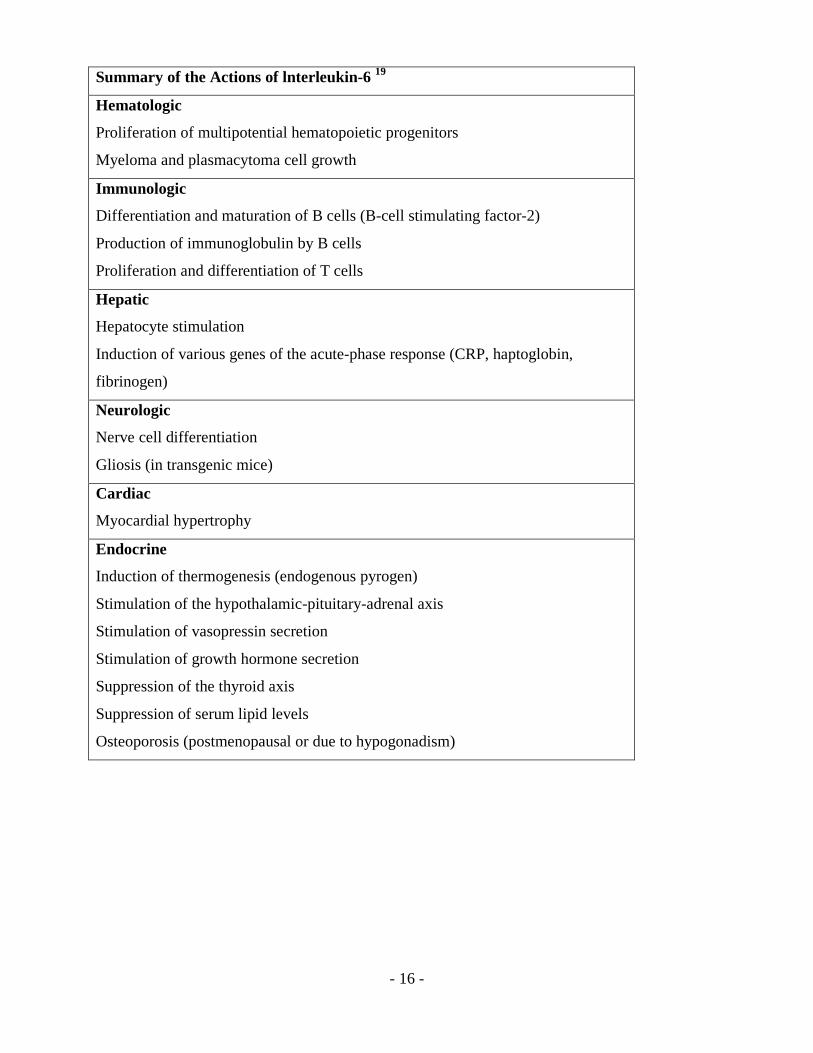
Summary of the Actions of lnterleukin-6 19
Hematologic
Proliferation of multipotential hematopoietic progenitors
Myeloma and plasmacytoma cell growth
Immunologic
Differentiation and maturation of B cells (B-cell stimulating factor-2)
Production of immunoglobulin by B cells
Proliferation and differentiation of T cells
Hepatic
Hepatocyte stimulation
Induction of various genes of the acute-phase response (CRP, haptoglobin,
fibrinogen)
Neurologic
Nerve cell differentiation
Gliosis (in transgenic mice)
Cardiac
Myocardial hypertrophy
Endocrine
Induction of thermogenesis (endogenous pyrogen)
Stimulation of the hypothalamic-pituitary-adrenal axis
Stimulation of vasopressin secretion
Stimulation of growth hormone secretion
Suppression of the thyroid axis
Suppression of serum lipid levels
Osteoporosis (postmenopausal or due to hypogonadism)

- 17 -
LITERATURE REVIEW:
Locally there are a few studies that have been conducted on adenotonsillar tissue. These include
studies on tonsil immunology 20, complications of adenotonsillectomy 4 and cardiopulmonary
effects of adenotonsillar hyperplasia.21. A prospective study in KNH in 1977 by Mulimba (20) on
histological, microbiological and immunological study of patients undergoing tonsillectomy for
recurrent sore throats showed that tonsils are immunologically active and competent as shown by
their response to phyto-hemoagglutination P immnunogen (PHA-P) stimulation. The levels of
immunoglobulin were generally lower than the international standard, though not too low as to
be used as explanation for frequent sore throats in these patients. There were no local figures
comparable for both age and sex comparison and a study to determine the local normal level was
thus recommended.20
A study by Desiderio et al (7) analysed structural and immunological aspects of tonsils and
adenoids in subjects who underwent adenotonsillectomy because of recurrent inflammatory
episodes with fever. Histological studies and analyses of the cytokine patterns were carried out in
palatine tonsils and adenoid samples from 105 patients who underwent adenoidectomy and
bilateral extracapsular tonsillectomy for chronic inflammatory hyperplasia of these organs; 46 of
the 105 cases examined presented hyperkeratosis of the crypt epithelium; in the remaining 59, the
epithelium was hyperplastic with no signs of keratosis. Titration of interleukin-1ß and tumor
necrosis factor alpha in serum and tissues demonstrated higher concentrations in the
adenotonsillar specimens, whereas the rise in interleukin-6 was more modest. 7
Sugiyami et al did a study on the influence of IL-6 on proliferation and differentiation of tonsillar
lymphocytes and detection of IL-6 producing cells in the tonsils. Tonsillar B cells were cultured
in the presence of exogenous interleukin-6, a small portion of them differentiated into plasma
cells which seemed to have IL-6 receptors on their surfaces. The number of plasma cells tended
to be greater in the tonsils of children than in the tonsils of adults. When tonsillar cells collected
by the Ficoll-Conray's method were cultured in medium containing IL-6, T cells differentiated
and the number of activated T cells increased. Of the four subsets of T cells, this effect of IL-6
was strongest on cytotoxic T cells. IL-6-mediated proliferation and activation of cytotoxic T cells
tended to be greater in the tonsils of children than in those of adults and the number of

- 18 -
macrophages producing IL-6 in tonsils of children tended to be greater than the number in tonsils
of adults. 22
Kheirandish-Gozal et al (23) did an invitro study on adenoid and tonsil tissue removed post
adenoidectomy. In the study the tissue was cultured in corticosteroids and the levels of cytokines
Tumour necrotic factor α, interleukins 6,and 8 measured. The study found that tonsils and
adenoids obtained from children with obstructive sleep apnoea undergoing tonsillectomy and
adenoidectomy displayed increased proliferative rates and proinflammatory cytokine production.
Furthermore, treatment with corticosteroids resulted in marked dose-dependent reductions in
proliferative rates, increased cellular apoptosis and diminished cytokine release. The relative
potency of the three corticosteroids used in the current study was highest for fluticasone and the
lowest for dexamethasone. Its findings supported the use of tonsillar or adenoidal tissue cell
cultures as a potentially useful approach for in vitro assessment of therapeutic efficacy of
corticosteroids and other candidate drugs in the treatment of the lymphoid hypertrophy that
underlies obstructive sleep apnoea in children.23
Rania Esteitie et al(24) studied the effect of fluticasone furoate on interleukin 6 secretion from
adenoid tissues in 24 children between the ages of 2 and 12 years who were undergoing
adenotonsillectomy for polysomnogram-documented obstructive sleep apnea syndrome. The
study was a randomized, prospective, exploratory study. The children were randomized to either
no treatment or treatment with fluticasone furoate nasal spray, for 2 weeks before
adenotonsillectomy. The study showed a reduction of IL-6, , in adenoid tissue obtained from
children with obstructive sleep apnea syndrome treated with fluticasone furoate nasal spray. The
aurthors believe that interleukin 6 an important predictor of cardiovascular risk.24
Krystal Revai et al (25) did a study on the association between cytokine gene polymorphisms
and risk for URTI and Acute Otitis Media in children. Two hundred and forty two children
between 6 to 35 months were prospectively followed for occurrences of URTI and AOM. Blood
or buccal mucosa samples were collected for DNA extraction to determine cytokine genotypes.
Active and passive surveillance was used to capture all URTI episodes during the one-year
follow-up period in order to study the rate of AOM following URI. Children who had IL-6 and

- 19 -
TNFα polymorphism had a higher susceptibility to URTI during the study period and were more
likely to meet established otitis susceptibility criteria (p<0.01). 25
Murat Ünal et al (26) did a study on serum interleukins (IL)-1β, IL-4, IL-6, IL-8 and tumor
necrosis factor (TNF)-α levels in 17 children aged 5–12 years (mean 7) with chronic tonsillitis
before and after tonsillectomy. Cytokine concentrations were measured by ELISA. IL-1β and IL-
6 levels were significantly higher than the control levels (p < 0.05) in preoperative serum
samples. Other cytokine levels were within normal limits. After tonsillectomy, IL-1β and IL-6
levels were significantly reduced (p < 0.05). It is suggested that IL-1β and IL-6 may be
mediators which have a role in chronic tonsillitis.26
t
None of the patients in this study were on topical antihistamines or leukotreine inhibitors but it
has recently been reported that montelukast, a cysteinyl leucotriene receptor antagonist, induced
considerable reductions in adenoidal size and respiratory-related sleep disturbances in children
with OSA.27 Currently there is ongoing research into the role of topical antihistamines on
adenoid tissue.
From the above literature review and paucity of published data, it is imperative that more work
be done on the molecular events that propagate the chronicity of andenotonsillitis and
hyperplasia. This will open new treatment strategies for proper targeted medical therapy as an
option before surgery.

- 20 -
JUSTIFICATION OF STUDY:
Adenotonsillar hyperplasia and chronic recurrent tonsillitis are known to be caused by chronic
inflammation which is mediated by cytokines at the local tissue level. The study has provided a
record of local interleukin 6 levels in adenotonsillar tissues in children. The study is a pioneer
study that will aid future studies aiming to modulate inflammmatory cytokines and develop new
less invasive non-surgical modes of treatment.
NULL HYPOTHESIS
Interleukin 6 does not play a role in adenotonsillar hyperplasia and chronic recurrent tonsillitis.
ALTERNATE HYPOTHESIS
Interleukin 6 plays a role in adenotonsillar hyperplasia and chronic recurrent tonsillitis.
AIMS AND OBJECTIVES:
BROAD OBJECTIVE:
The broad objective of the study was to determine the level of interleukin 6 in Adenotonsillar
tissue of patients obtained at adenotonsillectomy at KNH.
SPECIFIC OBJECTIVES:
1. To measure the level of the inflammatory cytokine interleukin 6 from adenotonsillar tissue.
2. To determine differences in Interleukin 6 in patients with obstructive symptoms only
(adenotonsillar hyperplasia) and in those with chronic recurrent tonsillitis, or both of the above
3. To correlate the level of interleukin 6 in adenotonsillar tissue of patients who have been on
medications such as antibiotics, topical nasal steroids and antihistamines within one month prior
to surgery.
STUDY METHODOLOGY
STUDY DESIGN:
The study was a prospective descriptive study

- 21 -
STUDY SITE
This study was conducted in Kenyatta National Hospital ENT department.
STUDY POPULATION
The patients recruited were sourced from the ENT department at KNH. This included patients on
follow up for adenotonsillar hyperplasia causing obstructive symptoms and patients on follow up
for chronic recurrent tonsillitis.
INCLUSION CRITERIA
This consisted of the following
1. Patients twelve years and below whom were to undergo adenoidectomy, tonsillectomy or
adenotonsillectomy for adenoid hyperplasia, adenotonsillar hyperplasia or chronic recurrent
tonsillitis at the satellite theater in the ENT department at KNH.
2. Patients whose parents or guardians gave informed consent to participate in the study.
EXCLUSION CRITERIA
This included the following;
1. Patients whose guardians declined to participate in the study
2. Patients undergoing tonsillectomy as a biopsy for suspected neoplastic lesions.
3. Patients older than 16 years of age were excluded as adenoids and tonsils involute by puberty
which varies from 12years to 16 years.
4. Patients who were found to have fever, neutrophilia of 12x109/l, and patients with respiratory
tract infections at least five days pre-operatively as most patients have hemograms done between
five days to seven days pre-operatively.
5. Patients with comorbidities such as rickets, heart disease sickle cell and overt asthma who did
not qualify for surgery in the satellite theatre
6. Patients undergoing branchial sinus surgery where tonsillectomy was done.
SAMPLE SIZE CALCULATION:
Yamen formula:
n = N
1+N(e)2

- 22 -
N is the population prevalence.
e is the error margin.
At a confidence interval of 95% and an error margin of 5% the sample size (n) was based on a
previous study by Desiderio et al8 where 105 cases will be the N (population size).
n= 105
1+105(0.05)2
n= 83.16
SAMPLING METHOD:
Consecutive sampling method was carried out to recruit 83 children who underwent
adenotonsillar surgery.
STUDY DURATION:
The study was conducted over a period of two months from March to April 2012.
PROCEDURES
The patients were recruited from the ENT clinic in KNH. Informed consent for participation in
the study was sought and participants who consented were recruited into the study. All patients
had a hemogram done pre-operatively as part of their routine pre-op work up.
The adenotonsillar tissue was collected after adenoidectomy by sharp curettage and extra-
capsular blunt dissection tonsillectomy. The adenotonsillar tissue was then homogenized in 2
milliliters of normal saline. All samples were centrifuged at 3,000 rpm, for 10 minutes and the
supernatant collected and refrigerated at minus 800C.
Levels of interleukin-6 were determined in the supernatant, with a quantitative enzyme-linked
immunosorbent assay which was collectively done by a single technician once the sample size
was achieved. The lowest detectable values in the standard curve were determined according to
the manufacturer.
CLINICAL EVALUATION:
Patient selection was done from the ENT consultant clinics at the Kenyatta National Hospital.
The relevant clinical history was be taken and this mainly include determining if the patient had
ATH causing features of OSA or CRT, any recent URTI or antibiotic use with the last month and

- 23 -
any medication the patient was on for the condition such as topical nasal steroids and
antihistamines. A physical examination included a general exam and a local ENT examination of
was carried out and the findings were recorded in the proforma.
MATERIALS AND EQUIPMENT:
The materials and equipment used for assessing the patients included interleukin-6 assay kit,
pippetes, ionized or distilled reagent water a centrifuge and refrigeration facilities.
QUALITY CONTROL:
All specimens were processed in the same laboratory at Kenya Aids Vaccine Initiative (KAVI)
and standard operating procedures for specimen handling, processing and analysis were followed
to ensure standardization. Guidelines on tissue handling followed the University of Nairobi
Laboratory testing protocols which are based on ISO certification.
ETHICAL CONSIDERATIONS:
1. The study was carried out only after approval of the KNH ethics and research committee.
2. Those included in the study were required to give informed consent either personally or by their
guardians in pediatric patients
3. Patients incurred no extra financial costs and their confidentiality was be maintained at all times.
4. Participants reserved the right to withdraw from the study at any time without any healthcare
penalties.
5. There was no monetary gain by the primary investigator from the study.
6. During the screening period patients requiring further referral such as patients with cormobidities
such as cardiac disease were referred accordingly.
DATA ENTRY:
The biodata of the patient, relevant history and the findings on the screening tests were recorded
in the proforma.
The data was then separated into different preformatted data sheets under the following
headings; age; sex; indication for surgery; adenotonsillar hyperplasia and CRT..
- 24 -
ANALYSIS:
Data was exported to SPSS17 and the cleaning was done in SPSS17. Independent t-test and
Analysis of variance (ANOVA) with LSD Post Hoc multiple comparisons were used.
Independent t-test was used to compare mean difference whenever a variable had two categories
for example IL-6 levels in patients with topical nasal steroids and those who were not (yes, no)
and interleukin level, ANOVA was used when the variable of interest had more than two
categories e.g. surgery and interleukin levels.
DATA PRESENTATION:
Data has been presented in form of tables and bargraphs and piecharts.
COMFOUNDING FACTORS
Topical steroids are given nasally and have maximal effect on the nasopharyngeal mucosa rather
than oropharyngeal mucosa thus affecting exposure of the tissue and achieving an adequate dose
within the adenoid and tonsil tissue. The effect of this factor between adenoid and tonsil tissue is
not ascertained in the study.

- 25 -
RESULTS
The study recruited a total of 83 children who underwent adenotonsillar surgery. The male to
female ratio was 1:1.1
Figure 1: Gender
Table 1: Age and interleukins
Age Interleukin level (pg/ml)
N 83 79
Mean 4.96 0.43
Median 4 0.23
Std. Deviation 2.62 0.26
Minimum 1.50 0.104
Maximum 11.00 1.029
- 26 -
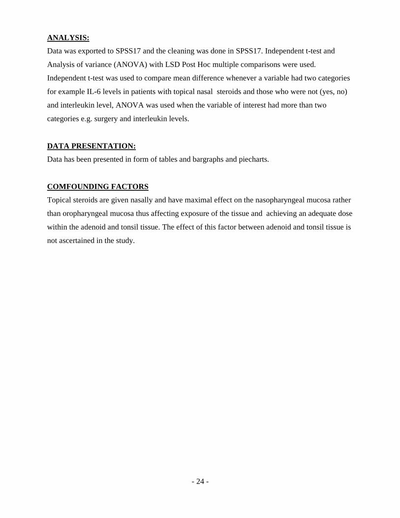
Table 1: Age and interleukins
Age Interleukin level (pg/ml)
N 83 79
Mean 4.96 0.43
Median 4 0.23
Std. Deviation 2.62 0.26
Minimum 1.50 0.104
Tonsillar Grade
Figure 2: Tonsil grade
Table 2: Tonsillar Grade
Grade N Mean(SD) Median 95% C.I. Mean Minimum-Maximum
1 16 0.27 (0.13) 0.231 0.196-0.34 0.157-0.67 pg/ml
2 10 0.40(0.26) 0.324 0. 21-0.58 0.139-0.89 pg/ml
3 33 0.42 (0.23) 0.340 0.33-0.50 0. 104-0.87 pg/ml
4 21 0.61(0.28) 0.589 0.48-0.74 0.242-1.029 pg/ml
Figure 3: Mean plot of interleukin level by tonsillar grade
- 27 -
The different tonsillar grades and the level of IL-6 were compared and the p values were as
follows: Grade 1 versus grade 3 (p=0.044); grade 1 versus grade 4(p<0.001), grade 2 versus
grade 4(p=0.022) and grade 3 versus grade 4 (p=0.005)
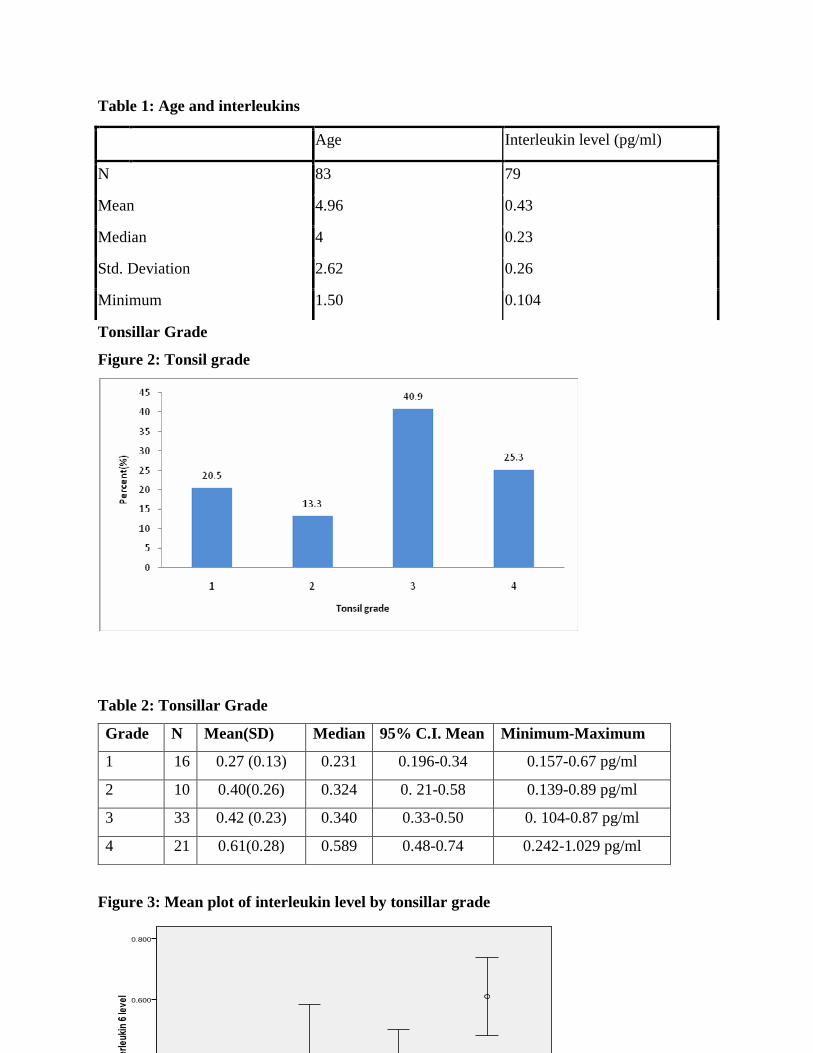
DIAGNOSIS AND SURGERY
Figure 4: Diagnosis
Figure 5: Surgeries
Key-: TS: Tonsillectomy ATS: Adenotonsillectomy AS: Adenoidectomy

- 28 -
Table 3: Summary of interleukin level by surgery
Surgery N Mean(SD) Median 95% C.I.
Mean
Minimum-Maximum
Tonsillar 13 0.523(0.280) 0.492 0.354-0.692 0.227-1.029 pg/ml
Adenotonsillar 46 0.495(0.265) 0.449 0. 417-0.574 0.104-1.022 pg/ml
Adenoid 21 0.247(0.124) 0.218 0.191-0.303 0.139-0.665 pg/ml
Figure 6: Mean plot of interleukin 6 level by surgery
The mean difference between Tonsillar Surgery and Adenoid Surgery was significant (p=0.002).
Mean interleukin levels for patients who had tonsillar Surgery was 0.52pg/ml compared to those
who had adenoid Surgery whose mean interleukin level was 0.25. The mean difference of the
above two which was 0.028 is statistically significant. Patients who had adenotonsillar surgery
had higher interleukin compared to those who had adenoid Surgery only. The Mean difference
(0.063) between Adenotonsillar Surgery and Adenoid Surgery is significant (p<0.001).

- 29 -
MEDICATIONS AND URTIs
Figure 7: Use of Topical steroids
Table 3: Topical Steroids Statistics
Topical steroids n Mean(SD) Median 95% C.I. Mean Minimum-Maximum
Yes 38 0.285(0.177) 0.243 0.226-0.343 0.104-0.798
No 42 0.571(0.253) 0.532 0.492-0.649 0.218-1.029
Mean difference is significant (p<0.001)
Figure 8: Mean plot of interleukin 6 level by use of topical steroids

- 30 -
Figure 9: Use of Antihistamines
Table 4: Statistics of Patients on Antihistamines with the last one month
Antihistamines n Mean(SD) Median 95% C.I. Mean Minimum-Maximum
Yes 22 0.395(0.24) 0.31 0.29-0.50 0.10-0.97
No 58 0.45(0.269) 0.32 0.38-0.52 0.14-1.029
The mean difference of IL- 6 levels in patients who were on antihistamines and those who were
not on antihistamines was not significant (p= 0.41)
Figure 10: Mean plot of interleukin 6 level by use of antihistamines

- 31 -
Figure 11: Use of Antibiotics within last 1month
Figure 12: Patients who had URTIs within the last 1month

- 32 -
21 children representing 25% of the children were reported to have had URTIs within the month
prior to surgery but only 18 (22%) were treated with antibiotics. 3 of the patients were managed
as URTI of viral etiology and were not treated with antibiotics.
Table 5: URTIs within last 1month and IL-6 levels
URTI last 1 month- IL-6 level pg/ml No URTI last 1 month- IL-6 level pg/ml
Median 0.59 0.23
Std. Deviation
Mean
0.25
0.62
0.14
0.37
Minimum 0.24 0.10
Maximum 1.029 0.97

- 33 -
DISCUSSION
In the descriptive analysis a slight female preponderance is noted. This was not found to be
statistically significant and research does not support any consistent findings of a particular
preponderance of sex in children with adenotonsillar hyperplasia and chronic recurrent tonsillitis.
In prepubertal children the size of the upper airways are similar in males and females after
puberty the upper airways become longer in boys than girls partially due to laryngeal descent
leading to higher prevalence of OSA in post-pubertal boys and adult men.28
Age associated changes in levels of serum IL-6 have only been studied in adults especially
geriatrics in healthy subjects, increased production of IL-6 is not a normal consequence of aging
but there is no research into the changes in IL-6 levels in children.29
The mean age of children who had only adenoid hyperplasia was 2.5 years while the mean age of
children with adenotonsillar hyperplasia and adenotonsillar hyperplasia with CRT was 4.6 and
6.1 years respectively. These finding correlate with studies using upper airway endoscopy under
anesthesia that have shown that in younger children the anatomic region of maximal narrowing is
at the level of the adenoids and soft palate. The tonsils proliferate between 4 to 10 years of age
thus the maximal narrowing of the upper airway within this age group occurs in the retropalatal
region, where the soft palate, adenoids and tonsils overlap in the three-dimensional space.30
In the study patients 62.6% of the patients had tonsillar grade 3 and 4 and those with higher
tonsillar grades had higher IL-6 levels as compared to the lower tonsillar grades that is 1 and 2.
This is due to the more severe obstructive symptoms associated with grade 3 and 4 tonsils.
Previous studies have shown IL-6 levels to be higher in severe as compared to mild OSA.31
Research indicates that IL-6 mRNA production in upper airway tissues is higher in patients with
severe compared to mild OSA. 31 The higher IL-6 levels with grade 3 and 4 tonsils may also be
due to increased tissue bulk as hyperplastic tissue maintains most of its immunologic function
but this is a postulation not supported by fact.
In the study most of the patients had surgery due to obstructive symptoms secondary to adenoid
hyperplasia and adenotonsillar hyperplasia rather than recurrent infections due to chronic
recurrent tonsillitis. 86.4% of the patients cumulatively had adenoid hyperplasia and

- 34 -
adenotonsillar hyperplasia Patients with adenoid hyperplasia tended to have lower levels of IL-6
as compared to those with adenotonsillar hyperplasia. This may be due to the duration of the
disease. The higher IL-6 levels may a mark of disease chronicity as the mean IL6 level in AH,
ATH, ATH and CRT was 0.25, 0.42 and 0.78 respectively. Patients with CRT had a mean age of
9.34 years and a mean IL6 level of 0.52. Thus patients who had a combination of obstructive
symptoms and recurrent infections had higher IL6 than those with recurrent tonsillitis only. It is
postulated that the mode of inflammation and cytokine production differs in obstructive disease
versus recurrent adenoiditis and tonsillitis thus cumulatively leading to higher tissue IL-6 levels.
Patients who were on steroid nasal sprays had significantly lower IL6 levels than those who were
not on IL6 with a mean of 0.285 and 0.569 respectively and a p<0.001 which was significant.
This correlates with a study by Kheirandish-Gozal et al which was a invitro study on adenoid and
tonsil tissue removed post adenoidectomy and cultured in corticosteroids. In that study treatment
with corticosteroids resulted in marked dose-dependent reductions in proliferative rates,
increased cellular apoptosis and diminished cytokine release. The relative potency of the three
corticosteroids used in the current study was highest for fluticasone and the lowest for
dexamethasone23.A study by Rania Esteitie et al on the effect of fluticasone furoate on
interleukin 6 secretion from adenoid tissues in children who were undergoing
adenotonsillectomy for polysomnogram-documented OSA showed a reduction of IL-6 in adenoid
tissue treated with fluticasone furoate nasal spray24.
It is possible that some of the patients especially those with adenoid hyperplasia only might have
benefited from optimum medical therapy and avoided surgery. This would involve a delicate
balancing act of patient education to improve compliance on medications and patient follow up
to monitor response to medication so as to avoid the dangerous sequelae of adenotonsillar
hypertrophy and OSA such as pulmonary hypertension.
In this study the difference in IL6 levels in patients on oral antihistamines and those who were
not on them was no significant with a p value of 0.44.There were no patients on topical
antihistamines or oral leukotreine inhibitors. There are no studies showing the effect of oral
antihistamines on cytokines in adenotonsillar tissues.

- 35 -
Mean IL6 levels in patient who had URTIs was 0.62 and mean IL6 levels in patient who did not
have URTIs was 0.37. Recurrent inflammatory episodes with fever have been found to lead to
higher cytokines levels including IL68. This finding correlates with an experimental study by
Gentile D.A et al that intranasally inoculated a safety-tested clinical isolate of RV-39 into
healthy patients. Nasal lavages were submitted for viral culture and assayed for cytokine protein
levels by ELISA. During infection, significant increases in mean levels of nasal IL-6) and IL-1
were observed in symptomatic but not asymptomatic subjects.
CONCLUSION
Interleukin 6 levels in adenotonsillar tissue was more elevated in patients with both chronic
recurrent tonsillitis and adenotonsillar hypertrophy causing obstruction than in patients with
adenoid hypertrophy. Thus elevated interleukin 6 levels are a mark of chronicity of the disease.
The use of topical nasal steroids leads to reduced IL6 levels thus are an effective medical
treatment for adenoid hypertrophy. The use of oral antihistamines did not significantly affect IL6
levels. The study has introduced local levels of interleukin 6 in adenotonsillar tissue that can be
used as for future reference.
RECOMMENDATIONS
A follow up study should be carried out with several proinflamatory and anti-inflamatory
cytokines to get a wider picture of the subcellular events to allow for future immunomodulation
that would lead to alternative medical treatment and reduced number of patients undergoing
surgery. A follow up study on the relationship between atopy and adenotonsillar hyperplasia and
chronic recurrent tonsillitis should be carried out possibly using tissue and serum inflammatory
cytokines and skin prick tests. In this study pre and post operative tissue and serum cytokine
levels should be measured to evaluate the effect of adenotonsillectomy on cytokine levels and
local imunity.
Studies should be carried out on inflammation in the tonsils and the specific effect of medication
to tonsillar size separate from adenoid hypertrophy as nasal steroids are most effective on

- 36 -
adenoid tissue rather than palatine tonsils. This might pave the way for topical medication for
tonsillar hypertrophy and chronic recurrent tonsillitis.
Studies have shown that intranasal antihistamines such as azelestamine and leukotrine inhibitors
such as monteleukast are effective in treating adenoid hypertrophy. These medications should be
made more widely available and patients educated on their benefits.

- 37 -
APPENDIX 1
CLIENT INFORMATION
Adenoids are tissue located behind the nose while tonsils are found inside the mouth. They are
useful in protecting the body against infection and allergies. Their surgical removal is
necessitated when they significantly increase in size causing difficulty in breathing which is
referred to as adenotonsillar hyperplasia. They may also get repeated infections which is referred
to as chronic recurrent tonsillitis.
1. Why is the study being done?
The study is being undertaken to help us to better understand the factors that contribute to the
causation and worsening adenoid and tonsillar enlargement and recurrent tonsillitis. Interleukin
6 is a substance produced by cells that aids in fighting constant tissue swelling due to recurrent
infection and allergies. The data will contribute to research activities that may one day
contribute to preventive treatment and earlier diagnosis of patients for surgical treatment and
those in whom medical treatment is sufficient.
2. Who will participate in the study?
Patients undergoing adenotonsillar surgery due to obstructive symptoms or Chronic recurrent
tonsillitis.
3. How many people will participate?
83 patients will be enrolled in the study
4. What does this study involve?
If you agree to participate in the study; you will be required to answer a few questions by an
ENT resident and undergo physical examination. After this you will undergo the surgery;
adenoidectomy, adenotonsillectomy or tonsillectomy as indicated. You will be allotted a study
number. The extracted tissue will then be used to assess the level of interleukin 6 and its role in
adenotonsillar enlargement and recurrent tonsillitis analyzed.
5. What are the risks of the study, and will your participation affect your child?
.The study does not affect the child negatively in any way because:
1. All the information you give will be confidential
- 38 -
2. The study does not reveal individual identity
3. The conclusions drawn from the study shall be useful to improve the management
adenotonsillar enlargement and recurrent tonsillitis.
Are there any hidden dangers in your participation or non-participation?
1. None whatsoever
2. Objecting to any part or whole of this study will not affect the quality of care you receive.
What do we do with the information we get
1. The information we get may not be of immediate benefit to you but it will help us in the long
run in managing the condition better.
2. Like all scientific information we will seek to share our findings with other people
undertaking similar studies. Therefore we may publish our findings in scientific journals or
present them in scientific meetings.
3. If you require discussing this matter with the family or friend you are free to do so and we
will be ready to answer any questions. If you are satisfied with our explanation and willing to
participate, then please sign the consent form below
4. In any case a child is found with some issues related to his health,this matter will be taken
into consideration. A recommendation to the Doctor specialist will be given for close follow-up.

- 39 -
6. What are the benefits of participating in the study?
The study may not benefit you directly but it will enable researchers to develop improved less
invasive treatment modalities for adenotonsillar enlargement and recurrent tonsillitis.
Participants in the study are entirely voluntary and you can get out at any stage of the study
without you having to justify your decision. No victimization of any participant who withdraws
from the study will be permited.

- 40 -
APPENDIX 3: KIELEZO
Madhumuni ya utafiti huu
Utafiti huu unafanywa kuangazia chanzo cha kuvimba kwa tonsils na adenoids na haswa
maaambukizi yake sugu. Interleukin 6 inazalishwa na vyembe vya tonsils na adenoids ili
kusuaidia kupambaae na uvimbe unaosababishwa na virusi na vijidudu nyingine
vinavyohusika.Utafiti huu utasaidia kuchangia kwa kuelewa chanzo na kiini cha maambukizi
haya na kuangaza njia ya kutambua mapema na kuzuia ugonjwa wa maambukizi ya tonsils na
adenoids.
Watu gani wanapaswa kushiriki kwa utafiti huu?
Wale ambao wanakisiwa kushiriki kwa utafiti huu ni wale wanaougua mara kwa mara au kisugu
na ugonjwa wa tonsils na adenoids. Lakini kushiriki kwao ni kwa hiari yao.
Kiwango Cha watu watakaoshiriki kwenye utafiti huu
Ni watu wasiopungua 83 watakaohitajiwa kushiriki kwenye utafiti huu lakini itakuwa ni kwa
hiari yao.
Sera Za Utafiti huu
Ukikubali kushiriki katika utafiti huu kwa kutoa kibali chako mwenyewe au kwa hisani ya
mwana, utatatarajiwa kujibu maswali kadhaa yatakayoulizwa na daktari. Utatazamwa kiafya
kikamilifu na kufanyiwa ukaguzi wa kawaida wa damu wa wale wanaotarajiwa kushiriki
upasuaji. Baada ya upasuaji wa adenoids na tonsils, vipande hivyo vya nyama vilivyongolewa
vitaelekezwa kwa chumba cha ukaguzi cha laboratory na kupekuliwa na kukaguliwa kuhusu
kiwango cha IL6 na uhusiano wake na ugonjwa wa tonsils na adenoids.
Je, kuna dhara au faida yoy yote inayohusika na utafiti huu ?
Hakuna yoyote atakayo adhiriwa na sera na mikakati ya utafiti huu. Kili mhusika anaweza
kujiondoa kwenye utafiti wakati wowote, au kutoshiriki kamwe. Isitoshe, hakuna lawama au
adabu yo yote itakayomkabili ye ypte atakaye thubutu kupinga utafiti huu. Licha ya haya, kila
mgonwa awe au asiye mshiriki atapata matibabu sawa na ya ugonwa wa tonsils na adenoids na
matibabu haya yatakuwa sare kwa kila mtu ahusike asihusike.
Habari za uvumbuzi wo wote zitakazotokea kwenye utafiti huu pengine haitakufaidi kibinafsi
lakini itawapa madaktari maarifa zaidi yatakayoboresha na kuendeleza matibabu ya kuvimba

- 41 -
kwa tonsils na adenoids, kukoroma na hata kushindwa kupumua wakati walioadhiriwa wakiwa
usingizini.
Kuna uwezekano kwamba matokeo ya utafiti huu utachapishwa kwa majarida ya kisayansi au
kujadiliwa kwenye kongamano ya madaktari. Licha ya hayo, washika dao wote, kama wewe,
wanapata fursa ya kudokezwa kibinafsi au kwenye magazeti ya kawaida, kwa njia ta radio,
televisheni au mtandao wa internet, au barua pepe kuhusu matokeo haya.
Ukihitaji majadiliano zaidi na jamaa yako, familia au marafiki zako kuhusu utafiti huu,una uhuru
wa kufanya hivyo kwani kushiriki kwenye utafiti huu ni kwa hiari yako na dhati na niko tayari
kujibu maswali yo yote yatakayotokea. Ukiridhika na maelezo yangu na kuamua kushiriki,
utapaswa kunipa idhini ya kukuhusisha kwenye utafiti huu kwa kuweka sahiihi kwenye form
spesheli iliyowekwa kwa madhumini ya huu utafiti.
Igundulikana kwamba mshiriki kwa bahati mbaya amepatikana na shida ingine ya afya
usiyotarajiwa, shida hilo, liwalo lile, litashugulikiwa na daktari gwiji wa ugonjwa huo mpya bila
ubaguzi wowote.

- 42 -
APPENDIX: 4
CONSENT FORM:
Patient number:………………………
CONSENT BY PATIENT:
I………………………of………………………………………hereby give consent to be
included in this study. I understand that the material collected may be used for future research. I
have understood the objectives of this study as well as my role as a participant. They have been
explained to me and I have had the opportunity to ask questions and any concerns I had been
adequately explained to me by Dr. ………………………..
Date………………. Signed………………..
I Dr.……………………..confirm that I have explained to the patient the nature of the study and
that the material collected may be used for future research.
Date………………..signed……………………
KUKUBALI KWA MGONJWA:
(Watu wazima) Mimi……………………kutoka……………………………..
(Watoto) Mimi …………………..mzazi wa …………………………
nakubali kushirikishwa kwa utafiti huu ambao lengo lake nikufanya utafiti kwa viongo vya
adenoid na tonsil. Nimeelezewa kiunaga na daktari. Nimeelewa wazi kwamba vipande vya
nyama vitakavyotolewa kutoka koo vitatumiwa kwa utafiti. Nimepewa fursa ya kuuliza maswali
yanayohusu utafiti huu na kafafanuliwa wajibu wangu na haki zangu katika utafiti huu.
Nime elezewa na dakatri……………….
Tarehe:…………………..sahihi……………….
Mimi daktari………………. nahakikisha ya kwamba nimemelezea mgonjwa juu ya utafiti huu.
Tarehe………………. Sahihi………………….
APPENDIX 3: HISTORY AND EXAMINATION

- 43 -
A. BIODATA
1. Initials:…………………….. ID number
2. Sex male female
3. Age
B. HISTORY:
1. Recent URTI 1 week prior to surgery YES NO
2. Medication use last 1 month prior to surgery:
a) Topical nasal steroids:
YES NO
b) Antibiotics:
YES NO
c) Antihistamines
YES NO
1. General examination
Good condition FGC
Pallor
YES NO
Jaundice
YES NO
Cyanosis
YES NO
Edema
YES NO

- 44 -
2. Tonsil grade: 1 2 3 4
INDICATION FOR SURGERY:
A) adenoid hyperplasia adenotonsillar hyperplasia chronic recurrent tonsillitis

- 45 -
APPENDIX 4
IMPLEMENTATION TIMETABLE:
PERIOD ACTIVITY
September to November 2011 Proposal writing
November to December 2011 Presentation of the proposal/ethical approval
January 2012 to April 2012 Data collection, analysis and report writing
May 2012 Presentation of results and submission.
BUDGET:
CONSIDERATION UNIT QUANTITY UNIT COST
(Ksh)
TOTAL COST
(Ksh)
Biostatician 25,000/=
Printing paper 20 400 10000/=
Immunoserological tests 700$ 90000
Contingency 25000/=
Total 150000

- 46 -
APPENDIX 5
AssayMax Human Interleukin-6 (IL-6) ELISA Kit
Principal of the Assay
The AssayMax Human IL-6 ELISA kit is designed for detection of IL-6 in human plasma,
serum or cell culture supernatants. This assay employs a quantitative sandwich enzyme
immunoassay technique that measures IL-6 in less than 5 hours. A murine monoclonal antibody
specific for human IL-6 has been pre-coated onto a microplate. IL-6 in standards and samples is
sandwiched by the immobilized antibody and a biotinylated polyclonal antibody specific for
human IL-6, which is recognized by a streptavidin-peroxidase conjugate. All unbound material
is then washed away and a peroxidase enzyme substrate is added. The color development is
stopped and the intensity of the color is measured.
Caution and Warning
• Prepare all reagents (working diluent buffer, wash buffer, standards,
biotinylatedantibody, and SP conjugate) as instructed, prior to running the assay.
• Prepare all samples prior to running the assay. The dilution factors for the samples are
suggested in this protocol. However, the user should determine the optimal dilution factor.
• Spin down the SP conjugate vial and the biotinylated-antibody vial before opening and
using contents.
• This kit is for research use only. • The kit should not be used beyond the expiration date. • The
Stop Solution is an acid solution.
Reagents
• IL-6 Microplate: A 96 well polystyrene microplate (12 strips of 8 wells) coated with a murine
monoclonal antibody against IL-6.
• Sealing Tapes: Each kit contains 3 pre-cut, pressure-sensitive sealing tapes that can be cut to
fit the format of the individual assay.
• IL-6 Standard: Human IL-6 in a buffered protein base (2 ng, lyophilized).
• Biotinylated IL-6 Antibody (50x): A 50-fold concentrated biotinylated polyclonal antibody
against IL-6 (120 µl).
• MIX Diluent Concentrate (10x): A 10-fold concentrated buffered protein base (30 ml).

- 47 -
• Wash Buffer Concentrate (20x): A 20-fold concentrated buffered surfactant (30 ml, 2
bottles).
• Streptavidin-Peroxidase Conjugate (SP Conjugate): A 100-fold concentrate (80 µl).
• Chromogen Substrate: A ready-to-use stabilized peroxidase chromogen substrate
tetramethylbenzidine (8 ml).
• Stop Solution: A 0.5 N hydrochloric acid to stop the chromogen substrate reaction (12 ml).
Storage Condition
• Store components of the kit at 2-80C or -200C upon arrival up to the expiration date.
• Store SP Conjugate and Biotinylated Antibody at -200C
• Store Microplate, Diluent Concentrate (10x), Wash Buffer, Stop Solution, and Chromogen
Substrate at 2-80C
• Opened unused microplate wells may be returned to the foil pouch with the desiccant packs.
Reseal along zip-seal. May be stored for up to 1 month in a vacuum desiccator.
• Diluent (1x) may be stored for up to 1 month at 2-80C.
• Store Standard at 2-80C before reconstituting with Diluent and at -200C after reconstituting
with Diluent.
Other Supplies Required
• Microplate reader capable of measuring absorbance at 450 nm
• Pipettes (1-20 µl, 20-200 µl, 200-1000 µl and multiple channel)
• Deionized or distilled reagent grade water
Sample Collection, Preparation and Storage
• Plasma: Collect plasma using one-tenth volume of 0.1 M sodium citrate as an
anticoagulant.
Centrifuge samples at 2000 x g for 10 minutes and assay. The undiluted samples can be stored at
-200C or below for up to 3 months. Avoid repeated freeze-thaw cycles. (EDTA or Heparin can
also be used as anticoagulant.)
• Serum: Samples should be collected into a serum separator tube. After clot formation,
centrifuge samples at 2000 x g for 10 minutes. Remove serum and assay. Store serum at -

- 48 -
200C or below. The undiluted samples can be stored at -200C or below for up to 3 months.
Avoid repeated freeze-thaw cycles.
• Cell Culture Supernatants: Centrifuge cell culture media at 2000 x g for 10 minutes to
remove debris. Collect supernatants and assay. Store samples at -200C or below. Avoid
repeated freeze-thaw cycles.
Reagent Preparation
• Freshly dilute all reagents at room temperature before use.
MIX Diluent Concentrate (10x): If crystals have formed in the concentrate, mix gently until
the crystals have completely dissolved. Dilute the MIX Diluent Concentrate 1:10 with reagent
grade water. Store for up to 1 month at 2-80C.
• Standard Curve: Reconstitute the 2 ng of human IL-6 Standard with 2 ml of MIX Diluent to
generate a solution of 1 ng/ml. Allow the standard to sit for 10 minutes with gentle agitation
prior to making dilutions. Prepare duplicate or triplicate standard points by serially diluting
the IL-6 standard solution 1:2 with equal volume of MIX Diluent to produce 0.5, 0.25, 0.125,
0.063, 0.031, 0.016 and 0.0078 ng/ml. MIX Diluent serves as the zero standard (0 ng/ml).
Any remaining solution should be frozen at -200C.
Standard Point Dilution [IL-6] (ng/ml) P1 Standard (1 ng/ml) + 1 part MIX Diluent 0.500 P2 1 part P1 + 1 part MIX Diluent 0.250 P3 1 part P2 + 1 part MIX Diluent 0.125 P4 1 part P3 + 1 part MIX Diluent 0.063 P5 1 part P4 + 1 part MIX Diluent 0.031 P6 1 part P5 + 1 part MIX Diluent 0.016 P7 1 part P6 + 1 part MIX Diluent 0.008 P8 MIX Diluent 0.000
• Biotinylated IL-6 Antibody (50x): Spin down the antibody briefly and dilute the desired
amount of the antibody 1:50 with MIX Diluent. Any remaining solution should be frozen at -
200c
• Wash Buffer Concentrate (20x): Dilute the Wash Buffer Concentrate 1:20 with reagent
grade water.
• SP Conjugate (100x): Spin down the SP Conjugate briefly and dilute the desired amount of
- 49 -
the conjugate 1:100 with MIX Diluent. Any remaining solution should be frozen at -200C.
Assay Procedure
• Prepare all reagents, working standards and samples as instructed. Bring all reagents to room
temperature before use. The assay is performed at room temperature (20-300C).
• Remove excess microplate strips from the plate frame and return them immediately to the
foil pouch with desiccant inside. Reseal the pouch securely to minimize exposure to water
vapor and store in a vacuum desiccator.
• Add 50 µl of Standard or sample per well. Cover wells and incubate for two hours. Start the
timer after the last sample addition.
• Wash five times with 200 µl of Wash Buffer manually. Invert the plate each time and decant
the contents; hit it 4-5 times on absorbent paper towel to completely remove the liquid.
If using a machine wash six times with 300 µl of Wash Buffer and then invert the plate,
decant the contents; hit it 4-5 times on absorbent paper towel to completely remove the
liquid.
• Add 50 µl of Biotinylated IL-6 Antibody to each well and incubate for two hours.
• Wash the microplate as described above.
• Add 50 µl of Streptavidin-Peroxidase Conjugate per well and incubate for 30 minutes. Turn
on the microplate reader and set up the program in advance.
• Wash the microplate as described above.
• Add 50 µl of Chromogen Substrate per well and incubate for approximately 12 minutes or till
the optimal blue color density develops. Gently tap the plate to ensure thorough mixing and
break the bubbles in the well with pipette tip.
• Add 50 µl of Stop Solution to each well. The color will change from blue to yellow.
• Read the absorbance on a microplate reader at a wavelength of 450 nm immediately. If
wavelength correction is available, subtract readings at 570 nm from those at 450 nm to correct
optical imperfections. Otherwise, read the plate at 450 nm only. Please note that some unstable
black particles may be generated at high concentration points after stopping the reaction for
about 10 minutes, which will reduce the readings.
Data Analysis
• Calculate the mean value of the duplicate or triplicate readings for each standard and

- 50 -
sample.
• To generate a Standard Curve, plot the graph using the standard concentrations on the x-axis
and the corresponding mean 450 nm absorbance on the y-axis. The best-fit line can be
determined by regression analysis of the linear portion of the curve.
• Determine the unknown sample concentration from the Standard Curve and multiply the
dilution factor.
Standard Curve
A standard curve should be generated each time the assay is performed.
Performance Characteristics
• The minimum detectable dose of IL-6 is typically ~ 0.008 ng/ml.
• Intra-assay and inter-assay coefficients of variation were 4.9% and 7.5% respectively.
• This assay recognizes both natural and recombinant human IL-6.
Linearity
Average Percentage of Expected Value Sample Dilution Plasma Serum No Dilution 96% 97% 1:2 99% 102% 1:4 102% 103%
Recovery
Standard Added Value 0.01 – 0.2 ng/ml Recovery % 89-1112% Average Recovery % 97 %
Cross-Reactivity
Species % Cross Reactivity Canine None Bovine < 2 Monkey None Mouse None Rat None Swine None Rabbit None

- 51 -
REFERENCES
1. Obura HO, Indenya M, Frequency of adenotonsillectomy on some Nairobi hospitals. EA Medical
journal 2001:78
2. Van Den Akker EH, Hoes AW, Burton MJ, Schilder AG. Large international differences in
(adeno)tonsillectomy rates. Clin Otolaryngol Allied Sci. 2004;29
3. Darrow DH, Siemens C 2002 Indications for tonsillectomy and adenoidectomy. Laryngoscope
112:6-10
4. Masinde P.W. Indications and complications of adenoidectomy and tonsillectomy at KNH from
1987 to 1992 – A six year study. Mmed ENT Surgery Dissertation, 1995.
5. Chole RA, Faddis BT. Anatomical evidence of microbial biofilms in tonsillar tissues: a possible
mechanism to explain chronicity. Arch Otolaryngol Head Neck Surg. 2003;129(6):634-636.
6. Khalid A. Al-Mazrou; Abdulaziz S. Al-Khattaf, Adherent Biofilms in Adenotonsillar Diseases in
Children, Arch Otolaryngol Head Neck Surg. 2008;134(1):20-23
7. Desiderio Passàli, Valerio Damiani, Giulio Cesare Passàli, Francesco Maria Passàli, Antonio
Boccazzi, Luisa Bellussi, Structural and Immunological Characteristics of Chronically Inflamed
Adenotonsillar Tissue in Childhood, Clin Diagn Lab Immunol. 2004; 11(6): 1154–1157
8. Arens R, Marcus CL: Pathophysiology of upper airway obstruction: A developmental
perspective. Sleep, 2004;27:997-1019
9. Arens R, McDonough JM, Corbin AM, et al: Linear dimensions of the upper airway structure
during development: Assessment by magnetic resonance imaging. Am J Respir Crit Care Med
2002;165:117-122
10. Serpero LD, Kheirandish-Gozal L, Dayyat E, Goldman JL, Kim J, Gozal D. A mixed cell
culture model for assessment of proliferation in tonsillar tissues from children with obstructive
sleep apnea or recurrent tonsillitis. Laryngoscope 2009;119, 1005–1010
11. Kim J, Bhattacharjee R, Dayyat E et al. Increased cellular proliferation and inflammatory
cytokines in tonsils derived from children with obstructive sleep apnea. Pediatr. Res. 2009; 66,
423–428
12. Goldbart AD, Krishna J, Li RC et al. Inflammatory mediators in exhaled breath condensate of
children with obstructive sleep apnea syndrome. Chest 2006;130, 143–148
13. Goeringer GC, Vidic SD. The Embryogenesis and Anatomy of Waldeyer’s ring.
Otolaryngology Clinics of North America 1987; 20(2):207-217.

- 52 -
14. Brodsky L, Moore L, Stanievich JF: A comparison of tonsillar size and oropharyngeal
dimensions in children with obstructive adenotonsillar hypertrophy. Int J Pediatr
Otorhinolaryngol 1987; 13:149.
15. Gordon Shields, MD, Ronald Deskin, MD, The Tonsils and Adenoids in Pediatric Patients,
Grand Rounds Presentation, UTMB, Dept. of Otolaryngology, 2002; June 19
16. Abbas AK et al: Cellular and Molecular Immunology, 2nd ed. Saunders, 2000.
17. Immune system imbalance (Th2) dominance. The Analyst 13th Feb 2004; 1-11
18. Odeh, M. Clin. Immunol. Immunopathol. 1997;83:103
19. Mulimba JAO, A histological, microbiological and immunologic study of patients undergoing
tonsillectomy for recurrent sore throats at Kenyatta national hospital. 1977. Thesis
20. Gathere S.K ECG and chest Xray changes in children with adenotonsillar hypertrophy. Thesis
21. Sugiyama M, Uekawa M, Yamane H, Takeda M, Sakamoto H, Nishimoto A, Nakai Y.
Department of Otorhinolaryngology, Osaka Municipal Shirokita Citizen's Hospital, Japan. Acta
Otolaryngol Suppl. 1991;486:245-53.
22. L. Kheirandish-Gozal, L.D. Serpero, E. Dayyat, J. Kim, J.L. Goldman, A. Snow, R.
Bhattacharjee and D. Gozal, Corticosteroids suppress in vitro tonsillar proliferation in children
with obstructive sleep apnoea, Eur Respir J 2009; 33: 1077–1084
23. Rania Esteitie, Janaki Emani, Shilpy Sharma, Dana L. Suskind, Fuad M. Baroody, Effect of
fluticasone furoate on interleukin 6 secretion from adenoid tissues in children with obstructive
sleep apnea. Arch Otolaryngol Head Neck Surg. 2011;137(6):576-582. doi:10.1001/archoto
24. Krystal Revai, Janak A Patel, James J Grady, Sangeeta Nair, Reuben Matalon, Tasnee
Chonmaitree, Association between Cytokine Gene Polymorphisms and Risk for Upper
Respiratory Tract Infection and Acute Otitis Media. Clin Infect Dis. 2009 July 15; 49(2): 257–
261.
25. Murat Ünal, Candan Öztürk, Kemal Görür,Effect of Tonsillectomy on Serum Concentrations of
Interleukins and TNF-α in Patients with Chronic Tonsillitis, ORL 2002;64:254-256 (DOI:
10.1159/000064136)
26. Kimoff RJ, Hamid Q, Divangahi M, Hussain S, Bao W, Naor N, Payne RJ, Ariyarajah A,
Mulrain K, Petrof BJ. Increased upper airway cytokines and oxidative stress in severe
obstructive sleep apnoea. Eur Respir J. 2010 Sep 16
27. Goldbart AD, Goldman GL, Veling MC, et al. Leukotriene modifier therapy for mild sleep-
disordered breathing in children. Am J Respir Crit Care Med. 2005;172:364–70.

- 53 -
28. Fregosi RF, Quan SF, Kaemingk KL et al. Sleep-disordered breathing, pharyngeal size and soft
tissue anatomy in children. J. Appl. Physiol. 2003; 95, 2010–2038
29. Ahluwalia N, Mastro AM, Ball R, Miles MP, Rajendra R, Handte G. Cytokine production by
stimulated mononuclear cells did not change with aging in apparently healthy, well-nourished
women. Mech Ageing Dev. 2001 Sep; 122(12):1269-79.
30. Mehra R, Redline S. Sleep apnea: a proinflammatory disorder that coaggregates with obesity. J
Allergy Clin Immunol. 2008;121:1096–102.
31. Deborah A. Gentile, Eliseo Villalobos, Betty Angelini, David Skoner, Cytokine levels during
symptomatic viral upper respiratory tract infection, Annals of Allergy, Asthma & Immunology,
2003 Oct ;91(4):362-7.


![Bond University Research Repository Tonsillectomy or ... · [Intervention Review] Tonsillectomy or adenotonsillectomy versus non-surgical treatment for chronic/recurrent acute tonsillitis](https://img.pdfslide.us/doc/110x75/5f022a457e708231d402e4ef/bond-university-research-repository-tonsillectomy-or-intervention-review-tonsillectomy.jpg)