Embed Size (px)
Citation preview

UC BerkeleyUC Berkeley Electronic Theses and Dissertations
TitleInvestigating the Role of Conserved Coding-Region Regulatory RNA Elements in Modulating the Dengue Viral Life Cycle
Permalinkhttps://escholarship.org/uc/item/5m017928
AuthorGroat Carmona, Anna Maria
Publication Date2011 Peer reviewed|Thesis/dissertation
eScholarship.org Powered by the California Digital LibraryUniversity of California
Investigating the Role of Conserved Coding‐Region Regulatory RNA Elements in Modulating the Dengue Viral Life Cycle
by
Anna Maria Groat Carmona
A dissertation submitted in partial satisfaction of the
requirements for the degree of
Doctor of Philosophy
in
Infectious Diseases and Immunity
in the
Graduate Division
of the
University of California, Berkeley
Committee in charge:
Professor Eva Harris, Chair Professor Britt Glaunsinger Professor Kathleen Collins
Fall 2011

1
ABSTRACT
Investigating the Role of Conserved Coding‐Region Regulatory RNA Elements in Modulating the Dengue Viral Life Cycle
by
Anna Maria Groat Carmona
Doctor of Philosophy in Infectious Disease and Immunity
University of California, Berkeley
Professor Eva Harris, Chair
Dengue (DENV) is the most important mosquito‐borne virus affecting humans and is transmitted by Aedes aegypti and Ae. albopictus mosquitoes. DENV is an enveloped virus in the family Flaviviridae, with a positive‐strand RNA genome that contains a single open reading frame, which is flanked by highly structured 5' and 3' untranslated regions (UTRs). Thus far, investigations of cis‐acting regulatory RNA elements have focused on those within the UTRs, though we have recently described an RNA element in the DENV coding‐region, the capsid‐coding hairpin, which has been shown to regulate both viral translation and RNA synthesis. Using sequence alignments and secondary structure prediction algorithms, additional coding‐region RNA elements were identified and characterized for their ability to modulate the viral life cycle in mammalian and mosquito cells. Three candidate RNA structure elements were identified in the capsid‐ and NS5‐coding region, termed capsid‐1 (C‐1), C‐2 and NS5‐2. Three RNA sequence elements were identified in the capsid‐, prM‐ and NS5‐coding region of the genome, termed the prM‐coding conserved region 1 (prMCR1), the NS5‐coding conserved region 1 (NS5CR1) and the conserved capsid‐coding region 1 (CCR1). Mutations were introduced into a DENV2 infectious clone to disrupt either the conserved predicted secondary structures or the primary nucleotide sequence while maintaining reading frame, amino acid R groups, adjacent predicted secondary structures, and codon usage bias in both baby hamster kidney (BHK) and Ae. albopictus mosquito (C6/36) cells. Initial infectivity screens showed that neither C‐1, C‐2, NS5‐2, prMCR1 nor NS5CR1 were critical for the viral life cycle. However, changes to the primary nucleotide sequence of CCR1 were shown to decrease viral titer in both BHK and C6/36 cells, though its effects were shown to have a more critical role in mosquito cells. The defects in viral replication in C6/36 cells were confirmed in vivo, where mutations to CCR1 decrease viral replication in Ae. aegypti mosquito bodies, leading to a defect in viral dissemination to the salivary glands. Furthermore, CCR1 was shown not to regulate viral translation, RNA synthesis or virion retention in BHK or C6/36 cells but rather to modulate viral assembly, as mutations resulted in the release of non‐infectious virions. Whereas most of the identified RNA elements in DENV have been shown to regulate RNA synthesis or viral translation, our data has implicated CCR1 as an assembly signal whose function is more important for the mosquito vector rather than the mammalian host. Understanding the role of cis‐acting regulatory RNA elements in the DENV coding region will provide insight into viral replication strategies and may uncover novel anti‐viral drug targets.

i
Dedicated to my grandparents who sacrificed everything so that their children and their children's children could have a better life.
Esteban and Cruz Galindo & Eligio and María del Carmen Carmona Gracias por creer en mi.
Eligio Carmona Solano (11/13/1918 – 2/14/2009) You will always be with me.

ii
TABLE OF CONTENTS Chapter 1: Introduction 1 The Epidemiology of Dengue 2 The Mosquito Vector 3 The Dengue Virus Life Cycle 4 Host Infection 4 Vector Infection 5 RNA Elements in the DENV UTRs 6 Protein‐RNA Interactions 7 Coding‐Region RNA Elements 8 Coding‐Region RNA Elements in Other Viruses 8 Novel Antiviral Therapies 10 Objectives 10 Figures 11 References 14 Chapter 2: Identification and Characterization of Coding‐Region RNA Elements 24 Introduction 25 Materials and Methods 26 Results 28 The Identification of Novel RNA Elements in the DENV Coding‐Region Based on Sequence and Secondary Structure Conservation 28 Designing Mutant Constructs and Optimizing the Transfection Protocol on BHK Cells 29 Candidate RNA Elements are Examined for Viral Infectivity in BHK Cells 31 Examining whether the Putative RNA Elements Play a Role in the Viral Life Cycle in C6/36 Cells 32 CCR1 and NS5‐2 have no Effect on Viral Translation or RNA Synthesis in BHK Cells 33 Discussion 34 Figures 36 Tables 48 References 51

iii
Chapter 3: A Novel Coding‐Region RNA Element Modulates Infectious Dengue Virus Particle Production in Both Mammalian and Mosquito Cells 54 Introduction 55 Materials and Methods 56 Results 59 A Novel RNA Element is Predicted to be Conserved Both in Sequence and SecondaryStructure in the DENV and TBEV Serogroups 59 CCR1 Modulates the Viral Life Cycle in Both BHK and C6/36 Cells 60 Altering CCR1 Impairs Viral Replication in Ae. aegypti Mosquitoes 61 Mutating CCR1 has no Effect on Viral Translation or RNA Synthesis in Either BHK or C6/36 Cells 62 Observed Effects of CCR1 Mutant Constructs on Viral Titer are not due to RNA Instability 63 CCR1 Regulates Infectious Particle Production in BHK and C6/36 Cells 63 Discussion 64 Figures 67 Tables 77 References 78 Chapter 4: “Tying Up Loose Ends” 82 “Tying Up Loose Ends” 83 Identification of Novel Coding‐Region RNA Elements 83 Sequencing vRNA from Infected BHK Cells 83 Future Directions 85 Determining the Mechanism by which CCR1 Regulates Infectious Particle Production 85 Conclusions 86 Materials and Methods 87 Figures 88 Tables 90 References 91

iv
FIGURES Chapter 1: Introduction Figure 1.1: Schematic diagram of the DENV genome. 11 Figure 1.2: Schematic representation of the DENV intracellular life cycle. 11 Figure 1.3: The DENV infection cycle in Aedes mosquitoes. 12 Figure 1.4: The life cycle of the Aedes mosquitoes. 12 Figure 1.5: Schematic for the synthesis of negative‐strand vRNA during viral replication. 13 Chapter 2: Identification and Characterization of Coding‐Region RNA Elements Figure 2.1: Putative RNA sequence elements are identified in the capsid‐, prM‐ And NS5‐coding region based on sequence conservation in the DENV And JEV serogroups. 36 Figure 2.2: Putative RNA structural elements are identified in the capsid‐ and NS5‐coding region based on structure conservation in the DENV, JEV and TBEV serogroups. 38 Figure 2.3: Generation of mutant constructs for the putative RNA structure elements. 40 Figure 2.4: Optimizing viral infectivity analysis. 42 Figure 2.5: Examination of the putative RNA elements in BHK cells. 43 Figure 2.6: Infection of BHK and C6/36 cells with mutant viruses reveals a role for CCR1 in C6/36 cells. 45 Figure 2.7: Examination of the putative RNA elements in C6/36 cells. 46 Figure 2.8: CCR1 has no effect on viral translation or RNA synthesis in BHK cells. 47 Chapter 3: A Novel Coding‐Region RNA Element Modulates Infectious Dengue Virus Particle Production in Both Mammalian and Mosquito Cells Figure 3.1: A conserved RNA element is identified in the capsid coding‐region of the DENV and TBEV serogroups. 67 Figure 3.2: CCR1 acts as an RNA sequence element in mediating the viral life cycle in BHK and C6/36 cells. 69 Figure 3.3: Infecting BHK and C6/36 cells with mutant viruses confirms that CCR1 plays a greater role in C6/36 cells. 71 Figure 3.4: CCR1 modulates viral replication and dissemination to the salivary glands in Ae. aegypti mosquitoes. 72 Figure 3.5: CCR1 has no effect on viral translation, RNA synthesis or RNA stability in BHK and C6/36 cells. 73 Figure 3.6: CCR1 mediates infectious viral particle production. 74 Figure 3.7: CCR1 has no effect on virion retention in BHK and C6/36 cells. 76 Chapter 4: “Tying Up Loose Ends” Figure 4.1: Putative RNA structural elements are identified in the NS5‐coding region based on structure conservation in the DENV, JEV and TBEV serogroups. 88

v
TABLES Chapter 2: Identification and Characterization of Coding‐Region RNA Elements Table 2.1: SOE‐PCR cloning primers for the generation of DNA mutant constructs. 48 Table 2.2: qRT‐PCR primer/probe sets directed against the 3’ UTR or the viral protein NS5. 49 Table 2.3: Sequences for the WT and mutant C‐1 constructs. 49 Table 2.4: Sequences for the WT and mutant C‐2 constructs. 49 Table 2.5: Sequences for the WT and mutant NS5‐2 constructs. 49 Table 2.6: Sequences for the WT and mutant prMCR1 constructs. 49 Table 2.7: Sequences for the WT and mutant NS5CR1 constructs. 50 Table 2.8: Sequences for the WT and mutant 5’/3’ CS controls. 50 Chapter 3: A Novel Coding‐Region RNA Element Modulates Infectious Dengue Virus Particle Production in Both Mammalian and Mosquito Cells Table 3.1: SOE‐PCR cloning primers for the generation of DNA mutant constructs. 77 Table 3.2: qRT‐PCR primer/probe set directed against the viral protein NS5. 77 Table 3.3: Sequence of WT CCR1 and CCR1 mutants. 77 Chapter 4: “Tying Up Loose Ends” Table 4.1: Sequencing results from BHK cells infected with the “CCR1orig mut” mutant virus at 48 hours post‐infection. 90 Table 4.2: Sequencing primers used for the generation of cDNA amplicons using RT‐PCR. 90

vi
ACKNOWLEDGEMENTS This isn’t in any particular order. The saying is that “Rome wasn’t built in a day.” While it’s not something people talk about, Rome was also not built by one person but rather a somewhat randomly linked network of people with a diverse set of backgrounds, affiliations, interests and a whole lot of slaves. I would like to say that no slaves were harmed in the making of this dissertation. Berkeley. Well I’d be lying if I said I had fun. =) I think graduate school is designed to be a series of “ups” and “downs.” How else do you find out what you are capable of? For the “ups,” I’m thankful; for the “downs,” give me a few months to buy new rose colored glasses. I do want to thank all the students (past and present) and faculty in the IDI department, it’s been one hell of a ride! I’d like to give a special thanks to Dr. Britt Glausinger, Dr. Kathy Collins and Dr. Caroline Kane for agreeing to be on my Qual and/or Dissertation committee and for helping me through some of the tougher times in my graduate career. The Harris Lab! Sondra and Milton Schlesinger, I can’t thank you enough for all your questions, thoughts, ideas and suggestions that you've given me over the years. You’ve been an invaluable part of my education in the Harris lab. Dr. Karen Clyde, my (tor)mentor who introduced me to the wondrously difficult world of RNA elements, I don’t think I could thank you enough for what you’ve taught me nor can I imagine my grad career without your advice and guidance. Thank you for everything. I have nothing but happy thoughts for all “Z” past Harris lab members but Dr. Jennifer Kyle, Dr. Suman Paranjape, Diana Flores and Ritela Gonzalez; you may have moved on but you are not forgotten and I can’t imagine my life in this lab without having known you. For my undergrad, Dipti Banerjee, thank you for all your help (and company) this past year and a half, I couldn’t have asked for a better undergrad. For Kate Williams (my fellow comrade in arms), Susana Orozco, Mary Pohl, Claire Quiner, Dr. Kristie Ibarra, Dr. Molly OhAinle, Dr. Poornima Parameswaran, Dr. Micha Schmid, Dr. Simona Zompi, Carolyn Winfrey, (“Sargent”) Rachel Sargent, Dr. Robert Beatty, Dr. Josefina Coloma and the hoard of undergrads that roam the TC room‐you have all been a riot through and through! The beauty of this group is that I can talk science, laugh at the general nerdiness, learn German and debate “inappropriate," "taboo," and "controversial” topics while simultaneously pipetting a 96‐well plate at my bench within the span of a standard enzymatic digestion. Special thanks to my postdoc, Dr. Peter Friebe, for not killing me during the course of his tenure in the lab. Wouldn’t have been the same without you Pit! To my mentor, Dr. Eva Harris, thank you for providing me the opportunity to grow as a scientist and teaching me that I am tougher than I thought. Through the good and through the bad, thank you for not giving up on me. Your guidance and support throughout these years has been invaluable. For my family and friends, thank you for putting up with the late night phone calls, random text messages, obscure tales of woe and my complete lack of a social life. To my brothers, Victor and Carlos, I thank you for reminding me that there is a light at the end of the tunnel (even if we sometimes have to forge a spoon out of clay and dig through the wall of the tunnel to get to the light). To my parents, who have always been there to support me (even when I was digging for bones out in the dunes behind our house in the vain hope that I would become an archeologist), I cannot express in words how much you mean to me and how grateful I am for all your sacrifices. As for the “Carmona,” “Galindo” and “Groat” clans, stay crazy and thank you for all your support and encouragement. I am in your debt. For my husband, LeRoy, you mean the world to me and I can’t fathom how I could have done this without you by my side or imagine my life without you in it. I am grateful to all my friends around the world and across the continent but I want to give a shout out to my Berkeley crew. Sara Irene Ruiz, Danielle Kristy Augustin and Alexander Chun‐Hao Chang; you three have been the cornerstone to my sanity and I love you dearly for everything. Thank you for being there through the laughter, the tears and the homicidal rages; I truly could never have done it without you guys. Long live Fight Club!

1
CHAPTER ONE
INTRODUCTION

2
THE EPIDEMIOLOGY OF DENGUE Dengue virus (DENV) is the most prevalent arthropod‐borne viral illness affecting humans and is transmitted by the Aedes aegypti and Ae. albopictus mosquitoes (69). The four DENV serotypes (DENV1‐4) are members of the family Flaviviridae, which includes other major public health concerns such yellow fever virus (YFV), hepatitis C virus (HCV) and West Nile virus (WNV) (18, 69). Flaviviruses are grouped into eight antigenic complexes based on reactivity in neutralization assays, including the DENV serogroup, the Japanese encephalitis virus (JEV) serogroup and the tick‐borne encephalitis virus (TBEV) serogroup (21). DENV causes a spectrum of clinical disease ranging from an acute debilitating, self‐limited febrile illness (dengue fever, DF) to a life‐threatening vascular leakage syndrome (dengue hemorrhagic fever, DHF) as well as hypotensive shock and circulatory failure (dengue shock syndrome, DSS). Despite being within the same serogroup and displaying similar disease phenotypes, DENV1‐4 only share 62‐67% amino acid homology (70), which means that they are four genetically distinct viruses; a fact that must be considered when discussing the epidemiology and immunopathogenesis of DENV. Symptomatic illness due to DENV has been described as the tip of an iceberg (97), given that less than 10% of symptomatic cases are reported (2) and 50‐90% of all viral infections are asymptomatic (10, 19, 47, 158). Clinical descriptions of dengue‐like syndrome have been recorded as far back as 992 A. D. in China, though the first documented dengue‐like epidemics occurred in 1779‐1780 (67), indicating that dengue has been a major public health concern for quite some time. DENV causes an estimated 50 million new cases of DF and 250,000‐500,000 cases of DHF/DSS annually worldwide, with a case fatality rate as high as 10‐20% depending on availability of supportive care (1, 61). Apparent disease as a result of DENV infection is often immune mediated and the incidence of DHF/DSS varies depending on geographic region. The risk factors for DHF/DSS include a previous infection with a distinct DENV serotype (secondary heterotypic infection) (19, 47, 64, 72, 76, 145, 158), increased time between infections (host immunity), younger age, race, socioeconomic status, host genetics, viral sequence and viral genotype (66, 75). Serotype‐specific immunity is believed to last a lifetime, though complete cross‐protective immunity only lasts 1‐2 months after the primary infection and partial cross‐protective immunity only lasts 6‐9 months (143). Most symptomatic cases occur as a result of a primary or secondary infection, though a secondary heterotypic infection is the greatest risk factor for DHF/DSS (76). DENV is associated with explosive urban epidemics, and the incidence of DHF/DSS has increased over 500‐fold in the last 50 years, affecting over 100 countries (98), including tropical Asia, Africa, Australia and the Americas, where the Ae. aegypti vector is present (30, 48, 163, 164). It has been estimated that in order to sustain DENV transmission within a given population, the mosquito vector needs access to ~10,000 to 1 million susceptible people (96, 150). Based on vector distribution, approximately 3.5 billion of the world's population is at risk for DENV infection, often as a result of increases in the human population, increased urbanization and greater ease in international shipping and travel (66, 68, 98, 138). Despite vector control methods, dengue remains a major public health concern given the robustness of the mosquito vector, which can recover and adapt to diverse ecological changes in a short period of time, and the emergence of dengue in the continental United States (121). Since 1980, there have been few cases of locally acquired dengue infections along the Texas‐Mexico border, which are temporally associated with large outbreaks in the neighboring Mexican cities (22, 23, 25). Recently, a dengue outbreak was described in Key West (2009‐2010) and represents the first case of dengue acquired outside of the Texas‐Mexico border in the continental United States since 1945 and the first locally acquired outbreak in Florida since 1934 (24). Since the Ae. albopictus mosquito is now endemic in the Southeastern United States, DENV has been recognized as a Category A priority pathogen (66, 77) and classified as a domestic re‐emerging disease (73).

3
The Mosquito Vector. Ae. aegypti is the primary vector responsible for DENV transmission. Originally from Africa, the spread of the vector to the American continent has been hypothesized to coincide with the slave trade in the 17th to 19th centuries (70, 150). Ae. aegypti mosquitoes are efficient viral vectors for many reasons, such as the preference to lay eggs in clean water and in artificial containers in and around human habitations, feeding on humans principally during the daytime and remaining indoors (70). Since Ae. aegypti mosquitoes commonly exhibit interrupted feeding behavior, most female mosquitoes feed multiple times between egg laying, which contributes to the explosive nature of dengue epidemics (147, 154). Furthermore, Ae. aegypti eggs can withstand desiccation for several months until the next rainfall or flooding induces hatching, but their eggs are not as adapted for colder climates as those of the secondary DENV vector, Ae. albopictus (79, 137) and less easily infected (70, 88, 119, 141). The low susceptibility of Ae. aegypti to oral infection results in the requirement for high DENV titers in the blood of infected human hosts in order to contract the virus (exceeding 105/mL) (18). Thus, it is likely that the Ae. aegypti vector is an important selection mechanism for DENV strains with higher viremia and possibly for maintenance of virulent strains within a population. Genetic differences between mosquitoes may also account for the varying susceptibilities to DENV infection (18, 70, 98, 119). In the absence of Ae. aegypti, Ae. albopictus has been identified as the primary vector in several dengue epidemics (65). Ae. albopictus is a peridomestic tree‐hole dwelling mosquito, which serves as the secondary vector for DENV and is believed to be an important means for carrying the virus through the inter‐epidemic period. Ae. albopictus is indigenous to South East Asia and has spread to Africa, the Middle East, Europe, the Caribbean, as well as South and North America (65). Within the continental United States, Ae. aegypti is predominantly found in the southern and southeastern states, however, since its introduction in 1985, Ae. albopictus has also spread throughout the southeastern states. Though Ae. albopictus feeds predominantly on mammalian hosts (136), the host‐feeding pattern of this mosquito is a significant limiting factor in its vector potential for arboviral transmission since it predominately feeds on non‐human mammals (65, 136). While the opportunistic feeding behavior of Ae. albopictus reduces its ability to acquire or transmit DENV, it also allows the mosquito to take advantage of available hosts (136). Ae. albopictus was found to be responsible for a dengue outbreak in Hawaii in 2001, but this outbreak most likely occurred because the virus was introduced by a Hawaii resident who had returned from Tahiti (44). Indeed, many travelers who are still viremic, return to the United States, and the potential to introduce DENV into a community that has the mosquito vector is a major concern (24). DENV infections show a seasonality in tropical and subtropical climates, which peaks in rainy season, indicating that the virus must be maintained in either infected mosquitoes or asymptomatic hosts during the interepidemic period (52, 70, 74). Vertical transmission of DENV in Aedes species has been experimentally documented. DENV has been isolated from field collected larvae of Ae. aegypti and male Ae. furcifer‐taylori mosquitoes in West Africa, and vertical transmission is believed to be an important mechanism for overwinter survival for certain mosquito‐ and tick‐borne flaviviruses (18, 139). Though described as vertical transmission, flaviviruses actually infect the genital tract of the female mosquitoes and enter the fully developed egg through the micropyle at the time of fertilization and oviposition (18, 139). Sexual transmission can also occur in male Aedes species with inherited infections to susceptible females, which may subsequently pass the virus to their progeny (18, 139) though Ae. aegypti is not as efficient at vertical transmission as Ae. albopictus (142). Additionally, there is evidence for a DENV sylvatic cycle whereby jungle primates and mosquito vectors perpetuate the virus, a well known mechanism for maintenance of YFV within a population (37). However, DENV transmission requires viral adaptation to the vector rather that to the host to change from a sylvatic cycle to an urban cycle (37). Currently, there are no effective antiviral therapies or vaccines available for dengue, and treatment is largely supportive; therefore, decreasing the mosquito population in order to reduce

4
transmission is the best defense. While non are currently available, there are several candidate vaccines in various stages of advanced development, with clinical trials currently in Phase IIb and recruitment for Phase III is underway. As with YFV, DENV introductions from a sylvatic cycle may require future vaccine programs continue indefinitely (37). Despite the worldwide morbidity and mortality associated with DENV infection, neither the molecular virology or pathogenesis is completely understood; both of which are necessary to develop effective vaccines and novel targets for antivirals.
THE DENGUE VIRUS LIFE CYCLE DENV is a small (~50 nanometer), enveloped virus with a positive‐strand RNA genome of ~11 kilobases that encodes ~3300 amino acids in one open reading frame (ORF) (Fig. 1.1). Its genome is structured much like a host cellular mRNA, containing a 5' type 1 7‐methyl‐G cap and an ORF that is flanked by highly structured 5' and 3' untranslated regions (UTRs), though unlike cellular mRNAs, DENV lacks a poly(A) tail (27) (Fig. 1.1). The 5' UTR is approximately 100 nucleotides in length, starting with a conserved AG, while the 3' UTR ends with a conserved CU and is of variable length (~450‐600 nucleotides) (Fig. 1.1). The ORF is translated as a single polyprotein that is cleaved co‐ and post‐translationally by both viral and host proteases (8). The virus encodes three structural (capsid‐C, premembrane/membrane‐prM/M and envelope‐E) and seven nonstructural (NS) proteins (NS1, NS2A, NS2B, NS3, NS4A, NS4B and NS5) (Fig. 1.1). Host Infection. Infection begins when the mosquito vector takes a blood meal and the virus is introduced into the human host. It is the general consensus that cells of the mononuclear phagocyte lineage (monocytes, macrophages and dendritic cells‐DC) are the primary targets of DENV (11, 40, 87, 175), specifically Langerhans cells (skin‐resident DCs) (175). DENV gains entry into the target cell by receptor‐mediated endocytosis (RME) (102), and a number of low‐affinity receptors have been proposed to be involved, including DC‐specific intercellular adhesion molecule 3 (ICAM‐3)‐grabbing nonintegrin (DC‐SIGN) (109, 123, 157) (Fig. 1.2). DC‐SIGN is an attachment factor that can facilitate infection of all four DENV serotypes, and the ectopic expression of DC‐SIGN confers permissiveness to non‐permissive cell lines (157). In a DENV infection, RME involves an additional high‐affinity receptor that mediates entry after the low‐affinity receptor captures the virus at the cell surface (34). Recent work has implicated the stage of the cell cycle as a factor contributing to determination of cellular tropism (82, 133). Human hepatocyte‐derived cells have been shown to be more permissive to DENV and produce higher viral titers when stalled in the G2 phase of the cell cycle, resulting in increased infectivity (133). On the other hand, viral assembly in Ae. albopictus mosquito (C6/36) cells is enhanced when the cells are stalled in the S phase of the cell cycle, causing a 30‐fold increase in viral titers (82). The viral E protein mediates virus attachment, and once inside the endosome, low pH conditions induce the E protein to undergo an irreversible trimerization (118) that exposes the fusion peptide, which mediates endosomal fusion of the viral envelope and vesicular membranes (80, 81, 118). After fusion, the nucleocapsid enters the cytoplasm and the viral genome is uncoated (80, 81) (Fig. 1.2). Much like with poliovirus (128), flavivirus translation is coupled to RNA synthesis (91, 92, 128), and viral RNAs must be translated in order to be replicated. Translation of the input strand takes place (early) until the replication complex has been assembled in association with the endoplasmic recticulum (ER)‐derived membranes and the viral polyprotein is processed by the viral serine protease NS3/NS2B as well as host proteases (8) (Fig. 1.2). The small viral hydrophobic proteins NS2A, NS4A and NS4B are not well characterized but have been implicated in localizing the replication complex to the sites of replication (103). According to a model that has been proposed for the flavivirus Kunjin (KUN), the replication complex is initiated during translation when NS3 and NS2A bind to conserved regions in the

5
NS5 protein, the RNA‐dependent RNA polymerase (RdRp) (89). After translation takes place, the partially assembled replication complex binds to the 3'UTR of the genome via NS2A (110), NS3 and NS5, and is transported to the replication sites in the ER (92). Electron tomography (ET) experiments have shown that DENV‐induced membrane structures are a part of the ER‐derived network containing vesicle pores that could enable the release of newly synthesized viral RNA (173). Given that the vesicle pores were found to be directly opposed to budding DENV particles on ER membranes, it is likely that the DENV‐induced modifications to the ER membrane structure coordinate viral replication and efficient assembly (173). Once at the ER, the structural proteins and NS1 undergo co‐translational translocation and membrane‐associated cleavage, whereas the remaining NS proteins remain in the cytoplasm (49, 113). Besides the ER‐resident form that co‐localizes with the viral replication complex, NS1 exists as a membrane anchored form and a secreted form (4, 9). Interestingly, flavivirus translation can occur even in conditions where cellular translation is inhibited (43). After early translation and assembly of the viral replication complex, the virus switches from translation to synthesis of a negative‐strand intermediate that serves as a template for the production of positive‐strand viral RNA (vRNA) (Fig. 1.2). NS3 exhibits the nucleoside triphosphatase and helicase functions required for replication as well as the 5' triphosphatase activity that is necessary for 5' 7mG capping (12, 13, 100). NS5 acts as the RdRp as well as the methyl transferase, which is also necessary for 5' capping (45, 127, 155). The positive‐strand vRNA is produced in excess with respect to the negative‐strand intermediate and serves as template for negative‐strand synthesis, RNA translation and viral packaging. Successive rounds of translation (late) produce high levels of the viral structural proteins (C, prM/M and E), which are required for the assembly of the virion. NS3 has been shown to interact with nuclear receptor binding proteins, which implicates it in the intracellular trafficking of the progeny noninfectious virions between the ER and the Golgi compartment (31), where they undergo maturation before being exocytosed via the secretory pathway (80) (Fig. 1.2). The virion consists of a nucleocapsid core containing the vRNA, surrounded by an ER‐derived lipid bilayer that includes the viral E and prM/M proteins. During maturation in the trans Golgi compartment, the prM/M protein is processed to the mature M protein by the host protease furin, a necessary step in order to expose the E receptor binding domain that confers viral infectivity (152, 184) (Fig. 1.2). As infection proceeds, virus‐induced hypertrophy of intracellular membranes continues, and vesicle packets that are thought to be sites of viral replication accumulate (173, 174). Vector Infection. Biological transmission of flaviviruses by arthropods depends on ingesting a blood meal with sufficient virus to establish an infection in the epithelial cells lining the mesenteron (midgut) (18, 140) (Fig. 1.3). The virus must escape from the midgut epithelium into the hemocele (body cavity) in order to spread to the brain, fat body and salivary glands of the infected mosquito (18, 140) (Fig. 1.3). Additional replication occurs in the salivary glands, and the virus is secreted in the saliva to be spread to a susceptible host (18, 140) (Fig. 1.3). Viral transmission to a susceptible host occurs during feeding, and salivary virus is deposited principally in the extravascular tissues of the host (18). Many flaviviruses exhibit a high degree of specificity in their ability to infect and be transmitted by arthropod species (18, 141), though maintenance of the virus within the mosquito population can be achieved via a number of mechanisms. Female mosquitoes lay their eggs on the inner, wet walls of containers that are filled with water, and the larvae hatch when inundated with water (Fig. 1.4). Once hatched, larvae begin to feed until they reach sufficient size, at which point metamorphosis is triggered and the larvae transform into pupae (Fig. 1.4). Pupae do not feed, but gradually molt into the body of an adult mosquito, which emerges from the water after breaking the pupal skin (Fig. 1.4). The entire life cycle lasts 8‐10 days at room temperature, depending on the level of feeding. Unlike mosquitoes infected with alphaviruses, there are no detectable pathologic changes that can be found in flavivirus‐

6
infected mosquitoes (18). Additionally, infected mosquitoes remain infectious for life (1‐2 weeks to ~174 days); a fact that contributes to the persistence of dengue within a population (70).
RNA ELEMENTS IN THE DENV UTRS As with other positive‐strand RNA viruses, the 5' and 3' UTRs play a key role in regulating viral translation and RNA synthesis (102). Both the 5’ and 3’ UTRs contain several conserved RNA elements (sequences and secondary structures) that have various regulatory roles in the DENV viral life cycle. The 5' UTR nucleotide sequence is homologous among DENV1‐4 and contains the conserved secondary structures stem loop (SL) A (SLA) and SLB, the latter of which terminates in the first translation initiator AUG (Fig. 1.1). SLB contains within it the 5' upstream AUG region (UAR) sequence, which has a complementary sequence, termed the 3’ UAR, located within the 3’ UTR (Fig. 1.1). While the 5’ UAR sequence is required for RNA synthesis, the SLB structure is not required and may be conserved in DENV1‐4 because the structure is conserved (6). The 5' conserved sequence (CS) is located within the capsid‐coding sequence and also has a complementary sequence in the 3’ UTR, called the 3’ CS (Fig. 1.1). Recently, an additional set of complementary sequences, termed the 5' downstream AUG region (DAR) was identified just downstream of the initiating AUG within the capsid‐coding region with a complementary sequence in the 3' UTR, termed the 3’ DAR (55, 56) (Fig. 1.1). Despite the fact that the 5’/3’ CS and 5’/3’ DAR contain a 5’ sequence located within the coding‐region, they are still considered when discussing the RNA elements in the UTRs. The 5'/3' DAR acts together with the 5’/3’ CS and 5'/3' UAR in order to circularize the viral genome, which is a requirement for efficient RNA synthesis but is not involved in translation (5‐7, 29, 55, 56, 90) (Fig. 1.5). It is known that many viral RNA genomes circularize in order to coordinate the switch between the initiation of translation and RNA synthesis at the 5' and 3' ends of the genome mediated by protein‐RNA and/or RNA‐RNA interactions, though in DENV, replication is mediated solely by RNA‐RNA interactions (42, 90) (Fig. 1.5). Complementarity between the 5’/3’ CS, 5’/3’ UAR and 5'/3' DAR is required but is not sequence‐specific since changing the primary nucleotide sequence while maintaining base pairing rescues defects in RNA synthesis though not always to wild type (WT) levels (6, 7, 55, 90). Recently, it has been shown that viral replication requires a balance between the linear and circular conformations since shifting the equilibrium towards either the linear or circular conformation results in decreased viral infectivity (168, 169). The oscillation between alternative conformations may act as a mechanism for organizing multiple functions of the viral genome, although to date, no mechanism for coordinating the molecular switch between viral translation and RNA synthesis has been described (168). The SLA stem structure rather than the primary nucleotide sequence has been shown to be vital for RNA synthesis (20, 51) (Fig. 1.1 and 1.5). Two helical regions were identified in the SLA, a side stem loop, a top loop and a U bulge (108). While the SLA loop sequence and a conserved oligo(U) track present downstream of SLA were shown to be important for RNA synthesis, the disruption of the SLA side loop, or bulge, had no effect (20, 51, 108). It is believed that when the genome circularizes as a result of RNA‐RNA interactions (5’/3’ CS, 5’/3’ UAR and 5'/3' DAR), the SLA acts as the promoter element by providing the RdRp access to the 3' end after it binds to the SLA secondary structure and directs the synthesis of the negative‐strand intermediate (7, 51) (Fig. 1.5). In WNV, an RNA secondary structure at the 3’ end of the negative‐strand has been shown to be important in the subsequent asymmetric amplification of vRNA from the negative‐strand template (46, 148). RNA elements in the 3’ UTR are involved in modulating 5’‐end dependent initiation of RNA translation (5, 29, 84, 171) and RNA synthesis (5, 41) (Fig. 1.1). The 3’ UTR also contains a number of RNA elements, including the 3’ stem loop (SL), dumbbell structures (DB) 1 and 2, the variable region (VR)

7
as well as the aforementioned 3’ CS, 3’ UAR and 3' DAR (Fig. 1.1). In the context of a DENV replicon, deleting the 3' UTR altogether had no effect on early translation events (5, 41) despite the role of the 3’ SL in enhancement of translation in reporters and replicons (84, 85). More recently, it was found that RNA elements in the 3’ UTR can positively and negatively regulate translation, potentially explaining the differences observed between these studies (171). Additional RNA elements in the 3' UTR include DB1 and DB2, which contain the sequences CS2 and repeated CS2 (RCS2) respectively (Fig. 1.1). Also present is the VR, which is not conserved among flaviviruses or even among viruses within the same DENV serotype and varies in sequence and length, unlike the CS2 and RCS2 (Fig. 1.1). Deleting DB1, DB2 and the VR significantly decreases DENV translational activity in both C6/36 and baby hamster kidney (BHK‐21, clone 15; hereafter BHK) cells (5, 29). Additionally, deleting the 3' UTR, DB1, DB2 or both DB1/2 abolishes RNA synthesis in C6/36 and BHK cells (5). However, the functions of the dumbbells are most likely redundant since the deletion of both DB1 and DB2 has a more deleterious effect than the deletion of either structure alone (5). The DB structures have the ability to form two potential pseudoknots between the identical five‐nucleotide terminal loops 1 and 2 (TL1 and TL2) and their complementary pseudoknot motifs (PK2 and PK1), which are proximally located in the 3’ UTR (112). All four motifs as well as and CS2 are important for viral replication but only TL2 has a modest effect on viral translation (112). Since viral translation is only reduced by 60% when TL1 and TL2 are mutated, it is possible that TL1 exhibits a cooperative synergy with TL2, however, complementarity between TL1/PK2 and TL2/PK1 can maintain WT levels of viral translation even when noncanonical translation initiation is inhibited (112). Recently, small structured non‐coding RNAs derived from the flavivira WNVl 3’UTR (sfRNAs) were shown to accumulate within the cell (101, 111, 134) and have been implicated in virus‐induced cytopathic effects in cell culture and in influencing viral pathogenicity in mice (134). Overall, studies have demonstrated that mutations in the DENV 5' and 3' UTRs reduce viral replication in cell culture and mosquitoes, neurovirulence in mice and viremia in monkeys (20, 116, 182). Interestingly, deleting the VR reduces RNA synthesis in BHK cells but slightly amplifies RNA synthesis in C6/36 cells, indicating that this sequence has differential effects depending on the cell type (5). Similarly, changes to the bottom stem structure of the 3’ SL were also shown to have cell type‐associated differential effects, resulting in WT replication levels in BHK cells but not in C6/36 cells (182). To date, the VR and the bottom portion of the 3’ SL are the only known RNA elements that have differential effects on the viral life cycle in a cell‐type dependent manner. Elucidating of host‐specific protein interactions is vital in understanding how DENV differentially modulates its life cycle in the human host and the mosquito vector, though more research is necessary in order to elucidate these global molecular regulatory mechanisms. Protein‐RNA Interactions. While the flavivirus 5' UTRs are longer and more structured than many cellular capped 5' UTRs, they are shorter and less structured than the uncapped internal ribosome entry site (IRES)‐containing 5' UTRs characteristic of the other members of the Flaviviridae family like the hepaci‐ or pestiviruses. It is presumed that most flavivirus translation is cap‐dependent and the DENV 5' UTR does not appear to contain IRES activity, although DENV can replicate under noncanonical mechanisms when cellular cap‐dependent translation is inhibited (42, 43). In cap‐dependent translation, the m7G cap associates with elongation initiation factor 4F (eIF4F); the cap‐binding protein eIF4E, the scaffold protein eIF4G, and eIF4A, which functions as a helicase in conjunction with eIF4B (59, 144, 156). Several other mammalian proteins that have been shown to interact with the DENV 3’ UTR include the poly(A) binding protein (PABP), poly‐pyrimidine tract binding protein (PTB), Y‐box binding protein 1 (YB‐1), and heterogenous nuclear ribonucleoproteins (hnRNP A1, hnRNP A2/B1 and hnRNP Q) (14, 15, 35, 36, 131, 135, 178, 179). YB‐1 binds to the terminal loop of the 3’ SL and has an anti‐viral effect on DENV infection, mediated in part by inhibiting input strand translation (131). YB‐1‐mediated translational repression is

8
not observed with reporter constructs in vitro or in cultured cells (131) and is not affected by providing DENV proteins in trans, implying that translational repression requires genomic sequences and/or viral proteins provided in cis. The mosquito and human La autoantigen (La) has been shown to interact with both the DENV UTRs (60, 178, 179), as well as with the negative‐strand 3’UTR, suggesting it might play a role in RNA synthesis (36, 60, 178, 179). The T‐cell intracellular antigen‐related (TIAR) protein and T‐cell intracellular antigen‐1 (TIA‐1) have been shown to interact with the 3’ SL on the WNV RNA negative‐strand (46, 148) and the TIAR binding sites have been mapped to short AU sequences (UAAUU) located in two internal loops of the 3' SL RNA structure (46). Based on studies with WNV, KUN and JEV, the 3' SL has been shown to mediate binding of NS5, NS3 (28), NS2A (110), eLF1A (15), and other host proteins (14). Binding of NS5 and cellular GAPDH to the 3’ end of the negative‐strand has been shown in JEV (176). All in all, much is known about the roles of RNA elements in the UTRs in regulating the viral life cycle by either RNA‐RNA or RNA‐protein interactions, but there is a great deal less information available as to the roles of putative RNA elements located within the DENV coding‐region.
CODING‐REGION RNA ELEMENTS The DENV start codon is located in a poor initiation context and is conserved among DENV1‐3, as well as members of the JEV serogroup. To compensate for the poor initiation context, it was hypothesized that a downstream (~14 nts) RNA secondary structure stalled the ribosome in order to enhance the selection of the first AUG (94, 95). This structure, termed the capsid‐coding region hairpin (cHP), was found to enhance selection of the first AUG in a position‐dependent, sequence‐independent manner proportional to its stability (33) (Fig. 1.1). Further work has shown that the cHP additionally functions in RNA synthesis and that these effects are independent of sequence for both DENV and WNV (32). Thus far, little is known about the existence of potential coding‐region RNA elements in DENV. Based on structure prediction algorithms that have been published, the DENV coding‐sequence has been reported to be somewhat unstructured (83, 159), although no functional proof exists that there are no additional putative coding‐region RNA sequence elements. Coding‐Region RNA Elements in Other Viruses. There are a number of animal and plant viruses with known coding‐region RNA elements that modulate various aspects of the viral life cycle including viral translation, RNA synthesis and viral assembly as well as other less well characterized replication intermediates (106, 117). With respect to mediating viral replication, one of the most well characterized is the cis‐acting replicating element (CRE), which is a 61‐nt stem‐loop found within the coding sequence of poliovirus (PV) protein 2C (63). Within the Picornaviridae family, the CREs can vary in length though they are preferentially located within the coding‐region, the only exception being the foot‐and‐mouth disease virus (FMDV) in which the CRE is located in the 5’ nontranslated region (NTR) (114, 132, 153). Adenosines in the loop of the CRE RNA structure function as the template for the uridylylation of the viral protein VPg, which in turn serves as the primer for the PV RdRp. It has been shown that inhibiting CRE mediated uridylylation can inhibit the synthesis of the positive‐strand but not the negative‐strand vRNA (120, 122). HCV contains several evolutionarily conserved secondary structures within the viral coding‐region, including the SLV and SLVI within the core protein that are thought to be required for translation and replication (115, 166), as well as the SL9266 within the NS5B‐coding sequence, which is involved in RNA synthesis (54, 99, 180, 181). It has been proposed that the SLV and SLVI might stimulate HCV IRES function by reducing inhibitory interactions between the 5’ NTR and the core region (86, 170) though an interaction between SLVI and the 24‐38 nt of the 5’ NTR are predicted to be detrimental to IRES dependent translation (93). The SL9266 has been implicated in a kissing loop interaction with a

9
conserved SL in the 3' NTR (54, 180) as well as with an upstream RNA sequence located at position 9110 in the NS5B coding‐region, which is involved in viral replication (38). This cooperative binding of SL9266 with both its 5’ and 3’ sequences increases its stability by creating a pseudoknot, though it is presently unknown if the functions of these kissing loops are connected (38). A 34‐nt sequence located within the ORF7 of the porcine reproductive syndrome virus is required for negative‐strand synthesis and while it is predicted to form a hairpin (167) the kissing loop interaction with the 7‐nt sequence in the loop of this structure with the loop of a hairpin in the 3’ NTR is more important for its function (167). In cardioviruses, Theiler’s murine encephalomyelitis virus and mengovirus encode a 9‐nt RNA sequence element located in a bulge structure within the VP2 capsid‐coding sequence, which has been implicated in RNA synthesis (107). The RNA sequence and bulge structure were both found to be conserved amongst various cardioviruses and potentially throughout the Picornaviridae family, although its location within the genome varies (107). In the bovine coronavirus (BCoV), there are two stem loops (SLV and SLVI) in the nsp1‐coding sequence that are required for replication of the full‐length viral genome (17, 71). The CRE element of hepatitis A virus was recently identified, consisting of a large 53‐nt stem‐loop structure located near the end of the 3Dpol coding‐region, which appears to be vital for the replication of a subgenomic (sg) replicon (177). Likewise in mediating synthesis of a sgRNA, in the alphavirus Sindbis (SIN) coding‐region, there lies an RNA element in the “junction region” of the genomic RNA preceding and including the beginning of the 26S sgRNA. However, its functions as the promoter for the synthesis of the sgRNA is in the context of the negative‐strand (129). The promoter for the sgRNA on the negative‐strand of the rubella virus is at the junction UTR (J‐UTR) separating the two ORFs in the viral genome (162). A stem‐loop located in the negative‐strand just 6‐nt upstream from the transcription site of the sgRNA in all caliciviruses acts as a promoter element for the synthesis of the sgRNA, functioning in a sequence‐independent but structure‐dependent manner (149). Translation of different overlapping ORFs can also be mediated by coding‐region RNA elements. Consider that the two replicase proteins of the SARS‐CoV are produced by ribosomal frameshifting mediated by a heptanucleotide sequence (UUUAAAC) and a closely spaced pseudoknot located in the replicase ORF (16, 39). ORF1b translation in arteriviruses requires a ribosomal frameshift just before termination of ORF1a and the sequence (GUUAAC) and downstream pseudoknot structure that promote this function are located within the overlap region (62, 151). The functional role of coding‐region RNA elements is not limited to viral translation and RNA synthesis, but rather can be involved in cellular localization of the vRNA as well as mediate viral assembly. Tombusviruses code for two overlapping replication proteins, the p33 auxiliary protein and the p92 polymerase, but in the cucumber necrosis virus, p33 recruitment of the positive‐ and negative‐strand to peroxisomal membranes is mediated via binding to a coding‐region RNA element (130). In flock house viruses an element in RNA1 (position 68‐205) is predicted to form two stem‐loops with nearly identical sequences that are required both for the recruitment of RNA1 to the outer mitochondrial membranes and for positive‐ and negative‐strand RNA synthesis (165). In retroviruses, the packaging signal (designated Ψ) is usually located in the 5’ end between the 5’ UTR and the Gag ORF (26, 105). However, in the case of SIN, the 132‐nt long segment of the nsp1‐coding sequence (position 944‐1076) is predicted to form four stem loop structures (58, 104, 172) but only two purine rich loops were found to be essential for vRNA encapsidationl (104). In the ORF1b of group 2 CoV mouse hepatitis virus there is a 69‐nt bulged stem‐loop that is required for viral packaging (53). In nodaviruses, the packaging signal for RNA2 is located within a 32‐nt region of RNA2 (nt 186‐217) (183). Other less conventional aspects of the viral life cycle that are mediated by coding‐region RNA elements can be observed in coxsackie A viruses (78). It has been observed that PV is more resistant to degradation by RNase L, a latent endoribonucleoase in an interferon‐regulated, double stranded RNA‐activated pathway, compared to other positive‐strand RNA viruses like HCV. Inhibition of RNase L

10
degradation is mediated by stem‐loops 1 (position 5742–5824) and 4 (position 5906–5967) located in the 3C coding‐region of the genome (78, 160), which are predicted to be involved in a putative “kissing loop” interaction (160). RNA elements can also explain cell type‐specific interactions, which are of particular importance when discussing differential regulation of the arboviral life cycle within the mammalian host and the viral vector. Within the SIN genome, there exists a 51‐nt long conserved sequence element (CSE) in the nsP1‐coding sequence (position 155‐205) that is predicted to form two smaller stem‐loop structures (SL3 and SL4). These structures act as replication enhancers though these effects are more important in mosquito cells than in mammalian cells (50, 57, 126). Studies such as these provide evidence that RNA elements located within the coding‐region can have diverse regulatory roles in the viral life cycle. Thus far, there have not been many efforts to elucidate the roles of coding‐region RNA elements within the DENV genome.
NOVEL ANTIVIRAL THERAPIES Conventional drugs are often specifically directed against the viral proteins though for most viruses, resistances can develop quickly. Therefore it is important to expand our knowledge of possible therapeutic compounds and approaches by targeting the vRNA and potentially using known RNA elements to our advantage. RNA interference based strategies have been successfully developed for many viruses, including DENV (125, 161). The antiviral use of phosphorodiamidate morpholino oligomers and ribozyme in knocking down DENV replication has been demonstrated (85, 124). The effective use of aptamers as antivirals has been demonstrated to interfere with HCV replication (146). Alternatively, RNA decoys or compounds that bind to viral RNA element can out‐compete natural binding partners and have been implicated as useful antiviral tools (3, 146). Since most RNA based antiviral therapies depend on a high complementarity to the viral target sequence, coding‐region RNA elements provide ideal targets because conserved motifs are also required for amino acid conservation. If there are no additional restrictions responsible for the conservation of the primary nucleotide sequence, viral escape mutants are likely to emerge, however, coding‐region RNA elements minimize this possibility. Thus far, only the 5’ CS, 5’ DAR and the cHP have been identified as known RNA elements within the DENV coding‐region, but in this study we hope to identify other putative coding‐region RNA regulatory elements that modulate the viral life cycle.
OBJECTIVES Despite the fact that a number coding‐region RNA regulatory elements have been identified in other RNA viruses, the cHP is the only known DENV coding‐region RNA element ‐‐ given that current research has focused on those elements located in the UTRs. As discussed in Chapter 2, the goal of this study is to identify and characterize additional cis‐acting RNA regulatory elements in the DENV coding‐region that regulate the viral life cycle in mammalian and mosquito cells. In Chapter 3, we describe the role of a novel coding‐region regulatory RNA element, termed the conserved capsid‐coding region 1 (CCR1), in modulating infectious particle production in both mammalian and mosquito cells, though its effects are shown to be more dramatic in the mosquito vector both in vitro and in vivo. In Chapter 4, we discuss the possible mechanism of action through which CCR1 might act as an assembly signal for DENV and propose how future studies might be pursued. The identification and characterization of novel RNA regulatory elements in the DENV coding region will improve our understanding of the viral life cycle and provide possible targets for novel therapeutics, since the evolution of such sequences towards resistance is constrained by both their RNA regulatory function and amino acid coding capacity.

11
FIGURES
Figure 1.1: Schematic diagram of the DENV genome. Outline of the DENV RNA genome, including the viral proteins encoded in the single open reading frame (ORF) and the known cis‐acting regulatory RNA elements located in the 5’ and 3’ untranslated region (UTR), as well as in the capsid‐coding sequence. SLA, stem‐loop A; SLB, stem‐loop B; UAR, upstream AUG region; CS, conserved sequence; DAR, downstream AUG region; cHP, capsid‐coding hairpin; VR, variable region; DB, dumbbell; 3’ SL, 3’ stem‐loop; CS2, conserved sequence 2; RCS2, repeated conserved sequence 2; C, capsid protein; prM/M, premembrane/membrane protein; E, envelope protein; NS1‐5, nonstructural proteins 1‐5. Adapted from Dr. Charlotta Polacek (Dr. Eva Harris’ Laboratory, 2008).
Figure 1.2: Schematic representation of the DENV intracellular life cycle (34). DENV (1) binds and (2) enters target cells by receptor‐mediated endocytosis through an as yet unknown receptor(s). (3) Acidification of the endosome allows for a conformational change in the viral E protein, which allows for fusion and release of the nucleocapsid core into the cytoplasm. Once uncoated, the vRNA is trafficked to the ER where (4) the viral genome is translated and the viral replication complex is assembled. (5) DENV replication occurs through a negative‐strand intermediate and (6) subsequent rounds of viral translation and RNA synthesis lead to the assembly of immature virions. (7) Virion maturation occurs during transit through the trans Golgi compartment where prM/M is cleaved by the host protease furin and mature infectious virions are secreted from the cell via the exocytic pathway. Copyright 2006 by American Society for Microbiology; reprinted with permission.

Figure 1.3:(1) the inglining the hemocele undergo ain the salivc/spip.php
Figure 1.4terrestrial hatch whefor severalmetamorpform until breaking th
: The DENV ingestion of a blomesenteron (m(body cavity) tnother round va to be transmp?article80.
: The life cyclform. Female en the eggs arel days and aftephosis is triggethe body of thhe pupal skin.
nfection cycle iood meal whemidgut). The to eventually (of replication mitted to a susc
le of Aedes mmosquitoes (1e inundated wer three separaered and the lahe adult, flyingReprinted fro
n Aedes mosqere sufficient qvirus must (2)(4) disseminatin the salivary ceptible host.
osquitoes. Th1) lay their eggith water. Larate moltings, tarvae transfor mosquito is fom CDC, http://
12
quitoes. Aedesquantities of v) escape from e to the brain,glands so thatAdapted from
he Aedes mosqgs on the wet wrvae feed on mthey grow fromm into pupaeormed. The ne/www.cdc.gov/
s mosquitoes bvirus establish the midgut e, fat body andt (6) sufficient Pasteur Instit
quito life cyclewalls of water‐microorganismsm first to fourt. Pupae do noewly formed a/dengue/ ento
become infectean infection inpithelium and salivary gland amounts of vute, http://ww
has two stagefilled containes and particulath instars. In tot feed and indult emerges fomologyEcolog
ed with DENV dn the epitheliad (3) replicate ds. (5) The virirus can be secww.institutpast
es, an aquatic ers and (2) the ate inorganic mthe fourth instnstead (4) chafrom the wategy/m_lifecycle.
during al cells in the us will creted teur.n
and a larvae matter tar, (3) nge in r after html.

13
Figure 1.5: Schematic diagram of the synthesis of negative‐strand vRNA during DENV replication (51). The DENV genome circularizes due to RNA‐RNA interactions, independent of protein binding. The process of circularization is mediated by the basepairing interaction of the 5’/3’ UAR, 5’/3’ CS and 5’/3’ DAR. The viral protein NS5 is the RNA‐dependent RNA polymerase (RdRp), which binds to SLA and gains access to the 3’ end of the genome during circularization to initiate synthesis of the negative‐strand intermediate, which in turn serves as the template for synthesis of the positive‐strand RNA. Copyright 2006 by Cold Spring Harbor Laboratory Press; reprinted with permission.

14
REFERENCES 1. 1997. Dengue Haemorrhagic Fever: Diagnosis, treatment, prevention, and control. Second
Edition. World Health Organization. 2. 2000. Strengthening implementation of the global strategy for dengue fever/dengue
hemorrhagic fever prevention and control. World Health Organization. 3. Akkina, R., A. Banerjea, J. Bai, J. Anderson, M. J. Li, and J. Rossi. 2003. siRNAs, ribozymes and
RNA decoys in modeling stem cell‐based gene therapy for HIV/AIDS. Anticancer Res 23:1997‐2005.
4. Alcon‐LePoder, S., M. T. Drouet, P. Roux, M. P. Frenkiel, M. Arborio, A. M. Durand‐Schneider, M. Maurice, I. Le Blanc, J. Gruenberg, and M. Flamand. 2005. The secreted form of dengue virus nonstructural protein NS1 is endocytosed by hepatocytes and accumulates in late endosomes: implications for viral infectivity. J Virol 79:11403‐11411.
5. Alvarez, D. E., A. L. De Lella Ezcurra, S. Fucito, and A. V. Gamarnik. 2005. Role of RNA structures present at the 3'UTR of dengue virus on translation, RNA synthesis, and viral replication. Virology 339:200‐212.
6. Alvarez, D. E., C. V. Filomatori, and A. V. Gamarnik. 2008. Functional analysis of dengue virus cyclization sequences located at the 5� and 3�UTRs. Virology 375:223‐235.
7. Alvarez, D. E., M. F. Lodeiro, S. J. Ludueña, L. I. Pietrasanta, and A. G. Gamarnik. 2005. Long‐range RNA‐RNA interactions circularize the dengue virus genome. J Virol 79:6631‐6643.
8. Arias, C. F., F. Preugschat, and J. H. Strauss. 1993. Dengue 2 virus NS2B and NS3 form a stable complex that can cleave NS3 within the helicase domain. Virology 193:888‐899.
9. Avirutnan, P., L. Zhang, N. Punyadee, A. Manuyakorn, C. Puttikhunt, W. Kasinrerk, P. Malasit, J. P. Atkinson, and M. S. Diamond. 2007. Secreted NS1 of dengue virus attaches to the surface of cells via interactions with heparan sulfate and chondroitin sulfate E. PLoS Pathog 3:e183.
10. Balmaseda, A., S. N. Hammond, Y. Tellez, L. Imhoff, Y. Rodriguez, S. Saborio, J. C. Mercado, L. Perez, E. Videa, E. Almanza, G. Kuan, M. Reyes, L. Saenz, J. J. Amador, and E. Harris. 2006. High seroprevalence of antibodies against dengue virus in a prospective study of schoolchildren in Managua, Nicaragua. Trop Med Intl Health 11:935‐942.
11. Balsitis, S. J., J. Coloma, G. Castro, A. Alava, D. Flores, J. H. McKerrow, P. R. Beatty, and E. Harris. 2009. Tropism of dengue virus in mice and humans defined by viral nonstructural protein 3‐specific immunostaining. Am J Trop Med Hyg 80:416‐424.
12. Barthelma, G., and R. Padmanabhan. 2002. Expression, purification, and characterization of the RNA 5'‐triphosphatase activity of dengue virus type 2 nonstructural protein 3. Virology 299:122‐132.
13. Benarroch, D., B. Selisko, G. A. Locatelli, G. Maga, J. L. Romette, and B. Canard. 2004. The RNA helicase, nucleotide 5'‐triphosphatase, and RNA 5'‐triphosphatase activities of Dengue virus protein NS3 are Mg2+‐dependent and require a functional Walker B motif in the helicase catalytic core. Virology 328:208‐218.
14. Blackwell, J. L., and M. A. Brinton. 1995. BHK cell proteins that bind to the 3' stem‐loop structure of the West Nile Virus genome RNA. J Virol 69:5650‐5658.
15. Blackwell, J. L., and M. A. Brinton. 1997. Translation elongation factor‐1 alpha interacts with the 3' stem‐loop region of West Nile Virus genomic RNA. J Virol 71:6433‐6444.
16. Brierley, I., M. E. Boursnell, M. M. Binns, B. Bilimoria, V. C. Blok, T. D. Brown, and S. C. Inglis. 1987. An efficient ribosomal frame‐shifting signal in the polymerase‐encoding region of the coronavirus IBV. EMBO 6:3779‐3785.

15
17. Brown, C. G., K. S. Nixon, S. D. Senanayake, and D. A. Brian. 2007. An RNA stem‐loop within the bovine coronavirus nsp1 coding region is a cis‐acting element in defective interfering RNA replication. J Virol 81:7716‐7724.
18. Burke, D. S., and T. P. Monath. 2001. Flaviviruses, p. 1043‐1126. In D. M. Knipe and P. M. Howley (ed.), Fields Virology, 4th ed, vol. 1. Lippincott Williams and Wilkins, Philadelphia.
19. Burke, D. S., A. Nisalak, D. E. Johnson, and R. M. Scott. 1988. A prospective study of dengue infections in Bangkok. Am J Trop Med Hyg 38:172‐180.
20. Cahour, A., A. Pletnev, M. Vazeille‐Flacoz, L. Rosen, and C.‐J. Lai. 1995. Growth‐restricted dengue virus mutants containing deletions in the 5' noncoding region of the RNA genome. Virology 207:68‐76.
21. Calisher, C. H., N. Karabatsos, J. M. Dalrymple, R. E. Shope, J. S. Porterfield, E. G. Westaway, and W. E. Brandt. 1989. Antigenic relationships between Flaviviruses as determined by cross‐neutralization tests with polyclonal antisera. J Gen Virol 70:37‐43.
22. CDC. 1996. Dengue fever at the US‐Mexico border, 1995‐1996. MMWR 45:841‐844. 23. CDC. 2007. Dengue hemorrhagic fever‐‐‐US‐Mexico border, 2005. MMWR 56:785‐789. 24. CDC. 2010. Locally acquired dengue‐‐‐Key West, Florida, 2009‐2010. MMWR 59:577‐581. 25. CDC. 2001. Underdiagnosis of dengue‐‐‐Laredo, Texas, 1999. MMWR 50:57‐59. 26. Chadwick, D. R., and A. M. Lever. 2000. Antisense RNA sequences targeting the 5' leader
packaging signal region of human immunodeficiency virus type‐1 inhibits viral replication at post‐transcriptional stages of the life cycle. Gene Ther 7:1362‐1368.
27. Chambers, T. J., C. S. Hahn, R. Galler, and C. M. Rice. 1990. Flavivirus genome organization, expression, and replication. Ann Rev Microbiol 44:649‐688.
28. Chen, C. J., M. D. Ku, L. J. Chien, S. L. Hsu, Y. M. Wang, and J. H. Lin. 1997. RNA‐protein interactions: involvement of NS3, NS5, and 3' noncoding regions of Japanese encephalitis virus genomic RNA. J Virol 71:3466‐3473.
29. Chiu, W. W., R. M. Kinney, and T. W. Dreher. 2005. Control of translation by the 5'‐ and 3'‐terminal regions of the dengue virus genome. J Virol 79:8303‐8315.
30. Chow, V. T. K., Y. C. Chan, R. Yong, K. M. Lee, L. K. Lim, Y. K. Chung, S. G. Lam‐Phua, and B. T. Tan. 1998. Monitoring of dengue viruses in field‐caught Aedes aegypti and Aedes albopictus mosquitoes by a type‐specific polymerase chain reaction and cycle sequencing. Am J Trop Med Hyg 58:578‐586.
31. Chua, J. J., M. M. Ng, and V. T. Chow. 2004. The non‐structural 3 (NS3) protein of dengue virus type 2 interacts with human nuclear receptor binding protein and is associated with alterations in membrane structure. Virus Res 102:151‐163.
32. Clyde, K., J. Barrera, and E. Harris. 2008. The capsid‐coding region hairpin element (cHP) is a critical determinant of dengue virus and West Nile virus RNA synthesis. . Virology 379:314‐323.
33. Clyde, K., and E. Harris. 2006. RNA secondary structure in the coding region of dengue virus type 2 directs translation start codon selection and is required for viral replication. J Virol 80:2170‐2182.
34. Clyde, K., J. L. Kyle, and E. Harris. 2006. Recent advances in deciphering viral and host determinants of dengue virus replication and pathogenesis. J Virol 80:11418‐11431.
35. Davis, W. G., J. L. Blackwell, P. Y. Shi, and M. A. Brinton. 2007. Interaction between the cellular protein eEF1A and the 3'‐terminal stem‐loop of West Nile virus genomic RNA facilitates viral minus‐strand RNA synthesis. J Virol 81:10172‐10187.
36. De Nova‐Ocampo, M., N. Villegas‐Sepulveda, and R. M. del Angel. 2002. Translation elongation factor‐1alpha, La, and PTB interact with the 3' untranslated region of dengue 4 virus RNA. Virology 295:337‐347.

16
37. Diallo, M., A. A. Sall, A. C. Moncayo, Y. Ba, Z. Fernandez, D. Ortiz, L. L. Coffey, C. Mathiot, R. B. Tesh, and S. C. Weaver. 2005. Potential role of sylvatic and domestic African mosquito species in dengue emergence. Am J Trop Med Hyg 73:445‐449.
38. Diviney, S., A. Tuplin, M. Struthers, V. Armstrong, R. M. Elliott, P. Simmonds, and D. J. Evans. 2008. A hepatitis C virus cis‐acting replication element forms a long‐range RNA‐RNA interaction with upstream RNA sequences in NS5B. 82 18.
39. Dos Ramos, F., M. Carrasco, T. Doyle, and I. Brierley. 2004. Programmed ‐1 ribosomal frameshifting in the SARS coronavirus. Biochem Soc Trans 32:1081‐1083.
40. Durbin, A., M. J. Vargas, B. Thumar, S. N. Hammond, G. Gordon, C. Rocha, A. Balmaseda, and E. Harris. 2008. Phenotyping of peripheral blood mononuclear cells during acute dengue illness demonstrates infection and increased activation of monocytes in severe cases compared to classic dengue fever. Virology 376:429‐435.
41. Edgil, D., M. S. Diamond, K. L. Holden, S. M. Paranjape, and E. Harris. 2003. Translation efficiency determines differences in cellular infection among dengue virus type 2 strains. Virology 317:275‐290.
42. Edgil, D., and E. Harris. 2006. End‐to‐end communication in the modulation of mammalian RNA virus translation. Virus Res 119:43‐51.
43. Edgil, D., C. Polacek, and E. Harris. 2006. Dengue virus utilizes a novel strategy for translation initiation when cap‐dependent translation is inhibited. J Virol 80:2976‐2986.
44. Effler, P. V., I. Pang, and P. Kitsutani. 2005. Dengue fever, Hawaii, 2001‐2002. Emerg Infect Dis 11:742‐749.
45. Egloff, M. P., D. Benarroch, B. Selisko, J. L. Romette, and B. Canard. 2002. An RNA cap (nucleoside‐2'‐O‐)‐methyltransferase in the flavivirus RNA polymerase NS5: crystal structure and functional characterization. EMBO J 21:2757‐2768.
46. Emara, M. M., H. Liu, W. G. Davis, and M. A. Brinton. 2008. Mutation of mapped TIA‐1/TIAR binding sites in the 3' terminal stem‐loop of West Nile virus minus‐strand RNA in an infectious clone negatively affects genomic RNA amplification. J Virol 82:10657‐10670.
47. Endy, T. P., S. Chunsuttiwat, A. Nisalak, D. H. Libraty, S. Green, A. L. Rothman, D. W. Vaughn, and F. A. Ennis. 2002. Epidemiology of inapparent and symptomatic acute dengue virus infection: A prospective study of primary school children in Kamphaeng Phet, Thailand. Am J Epidemiol 156:40‐51.
48. Failloux, A. B., M. Vazeille, and M. A. Rodgers. 2002. Geographic genetic variation in populations of the dengue virus vector Aedes aegypti. J Mol Evol 55:653‐663.
49. Falgout, B., and L. Markoff. 1995. Evidence that flavivirus NS1‐NS2A cleavage is mediated by a membrane‐bound host protease in the endoplasmic reticulum. J Virol 69:7232‐7243.
50. Fayzulin, R., and I. Frolov. 2004. Changes of the secondary structure of the 5' end of the Sindbis virus genome inhibit virus growth in mosquito cells and lead to accumulation of adaptive mutations. J Virol 78:4953‐4964.
51. Filomatori, C. V., M. F. Lodeiro, D. E. Alvarez, M. M. Samsa, L. Pietrasanta, and A. V. Gamarnik. 2006. A 5' RNA element promotes dengue virus RNA synthesis on a circular genome. Genes Dev 20:2238‐2249.
52. Focks, D., and R. Barrera. 2007. Dengue transmission dynamics: assessment and implications for control. World Health Organization.
53. Fosmire, J. A., K. Hwang, and S. Makino. 1992. Identification and characterization of a coronavirus packaging signal. J Virol 66:3522‐3530.
54. Friebe, P., J. Boudet, J. P. Simorre, and R. Bartenschlager. 2005. Kissing‐loop interaction in the 3' end of the hepatitis C virus genome essential for RNA replication. J Virol 79:380‐392.

17
55. Friebe, P., and E. Harris. 2010. Interplay of RNA elements in the dengue virus 5' and 3' ends required for viral RNA replication. J Virol 84:6103‐6118.
56. Friebe, P., P. Y. Shi, and E. Harris. 2011. The 5' and 3' downstream AUG region elements are required for mosquito‐borne flavivirus RNA replication. J Virol 85:1900‐1905.
57. Frolov, I., R. Hardy, and C. M. Rice. 2001. Cis‐acting RNA elements at the 5' end of Sindbis virus genome RNA regulate minus‐ and plus‐strand RNA synthesis. RNA 7:1638‐1651.
58. Frolova, E., I. Frolov, and S. Schlesinger. 1997. Packaging signals in alphaviruses. J Virol 71:248‐258.
59. Gale, M., S.‐L. Tan, and M. G. Katze. 2000. Translational control of viral gene expression in eukaryotes. Microbiol. Mol Biol Reviews 64:239‐280.
60. Garcia‐Montalvo, B. M., F. Medina, and R. M. Del Angel. 2004. La protein binds to NS5 and NS3 and to the 5' and 3' ends of Dengue 4 virus RNA. Virus Res 102:141‐150.
61. Gibbons, R. V., and D. W. Vaughn. 2002. Dengue: an escalating problem. BMJ 324:1563‐1566. 62. Giedroc, D. P., and P. V. Cornish. 2009. Frameshifting RNA pseudoknots: structure and
mechanism. Virus Res 139:193‐208. 63. Goodfellow, I., Y. Chaudhry, A. Richardson, J. Meredith, J. W. Almond, W. Barclay, and D. J.
Evans. 2000. Identification of a cis‐acting replication element within the poliovirus coding region. J Virol 74:4590–4600.
64. Graham, R. R., M. Juffrie, R. Tan, C. G. Hayes, I. Laksono, C. Ma'roef, Erlin, Sutaryo, K. R. Porter, and S. B. Halstead. 1999. A prospective seroepidemiologic study on dengue in children four to nine years of age in Yogyakarta, Indonesia I. Studies in 1995‐1996. Am J Trop Med Hyg 61:412‐419.
65. Gratz, N. G. 2004. Critical review of the vector status of Aedes albopictus. Med Vet Entomol 18:215‐227.
66. Gubler, D. J. 1998. Dengue and dengue hemorrhagic fever. Clin Microbiol Reviews 11:480‐496. 67. Gubler, D. J. 1997. Dengue and dengue hemorrhagic fever: Its history and resurgence as a global
public health problem. In D. J. Gubler and G. Kuno (ed.), Dengue and Dengue Hemorrhagic Fever. CAB International, New York.
68. Gubler, D. J. 2002. Epidemic dengue/dengue hemorrhagic fever as a public health, social and economic problem in the 21st century. Trends Microbiol 10:100‐103.
69. Gubler, D. J. 2002. The global emergence/resurgence of arboviral diseases as public health problems. Arch Med Res 33:330‐342.
70. Gubler, D. J., and G. Kuno. 1997. Dengue and dengue hemorrhagic fever. CAB International. 71. Gustin, K. M., B. J. Guan, A. SDziduszko, and D. A. Brian. 2009. Bovine coronavirus
nonstructural protein 1 (p28) is an RNA binding protein that binds terminal genomic cis‐replication elements. J Virol 83:6087‐6097.
72. Guzman, M. G., G. Kouri, L. Valdes, J. Bravo, S. Vazquez, and S. B. Halstead. 2002. Enhanced severity of secondary dengue‐2 infections: death rates in 1981 and 1997 Cuban outbreaks. Revista Panamericana de Salud Publica 11:223‐227.
73. Halstead, S. B. 2007. Dengue. Lancet 370:1644‐1652. 74. Halstead, S. B. 2008. Dengue virus‐mosquito interactions. Annu Rev Entomol 53:273‐291. 75. Halstead, S. B. 1997. Epidemiology of dengue and dengue hemorrhagic fever. In D. J. Gubler and
G. Kuno (ed.), Dengue and Dengue Hemorrhagic Fever. CAB International, New York. 76. Halstead, S. B. 1970. Observations related to pathogensis of dengue hemorrhagic fever. VI.
Hypotheses and discussion. Yale J Biol Med 42:350‐362. 77. Halstead, S. B., J. A. Suaya, and D. S. Shepard. 2007. The burden of dengue infection. Lancet
369:1410‐1411.

18
78. Han, J. Q., H. L. Townsend, B. K. Jha, J. M. Paranjape, R. H. Silverman, and D. J. Barton. 2007. A phylogenetically conserved RNA structure in the poliovirus open reading frame inhibits the antiviral endoribonuclease RNase L. J Virol 81:5561–5572.
79. Hawley, W. A., P. Reiter, R. S. Copeland, C. B. Pumpuni, and G. B. J. Craig. 1987. Aedes albopictus in North America: probable introduction in used tires from northern Asia. Science 236:1114‐1116.
80. Heinz, F., G. Auer, K. Stiasny, H. Holzmann, C. Mandl, F. Guirakhoo, and C. Kunz. 1994. The interactions of the flavivirus envelope proteins: implications for virus entry and release. Arch Virol 9(S):339‐348.
81. Heinz, F., K. Stiasny, G. Puschner‐Auer, H. Holzmann, S. Allison, C. Mandl, and C. Kunz. 1994. Structural changes and functional control of the tick‐borne encephalitis virus glycoprotein E by the heterodimeric association with the protein prM. Virology 198:109‐117.
82. Helt, A.‐M., and E. Harris. 2005. S‐phase‐dependent enhancement of dengue virus 2 replication in mosquito cells, but not in human cells. J Virol 79:7291‐7299.
83. Hofacker, I. L., W. Fontana, P. F. Stadler, S. Bonhoeffer, M. Tacker, and P. Schuster. 1994. Fast folding and comparison of RNA secondary structures. Monatsh Chem 125:167‐188.
84. Holden, K. L., and E. Harris. 2004. Enhancement of dengue virus translation: Role of the 3’untranslated region and the terminal 3’ stem‐loop domain. Virology 329:119‐133.
85. Holden, K. L., D. Stein, T. C. Pierson, A. Ahmed, K. Clyde, P. Iverson, and E. Harris. 2006. Inhibition of dengue virus translation and RNA synthesis by a morpholino oligomer to the top of the 3’ stem‐loop structure. Virology 344:439‐452.
86. Honda, M., M. R. Beard, L. H. Ping, and S. M. Lemon. 1999. A phylogenetically conserved stem‐loop structure at the 5' border of the internal ribosome entry site of hepatitis C virus is required for cap‐independent viral translation. J Virol 73:1165‐1174.
87. Jessie, K., M. Y. Fong, S. Devi, S. K. Lam, and K. T. Wong. 2004. Localization of dengue virus in naturally infected human tissues, by immunohistochemistry and in situ hybridization. J Infect Dis 189:1411‐1418.
88. Jumali, Sunarto, D. J. Gubler, S. Nalim, S. Eram, and J. Sulianti Saroso. 1979. Epidemic dengue hemorrhagic fever in rural Indonesia. III. Entomological studies. Am J Trop Med Hyg 28:717‐724.
89. Kapoor, M., L. Zhang, M. Ramachandra, J. Kusukawa, K. E. Ebner, and R. Padmanabhan. 1995. Association between NS3 and NS5 proteins of dengue virus type 2 in the putative RNA replicase is linked to differential phosphorylation of NS5 J Biol Chem 270:19100‐19106.
90. Khromykh, A. A., H. Meka, K. J. Guyatt, and E. G. Westway. 2001. Essential role of cyclization domains in flavivirus RNA replication. J Virol 75:6719‐6728.
91. Khromykh, A. A., P. L. Sedlak, and E. G. Westaway. 2000. cis‐ and trans‐acting elements in flavivirus RNA replication. J Virol 74:3253‐3263.
92. Khromykh, A. A., P. L. Sedlak, and E. G. Westaway. 1999. trans‐Complementation analysis of the flavivirus Kunjin ns5 gene reveals an essential role for translation of its N‐terminal half in RNA replication. J Virol 73:9247‐9255.
93. Kim, Y. K., S. H. Lee, C. S. Kim, S. K. Seol, and S. K. Jang. 2003. Long‐range RNA‐RNA interaction between the 5' nontranslated region and the core‐coding sequences of hepatitis C virus modulates the IRES‐dependent translation. RNA 9:599‐606.
94. Kozak, M. 1990. Downstream secondary structure facilitates recognition of initiator codons by eukaryotic ribosomes. Proc Natl Acad Sci USA 87:8301‐8305.
95. Kozak, M. 1991. Structural features in eukaryotic mRNAs that modulate the initiation of translation. J Biol Chem 266:19867‐19870.
96. Kuno, G. 1997. Factors influencing the tranmsission of dengue viruses. In D. J. Gubler and G. Kuno (ed.), Dengue and Dengue Hemorrhagic Fever. CAB International, New York.

19
97. Kurane, I., and F. A. Ennis. 1992. Immunity and immunopathology in dengue virus infections. Sem Immunol 4:121‐127.
98. Kyle, J. L., and E. Harris. 2008. Global persistence and spread of dengue. Annu Rev Microbiol 62:71‐92.
99. Lee, H., H. Shin, E. Wimmer, and A. V. Paul. 2004. cis‐acting RNA signals in the NS5B C‐terminal coding sequence of the hepatitis C virus genome. J Virol 78:10865‐10877.
100. Li, H., S. Clum, S. You, K. E. Ebner, and R. Padmanabhan. 1999. The serine protease and RNA‐stimulated nucleoside triphosphatase and RNA helicase functional domains of dengue virus type 2 NS3 converge within a region of 20 amino acids. J Virol 73:3108‐3116.
101. Lin, K. C., H. L. Chang, and R. Y. Chang. 2004. Accumulation of a 3'‐terminal genome fragment in Japanese encephalitis virus‐infected mammalian and mosquito cells. J Virol 78:5133‐5138.
102. Lindenbach, B. D., and C. M. Rice. 2001. Flaviviridae: The viruses and their replication, p. 991‐1041. In D. M. Knipe and P. M. Howley (ed.), Fields Virology, 4th ed. Lippincott Williams and Wilkins, Philadelphia.
103. Lindenbach, B. D., and C. M. Rice. 2003. Molecular biology of flaviviruses. Adv Virus Res 59:23‐61.
104. Linger, B. R., L. Kunovska, R. J. Kuhn, and B. L. Golden. 2004. Sindbis virus nucleocapsid assembly: RNA folding promotes capsid protein dimerization. RNA 10:128‐138.
105. Linial, M. L., and A. D. Miller. 1990. Retroviral RNA packaging: sequence requirements and implications. Curr Top Microbiol Immunol 157:125‐152.
106. Liu, Y., E. Wimmer, and A. V. Paul. 2009. Cis‐acting RNA elements in human and animal plus‐strand RNA viruses. Biochim Biophys Acta 1789:495‐517.
107. Lobert, P. E., N. Escriou, J. Ruelle, and T. Michiels. 1999. A coding RNA sequence acts as a replication signal in cardioviruses. Proc Natl Acad Sci USA 96:11560–11565.
108. Lodeiro, M. F., C. V. Filomatori, and A. V. Gamarnik. 2009. Structural and functional studies of the promoter element for dengue virus RNA replication. J Virol 83:993‐1008.
109. Lozach, P. Y., L. Burleigh, I. Staropoli, E. Navarro‐Sanchez, J. Harriague, J. L. Virelizier, F. A. Rey, P. Despres, F. Arenzana‐Seisdedos, and A. Amara. 2005. Dendritic cell‐specific intercellular adhesion molecule 3‐grabbing non‐integrin (DC‐SIGN)‐mediated enhancement of dengue virus infection is independent of DC‐SIGN internalization signals. J Biol Chem 280.
110. Mackenzie, J. M., A. A. Khromykh, M. K. Jones, and E. G. Westaway. 1998. Subcellular localization and some biochemical properties of the flavivirus Kunjin nonstructural proteins NS2A and NS4A. Virology 245:203‐215.
111. Maeda, A., J. Maeda, H. Takagi, and I. Kurane. 2008. Detection of small RNAs containing the 5'‐ and the 3'‐end sequences of viral genome during West Nile virus replication. Virology 371:130‐138.
112. Manzano, M., E. D. Reichert, S. Polo, B. Falgout, W. Kasprzak, B. A. Shapiro, and R. Padmanabhan. 2011. Identification of cis‐acting elements in the 3'‐untranslated region of the dengue virus type 2 RNA that modulate translation and replication. J Biol Chem 286:22521‐22534.
113. Markoff, L., A. Chang, and B. Falgout. 1994. Processing of flavivirus structural glycoproteins: stable membrane insertion of premembrane requires the envelope signal pepetide. Virology 204:526‐540.
114. Mason, P. W., S. V. Bezborodova, and T. M. Henry. 2002. Identification and characterization of a cis‐acting replication element (cre) adjacent to the internal ribosome entry site of foot‐and‐mouth disease virus. J Virol 76:9686‐9694.

20
115. McMullan, L. K., A. Grakoui, M. J. Evans, K. Mihalik, M. Puig, A. D. Branch, S. M. Feinstone, and C. M. Rice. 2007. Evidence for a functional RNA element in the hepatitis C virus core gene. Proc Natl Acad Sci USA 104:2879‐2884.
116. Men, R., M. Bray, D. Clark, R. M. Chanock, and C.‐J. Lai. 1996. Dengue type 4 virus mutants containing deletions in the 3' noncoding region of the RNA genome: analysis of growth restriction in cell culture and altered viremia pattern and immunogenicity in Rhesus monkeys. J Virol 70:3930‐3937.
117. Miller, W. A., and K. A. White. 2006. Long‐distance RNA‐RNA interactions in plant virus gene expression and replication. Annu Rev Phytopathol 44:447‐467.
118. Modis, Y., S. Ogata, D. Clements, and S. C. Harrison. 2004. Structure of the dengue virus envelope protein after membrane fusion. Nature 427:313‐319.
119. Moncayo, A. C., Z. Fernandez, D. Ortiz, M. Diallo, A. Sall, S. Hartman, C. T. Davis, L. Coffey, C. C. Mathiot, R. B. Tesh, and S. C. Weaver. 2004. Dengue emergence and adaptation to peridomestic mosquitoes. Emerg Infect Dis 10:1790‐1796.
120. Morasco, B. J., N. Sharma, J. Parilla, and J. B. Flanegan. 2003. Poliovirus cre(2C)‐dependent synthesis of VPgpUpU is required for positive‐ but not negative‐strand RNA synthesis. J Virol 77:5136–5144.
121. Morens, D. M., and A. S. Fauci. 2008. Dengue and hemorrhagic fever: a potential threat to public health in the United States. JAMA 299:214‐216.
122. Murray, K. E., and D. J. Barton. 2003. Poliovirus CRE‐dependent VPg uridylylation is required for positive‐strand RNA synthesis but not for negative‐strand RNA synthesis. J Virol 77:4739–4750.
123. Navarro‐Sanchez, E., R. Altmeyer, A. Amara, O. Schwartz, F. Fieschi, J. L. Vierelizier, R. Arenazana‐Seisdedos, and P. Despres. 2003. Dendritic‐cell‐specific ICAM3‐grabbing non‐integrin is essential for the productive infection of human dendritic cells by mosquito‐cell‐derived dengue viruses. EMBO Reports 4:723‐729.
124. Nawtaisong, P., J. Keith, T. Fraser, V. Balaraman, A. Kolokoltsov, R. A. Davey, S. Higgs, A. Mohammed, Y. Rongsriyam, N. Komalamisra, and M. J. Fraser, Jr. 2009. Effective suppression of Dengue fever virus in mosquito cell cultures using retroviral transduction of hammerhead ribozymes targeting the viral genome. Virol J 6:73.
125. Ng, C. Y., F. Gu, W. Y. Phong, Y. L. Chen, S. P. Lim, A. Davidson, and S. G. Vasudevan. 2007. Construction and characterization of a stable subgenomic dengue virus type 2 replicon system for antiviral compound and siRNA testing. Antiviral Res 76:222‐231.
126. Niesters, H. G., and J. H. Strauss. 1990. Mutagenesis of the conserved 51‐nucleotide region of Sindbis virus. J Virol 64:1639‐1647.
127. Nomaguchi, M., M. Ackermann, C. Yon, S. You, and R. Padmanabhan. 2003. De novo synthesis of negative‐strand RNA by Dengue virus RNA‐dependent RNA polymerase in vitro: nucleotide, primer, and template parameters. J Virol 77:8831‐8842.
128. Novak, J. E., and K. Kirkegaard. 1994. Coupling between genome translation and replication in an RNA virus. Genes Dev 8:1726‐1737.
129. Ou, J. H., C. M. Rice, L. Dalgarno, E. G. Strauss, and J. H. Strauss. 1982. Sequence studies of several alphavirus genomic RNAs in the region containing the start of the subgenomic RNA. Proc Natl Acad Sci USA 79:5235‐5239.
130. Panavas, T., C. M. Hawkins, Z. Panaviene, and P. D. Nagy. 2005. The role of the p33:p33/p92 interaction domain in RNA replication and intracellular localization of p33 and p92 proteins of Cucumber necrosis tombusvirus. Virology 338:81‐95.
131. Paranjape, S. M., and E. Harris. 2007. Y‐box‐binding protein‐1 binds to the dengue virus 3'‐untranslated region and mediates antiviral effects. J Biol Chem. 282:30497‐30508.

21
132. Paul, A. V. 2002. Possible unifying mechanism of picornavirus genome replication. ASM Press, Washington DC.
133. Phoolcharoen, W., and D. R. Smith. 2004. Internalization of the dengue virus is cell cycle modulated in HepG2, but not vero cells. J Med Virol 74.
134. Pijlman, G. P., A. Funk, N. Kondratieva, J. Leung, S. Torres, L. van der Aa, W. J. Liu, A. C. Palmenberg, P. Y. Shi, R. A. Hall, and A. A. Khromykh. 2008. A highly structured, nuclease‐resistant, noncoding RNA produced by flaviviruses is required for pathogenicity. Cell Host Microbe 4:579‐591.
135. Polacek, C., P. Friebe, and E. Harris. 2009. Poly(A)‐binding protein binds to the non‐polyadenylated 3' untranslated region of dengue virus and modulates translation efficiency. J Gen Virol 90:687‐692.
136. Richards, S. L., L. Ponnusamy, T. R. Unnasch, H. K. Hassan, and C. S. Apperson. 2006. Host‐feeding patterns of Aedes albopictus (Diptera:Culicidae) in relation to availability of human and domestic animals in suburban landscapes of central North Carolina. J Med Entomol 43:543‐551.
137. Romi, R. 1995. History and updating on the spread of Aedes albopictus in Italy. Parassitologia 37:99‐103.
138. Rosen, L. 1977. The Emperor's New Clothes revisited, or reflections on the pathogenesis of dengue hemorrhagic fever. Am J Trop Med Hyg 26:337‐343.
139. Rosen, L. 1987. Sexual transmission of dengue viruses by Aedes albopictus. Am J Trop Med Hyg 37:398‐402.
140. Rosen, L., and D. Gubler. 1974. The use of mosquitoes to detect and propagate dengue viruses. Am J Trop Med Hyg 23:1153‐1160.
141. Rosen, L., L. E. Roseboom, D. J. Gubler, J. C. Lien, and B. N. Chaniotis. 1985. Comparative susceptibility of mosquito species and strains to oral and parenteral infection with dengue and Japanese encephalitis viruses. Am J Trop Med Hyg 34:603‐615.
142. Rosen, L., D. A. Shroyer, R. B. Tesh, J. E. Freier, and J. C. Lien. 1983. Transovarial transmission of dengue viruses by mosquitoes: Aedes albopictus and Aedes aegypti. Am J Trop Med Hyg 32:1108‐1119.
143. Sabin, A. B. 1950. The dengue group of viruses and its family relationships. Bacteriol Rev 14:225‐232.
144. Sachs, A. 2000. Physical and functional interactions between the mRNA cap structure and the poly(A) tail, p. 447‐465. In N. Sonenberg, J. W. B. Hershey, and M. B. Matthews (ed.), Translational Control of Gene Expression, 2nd ed. Cold Spring Harbor Laboratory Press, Cold Spring Harbor, NY.
145. Sangkawibha, N., S. Rojanasuphot, S. Ahandrik, S. Viriyapongse, S. Jatanasen, V. Salitul, B. Phanthumachinda, and S. B. Halstead. 1984. Risk factors in dengue shock syndrome: a prospective epidemiologic study in Rayong, Thailand. I. The 1980 outbreak. Am J Epidemiol 120:653‐669.
146. Scherer, L., J. J. Rossi, and M. S. Weinberg. 2007. Progress and prospects: RNA‐based therapies for treatment of HIV infection. Gene Ther 14:1057‐64.
147. Scott, T. W., A. Naksathit, J. F. Day, P. Kittayapong, and J. D. Edman. 1997. A fitness advantage for Aedes aegypti and the viruses it transmits when females feed only on human blood. Am J Trop Med Hyg 57:235‐239.
148. Shi, P. Y., W. Li, and M. A. Brinton. 1996. Cell proteins bind specifically to West Nile virus minus‐strand 3' stem‐loop RNA. J Virol 70:6278‐6287.
149. Simmonds, P., I. Karakasiliotis, D. Bailey, Y. Chaudhry, D. J. Evans, and I. G. Goodfellow. 2008. Bioinformatic and functional analysis of RNA secondary structure elements among different genera of human and animal caliciviruses. Nucleic Acids Res 36:2530‐2546.

22
150. Smith, C. E. 1956. The history of dengue in tropical Asia and its probable relationship to the mosquito Aedes aegypti. J Trop Med Hyg 59:243‐251.
151. Snijder, E. J., and J. J. Meulenberg. 1998. The molecular biology of arteriviruses. J Gen Virol 79:961‐979.
152. Stadler, K., S. L. Allison, J. Schalich, and F. X. Heinz. 1997. Proteolytic activation of tick‐borne encephalitis virus by furin. J Virol 71:8475–8481.
153. Steil, B. P., and D. J. Barton. 2008. Poliovirus cis‐acting replication element‐dependent VPg Uridylylation lowers the Km of the initiating nucleoside triphosphate for viral RNA replication. J Virol 82:9400‐9408.
154. Strickman, D., and P. Kittayapong. 1993. Loboratory demonstration of oviposition by Aedes aegypti (Diptera: Culicidae) in covered water jars. J Med Entomol 30:947‐949.
155. Tan, B.‐H., J. Fu, R. J. Sugrue, E.‐H. Yap, Y.‐C. Chan, and Y. H. Tan. 1996. Recombinant dengue type 1 virus NS5 protein expressed in Escherichia coli exhibits RNA‐dependent RNA polymerase activity. Virology 216:317‐325.
156. Tarun, S. Z., and A. B. Sachs. 1996. Association of the yeast poly(A) tail binding protein with translation initiation factor eIF4G. EMBO Journal 15:7168‐7177.
157. Tassaneetrithep, B., T. H. Burgess, A. Granelli‐Piperno, C. Trumpfheller, J. Finke, W. Sun, M. A. Eller, K. Pattanapanyasat, S. Sarasombath, D. L. Birx, R. M. Steinman, S. Schlesinger, and M. A. Marovich. 2003. DC‐SIGN (CD209) mediates dengue virus infection of human dendritic cells. J Exp Med 197:823‐829.
158. Thein, S., M. M. Aung, T. N. Shwe, M. Aye, A. Zaw, K. Aye, K. M. Aye, and J. Aaskov. 1997. Risk factors in dengue shock syndrome. Am J Trop Med Hyg 56:566‐572.
159. Thurner, C., C. Witwer, I. L. Hofacker, and P. F. Stadler. 2004. Conserved RNA secondary structures in Flaviviridae genomes. J Gen Virol 85:1113‐1124.
160. Townsend, H. L., B. K. Jha, J. Q. Han, N. K. Maluf, R. H. Silverman, and D. J. Barton. 2008. A viral RNA competitively inhibits the antiviral endoribonuclease domain of RNase L. RNA 14:1026‐1036.
161. Travanty, E. A., Z. N. Adelman, A. W. Franz, K. M. Keene, B. J. Beaty, C. D. Blair, A. A. James, and K. E. Olson. 2004. Using RNA interference to develop dengue virus resistance in genetically modified Aedes aegypti. Insect Biochem Mol Biol 34:607‐613.
162. Tzeng, W. P., and T. K. Frey. 2002. Mapping the rubella virus subgenomic promoter. J Virol 76:3189‐3201.
163. Urdaneta‐Marquez, L., and A. B. Failloux. 2010. Population genetic structure of Aedes aegypti, the principal vector of dengue viruses. Infect Genet Evol 11:253‐261.
164. Urdaneta, L., F. Herrera, M. Pernalete, N. Zoghbi, Y. Rubio‐Palis, R. Barrios, J. Rivero, G. Comach, M. Jimenez, and M. Salcedo. 2005. Detection of dengue viruses in field‐caught Aedes aegypti (Dipterra: Culicidae) in Maracay, Aragua state, Venezuela by type‐specific polymerase chain reaction. Infect Genet Evol 5:177‐184.
165. Van Wynsberghe, P. M., and P. Ahlguist. 2009. 5' cis elements direct nodavirus RNA1 recruitment to mitochondrial sites of replication complex formation. J Virol 83:2976‐2988.
166. Vassilaki, N., P. Friebe, P. Meuleman, S. Kallis, A. Kaul, G. Paranhos‐Baccala, G. Leroux‐Roels, P. Mavromara, and R. Bartenschlager. 2008. Role of the hepatitis C virus core+1 open reading frame and core cis‐acting RNA elements in viral RNA translation and replication. J Virol 82:11503‐11515.
167. Verheije, M. H., R. C. Olsthoorn, M. V. Kroese, P. J. Rottier, and J. J. LMeulenberg. 2002. Kissing interaction between 3' noncoding and coding sequences is essential for porcine arterivirus RNA replication. J Virol 76:1521‐1526.

23
168. Villordo, S. M., D. E. Alvarez, and A. V. Gamarnik. 2010. A balance between circular and linear forms of the dengue virus genome is crucial for viral replication. RNA 16:2325‐2335.
169. Villordo, S. M., and A. V. Gamarnik. 2009. Genome cyclization as strategy for flavivirus RNA replication. Virus Res 139:230‐239.
170. Wang, T. H., R. C. Rijnbrand, and S. M. Lemon. 2000. Core protein‐coding sequence, but not core protein, modulates the efficiency of cap‐independent translation directed by the internal ribosome entry site of hepatitis C virus. J Virol 74:11347‐11358.
171. Wei, Y., C. Qin, T. Jiang, X. Li, H. Zhao, Z. Liu, Y. Deng, R. Liu, S. Chen, M. Yu, and E. Qin. 2009. Translational regulation by the 3' untranslated region of the dengue type 2 virus genome. Am J Trop Med Hyg 81:817‐824.
172. Weiss, B., U. Geigenmuller‐Gnirke, and S. Schlesinger. 1994. Interactions between Sindbis virus RNAs and a 68 amino acid derivative of the viral capsid protein further defines the capsid binding site. Nucleic Acids Res 22:780‐786.
173. Welsch, S., S. Miller, I. Romero‐Brey, A. Merz, C. K. Bleck, P. Walther, S. D. Fuller, C. Antony, J. Krijnse‐Locker, and R. Bartenschlager. 2009. Composition and three‐dimensional architecture of the dengue virus replication and assembly sites. Cell Host Microbe 5:365‐375.
174. Westaway, E. G., A. A. Khromykh, and J. M. Mackenzie. 1999. Nascent flavivirus RNA colocalized in situ with double‐stranded RNA in stable replication complexes. Virology 258:108‐117.
175. Wu, S. J., G. Grouard‐Vogel, W. Sun, J. R. Mascola, E. Brachtel, R. Putvatana, M. K. Louder, L. Filgueira, M. A. Marovich, H. K. Wong, A. Blauvelt, G. S. Murphy, M. L. Robb, B. L. Innes, D. L. Birx, C. G. Hayes, and S. S. Frankel. 2000. Human skin Langerhans cells are targets of dengue virus infection. Nature Med 6:816‐820.
176. Yang, S. H., M. L. Liu, C. F. Tien, S. J. Chou, and R. Y. Chang. 2009. Glyceraldehyde‐3‐phosphate dehydrogenase (GAPDH) interaction with 3' ends of Japanese encephalitis virus RNA and colocalization with the viral NS5 protein. J Biomed Sci 16:40.
177. Yang, Y., M. Yi, D. J. Evans, P. Simmonds, and S. M. Lemon. 2008. Identification of a conserved RNA replication element (cre) within the 3Dpol‐coding sequence of hepatoviruses. J Virol 82:10118‐10128.
178. Yocupicio‐Monroy, M., R. Padmanabhan, F. Medina, and R. M. del Angel. 2007. Mosquito La protein binds to the 3' untranslated region of the positive and negative polarity dengue virus RNAs and relocates to the cytoplasm of infected cells. Virology 357:29‐40.
179. Yocupicio‐Monroy, R. M., F. Medina, J. Reyes‐del Valle, and R. M. del Angel. 2003. Cellular proteins from human monocytes bind to dengue 4 virus minus‐strand 3' untranslated region RNA. J Virol 77:3067‐3076.
180. You, S., and C. M. Rice. 2008. 3' RNA elements in hepatitis C virus replication: kissing partners and long poly(U). J Virol 82:184‐195.
181. You, S., D. D. Stump, A. D. Branch, and C. M. Rice. 2004. A cis‐acting replication element in the sequence encoding the NS5B RNA‐dependent RNA polymerase is required for hepatitis C virus RNA replication. J Virol 78:1352‐1366.
182. Zeng, L., B. Falgout, and L. Markoff. 1998. Identification of specific nucleotide sequences within conserved 3'‐SL in the dengue type 2 virus genome required for replication. J Virol 72:7510‐7522.
183. Zhong, W., R. Dasgupta, and R. Rueckert. 1992. Evidence that the packaging signal for nodaviral RNA2 is a bulged stem‐loop. Proc Natl Acad Sci USA 89:11146‐11150.
184. Zybert, I. A., H. van der Ende‐Metselaar, J. Wilschut, and J. M. Smit. 2008. Functional importance of dengue virus maturation: infectious properties of immature virions. J Gen Virol 89:3047‐3051.

24
CHAPTER TWO
IDENTIFICATION AND CHARACTERIZATION OF CODING‐REGION RNA ELEMENTS

25
INTRODUCTION Dengue virus (DENV) is a member of the family Flaviviridae (5, 21) and causes a spectrum of clinical disease, ranging from an acute febrile illness known as dengue fever (DF) to the more severe forms of the illness, dengue hemorrhagic fever (DHF) and dengue shock syndrome (DSS). DENV is an enveloped positive‐sense RNA virus whose genome is ~11 kilobases and encodes 10 viral proteins in a single open reading frame (ORF) that is flanked by highly structured 5' and 3' untranslated regions (UTRs) (7, 11, 32). While the DENV genome contains a 5' type 1 7‐methyl‐G cap, unlike cellular mRNAs, DENV lacks a poly(A) tail (7). The ORF is translated as a single polyprotein that is cleaved co‐ and post‐translationally by both viral and host proteases (4) into three structural (capsid‐C, premembrane/membrane‐prM/M and envelope‐E) and seven nonstructural (NS) proteins (NS1, NS2A, NS2B, NS3, NS4A, NS4B and NS5). As with other positive‐sense RNA viruses, the 5' (16) and 3' UTRs play a key role in regulating viral translation (1, 8, 14, 24, 47) and RNA synthesis (1‐3). In the 5’ UTR, stem‐loop A (SLA) acts as the promoter element for synthesis of the negative‐strand by providing the RNA‐dependent RNA polymerase (RdRp) access to the 3' end of the genome during circularization (2, 3, 16). The panhandle structure that forms during viral replication is mediated by three pairs of circularization sequences, the 5’/3’ conserved sequence (CS), 5’/3’ upstream AUG region (UAR) and 5'/3' downstream AUG region (DAR), which are required for RNA synthesis though they have no effect on viral translation (2, 3, 8, 18, 19, 27). Recently it has been shown that viral replication requires a balance between the linear and circular conformations (45, 46), which could act as a mechanism for coordinating the molecular switch between viral translation and RNA synthesis (45). Both the 3’ stem‐loop (SL) and the 3’ UTR are required for efficient RNA synthesis (1, 14, 15), and RNA elements in the 3’ UTR have been shown to positively and negatively regulate viral translation (8, 14, 24, 47). Recently, small structured non‐coding RNAs derived from the flaviviral 3’UTR (sfRNAs) were shown to accumulate in cells (31, 39), causing virus‐induced cytopathic effects in cell culture and contribute to viral pathogenicity in mice (39). The importance of the regulatory RNA elements present within the UTRs is best demonstrated by the fact that mutations in either the 5' and 3' UTR can reduce viral replication in cell culture and mosquitoes, neurovirulence in mice, and viremia in monkeys (6, 36). Published structure prediction algorithms have shown that the DENV coding‐sequence is rather unstructured (23, 43), though there has been no functional proof that there are no putative coding‐region RNA sequence elements. To date, only one RNA element has been identified in the DENV coding‐region, an RNA secondary structure known as the capsid‐coding hairpin (cHP), which regulates both viral translation and RNA synthesis (9, 10). Though the cHP is the only known DENV coding‐region RNA element, there are several coding‐region RNA elements in other animal and plant viruses that modulate various aspects of the viral life cycle including viral translation, RNA synthesis, vRNA intracellular trafficking, and viral assembly, as well as other less well characterized stages in viral replication (33, 37). The most well characterized coding‐region RNA element is the cis‐acting replicating element (CRE), which is a 61‐nt stem‐loop found within the 2C‐coding sequence of poliovirus (PV) (20). Within the Picornaviridae family, the CREs can vary in length, and with the exception of the foot‐and‐mouth disease virus, the CREs are preferentially located within the coding‐region (35, 38, 41). Hepatitis C virus contains several conserved secondary structures within the viral coding‐region, including the SL9266 within the NS5B‐coding sequence, which is involved in RNA synthesis (17, 30, 48, 49) and has been implicated in a kissing loop interaction with a conserved SL in the 3' UTR (17, 48) as well as with an upstream RNA sequence located at position 9110 in the NS5B coding‐region (13). The cooperative binding of SL9266 with both its 5’ and 3’ complementary sequences increases genomic stability by creating a pseudoknot, though it is presently unknown if the functions of these two kissing loops are connected (13). Thus far, there have not been many efforts to elucidate the roles of coding‐region RNA elements within the DENV
26
genome, though evidence from other positive‐strand RNA viruses suggests that coding‐region RNA elements can have diverse regulatory roles in the viral life cycle. This study sought to identify whether additional RNA elements were located within the DENV coding region with the goal of expanding our knowledge of how the intracellular viral life cycle is regulated. Using sequence alignments and secondary structure conservation algorithms as a platform for the identification of novel RNA elements, three candidate RNA structural elements and three candidate RNA sequence elements were identified in the DENV capsid‐, prM‐ and NS5‐coding regions. Initial screening was conducted in mammalian and mosquito cells in order to assess the mutant constructs for their impact on viral infectivity, though only one of the original six putative RNA elements was shown to modulate the viral life cycle. The novel RNA sequence element identified in these early screens was called the conserved capsid‐coding region 1 (CCR1), and preliminary evidence indicates that minor nucleotide changes to CCR1 impact viral spread in mammalian cells to some degree but can significantly delay viral replication in mosquito cells. Studies such as these are important for improving our understanding of cis‐acting determinants that differentially regulate the viral life cycle in both the human host and mosquito vector, which could help in the design of attenuated vaccine strains or serve as candidate targets for antiviral agents.
MATERIALS AND METHODS Sequence conservation and secondary structure prediction algorithms. For conserved sequence prediction, sequences were aligned using the Clustal W2 web software (42) from the European Bioinformatics Institute. Portions of the Clustal W2 alignments were processed by the RNAalifold web server (22) from the Institute of Theoretical Chemistry at the University of Vienna in order to generate the phylogenetically conserved secondary structure predictions. Individual RNA secondary structures were predicted using the mfold 3 web server (50) from the RNA Institute at the University at Albany. Reported ΔG values reflect enf refinement (the mfold free‐energy computation incorporating coaxial stacking and the Jacobson‐Stockmeyer theory for multibranched loops) (50). Construction of mutant DNA contructs. Mutations were introduced into either the capsid‐, prM‐ and NS5‐coding region or the 3’ UTR of an infectious clone (IC) of the DENV2 Thai strain 16681 (pD2/IC‐30P, hereafter pD2/IC; a gift of R. Kinney, Centers for Disease Control and Prevention, Fort Collins CO) using overlap extension‐PCR (SOE‐PCR). For mutations introduced into either the capsid‐ or prM‐coding sequence, the T7 promoter and the first 1,391 nucleotides (nts) of DENV2 were amplified, whereas for mutations introduced into the NS5‐coding sequence or the 3’ UTR, the last 1,867 nts of DENV2 were amplified. Resulting products were either digested with SacI/SphI (for mutations in capsid and prM) and ligated into a SacI/SphI‐digested pD2/IC as previously described (9, 10) or digested with BsrGI/XbaI (for mutations in NS5 and the 3’ UTR) and ligated into a BsrGI/XbaI‐digested pD2/IC. For the Renilla luciferase reporter constructs, the T7 promoter and the first 167 nts (for mutations in the first 72 nts of capsid) were amplified and resulting products were digested with NotI/MluI and ligated into a NotI/MluI‐digested pDRrep as previously described (9). For mutations in NS5 and the 3’ UTR, the last 5,298 nts of the mutant IC were digested with XhoI/XbaI and ligated into a XhoI/XbaI‐digested pDRrep. Primer sequences are provided in Table 2.1. Generation of RNA templates. For all DNA templates (pD2/IC, pDRrep and pDRrep‐RdRpmu (9)), linearized templates were generated by digestion with XbaI and purified using a 25:24:1 phenol/chloroform/isoamyl alcohol extraction (pH 6.7 + 0.2, Fischer Scientific). RNA templates were generated via in vitro transcription using the RiboMax Large Scale RNA Production System (T7, Promega)

27
with the following modifications to the manufacturer’s protocol: 5 mM each GTP, CTP and UTP; 1 mM ATP; 5 mM m7G(5’)ppp(5’)A cap analog (New England Biolabs) were incubated for 4 hours (h) at 30oC with the addition of 2 mM ATP after 30 minutes (min). RNAs were subsequently treated with 80 U/mL TURBO DNase (Ambion) for 15 min at 37oC and unincorporated nucleotides were removed from DENV RNAs by size exclusion chromatography using Micro Bio‐Spin P‐30 Tris columns (BioRad Laboratories). Cell culture. Baby hamster kidney (BHK‐21, clone 15; hereafter BHK) cells were grown in minimal essential medium‐alpha (MEMα, Gibco) with 5% fetal bovine serum (FBS; HyClone) at 37oC in 5% CO2. For infections and preliminary transfections in Ae. albopictus mosquito (C6/36, ATCC #CRL‐1660) cells were grown in Leibovitz’s medium (L‐15, Gibco) with 10% FBS at 28oC without CO2. For more efficient transfections in C6/36 cells, a high temperature (HT) variant was utilized (gift from A. Garmanik, Fundación Instituto Leloir, Buenos Aires Argentina) and grown in L‐15 medium with 10% FBS at 33oC without CO2. All media was supplemented with 10 mM HEPES (pH 7.5), 100 units/mL penicillin and 100 µg/mL streptomycin. Virus titration by plaque assay. Plaque assays using BHK cells for virus titration were conducted as described previously (9, 10, 12). Viral titer was calculated as plaque forming units (pfu) per mL. For transfections, viral titers were normalized to either the amount of viral RNA (vRNA) in the cells at 2 h post‐transfection (hpt, relative transfection efficiency) or at the time that the virus‐containing supernatants were harvested, as determined by quantitative reverse transcription (qRT)‐PCR. p‐values relative to the WT control were derived by Wicoxon rank sum test in Mstat. RNA transfections. BHK cells were seeded in 24‐well plates, grown to 50% confluence, and transfected with 0.5 µg IC or replicon RNA using 4X Lipofectamine 2000 (Invitrogen), followed by a 2‐h incubation at 37oC in 5% CO2. BHK cells were washed 3X in MEMα medium and incubated at 37oC with 5% CO2 until harvest at indicated timepoints. C6/36 cells were seeded in 12‐well plates, grown to 70% confluence and transfected with 1 µg IC RNA using 8X TransMessenger (Qiagen), followed by a 3‐h incubation at 28oC without CO2. C6/36 cells were washed 1X in L‐15 media and incubated at 28oC without CO2 until harvest at indicated timepoints. C6/36 HT cells were seeded in 12‐well plates, grown to 95% confluence, and transfected with 1 µg IC or replicon RNA using 6X Lipofectamine 2000 (Invitrogen), followed by a 3‐h incubation at 33oC without CO2. C6/36 HT cells were washed 1X in L‐15 media and incubated at 33oC without CO2 until harvest at indicated timepoints. Virus‐containing supernatants were collected at the indicated timepoints and viral titer was assessed by plaque assay. Cellular RNA was harvested from a duplicate well at either 2 hpt or at the same time as the virus‐containing supernatants and purified using the Mini RNA Isolation II Kit (Zymo Research) so that vRNA concentrations could be determined using qRT‐PCR. Viral infections. Virus‐containing supernatants were harvested at 72 hpt from IC‐transfected BHK cells. Virus‐containing supernatants were concentrated by ultracentrifugation at 89,500 g for 2 h, and viral titer was assessed by plaque assay. BHK and C6/36 cell monolayers were infected at an MOI of 0.01, as determined by plaque assay. Infections were incubated at 37oC with 5% CO2 (BHK) or 28
oC without CO2 (C6/36), and at 2 (BHK) or 3 (C6/36) hours post infection (hpi), cells were washed 1X with MEMα (BHK) or L‐15 (C6/36) medium and incubated at 37oC with 5% CO2 (BHK) or 28
oC without CO2 (C6/36) until harvest at indicated timepoints. Virus‐containing supernatants were collected, and viral titer was assessed by plaque assay. Luciferase assay. BHK cells transfected transfected with the Renilla luciferase reporter constructs were harvested at the indicated timepoints, and samples were analyzed with the Renilla Luciferase Assay

28
System (Promega) according to the manufacturer’s instructions on a GloMax‐96 Microplate Luminometer (Promega) with the following modifications to the manufacturer’s instructions: 50 µL cell lysate and 40 µL of 1X Renilla Luciferase Assay Substrate was used for each sample. Translation levels were normalized to the relative transfection efficiency at 2 hpt as determined by qRT‐PCR. p‐values relative to the WT control were derived by Wicoxon rank sum test in Mstat. Quantitative RT‐PCR. Cellular RNA was harvested at indicated timepoints from duplicate wells and purified using the Mini RNA Isolation II Kit (Zymo Research). qRT‐PCR was first conducted using a modification to the method of Houng et al. (26), which uses a FAM‐TAMRA labeled probe and primer set that is directed against the 3’ UTR (Table 2.2). Due to the discovery of sfRNAs (39), the qRT‐PCR procedure was modified using the Laue et al. (29) method, which employs a FAM‐TAMRA labeled probe and primer set that is directed against the viral protein NS5 (Table 2.2). qRT‐PCR was conducted using the TaqMan One‐Step Master Mix (Applied Biosystems) system with the following modifications to the protocol: the RT step was conducted at 48oC for 30 min, one denaturation cycle was included at 95oC for 10 min, annealing temperature was set at 60oC, and the reaction volume was fixed at 20 µL. For the purposes of determining transfection efficiency (2 hpt or at the time of virus‐containing supernatants), target vRNA was first normalized to the cellular 18S RNA using the TaqMan VIC‐MGB Primer Limited Eukaryotic 18S rRNA Endogenous Control (Applied Biosystems) in a parallel reaction. qRT‐PCR was conducted on a Sequence Detection System 7300 (Applied Biosystems). Statistical analysis. Wilcoxon rank sum test were performed in Mstat. Statistical significance was defined as p<0.05.
RESULTS The identification of novel RNA elements in the DENV coding‐region based on sequence and secondary structure conservation. The selection of candidate coding‐region RNA sequence elements was based on an alignment of sequences from the representative genomes of DENV1‐4, generated using Clustal W2 (42). Putative RNA sequence elements were defined by comparing the level of homology within a given stretch to that of the background sequences, which were defined as 30 nucleotides upstream and downstream of the putative element, requiring at least 70% homology in the region of interest. As a control, the 5’ and 3’ conserved sequence (CS, Fig. 2.1A and 2.1B) were analyzed for comparison, demonstrating 87% sequence homology in the 5’ region (~67% background, Fig. 2.1A) and 100% homology in its 3’ complementary sequence (~90% background, Fig. 2.1B). Based on the DENV1‐4 sequence alignment, two novel RNA sequence elements were identified. In the prM‐coding sequence, a region termed the prM‐coding conserved region 1 (prMCR1) was identified with 73% sequence homology (~59% background, Fig. 2.1C), whereas in the NS5‐coding sequence, a region called the NS5‐coding conserved region 1 (NS5CR1) was found with 83% sequence homology (~58% background, Fig. 2.1D). When an alignment of sequences from the representative genomes of viruses in the closely‐related Japanese encephalitis virus (JEV) serogroup was examined using Clustal W2 (42), the same candidate RNA sequence elements were identified, though the levels of sequence homology were reduced even among the control sequences. In the JEV serogroup alignment, the 5’ CS showed 73% homology (~32% background, Fig. 2.1E), while the 3’ CS had 56% homology (~42% background, Fig. 2.1F). Likewise, the candidate RNA sequence element prMCR1 displayed 60% homology (~40% background, Fig. 2.1G), which was only slightly lower than the candidate RNA sequence element NS5CR1, which showed 69% homology (~45% background, Fig. 2.1H). Although the putative RNA sequence elements were less conserved in the JEV serogroup than they were in the DENV serogroup,
29
they had comparable levels of homology when compared to the 5’ and 3’ CS control sequences. Based on the high degree of sequence conservation identified in both the DENV and JEV serogroups, prMCR1 and NS5CR1 were chosen as candidates RNA sequence elements (Fig. 2.1C and 2.1D). Since the function of viral RNA elements can be structure‐dependent rather than sequence‐dependent, analyzing the sequence alignments was not sufficient to identify novel coding‐region RNA elements. To this end, the sequence alignments were modified and examined for structural conservation using a prediction algorithm called RNAalifold (22). The closely‐related DENV, JEV and tick‐borne encephalitis virus (TBEV) serogroup alignments were modified so as to include either the 5’ UTR and a portion of the capsid‐coding region or a portion of the NS5‐coding region and the 3’ UTR to generate phylogenetically‐conserved structure predictions using RNAalifold (22). The conserved secondary structure predictions showed that within the DENV serogroup, there were two candidate RNA structural elements in the capsid‐coding region, termed capsid‐1 (C‐1) and C‐2 (Fig. 2.2A). Interestingly, both C‐1 and C‐2 were found to be conserved amongst members of the JEV (Fig. 2.2B) and TBEV (Fig. 2.2C) serogroups. Much like with the known RNA structure element cHP, the primary nucleotide sequence of C‐1 and C‐2 is not well conserved in either the DENV or JEV serogroups, despite the fact that the predicted secondary structures are conserved (data not shown). There was one candidate RNA structural element identified in the NS5‐coding region, called NS5‐2 (Fig. 2.2D), though it was not found to be conserved in either the JEV or TBEV serogroups (data not shown). The predicted secondary structures of C‐1, C‐2 and NS5‐2 showed the same level of covariance as the predicted secondary structures of cHP and to a lesser extent than the SLA (Fig. 2.2). Amino acid (aa) conservation alone is not sufficient to explain the observed sequence and structure conservation of all the identified candidate RNA elements in the DENV, JEV and TBE serogroups. Taken together, it is likely that the novel RNA elements that were identified are conserved in either their sequence or secondary structure because they may have a regulatory role in mediating the viral life cycle. Designing mutant constructs and optimizing the transfection protocol in BHK cells. To determine whether the chosen candidate RNA elements regulate the viral life cycle, various mutations were introduced into an infectious clone (IC) of the DENV2 Thai strain 16681 (28). To disrupt the predicted secondary structure of C‐1 with minimal changes to the primary nucleotide sequence, nine sequence changes were introduced into either side of the predicted stem structure, which created a side bulge as predicted by the mfold prediction software (50), however, these mutations also introduced conserved aa changes (“C1aa mut,” Fig. 2.3A, Table 2.3). Since even conserved aa changes will alter the protein‐coding sequence, it is possible that changes to the aa sequence could mask an effect that the altered secondary structure may have on the viral life cycle. Therefore, eight nucleotide mutations were introduced into C‐1 so as to restore the wild‐type (WT) stem‐loop structure and maintain the aa changes that were introduced into the parental “C1aa mut” construct (“C1aa cmut,” Fig. 2.3A, Table 2.3). By restoring the WT structure but maintaining the mutant aa sequence, it was possible to complement the structural defect to determine if the aa changes in "C1aa mut" are important for the viral life cycle. To determine whether the predicted secondary structure rather than the aa sequence of C‐1 is important for the viral life cycle, silent point mutations were introduced to disrupt the predicted secondary structure (“C1sil cmut” and “C1sil mut,” Fig. 2.3A, Table 2.3). However, one mutant construct was designed so it would have the same mutant structure as the parental “C1aa mut” construct (“C1sil cmut”) in order to complement the aa changes in the original mutant "C1aa mut" while examining the structural changes for their impact on the viral life cycle. The second structure mutant was designed so as to determine whether different structural changes in C‐1 would have different impacts on its role, if any, on the viral life cycle ("C1sil mut"). To disrupt the predicted secondary structure of C‐2 with minimal changes to the primary nucleotide sequence, ten mutations were introduced to minimize the predicted stem‐structure, though

30
these sequence changes also introduced conserved aa changes (“C2aa mut,” Fig. 2.3B, Table 2.4). Since C‐2 is directly downstream of another predicted stem‐loop, the "C2aa mut" construct was designed so that the mutations would not affect neighboring RNA elements but would stably disrupt the C‐2 structure as predicted by mfold (50). For NS5‐2, thirteen nucleotide changes were introduced to disrupt its predicted secondary structure by creating two smaller stem‐loops that share a common stem, though these changes also introduced conserved aa changes (“NS5‐2aa mut,” Fig. 2.3C, Table 2.5). Much like with C‐1, to determine whether the aa sequence rather than the predicted structure of NS5‐2 was important for the viral life cycle, eleven nucleotide changes were made in a complementary mutant, which restored the WT structure while maintaining the aa changes that were introduced into the parental “NS5‐2aa mut” construct (“NS5‐2aa cmut,” Fig. 2.3C, Table 2.5). To examine whether the predicted secondary structure of NS5‐2 is important for the viral life cycle, five silent point mutations were introduced to disrupt the predicted secondary structure without affecting the aa sequence (“NS5‐2sil mut,” Fig. 2.3C, Table 2.5). For the putative RNA sequence element, prMCR1, three nucleotide changes were introduced so as to disrupt the primary nucleotide sequence while introducing conserved aa changes (“prMaa mut,” Table 2.6). However, to examine the effect that conserved aa changes would have on the prM‐coding sequence, an additional mutant construct was designed with nine silent point mutations (“prMsil mut,” Table 2.6). A similar approach was taken with the putative RNA sequence element NS5CR1. However, there were eleven sequence changes introduced in the “NS5aa mut” construct, which contained conserved aa changes (Table 2.7) and thirteen silent point mutations in the “NS5sil mut” construct (Table 2.7). A series of control constructs were designed for these studies, introducing mutations into either the 5' or 3' CS. While three nucleotide changes were intended to be introduced into the 5’ CS so as to disrupt the 5' and 3' CS interaction (“5’CSmut,” Table 2.8), due to technical difficulties, two of these mutations occurred in a portion of the capsid‐coding sequence that is just downstream of the 5' CS. Since none of the mutations affected the aa sequence of capsid, a mutant construct was designed with one nucleotide change in the 3' CS, which would restore basepair complementarity with the “5’CS mut” (“3’CSmut,” Table 2.8). A third mutant construct was created that contained the mutations from both the "5'CSmut" and "3'CSmut" constructs, which would restore the 5’/3’ CS‐mediated circularization interaction (“CSmut comp,” Table 2.8). An additional mutant was created that contained the two nucleotide changes that were introduced downstream of the 5' CS in the “5'CSmut” construct ("CCR1orig mut," Table 2.8). For all the designed mutant constructs used in this study, reading frame and codon usage bias in baby hamster kidney (BHK) and Ae. albopictus mosquito (C6/36) cells were maintained while preserving the surrounding secondary structures as predicted by the mfold (50). RNA transcripts derived from the WT IC were transfected into BHK cell monolayers, and virus‐containing supernatants were harvested at 24, 48 and 72 hours post‐transfection (hpt) to be assayed for viral infectivity using plaque assays. Initially, the viral titers (pfu/mL) generated in the plaque assays were normalized to the relative transfection efficiency by extracting cellular RNA from BHK cells at 2 hpt. The cellular RNA extract was then purified and the amount of viral RNA (vRNA) in the cells quantified via qRT‐PCR using a primer/probe set directed against the 3’ UTR (1‐3, 9, 10). However, recent studies have shown that small structures RNAs derived from the 3’UTR (small flaviviral RNAs, sfRNAs) accumulate over time, which can lead to cytopathic effects in cells (39). While vRNA samples are harvested at 2 hpt, which is before RNA synthesis occurs (1), to avoid the possibility that normalizing viral titers using a primer/probe set directed against the 3’ UTR could skew results, a pilot study was conducted that compared the 3' UTR primer/probe set (26) with a primer/probe set directed against the viral NS5 gene (29). We determined that there is no difference in viral titers that have been normalized using a primer/probe set directed against either the 3’ UTR or the viral protein NS5 at 2 hpt (Fig. 2.4A), which implies that either primer/probe set could be used to normalize the transfection data in this study.

31
During the course of the normalization study, we found that there were minor inconsistencies in the calculated viral titer in independent experiments when normalizing the plaque assays to the relative transfection efficiency at 2 hpt regardless of which primer/probe set was utilized (Fig. 2.4B). While the variation was minor when compared to the raw viral titers (Fig. 2.4B), it is possible that small significant differences in viral titer could be obscured by the variability introduced by the normalization method. At the time of this study, there were four possible methods to normalize transfection experiments of this nature, including normalizing to the transfection efficiency at 2 hpt, normalizing to the amount of vRNA in the cells at the time that the virus‐containing supernatant is harvested, normalizing the viral titer to the amount of vRNA in the supernatant at the time of harvest, or making comparisons based off the raw viral titers. Raw viral titers were used as a standard ("Raw Titer") with which to compare the variability inherent to each of the different normalization techniques. We chose to compare raw viral titer to the standard normalization technique ("2 hpt") as well as to the technique that examined how much virus is actively being replicated by the cells at the time of supernatant harvest ("vRNA in Cells"). The alternate method of normalizing the viral titer to the amount vRNA in the virus‐containing supernatant at the time of harvest is used to calculate a particle‐to‐plaque forming unit (pfu) ratio, which is often used to determine how much of the viral supernatant is infectious rather than reflect the amount virus being produced. For this reason, this technique was not considered as a possible normalization technique. While normalizing viral titer to the transfection efficiency at 2 hpt did not display a significant difference compared to the other normalization method tested, it did slightly amplify the viral titers at all the timepoints tested. On the other hand, normalizing the viral titers to the amount of vRNA in the cells at the time that the viral supernatants are harvested showed less variability across all experiments and was therefore chosen as the new standard (Fig. 2.4B). Candidate RNA elements are examined for viral infectivity in BHK cells. RNA transcripts for the WT and mutant ICs were transfected into BHK cell monolayers, and virus‐containing supernatants were harvested at 24, 48 and 72 hpt to be assessed for viral infectivity using plaque assays. All mutant C‐1 constructs that had conserved aa changes (“C1aa mut” and “C1aa cmut”) had either a no‐plaque phenotype or a viral titer that was barely above the limit of detection (Fig. 2.5A). On the other hand, both mutant constructs where the predicted secondary structure was disrupted without affecting the protein‐coding sequence (“C1sil cmut” and “C1sil mut”) resulted in WT levels of replication (Fig. 2.5A), indicating that the predicted secondary structure of C‐1 is not important for the viral life cycle in BHK cells. However, these results indicate that even minor conserved aa changes in this region of the capsid‐coding sequence can have drastic consequences on the viral life cycle. In contrast, changes to both the aa sequence and the predicted secondary structure of C‐2 (“C2aa mut”) had no effect on viral titer, which implies that while the C‐2 stem structure is not a putative RNA element and that this portion of the capsid‐coding sequence is not as sensitive to aa changes as C‐1 (Fig. 2.5B). NS5‐2 behaved much like C‐1, in that the protein‐coding sequence of NS5‐2 was shown to be more important for the viral life cycle than its predicted secondary structure, since both the “NS5‐2aa mut” and “NS5‐2aa cmut” constructs had a no‐plaque phenotype and while the “NS5‐2sil mut” construct had no effect on viral titer (Fig. 2.5C). Similar results were observed when aa changes were introduced into the prMCR1 (“prMaa mut,” Fig. 2.5D) and NS5CR1 (“NS5aa mut,” Fig. 2.5E) regions of the genome, where mutations to the aa sequence resulted in a no‐plaque phenotype and this defect in viral titer was restored to WT levels when silent point mutations were introduced instead (“prMsil mut,” Fig. 2.5D; “NS5sil mut,” Fig. 2.5E), indicating that neither prMCR1 or NS5CR1 are putative RNA sequence elements in BHK cells. Changes to the 5’ CS had the expected result of producing a no‐plaque phenotype (“5’CSmut,” Fig. 2.5F), though the changes to the 3’ CS were not sufficient to completely diminish viral titer in BHK cells (“3’CSmut,” Fig. 2.5F), which supports the hypothesis that the 3’ circularization sequence is more forgiving to sequence changes than its 5’ complementary sequence (2, 3, 19, 27). As expected, when
32
the 5’ and 3’ mutant CS sequences were combined in one complementary mutant construct that restored base‐pair complementarity (“CSmut comp”), the viral titer was restored to close to WT levels (Fig. 2.5F) though unexpectantly, the resulting virus had a small plaque phenotype (Fig. 2.5G). Given that the 5’/3’ CS have been studied extensively (1, 27) and none of the associated mutants displayed a small plaque phenotype, it was thought that the diminished plaque size could be due to the two sequence changes that were introduced into the region just downstream of the 5’ CS. Analysis of the region just downstream of the 5' CS showed a high degree of sequence and secondary structure conservation in both the DENV and TBEV serogroups (addressed in Chapter 3), so it was termed the conserved capsid‐coding region 1 (CCR1). The two nucleotide changes that were outside of the 5’ CS region in the “5’CSmut” construct were isolated in separate mutant construct, termed “CCR1orig mut” (Table 2.8). While the “CCR1orig mut” construct had no effect on viral titer (Fig. 2.5F), it demonstrated the same small plaque phenotype observed with the “CSmut comp” construct (Fig. 2.5G), indicating that CCR1 might be a putative RNA sequence element, though these small sequence changes only affected plaque morphology in BHK cells. Examining whether the putative RNA elements play a role in the viral life cycle in C6/36 cells. As transfections bypass key steps in viral replication, such as entry and uncoating, the mutant constructs were examined in the context of viral infections. Since the "C2aa mut" construct did not affect the viral titer, despite having aa changes in the capsid‐coding region, it was not used for further investigation since it was not likely to be a putative RNA sequence element. Instead, only the candidate RNA structure elements (C‐1 and NS5‐2) that had mutant constructs with silent point mutations were examined. The "CCR1orig mut" construct was included in these studies since it was the only candidate RNA sequence element that showed a distinct phenotype in IC‐transfected BHK cells. Virus‐containing supernatants were harvested from IC‐transfected BHK cells at 72 hpt, concentrated, titered and used to infect BHK cell monolayers at an MOI of 0.01. Virus‐containing supernatants were harvested at 48 hours post‐infection (hpi) before being assayed for viral infectivity by plaque assay. Given the technical difficulties that existed at the time with respect to transfecting C6/36 cells, the infection experiments provided an opportunity to examine the effect of the putative RNA elements on the viral life cycle in mosquito cells. As DENV replication kinetics are slower in C6/36 cells, virus‐containing supernatants derived from infected C6/36 cells were assessed for viral infectivity at 120 hpi. While the “CCR1orig mut” virus maintains a small plaque phenotype in infected BHK cells, none of the remaining mutant viruses showed a significantly lower viral titer as compared to the WT in infected BHK cells (Fig. 2.6), implying that neither C‐1 or NS5‐2 plays a role in entry and uncoating. The infections in C6/36 cells confirm that NS5‐2 is not important for the viral life cycle in any of the tested cell lines, though the C‐1 results were conflicting (Fig. 2.6). The “C1sil cmut” virus had a significantly lower viral titer in infected C6/36 cells but the “C1sil mut” mutant virus had WT levels of replication (Fig. 2.6), indicating that this RNA element may have minor effects on viral titer in C6/36 cells. Since both mutant constructs (“C1sil mut” and “C1sil cmut”) have distinct structural changes but vary slightly in their ability to replicate in C6/36 cells, C‐1 may have a sequence‐dependent rather than structure‐dependent effect on early steps in the viral life cycle. In contrast, the “CCR1orig mut” mutant virus displayed a small plaque phenotype and a significantly lower viral titer that was barely above the limit of detection in infected C6/36 cells, implying that this region of the capsid‐coding sequence has a far more significant effect in mosquito cells (Fig. 2.6). As stated earlier, C‐1, NS5‐2, and CCR1 were examined for their effect on the viral life cycle in mosquito cells using infections due to technical difficulties associated with conducting transfections in C6/36 cells. During the course of this study, the procedure for transfecting BHK cells was successfully adapted for use in C6/36 cells (Fig. 2.7A), though this initial procedure was inconsistent and detectable virus could only be measured after 96 hpt. Infected C6/36 cells produced detectable viral titers at 48 hpi

33
though the levels are only slightly higher than the limit of detection at this time (data not shown), so the transfected C6/36 should produce detectable virus by 48 or 72 hpt. Furthermore, viral titers in the transfected C6/36 cells were ~2‐3 logs lower than the titers achieved during viral infections at the same timepoint (data not shown), indicating that viral production after the procedure was inefficient. A high temperature (HT) variant of the C6/36 cell line was shown to be more easily and efficiently transfected with ICs (40), though the transfection reagent caused massive cell death at 24 hpt, which resulted in inability to consistently reproduce the transfections. After additional modifications, the C6/36 HT cell line was transfected with the WT IC, producing detectable high quantities of viral titer at 72 hpt (Fig. 2.7A). Once the protocol was more efficient and reproducible, the remaining mutant constructs, “prMsil mut” and “NS5sil mut,” could be examined for their effect on the viral life cycle in C6/36 cells since they had not been tested in infected mosquito cells. prMCR1 and NS5CR1 were examined alongside the “CCR1orig mut” construct, which had showed promising results in the infected C6/36 cells. While neither the “prMsil mut” and “NS5sil mut” constructs had an effect on viral titer in the transfected C6/36 HT cells, the “CCR1 orig mut” construct showed a significantly lower viral titer at all timepoints tested (Fig. 2.7B) as well as a small plaque phenotype whenever detectable virus was measured. These results confirmed the infection experiments, indicating that CCR1 is a putative RNA sequence element though it effects are more significant in C6/36 cells. CCR1 and NS5‐2 have no effect on viral translation or RNA synthesis in BHK cells. The majority of the known DENV RNA elements regulate viral translation (1, 8, 10) or RNA synthesis (2, 3, 9, 16, 18, 19). Other than the 5'/3' CS controls, CCR1 was the only candidate RNA element that was shown to modulate the viral life cycle, so it was of interest to determine whether its effect on plaque phenotype was due to defects in viral translation or RNA synthesis. Since the C‐1, C‐2 and prMCR1 regions are not found on the currently available Renilla luciferase reporter construct (9), only CCR1, the 5'/3' CS controls and the NS5‐2 mutant construct that had no aa changes in the protein‐coding sequence were subcloned into the replicons. Neither the "NS5‐2sil mut" not the "NS5sil mut" construct were shown to modulate viral titer in BHK or C6/36 cells, so only one ("NS5‐2sil mut") was examined for its effects on viral translation and RNA synthesis, serving as an internal control. Another negative control that was utilized in this assay was the pDRrep‐RdRpmut construct (“GVD”), which is unable to replicate due to a mutation in the active site of the viral RdRp (9). As with other DENV and WNV replicons, the replication kinetics are slower compared to the infectious clone (1, 25, 34, 44). RNA transcripts were transfected into BHK cell monolayers and assayed for luciferase activity at 4, 8, 12, 24, 48 and 72 hpt. All constructs had WT levels of luciferase counts at early timpoints post‐transfection, indicating that input viral translation is not affected by the introduced mutations (Fig. 2.8). Aside from the GVD negative control, the only mutant construct that had a significantly lower luciferase count at later timepoints was the “5’CSmut” construct, indicating that this construct has a defect in RNA synthesis. This is not unexpected given that the "5'CSmut" mutant construct demonstrated a no‐plaque phenotype in IC‐transfected BHK cells (Fig. 2.8) and it is know that disruption of the 5'/3' CS interaction can inhibit RNA synthesis (1, 27). The "3'CSmut," “CSmut comp” and “NS5‐2sil mut” constructs behaved as expected, displaying WT levels of luciferase counts throughout the assay, indicating that they have no significant effect on viral translation or RNA synthesis (Fig. 2.8). Interestingly, the “CCR1orig mut” construct also had no significant difference in luciferase counts as compared to the WT at all timepoints tested, implying that CCR1 has no effect on viral translation and RNA synthesis in BHK cells (Fig. 2.8) despite the effect on plaque morphology in IC‐transfected BHK cells.

34
DISCUSSION Sequence and structure conservation was used as a platform for identifying putative RNA elements in the DENV coding‐region, since RNA elements are more likely to perform a function in the viral life cycle if they are conserved. All three candidate RNA structure elements (C‐1, C‐2 and NS5‐2) and the two candidate RNA sequence elements (prMCR1 and NS5CR1) were shown not to play a role in the viral life cycle in either BHK or C6/36 cells. However, this does not exclude the possibility that additional RNA elements exist within the DENV coding‐region. Continuation of the current study should include re‐examining the sequence alignments and phylogenetically conserved structure predictions and identifying additional candidate RNA elements that could be tested using the tools outlined in this study (discussed in Chapter 4), though it would be best if all mutant constructs were designed with silent point mutations since altering the protein‐coding sequence can have drastic effects on viral replication. It should be noted that while the C‐1, NS5‐2, prMCR1 and NS5CR1 protein‐coding sequences were shown to be important for virus production, the C‐2 aa sequence is dispensable. This is important when considering the effect that sequence changes may have on protein folding, implying that this portion of the capsid‐coding sequence can tolerate aa changes without affecting the role of capsid in viral replication. The “C1sil mut” and “C1sil cmut” constructs had distinct structural changes to C‐1, however, they varied slightly in their ability to replicate in C6/36 cells. Differences in viral replication could indicate that C‐1 has a sequence‐dependent rather than structure‐dependent effect on the viral life cycle in C6/36 cells. However, given the lack of sequence homology in this region of the genome in both the DENV and JEV serogroups (discussed in Chapter 4), it is unlikely that C‐1 would be a candidate RNA sequence element. It would be of interest to test this theory by introducing more specific sequence changes in C‐1, focusing on altering the primary nucleotide sequence rather than the predicted secondary structure and testing its effects on the viral life cycle in C6/36 cells. While it has been shown that C‐1, C‐2, NS5‐2, prMCR1 and NS5CR1 are not important cis‐acting regulatory RNA elements, aa changes introduced into C‐1, NS5‐2, prMCR1 and NS5CR1 did impact viral titer, which provides some insight into the requirement for their protein coding‐sequence in the viral life cycle, though these observations are outside the scope of this study. However, initial testing of the 5’/3’ CS control mutant constructs identified a putative RNA sequence element, termed CCR1. CCR1 is further analyzed in Chapter 3; however, preliminary characterization of the “CCR1orig mut” construct indicates that CCR1 may act as a cell‐type specific RNA element since it has a more dramatic phenotype in C6/36 cells. Considering that DENV is an arbovirus with distinct replication strategies in the mammalian host and mosquito vector, cell‐type dependent cis‐acting determinants can help explain how viral replication is differentially regulated. However, while two nucleotide changes were enough to significantly reduce viral titer in C6/36 cells, the “CCR1orig mut” construct only influenced plaque morphology in BHK cells. It is possible that CCR1 is important for viral replication in BHK cells, but that additional sequence changes need to be introduced to observe a more dramatic phenotype. Further investigation should explore which aspects of the primary nucleotide sequence for CCR1 are important for the viral life cycle in C6/36 cells, as well as further characterize the effect of CCR1 on mammalian cells. It is known from other positive‐strand RNA viruses that multiple coding‐region RNA elements can have important regulatory roles in modulating various aspects of the viral life cycle (33). Studies such as these can help identify additional RNA elements located within the coding‐region, which may provide insight into novel replication strategies, help elucidate the regulation of other stages in the viral life cycle besides viral translation and RNA synthesis, and potentially explain the differential regulation of the viral life cycle in the mammalian host and mosquito vector. Thus far, few efforts have been undertaken to elucidate the roles of coding‐region RNA elements within the DENV genome, which limits

35
our understanding of DENV replication. However, the potential role of CCR1 in modulating the viral life cycle in mosquito cells might provide insight into the viral replication strategies within the mosquito vector.

FIGURES
DEN1 ACGGGTCGACCGTCTTTCAATATGCTGAAACGCGCGAGAAACCGCGTGTCAACTGTTTCADEN3 ACGGGAAAACCGTCTATCAATATGCTGAAACGCGTGAGAAACCGTGTGTCAACTGGATCADEN2 GCGAAAAACACGCCTTTCAATATGCTGAAACGCGAGAGAAACCGCGTGTCGACTGTGCAADEN4 GTGGTTAGACCACCTTTCAATATGCTGAAACGCGAGAGAAACCGCGTATCAACCCCTCAA
* * ** ****************** ********* ** ** ** *
A
B
DEN1 CCCCGGATCACTGAGACGGAACCAGATGACGTTGACTGTTGGTGCAATGCCACGGAGACAC
DEN1 GGTGGTAAGGACTAGAGGTTAGAGGAGACCCCCCGCACAACAACAAACAGCATATTGACGDEN3 CTTGCAAAGGACTAGAGGTTAGAGGAGACCCCCCGCA-AATAA-AAACAGCATATTGACGDEN2 CGCTGGAAGGACTAGAGGTTAGAGGAGACCCCCCCGA-AACAAAAAACAGCATATTGACGDEN4 TGGTGGAAGGACTAGAGGTTAGAGGAGACCCCCCCAA-CACAA-AAACAGCATATTGACG
**************************** * * ** ****************
DEN3 CCCCACATTACCGAAGTGGAACCTGAAGACATTGACTGCTGGTGCAACCTTACATCAACADEN2 CCCCTTCTCAGGCAGAATGAGCCAGAAGACATAGACTGTTGGTGCAACTCTACGTCCACGDEN4 CCCCTACTGGTCAATACCGAACCTGAAGACATTGATTGCTGGTGCAACCTCACGTCTACC
**** * * ** ** ** *** * ** ** ******** ** **
DEN1 CAAACTTGGATACATACTCAGAGACATATCAAAGATTCCAGGGGGAAATATGTATGCAGADEN3 CAAGCTAGGCTACATATTAAGGGACATTTCCAAGATACCCGGAGGAGCCATGTATGCTGA
2
D
DEN2 CAAGCTAGGTTACATTCTAAGAGACGTGAGCAAGAAAGAGGGAGGAGCAATGTATGCCGADEN4 CAGATTGGGATATATCCTGGAGGAGATAGACAAGAAGGATGGAGACCTAATGTATGCTGA
** * ** ** ** * ** * **** ** * ******** **
DEN1 TGACACAGCCGGATGGGACACAAGAATAACAGAGGATGATCTTCAGAATGAGGCCAAAATDEN3 TGACACAGCTGGTTGGGACACAAGAATAACAGAAGATGACCTGCACAATGAGGAAAAGATDEN2 TGACACCGCAGGATGGGATACAAGAATCACACTAGAAGACCTAAAAAATGAAGAAATGGTDEN4 TGACACAGCAGGCTGGGACACAAGAATCACTGAGGATGACCTTCAAAATGAGGAACTGATDEN4 TGACACAGCAGGCTGGGACACAAGAATCACTGAGGATGACCTTCAAAATGAGGAACTGAT
****** ** ** ***** ******** ** ** ** ** * ***** * *
JEV C------CAGGAGGGCCC--GGTAAAAACCGGGCTATCAATATGCTGAAACGCGGCCTACMVEV C------CAGGAGGACCC--GGGAAGCCCCGGGTCGTCAATATGCTAAAACGCGGCATACWNV C------CAGGAGGGCCC--GGTAAAAACCGGGCTGTCAATATGCTAAAACGCGGTATGCKUN C------CAGGAGGGCCC--GGCAAAAGCCGGGCTGTCAATATGCTAAAACGCGGAATGCSLEV C------CAGGAAAACCC--GGTAGAAACCGGGTTGTCAATATGCTAAAACGCGGCGTAT
E
SLEV C CAGGAAAACCC GGTAGAAACCGGGTTGTCAATATGCTAAAACGCGGCGTATYFV TGTCTGGTCGTAAAGCTCAGGGAAAAACCCTGGGCGTCAATATGGTACGACGAGGAGTTC
* * * * ** * ** ** ******** * *** ** *
36

JEV GGAGACCCCGCATTT GCATCA AACA GCATATTGACACCTGGGAATAGACTF
G
JEV GGAGACCCCGCATTT----GCATCA-AACA-----GCATATTGACACCTGGGAATAGACTMVEV GGAGACCCCACTCTCAAAAGCATCA-AACAA--CAGCATATTGACACCTGGGAAAAGACTWNV GGAGACCCCGTGCCA-AAAACACCAAAAGAA-ACAGCATATTGACACCTGGGA-TAGACTKUN GGAGACCCCGTGCCGCAAAACACCACAACAACACAGCATATTGACACCTGGGA-TAGACTSLEV GGAGACCCCTTGCCG-TTAACGCAA--ACAA--CAGCATATTGACACCTGGAA—-AGAC-YFV GGAGACCCTCCAGGG-AACAAATAGTGGGAC-----CATATTGACGCCAGGGAAAGACCG
******** * ********* ** ** * *
JEV AAGCTCACCATGGGCAATGATCCAGAGGATGTGGATTGCTGGTG---TGACAACCAAGAAMVEV AAATTGGAAAGTGGAAATGACCCTGAAGACATTGACTGCTGGTG---TGACAAACAAGCTWNV GTCCTAGCTGCTGGAAATGACCCTGAAGACATTGACTGCTGGTG---CACGAAATCATCTKUN GTGTTGTCGGCCGGAAATGATCCAGAAGACATTGACTGCTGGTG---CACGAAGTTAGCASLEV GTGCTTTCAGCGGGAAATGATCCCGAGGACATTGACTGTTGGTG---TGACGTCGAAGAGYFV AATCTCAGTCCAAGAGAGGAGCCAGATGACATTGATTGCTGGTGCTATGGGGTGGAAAAC
* * * ** ** ** ** * ** ** ***** *
G
JEV TCCAAAAGCTGGGATACATCCTCCGTGACATAGCAGGAAAGCAAGGAGGGAAAATGTACGMVEV TTCAGAAGCTGGGATACATCTTGAGAGATGTGGCTCAAAAGCCTGGAGGGAAAATTTATGWNV TGCAGAAGCTCGGGTACATCTTGAAGGAAGTTGGAACAAAGCCTGGAGGAAAGGTTTACGKUN TCCAGAAATTAGGCTACATCCTGCGTGAAGTTGGCACCCGACCCGGAGGCAGAATCTACGSLEV TCCAGAAACTTGGATACATCCTGCAAGAAATCTCCCAAATTCCAGGAGGAAAGATGTACGYFV TACAATACCTAGGATATGTGATCAGAGACCTGGCTGCAATGGATGGTGGTGGATTCTACG
H
YFV TACAATACCTAGGATATGTGATCAGAGACCTGGCTGCAATGGATGGTGGTGGATTCTACG* ** * * ** ** * * ** * ** ** * ** *
JEV CTGATGACACCGCCGGATGGGACACTAGAATTACCAGAACTGATTTAGAAAATGAAGCCAMVEV CCGATGACACAGCTGGTTGGGACACCCGCATCACACAAGCTGACCTTGAGAACGAAGCTAWNV CTGATGATACCGCAGGCTGGGACACACGCATCACCAAAGCTGACCTCGAGAATGAAGCGAKUN CTGATGACACAGCCGGTTGGGACACCCGCATCACAAGAGCTGACCTGGAGAATGAAGCCASLEV CAGATGACACAGCCGGCTGGGACACCCGGATCACAAAAGAAGACTTGAAAAATGAAGCAAYFV CGGATGACACCGCTGGATGGGACACGCGCATCACAGAGGCAGACCTTGATGATGAACAGG
* ***** ** ** ** ******** * ** ** ** * * * ***
Figure 2.1: Putative RNA sequence elements identified in the capsid‐, prM/M‐ and NS5‐coding region basedon sequence conservation in the DENV and JEV serogroups. Partial alignment of the representative genomesfor the DENV serogroup (accession codes DENV1 U88535, DENV2 NC_001474, DENV3 AY099336 and DENV4GU289913) for the (A) 5’ CS (position 118‐177, numbering based on DENV2 Thai strain 16681), (B) 3’ CS) ( ) (p , g ), ( )(position 10572‐10630), (C) prMCR1 (position 598‐657) and (D) NS5CR1 (position 9108‐9227). Partial alignmentof the representative genomes for the JEV serogroup (accession codes JEV M18370, Murray Valley encephalitisvirus AF161266, WNV NC_001563, Kunjin virus AY274505, St. Louis encephalitis virus DQ525916 and YFVU17067) for the (E) 5’ CS (position 108‐159, numbering based on JEV), (F) 3’ CS (position 10840‐10889), (G)prMCR1 (position 639‐695) and (H) NS5CR1 (position 9217‐9336). Alignments were generated using Clustal W2(42). Positions of sequence homology are indicated by asterisks; the 5’ CS is underlined with a solid line; the 3’CS is underlined with a dashed line and the candidate RNA sequence elements prMCR1 and NS5CR1 are boxed.
37

A
C‐1SLA
5’3’
SLA B
5’ 3’cHP
C 2
CCR1
cHP
C‐2
C‐1
C‐2
C
’3’
5’
C‐1
SLA
cHP
C‐2
CCR1
38

D
5’3’
NS5‐2
3’ SL DB1
DB2
VR
Figure 2.2: Putative RNA structural elements identified in the capsid‐ and NS5‐coding region based onstructure conservation in the DENV, JEV and TBEV serogroups. Phylogenetic consensus structure based on (A)the 5’ UTR and the entire capsid‐coding sequence of DENV1‐4, (B) the 5’ UTR and portions of the capsid‐codingsequence from the JEV serogroup (position 1‐273, numbering based on JEV) or (C) the 5’ UTR and portions ofthe capsid‐coding sequence from the TBEV serogroup (position 1‐302, numbering based on TBEV; accessiond d k h h ) ( ) h lcodes TBEV U27495, Powassan virus NC_003687 and Omsk hemorrhagic virus AY193805). (D) Phylogenetic
consensus structure based on the 3’ UTR and portions of the NS5‐coding sequence from the DENV serogroup(position 9647‐10159, numbering based on DENV2 Thai strain 16681). Consensus structures were computedwith RNAalifold (22). Covariance is depicted as a colored spectrum ranging from red, to dark pink, lightpink, yellow and then green. Major known RNA elements are indicated as follows: stem‐loop A (SLA), capsid‐coding hairpin (cHP), conserved capsid‐coding region 1 (CCR1), variable region (VR), dumbbells 1 and 2 (DB1/2)and 3’ stem‐loop (3’ SL). Putative RNA structure elements C‐1 (red), C‐2 (black) and NS5‐2 (blue) are indicatedby colored circles.
39

A WTΔG = ‐24.6
C1sil mutΔG = ‐20.4
C1aa mutΔG = ‐13.9ΔG 24.6 ΔG 20.4ΔG 13.9
C1aa cmut C1sil cmutC1aa cmutΔG = ‐16.1
C1sil cmutΔG = ‐15.1
B WTΔG = ‐13.3
C2aa mutΔG = ‐6.5
C‐2CCR1 CCR1
C‐2
40

C WTΔG = 20
NS5‐2aa mutΔG = 18 11ΔG = ‐20 ΔG = ‐18.11
NS5‐2sil mutΔG = ‐23
NS5‐2aa cmutΔG = ‐18.5
Figure 2.3: Generation of mutant constructs for the putative RNA structural elements. mfold predictions (50)of the mutant constructs designed to disrupt the predicted secondary structures of putative RNA structuralelements (A) C‐1, (B) C‐2 and (C) NS5‐2. The stem‐loop structure of C‐1 was first mutated with conservedamino acid changes (“C1aa mut”) and then restored with a complementary mutant that had a WT structure butmutant amino acid sequence (“C1aa cmut”). Two additional mutants were designed with silent pointq ( ) g pmutations that disrupted the predicted structure of C‐1 (“C1sil cmut” and “C1sil mut”). The predicted stem‐loop of C‐2 was disrupted with conserved amino acid changes (“C2aa mut”), as was the stem loop of NS5‐2(“NS5‐2aa mut”). The amino acid changes in “NS5‐2aa mut” were maintained in a complementary mutant thatrestored the WT structure of NS5‐2 (“NS5‐2aa cmut”). The predicted secondary structure of NS5‐2 was alsomutated with silent point mutations (“NS5‐2sil mut”). The positions of introduced mutations are indicated byboxes. Major known RNA elements are indicated as follows: CCR1, conserved capsid‐coding region 1.
41

6 5
A B
3
4
5
6
g(pfu/mL)
3' UTR Primer/Probe NS5 Primer/Probe
4.5
5
/mL)
Raw Titer 2 hpt vRNA in Cells
0
1
2
24 48 72
Log
H P t T f ti
3.5
4
24 48 72
Log(pfu/
H P t T f tiHours Post‐Transfection Hours Post‐Transfection
Figure 2.4: Optimization of viral infectivity analysis. Virus‐containing supernatants from IC‐transfected BHKcells were assessed for viral titer (pfu/mL) by plaque assay. (A) The normalization protocol was optimized byharvesting vRNA from IC‐transfected cells at 2 hpt and analyzed using qRT‐PCR with primer/probe sets directedagainst either the 3’ UTR or the viral protein NS5. (B) The normalization protocol was modified to examinewhether viral titers are better reflected by the transfection efficiency at 2 hpt ("2 hpt") or the amount of vRNAin the cells at the time that the virus containing supernatants were harvested ("vRNA in Cells") as they comparein the cells at the time that the virus‐containing supernatants were harvested ( vRNA in Cells ) as they compareto the raw viral titers ("Raw Titer"). Results were obtained from 3 independent experiments, conducted induplicate. The limit of detection is shown by a dashed line (1.9 pfu/mL). *p<0.05 relative to the WT. Errorsbars indicate standard deviation.
42

A B6
3
4
5
6
(pfu/m
L)
3
4
5
6
7
(pfu/m
L)
WT C1aa mutC1aa cmut C1sil cmutC1sil mut
0
1
2
WT C2aa mut
Log(
0
1
2
3
24 48 72
Log
H P t T f ti
* * **
C
6
7 WT NS5‐2aa mut
NS5‐2aa cmut NS5‐2sil mut
Hours Post‐Transfection
6
7WT prMaa mut prMsil mut
D
2
3
4
5
Log(pfu/mL)
* **2
3
4
5
Log(pfu/mL)
* **
0
1
24 48 72Hours Post‐Transfection
0
1
24 48 72Hours Post‐Transdection
43

E F
3
4
5
6
7
(pfu/m
L)
WT NS5aa mut NS5sil mut
3
4
5
6
7
g(pfu/mL)
WT 5'CSmut3'CSmut CSmut compCCR1orig mut
SP*SP*
0
1
2
3
24 48 72
Log(
Hours Post Transfection
* **
0
1
2
3
24 48 72
Log
Hours Post Transfection
* **
Hours Post Transfection Hours Post Transfection
G
WT SmallPlaque (SP)
Figure 2.5: Examination of the putative RNA elements in BHK cells. Virus‐containing supernatants from IC‐/transfected BHK cell monolayers were assessed for viral titer (pfu/mL) by plaque assay. The role of (A) C‐1, (B)
C‐2, (C) NS5‐2, (D) prMCR1, (E) NS5CR1, and (F) the control mutant constructs 5’/3’ CS were examined bycomparing the WT and mutant IC constructs. (G) Photographic representation of the small plaque phenotypeobserved with the “CSmut comp” and “CCR1orig mut” mutant constructs. Viral titers were normalized to theamount of vRNA present in the cells at the time of harvest as measured by qRT‐PCR using the primer/probe setdirected against the viral NS5 gene. Results were obtained from 3 independent experiments, conducted induplicate. Plaque phenotypes are indicated (SP, small plaque). The limit of detection is shown by a dashed line(1.9 pfu/mL). *p<0.05 relative to the WT. Errors bars indicate standard deviation. Note: prMCR1 and NS5CR1transfections in BHK cells were conducted by Dipti Banerjee UC Berkeley Senior Thesis 2011transfections in BHK cells were conducted by Dipti Banerjee, UC Berkeley Senior Thesis 2011.
44

7WT CCR1orig mut C1sil cmut C1sil mut NS5‐2sil mut
2
3
4
5
6
Log(pfu/mL) *SP
SP*
0
1
2
BHKs ‐ 48 hrs C6/36 ‐ 120 hrs
Figure 2.6: Infection of BHK and C6/36 cells with mutant viruses demonstrates a role for CCR1 in C6/36 cells.Mutant viruses were harvested from IC‐transfected BHK monolayers, concentrated, titered and used to infectBHK and C6/36 cell monolayers at an MOI of 0.01, and the viral titer was assessed by plaque assay at 48 (BHKcells) and 120 (C6/36 cells) hours post‐infection. Results were obtained from 3 independentexperiments, conducted in duplicate. Plaque phenotypes are indicated (SP, small plaque). The limit ofdetection is shown by a dashed line (1.9 pfu/mL). *p<0.05 relative to the WT. Errors bars indicate standarddeviation.
45

A B
5
6
7
8
9
10
pfu/mL)
WT prMsil mut
NS5sil mut CCR1orig mut
5
6
7
8
9
10
(pfu/m
L)
C6/36 C6/36 HTs
0
1
2
3
4
72 96 120 144
Log(p
Hours Post‐Transfection
0
1
2
3
4
72 96 120 144
Log
Hours Post‐Transfection
*
SP*
**
Hours Post‐TransfectionHours Post‐Transfection
Figure 2.7: Examination of the putative RNA elements in C6/36 cells. (A) C6/36 and C6/36 HT cells wereanalyzed for their ability to be transfected with ICs. (B) Virus‐containing supernatants from IC‐transfectedC6/36 HT cell monolayers were assessed for viral titer (pfu/mL) by plaque assay in order to determine the roleof CCR1, prMCR1 and NS5CR1 in the viral life cycle. Viral titers were normalized to the amount of vRNA presentin the cells at the time of harvest as measured by qRT‐PCR using the primer/probe set directed against the viralprotein NS5 Results were obtained from 3 independent experiments conducted in duplicate Plaqueprotein NS5. Results were obtained from 3 independent experiments, conducted in duplicate. Plaquephenotypes are indicated (SP, small plaque). The limit of detection is shown by a dashed line (1.9 pfu/mL).*p<0.05 relative to the WT. Errors bars indicate standard deviation. Note: prMCR1 and NS5CR1 transfectionsin C6/36 HT cells were conducted by Dipti Banerjee, UC Berkeley Senior Thesis 2011.
46

12
WT GVD 5'Csmut 3'Csmut Csmut comp CCR1orig mut NS5‐2sil mut
4
6
8
10
Log(rLUC)
* **
*
0
2
0 10 20 30 40 50 60 70 80Hours Post‐Transfection
**
Figure 2.8: CCR1 shows no effect on viral translation or RNA synthesis in BHK cells. Mutations weresubcloned into Renilla luciferase replicons and the mutant replicons were transfected into BHK cell monolayers.Cellular lysates were harvested at the indicated timepoints and assayed for luciferase activity. Results wereobtained from 3 independent experiments, conducted in duplicate. *p<0.05 relative to the WT replicon. Errorsbars indicate standard deviation.
47

48
TABLES Table 2.1: SOE‐PCR cloning primers for the generation of DNA mutant constructs. For cloning into the IC, the mutant forward (FWD) primers (listed) were used in conjunction with the WT reverse (REV) primer to generate the “A” fragment. The mutant REV primers are the reverse complement of the mutant FWD primer, and used in conjunction with the WT FWD primer to generate the “B” fragment. The “A” and “B” fragments were spliced together using the WT FWD and REV primers (“C” fragment), which was used to generate the inserts. Certain mutant constructs required two rounds of SOE‐PCR; primers used in the second round of SOE‐PCR are indicated by asterisks. Codons where mutations were introduced are in lower case. WT flanking primers are underlined. Backbone Construct Primer Sequence
IC
F1 (FWD) 5’-AACGACAGGAGCACGATCATGC-3’
C1aa mut (FWD) 5’-GATTCTCACTTGGAATGCTGaacGGACGAGGACCAatcAAACTGTTCATGGCCCTGG-3’
C1aa mut (FWD)* 5’-GACCAatcAAACTGTTCATGggcatcggcGCGTTCCTTCGTTTCCTAAC-3’
C1aa cmut (FWD) 5’-GATTCTCACTTGGAATGCTGaatGGACGAGGACCAataAAACTGTTCATGGCCCTGG-3’
C1aa cmut (FWD)* 5’-GACCAataAAACTGTTCATGggcatcggaGCGTTCCTTCGTTTCCTAAC-3’
C1sil cmut (FWD) 5’-GATTCTCACTTGGAATGCTGcagGGACGAGGACCActcAAACTGTTCATGGCCCTGG-3’
C1sil cmut (FWD)* 5’-GACCAcucAAACTGTTCATGgcgcuggucGCGTTCCTTCGTTTCCTAAC-3’
C1sil mut (FWD) 5’-CTTGGAATGCTGCAGGGAagaggcCCActgaagCTGTTCATGGCCCTGGTG-3’
C1sil mut (FWD)* 5’-GAagaggcCCActgaagCTGtttATGgctttggtagctTTCCTTCGTTTCCTAACAATC-3’
C2aa mut (FWD) 5’-CGCGAGAGAAACCGCGTGacctccGTGCAAaacatctccAAGAGATTCTCACTTGG-3’
5’CSmut (FWD) 5’-GCGAAAAACACGCCTTTCaacATGCTGaagcggGAGAGAAACCGCGTGTCG-3’
CCR1orig mut (FWD) 5’-GCCTTTCAATATGCTGaagcggGAGAGAAACCGCGTGTCG-3’
prMaa mut (FWD) 5’-CTTCTCAGGCAGAATGAGcccGAAgaaATAgaaTGTTGGTGCAACTCTACG-3’
prMsil mut (FWD) 5’-CCCCTTCTCAGGCAGAATgaacccgaggatatcgatTGTTGGTGCAACTCTACG-3’
prMsil mut (FWD)* 5’-gaacccgaggatatcgattgcTGGtgtaatTCTACGTCCACGTGGG-3’
ESphIR (REV) 5’-TTTCCGACTGCATGCTCTTC-3’
BsrGI (FWD) 5’-GAGCTGGTTGACAAGGAAAG-3’
NS5‐2aa mut (FWD) 5’-GAGACGGCCTGTTTGGGGcggaccTACGCCaacATGTGGAGCTTGATGTACTTC-3’
NS5‐2aa mut (FWD)* 5’-cTACGCCaacATGTGGAGCatcATGTACTTCCACaagaagGACCTCAGGCTGGCGGC-3’
NS5‐2aa cmut (FWD) 5’-GAGACGGCCTGTTTGGGGaggacuTACGCCaacATGTGGAGCTTGATGTACTTC-3’
NS5‐2aa cmut (FWD)* 5’-ctTACGCCaacATGTGGAGCataATGTACTTCCACaagaagGACCTCAGGCTGGCGGC-3’
NS5‐2sil mut (FWD) 5’-CTTTGCGGGAGACGGCCtgcctgggcAAGTCTTACGCCcagATGTGGAGCTTGATGTAC-3’
NS5‐2sil mut (FWD)* 5’-GCTTGATGTACTTCCACAGAcggGACCTCAGGCTGGCGGC-3’
NS5aa mut (FWD) 5’-GAAAGAGGGAGGAGCAATGtacggcgacgatACCggcGGATGGGATACAAGAATC-3’
NS5aa mut (FWD)* 5’-cggcgacgatACCggcGGATGGGATtccaggATCtccCTAGAAGACCTAAAAAATG-3’
NS5sil mut (FWD) 5’-GAAAGAGGGAGGAGCAATGtacgctgacgatactgccGGATGGGATACAAGAATC-3’
NS5sil mut (FWD)* 5’-GtacgctgacgatactgccggcTGGgacactcgcattaccCTAGAAGACCTAAAAAATG-3’
3’CSmut (FWD) 5’-CGAAACAAAAAACAGCATgTTGACGCTGGGAAAGACC-3’
XbaIR (REV) 5’-CGACTCTAGAGAACCTG-3’
Replicon SacIT7RepF (FWD) 5’-CGAGCTCGCGGCCGCAAATTTAATACGACTCAC-3’
MluICrepR (REV) 5’-CATACGCGTGGACACGCGGTTTCTCTCGCGTTTC-3’
CCR1orig mutR (REV) 5’-CATACGCGTGGACACGCGGTTTCTCTCccgcttC-3’

49
Table 2.2: qRT‐PCR primer/probe sets directed against the 3’ UTR or the viral NS5 gene. Directed Against… Construct Primer or Probe Sequence
3’ UTR DV10621F (FWD) 5’-CATATTGACGCTGGGAAAGA-3’ DV10723R (REV) 5’-AGAACCTGTTGATTCAACAGCACC-3’
DV‐3UTR‐F/T (Probe) 5’-FAM-CTGTCTCCTCAGCATCATTCCAGGCA-TAMRA-3’
NS5 DV9944F (FWD) 5’-ACAAGTCGAACAACCTGGTCCAT-3’ DV10121R (REV) 5’-GCCGCACCATTGGTCTTCTC-3’
DV‐NS5‐F/T(Probe) 5’-FAM-TGGGATTTCCTCCCATGATTCCACTGG-TAMRA-3’
Table 2.3: Sequences for the WT and mutant C‐1 constructs. Mutations are in lower case and amino acid changes are underlined.
Construct Nucleotide Sequence Amino Acid Sequence
WT 5’–CTTGGAATGCTGCAGGGACGAGGACCATTA AAACTGTTCATGGCCCTGGTGGCGTTCCTT–3’ 5’-LGMLQGRGPLKLFMALVAFL-3’
C1aa mut 5’-CTTGGAATGCTGaAcGGACGAGGACCAaTc AAACTGTTCATGGgCaTcGgcGCGTTCCTT-3’ 5’-LGMLNGRGPIKLFMGIGAFL-3’
C1aa cmut 5’-CTTGGAATGCTGaAtGGACGAGGACCAaTA AAACTGTTCATGGgCaTcGgaGCGTTCCTT-3’ 5’-LGMLNGRGPIKLFMGIGAFL-3’
C1sil cmut 5’-CTTGGAATGCTGCAGGGACGAGGACCAcTc AAACTGTTCATGGCgCTGGTcGCGTTCCTT-3’ 5’-LGMLQGRGPLKLFMALVAFL-3’
C1sil mut 5’-CTTGGAATGCTGCAGGGAaGAGGcCCAcTg AAgCTGTTtATGGCttTGGTaGCtTTCCTT-3’ 5’-LGMLQGRGPLKLFMALVAFL-3’
Table 2.4: Sequences for the WT and mutant C‐2 constructs. Mutations are in lower case and amino acid changes are underlined.
Construct Nucleotide Sequence Amino Acid Sequence WT 5’-GTGTCGACTGTGCAACAGCTGACAAAG-3’ 5’-VSTVQQLTK-3’
C2aa mut 5’-GTGaCctCcGTGCAAaAcaTctCcAAG-3’ 5’-VTSVQNISK-3’
Table 2.5: Sequences for the WT and mutant NS5‐2 constructs. Mutations are in lower case and amino acid changes are underlined.
Construct Nucleotide Sequence Amino Acid Sequence
WT 5’-ACGGCCTGTTTGGGGAAGTCTTACGCCCAAATG TGGAGCTTGATGTACTTCCACAGACGCGACCTC-3’ 5’-TACLGKSYAQMWSLMYFHRRDL-3’
NS5‐2aa mut 5’-ACGGCCTGTTTGGGGcgGaCcTACGCCaAcATG TGGAGCaTcATGTACTTCCACAagaagGACCTC-3’ 5’-TACLGRTYANMWSIMYFHKKDL-3’
NS5‐2aa cmut 5’-ACGGCCTGTTTGGGGAgGaCTTACGCCaAcATG TGGAGCaTaATGTACTTCCACAagaagGACCTC-3’ 5’-TACLGRTYANMWSIMYFHKKDL-3’
NS5‐2sil mut 5’-ACGGCCTGccTGGGcAAGTCTTACGCCCAgATG TGGAGCTTGATGTACTTCCACAGACGgGACCTC-3’ 5’-TACLGKSYAQMWSLMYFHRRDL-3’
Table 2.6: Sequences for the WT and mutant prMCR1 constructs. Mutations are in lower case and amino acid changes are underlined.
Construct Nucleotide Sequence Amino Acid Sequence WT 5’-GAGCCAGAAGACATAGACTGTTGGTGCAAC-3’ 5’-EPEDIDCWCN-3’
prMaa mut 5’-GAGCCcGAAGAaATAGAaTGTTGGTGCAAC-3’ 5’-EPEEIECWCN-3’ prMsil mut 5’-GAaCCcGAgGAtATcGAtTGcTGGTGtAAt-3’ 5’-EPEDIDCWCN-3’

50
Table 2.7: Sequences for the WT and mutant NS5CR1 constructs. Mutations are in lower case and amino acid changes are underlined.
Construct Nucleotide Sequence Amino Acid Sequence
WT 5’-ATGTATGCCGATGACACCGCA GGATGGGATACAAGAATCACA-3’ 5’-MYADDTAGWDTRIT-3’
NS5aa mut 5’-ATGTAcGgCGAcGAtACCGgc GGATGGGATtCcAGgATCtCc-3’ 5’-MYGDDTGGWDSRIS-3’
NS5sil mut 5’-ATGTAcGCtGAcGAtACtGCc GGcTGGGAcACtcGcATtACc-3’ 5’-MYADDTAGWDTRIT-3’
Table 2.8: Sequences for the WT and mutant 5’/3’ CS controls, and the putative RNA sequence element CCR1. The 5’ CS is highlighted. The "CSmut comp" construct contains the mutations that were introduced in the "5'CSmut" and "3'CSmut" constructs. Mutations are in lower case and amino acid changes are underlined.
Construct Nucleotide Sequence Amino Acid Sequence WT 5’ CS 5’-TTCAATATGCTGAAACGCGAG-3’ 5’-FNMLKRE-3’ 5’CSmut 5’-TTCAAcATGCTGAAgCGgGAG-3’ 5’-FNMLKRE-3’
WT 3’ CS 5’-AGCATATTGA-3’ 3’CSmut 5’-AGCATgTTGA-3’
CSmut comp 5’-TTCAAcATGCTGAAgCGgGAG-3’ 5’-FNMLKRE-3’
5’-AGCATgTTGA-3’
CCR1orig mut 5’-TTCAATATGCTGAAgCGgGAG-3’ 5’-FNMLKRE-3’
51
REFERENCES 1. Alvarez, D. E., A. L. De Lella Ezcurra, S. Fucito, and A. V. Gamarnik. 2005. Role of RNA structures
present at the 3'UTR of dengue virus on translation, RNA synthesis, and viral replication. Virology 339:200‐212
2. Alvarez, D. E., C. V. Filomatori, and A. V. Gamarnik. 2008. Functional analysis of dengue virus cyclization sequences located at the 5′ and 3′UTRs. Virology 375:223‐235.
3. Alvarez, D. E., M. F. Lodeiro, S. J. Ludueña, L. I. Pietrasanta, and A. G. Gamarnik. 2005. Long‐range RNA‐RNA interactions circularize the dengue virus genome. J Virol 79:6631‐6643.
4. Arias, C. F., F. Preugschat, and J. H. Strauss. 1993. Dengue 2 virus NS2B and NS3 form a stable complex that can cleave NS3 within the helicase domain. Virology 193:888‐899.
5. Burke, D. S., and T. P. Monath. 2001. Flaviviruses, p. 1043‐1126. In D. M. Knipe and P. M. Howley (ed.), Fields Virology, 4th ed, vol. 1. Lippincott Williams and Wilkins, Philadelphia.
6. Cahour, A., A. Pletnev, M. Vazeille‐Flacoz, L. Rosen, and C.‐J. Lai. 1995. Growth‐restricted dengue virus mutants containing deletions in the 5' noncoding region of the RNA genome. Virology 207:68‐76.
7. Chambers, T. J., C. S. Hahn, R. Galler, and C. M. Rice. 1990. Flavivirus genome organization, expression, and replication. Ann Rev Microbiol 44:649‐688.
8. Chiu, W. W., R. M. Kinney, and T. W. Dreher. 2005. Control of translation by the 5'‐ and 3'‐terminal regions of the dengue virus genome. J Virol 79:8303‐8315.
9. Clyde, K., J. Barrera, and E. Harris. 2008. The capsid‐coding region hairpin element (cHP) is a critical determinant of dengue virus and West Nile virus RNA synthesis. . Virology 379:314‐323.
10. Clyde, K., and E. Harris. 2006. RNA secondary structure in the coding region of dengue virus type 2 directs translation start codon selection and is required for viral replication. J Virol 80:2170‐2182.
11. Clyde, K., J. L. Kyle, and E. Harris. 2006. Recent advances in deciphering viral and host determinants of dengue virus replication and pathogenesis. J Virol 80:11418‐11431.
12. Diamond, M. S., D. Edgil, T. G. Roberts, B. Lu, and E. Harris. 2000. Infection of human cells by dengue virus is modulated by different cell types and viral strains. J Virol 74:7814‐7823.
13. Diviney, S., A. Tuplin, M. Struthers, V. Armstrong, R. M. Elliott, P. Simmonds, and D. J. Evans. 2008. A hepatitis C virus cis‐acting replication element forms a long‐range RNA‐RNA interaction with upstream RNA sequences in NS5B. 82 18.
14. Edgil, D., M. S. Diamond, K. L. Holden, S. M. Paranjape, and E. Harris. 2003. Translation efficiency determines differences in cellular infection among dengue virus type 2 strains. Virology 317:275‐290.
15. Edgil, D., and E. Harris. 2006. End‐to‐end communication in the modulation of mammalian RNA virus translation. Virus Res 119:43‐51.
16. Filomatori, C. V., M. F. Lodeiro, D. E. Alvarez, M. M. Samsa, L. Pietrasanta, and A. V. Gamarnik. 2006. A 5' RNA element promotes dengue virus RNA synthesis on a circular genome. Genes Dev 20:2238‐2249.
17. Friebe, P., J. Boudet, J. P. Simorre, and R. Bartenschlager. 2005. Kissing‐loop interaction in the 3' end of the hepatitis C virus genome essential for RNA replication. J Virol 79:380‐392.
18. Friebe, P., and E. Harris. 2010. Interplay of RNA elements in the dengue virus 5' and 3' ends required for viral RNA replication. J Virol 84:6103‐6118.
19. Friebe, P., P. Y. Shi, and E. Harris. 2011. The 5' and 3' downstream AUG region elements are required for mosquito‐borne flavivirus RNA replication. J Virol 85:1900‐1905.

52
20. Goodfellow, I., Y. Chaudhry, A. Richardson, J. Meredith, J. W. Almond, W. Barclay, and D. J. Evans. 2000. Identification of a cis‐acting replication element within the poliovirus coding region. J Virol 74:4590–4600.
21. Gubler, D. J. 2002. The global emergence/resurgence of arboviral diseases as public health problems. Arch Med Res 33:330‐342.
22. Hofacker, I. L., M. Fekete, and P. F. Stadler. 2002. Secondary structure prediction for aligned RNA sequences. J Mol Biol 319:1059‐1066.
23. Hofacker, I. L., W. Fontana, P. F. Stadler, S. Bonhoeffer, M. Tacker, and P. Schuster. 1994. Fast folding and comparison of RNA secondary structures. Monatsh Chem 125:167‐188.
24. Holden, K. L., and E. Harris. 2004. Enhancement of dengue virus translation: Role of the 3’untranslated region and the terminal 3’ stem‐loop domain. Virology 329:119‐133.
25. Holden, K. L., D. Stein, T. C. Pierson, A. Ahmed, K. Clyde, P. Iverson, and E. Harris. 2006. Inhibition of dengue virus translation and RNA synthesis by a morpholino oligomer to the top of the 3’ stem‐loop structure. Virology 344:439‐452.
26. Houng, H. H., D. Hritz, and N. Kanesa‐thasan. 2000. Quantitative detection of dengue 2 virus using fluorogenic RT‐PCR based on 3'‐noncoding sequence. J Virol Methods 86:1‐11.
27. Khromykh, A. A., H. Meka, K. J. Guyatt, and E. G. Westway. 2001. Essential role of cyclization domains in flavivirus RNA replication. J Virol 75:6719‐6728.
28. Kinney, R. M., S. Butrapet, G. J. Chang, K. R. Tsuchiya, J. T. Roehrig, N. Bhamarapravati, and D. J. Gubler. 1997. Construction of infectious cDNA clones for dengue 2 virus strain 16681 and its attenuated vaccine derivative, strain PDK‐53. Virology 230:300‐308.
29. Laue, T., P. Emmerich, and H. Schmitz. 1999. Detection of dengue virus RNA in patients after primary or secondary dengue infection by using the TaqMan automated amplification system. J Clin Microbiol 37:2543‐2547.
30. Lee, H., H. Shin, E. Wimmer, and A. V. Paul. 2004. cis‐acting RNA signals in the NS5B C‐terminal coding sequence of the hepatitis C virus genome. J Virol 78:10865‐10877.
31. Lin, K. C., H. L. Chang, and R. Y. Chang. 2004. Accumulation of a 3'‐terminal genome fragment in Japanese encephalitis virus‐infected mammalian and mosquito cells. J Virol 78:5133‐5138.
32. Lindenbach, B. D., H. J. Thiel, and C. M. Rice. 2007. Flaviviridae: the viruses and their replication, p. 1101‐1152. In D. M. Knipe and P. M. Howley (ed.), Fields Virology, 5th ed. Lippincott‐Raven Publishers, Philadelphia.
33. Liu, Y., E. Wimmer, and A. V. Paul. 2009. Cis‐acting RNA elements in human and animal plus‐strand RNA viruses. Biochim Biophys Acta 1789:495‐517.
34. Lo, M., M. Tilgner, K. A. Bernard, and P.‐Y. Shi. 2003. Functional analysis of mosquito‐borne flavivirus conserved sequence elements within 3’ untranslated region of West Nile Virus by use of a reporting replicon that differentiates between viral translation and RNA replication. J Virol 77:10004‐10014.
35. Mason, P. W., S. V. Bezborodova, and T. M. Henry. 2002. Identification and characterization of a cis‐acting replication element (cre) adjacent to the internal ribosome entry site of foot‐and‐mouth disease virus. J Virol 76:9686‐9694.
36. Men, R., M. Bray, D. Clark, R. M. Chanock, and C.‐J. Lai. 1996. Dengue type 4 virus mutants containing deletions in the 3' noncoding region of the RNA genome: analysis of growth restriction in cell culture and altered viremia pattern and immunogenicity in Rhesus monkeys. J Virol 70:3930‐3937.
37. Miller, W. A., and K. A. White. 2006. Long‐distance RNA‐RNA interactions in plant virus gene expression and replication. Annu Rev Phytopathol 44:447‐467.
38. Paul, A. V. 2002. Possible unifying mechanism of picornavirus genome replication. ASM Press, Washington DC.

53
39. Pijlman, G. P., A. Funk, N. Kondratieva, J. Leung, S. Torres, L. van der Aa, W. J. Liu, A. C. Palmenberg, P. Y. Shi, R. A. Hall, and A. A. Khromykh. 2008. A highly structured, nuclease‐resistant, noncoding RNA produced by flaviviruses is required for pathogenicity. Cell Host Microbe 4:579‐591.
40. Samsa, M. M., J. A. Mondotte, N. G. Iglesias, I. Assuncao‐Miranda, G. Barbosa‐Lima, A. T. Da Poian, P. T. Bozza, and A. V. Gamarnik. 2009. Dengue virus capsid protein usurps lipid droplets for viral particle formation. PLoS Pathog 5:e1000632.
41. Steil, B. P., and D. J. Barton. 2008. Poliovirus cis‐acting replication element‐dependent VPg Uridylylation lowers the Km of the initiating nucleoside triphosphate for viral RNA replication. J Virol 82:9400‐9408.
42. Thompson, J. D., T. J. Gibson, F. Plewniak, F. Jeanmougin, and D. G. Higgins. 1997. The CLUSTAL_X Windows interface: flexible strategies for multiple sequence alignment aided by quality analysis tools. Nucleic Acids Res 25:4876‐4882.
43. Thurner, C., C. Witwer, I. L. Hofacker, and P. F. Stadler. 2004. Conserved RNA secondary structures in Flaviviridae genomes. J Gen Virol 85:1113‐11124.
44. Tilgner, M., and P. Y. Shi. 2004. Structure and function of the 3' terminal six nucleotides of the West Nile virus genome in viral replication. J Virol 78:8159‐8171.
45. Villordo, S. M., D. E. Alvarez, and A. V. Gamarnik. 2010. A balance between circular and linear forms of the dengue virus genome is crucial for viral replication. RNA 16:2325‐2335.
46. Villordo, S. M., and A. V. Gamarnik. 2009. Genome cyclization as strategy for flavivirus RNA replication. Virus Res 139:230‐239.
47. Welsch, S., S. Miller, I. Romero‐Brey, A. Merz, C. K. Bleck, P. Walther, S. D. Fuller, C. Antony, J. Krijnse‐Locker, and R. Bartenschlager. 2009. Composition and three‐dimensional architecture of the dengue virus replication and assembly sites. Cell Host Microbe 5:365‐375.
48. You, S., and C. M. Rice. 2008. 3' RNA elements in hepatitis C virus replication: kissing partners and long poly(U). J Virol 82:184‐195.
49. You, S., D. D. Stump, A. D. Branch, and C. M. Rice. 2004. A cis‐acting replication element in the sequence encoding the NS5B RNA‐dependent RNA polymerase is required for hepatitis C virus RNA replication. J Virol 78:1352‐1366.
50. Zuker, M. 2003. Mfold web server for nucleic acid folding and hybridization prediction. Nucleic Acids Res 31:3406‐3415.

54
CHAPTER THREE
A NOVEL CODING‐REGION RNA ELEMENT MODULATES INFECTIOUS DENGUE VIRUS PARTICLE
PRODUCTION IN BOTH MAMMALIAN AND MOSQUITO CELLS
A. M. Groat Carmona, S. Orozco, A. Payne, L. Kramer and E. Harris. A Novel Coding‐Region RNA Element Modulates Infectious Dengue Virus Particle Production in both Mammalian and Mosquito Cells. (manuscript submitted). Permission to include Co‐Authored Material granted by UC Berkeley 2011.

55
INTRODUCTION Dengue virus (DENV) is a small, enveloped positive‐sense RNA virus whose 5' and 3' untranslated regions (UTR) contain several conserved RNA elements that regulate various aspects of viral translation and RNA synthesis. During the later stages of viral replication, large quantities of the viral structural proteins (C, prM/M and E) and the viral RNA (vRNA) are synthesized and subsequently assembled into immature noninfectious virions. The fully assembled flavivirus virion consists of a nucleocapsid core containing the vRNA, surrounded by a lipid bilayer derived from the endoplasmic recticulum (ER), which includes the viral E and prM/M proteins (36). Flavivirus virions are assembled in association with intracellular membranes (42), and mature virions are first observed in the ER lumen in vitro (20, 27, 31, 35, 40, 44, 47, 51) and in vivo (21, 52). However, while virions accumulate within membrane‐bound vesicles clear budding intermediates and naked nucleocapsids have not been observed (3, 58), indicating that viral assembly is rapid. Assembly is localized to the membranous sites of replication by the hydrophobic segments of the anchored capsid protein (46), and nucleocapsids acquire an envelope by budding into the ER lumen. The viral NS3 protein has been implicated in the intracellular trafficking of the progeny noninfectious virions from the ER through the Golgi compartment (6). During virion maturation in the trans Golgi compartment, the prM/M protein is processed to the mature M protein by the host protease furin, which exposes the E receptor binding domain that confers viral infectivity (53, 66). Intracellular M‐containing virions have not been detected, suggesting that prM/M cleavage occurs just before the release of mature virions (36, 43, 60). Virion transport from the sites of replication to the cell surface involves vesicles derived from the ER (9) and other components of the exocytic pathway (20, 22, 51). Despite the information available on viral assembly, the mechanism by which the vRNA is recognized by the capsid protein to form the nucleocapsid precursor is still unknown. Thus far, no candidate assembly signals have been identified within the DENV genome, and the ability of subgenomic replicons lacking the structural protein‐coding sequence to be assembled in trans (28, 29) suggests that the signal does not reside in this area of the genome. Much of what is known about RNA elements located in the UTRs has been shown to be equally important for viral replication in mammalian cells and mosquito cells, with few exceptions. Deleting the variable region (VR), which is located within the 3’ UTR, reduces RNA synthesis in the mammalian baby hamster kidney cell line (BHK), but slightly amplifies RNA synthesis in an Ae. albopictus mosquito cell line (C6/36) (1). On the other hand, changes to the bottom stem structure of the 3’ stem‐loop (SL), located at the 3’ end of the viral genome, were shown to have wild‐type (WT) replication levels in BHK cells but not in C6/36 cells (61). To date, these are the only regions within the DENV genome that display cell type‐associated differential effects on the viral life cycle, likely due to interactions with host‐dependent factors. Indeed, host‐specific protein interactions are vital for our understanding of how the virus differentially modulates its life cycle in the human host and the mosquito vector, though more research is necessary to elucidate these molecular regulatory mechanisms. There is little information available with respect to RNA elements located within the coding region, though recently an RNA secondary structure, termed the capsid‐coding hairpin (cHP), was found to regulate both viral translation and RNA synthesis (7, 8). Based on the structure prediction algorithms that have been published, the DENV coding sequence has been reported to be rather unstructured (25, 55), though no functional proof exists that the coding‐region lacks putative RNA sequence elements. While none have been identified in DENV, coding‐region RNA elements in a number of animal and plant viruses modulate various aspects of the viral life cycle besides viral translation and RNA synthesis, including trafficking the vRNA to the membrane sites of replication, viral assembly, and other less well characterized steps in viral replication (38, 41).
56
This study sought to identify additional RNA elements in the DENV coding region in order to expand our knowledge of how the viral life cycle is regulated. We describe a novel coding‐region regulatory RNA element termed the conserved capsid‐coding region 1 (CCR1), which regulates the viral life cycle in both mammalian and mosquito cells, though it displays a more prominent role in mosquito cells. Consistent with our in vitro data, CCR1 mutant viruses do not replicate as well as WT DENV2 in Ae. aegypti mosquitoes, resulting in a reduction of viral transmission to the salivary glands. We demonstrate that CCR1 plays a role in post‐RNA replication events, possibly acting as an assembly signal for DENV in a sequence‐dependent and cell type‐specific manner. These studies are important for improving our understanding of cis‐acting determinants that differentially regulate the viral life cycle in the human host and mosquito vector, potentially serving in the design of attenuated vaccine strains or as candidate targets for antiviral agents.
MATERIALS AND METHODS Sequence conservation and secondary structure prediction algorithms. For conserved sequence prediction, sequences were aligned using the Clustal W2 web software (54) from the European Bioinformatics Institute. Portions of the Clustal W2 alignments were processed by the RNAalifold web server (24) from the Institute of Theoretical Chemistry at the University of Vienna to generate the phylogenetically conserved secondary structure predictions. Individual RNA secondary structures were predicted using the mfold 3 web server (65) from the RNA Institute at the University at Albany. Reported ΔG values reflect enf refinement (the mfold free‐energy computation incorporating coaxial stacking and the Jacobson‐Stockmeyer theory for multibranched loops) (65). Construction of mutant DNA constructs. Mutations were introduced into the capsid‐coding region of an infectious clone (IC) of the DENV2 Thai strain 16681 (pD2/IC‐30P, hereafter pD2/IC; a gift from R. Kinney, Centers for Disease Control and Prevention, Fort Collins, CO) using splicing overlap extension‐PCR (SOE‐PCR), where the T7 promoter and the first 1,391 nucleotides (nts) of DENV2 were amplified and cloned into a pCR®2.1‐TOPO vector (Invitrogen). Resulting DNA constructs were digested with SacI/SphI and the inserts purified with NucleoSpin Extract II (Macherey‐Nagel) before being ligated into a SacI/SphI‐digested pD2/IC or a SacI/SphI‐digested pD2/IC‐RdRpmut (“GVD”), as previously described (7, 8). For the Renilla luciferase reporter constructs, the T7 promoter and the first 167 nts from the IC constructs were amplified, and resulting products were digested with NotI/MluI and ligated into a NotI/MluI‐digested pDRrep as previously described (7). For the “delta” replicon, CCR1 was removed from pDRrep using PCR. Primer sequences are listed in Table 3.1. Generation of RNA templates. For all DNA templates (pD2/IC, pD2/IC‐RdRpmut, pDRrep and pDRrep‐RdRpmut), linearized templates were generated by digestion with XbaI and purified using a 25:24:1 phenol/chloroform/isoamyl alcohol extraction (pH 6.7 + 0.2, Fischer Scientific). RNA templates were generated via in vitro transcription using the RiboMax Large Scale RNA Production System (T7, Promega) with the following modifications to the manufacturer’s protocol: 5 mM each GTP, CTP and UTP; 1 mM ATP; 5 mM m7G(5’)ppp(5’)A cap analog (New England Biolabs) were incubated for 4 hours (h) at 30oC with the addition of 2 mM ATP after 30 minutes (min). RNAs were subsequently treated with 80 U/mL TURBO DNase (Ambion) for 15 min at 37oC, and unincorporated nucleotides were removed from DENV RNAs by size exclusion chromatography using Micro Bio‐Spin P‐30 Tris columns (BioRad Laboratories). Cell culture. Baby hamster kidney cells (BHK‐21, clone 15; hereafter BHK) were grown in minimal essential medium‐alpha (MEMα, Gibco) with 5% fetal bovine serum (FBS; HyClone) at 37oC in 5% CO2.

57
For infections in Ae. albopictus mosquito cells (C6/36, ATCC #CRL‐1660), cells were grown in Leibovitz’s medium (L‐15, Gibco) with 10% FBS at 28oC without CO2. For transfections in C6/36 cells, a high temperature (HT) variant was utilized (gift from A. Garmanik, Fundación Instituto Leloir, Buenos Aires, Argentina) and grown in L‐15 medium with 10% FBS at 33oC without CO2. All media was supplemented with 10 mM HEPES (pH 7.5), 100 units/mL penicillin, and 100 µg/mL streptomycin. Virus titration by plaque assay. Plaque assays using BHK cells for virus titration were conducted as described previously (7, 8, 10). The viral titers in Ae. aegypti mosquito bodies and salivary glands were determined by plaque assay on African green monkey kidney cells (Vero, ATCC #CCL‐81) as previously described (48). Viral titer was calculated as plaque forming units (pfu) per mL, and the limit of detection was defined as the lowest titer of virus that could be accurately measured in BHK (1.9 pfu/mL) and Vero (1.4 pfu/mL) cells. For transfections, viral titers were normalized to the amount of viral RNA (vRNA) in the cells at the time that the virus‐containing supernatants were harvested, as determined by qRT‐PCR. p‐values relative to the WT control were derived by Wilcoxon rank sum test in Mstat. RNA transfections. BHK cells were seeded in 24‐well plates, grown to 50% confluence, and transfected with 0.5 µg IC or replicon RNA using 4X Lipofectamine 2000 (Invitrogen), followed by a 2‐h incubation at 37oC in 5% CO2. BHK cells were washed 3X in MEMα medium and incubated at 37oC with 5% CO2 until harvest at indicated timepoints. C6/36 HT cells were seeded in 12‐well plates, grown to 95% confluence, and transfected with 1 µg IC or replicon RNA using 6X Lipofectamine 2000, followed by a 3‐h incubation at 33oC without CO2. C6/36 HT cells were washed 1X in L‐15 media and incubated at 33oC without CO2 until harvest at indicated timepoints. Virus‐containing supernatants and intracellular virus were collected, and viral titer was assessed by plaque assay. Cellular RNA was harvested from a duplicate well, purified using the Mini RNA Isolation II Kit (Zymo Research), and vRNA concentrations were determined using quantitative reverse transcriptase (qRT)‐PCR. vRNA in the virus‐containing supernatants was isolated using the ZR Viral RNA kit (Zymo Research) and quantified using qRT‐PCR. Viral infections. Virus‐containing supernatants were harvested at 72‐h post‐transfection (hpt) from IC‐transfected BHK cells. Virus‐containing supernatants were concentrated by ultracentrifugation at 89,500 g for 2‐h using a 5% cold sucrose column, and viral titer was assessed by plaque assay. vRNA in the concentrated virus‐containing supernatants was isolated using the ZR Viral RNA kit and quantified using qRT‐PCR. BHK and C6/36 cell monolayers were infected at an MOI of 0.01, as determined by plaque assay or with approximately 2.5 genome equivalents (G‐Eq)/cell, as determined by qRT‐PCR. Infections were incubated at 37oC with 5% CO2 (BHK) or 28
oC without CO2 (C6/36) and at 2 (BHK) or 3 (C6/36) h post‐infection (hpi), cells were washed 1X with MEMα (BHK) or L‐15 (C6/36) medium and incubated at 37oC with 5% CO2 (BHK) or 28
oC without CO2 (C6/36) until harvest at indicated timepoints. Virus‐containing supernatants were collected, and viral titer was assessed by plaque assay. The vRNA in the virus‐containing supernatants was isolated using the ZR Viral RNA kit and quantified using qRT‐PCR. Ae. aegypti mosquitoes. The Ae. aegypti colony originated from eggs collected in Panama by J. Loaiza and were received at the Arbovirus Laboratory (Wadsworth Center) in 2007. Colonized mosquitoes were maintained on defibrinated rabbit blood with 10% sucrose (for egg laying) and given 10% sucrose ad libitum. Larvae were reared and adults maintained at 27°C with 70% relative humidity (RH) and under a 16:8 light:dark diurnal cycle in 30.5‐cm3 cages. Infections in Ae. aegypti. Virus‐containing supernatants were harvested at 72 hpt from IC‐transfected BHK cells. Virus‐containing supernatants were concentrated by ultracentrifugation at 89,500 g for 2‐h using a 5% cold sucrose column, and viral titer was assessed by plaque assay. Viruses were diluted to a

58
titer of 104 pfu/mL in mosquito diluent (MD; 20% heat‐inactivated FBS in PBS (Dulbecco), supplemented with 50 µg/mL penicillin/streptomycin, 50 µg/mL gentamicin, and 2.5 µg/mL fungizone). Two‐to‐five day‐old mosquitoes were inoculated intrathoracically (IT) with 0.1‐1 pfu under CO2 anesthesia using the FemtoJet microinjection system (Eppendorf) and held at 30°C with a 16:8 light:dark photoperiod for up to 20 days post‐inoculation (dpi). Mosquito bodies were placed into 1 mL MD, and samples were stored at −70°C until they were homogenized in a mixer mill (Qiagen) and clarified by centrifugation at 1,200 rpm for 3 min. Body titer was evaluated at indicated timepoints, and the viral load in infected mosquitoes was determined by plaque assay. Salivary glands were dissected from infected mosquitoes at day 10 post‐inoculation and placed into 0.3 mL MD, while the mosquito remnants were placed in 1 mL MD; samples were stored at −70°C. Viral load in the salivary glands was determined by plaque assay. Luciferase assay. BHK cells transfected transfected with the Renilla luciferase reporter constructs were harvested at the indicated timepoints, and samples were analyzed with the Renilla Luciferase Assay System (Promega) according to the manufacturer’s instructions on a GloMax‐96 Microplate Luminometer (Promega) with the following modifications to the manufacturer’s instructions: 50 µL cell lysate and 40 µL of 1X Renilla Luciferase Assay Substrate was used for each sample. Translation levels were normalized to the relative transfection efficiency at 2 hpt as determined by qRT‐PCR. p‐values relative to the WT control were derived by Wilcoxon rank sum test in Mstat. Quantitative RT‐PCR. Cellular RNA was harvested at indicated timepoints from duplicate wells and purified using the Mini RNA Isolation II Kit, and the vRNA in the virus‐containing supernatants was isolated using the ZR Viral RNA kit. qRT‐PCR was performed using a modification to the method of Laue et al. (34), which uses a FAM‐TAMRA labeled probe and primer set that is directed against the viral protein NS5 (Table 3.2). The TaqMan One‐Step Master Mix (Applied Biosystems) system was used with the following modifications to the protocol: the RT step was conducted at 48oC for 30 min, the initial denaturation step was performed at 95oC for 10 min, the annealing temperature was set at 60oC, and the reaction volume was fixed at 20 µL. For the purposes of determining transfection efficiency, target vRNA was first normalized to the cellular 18S RNA using the TaqMan VIC‐MGB Primer Limited Eukaryotic 18S rRNA Endogenous Control (Applied Biosystems) in a parallel reaction. qRT‐PCR was conducted on a Sequence Detection System 7300 (Applied Biosystems). Determination of vRNA half‐life. Total cellular RNA was harvested from BHK cells transfected with pD2/IC‐RdRp constructs ("GVD") at 2, 4, 8, 24, 48 and 72 hpt, treated with DNase and quantified by NS5 qRT‐PCR as described above. Values were graphed, and an exponential trendline was fit to the data in Microsoft Excel 2007. The half‐life of each variant in each experiment was determined from the exponential trendline equation, averaged and shown in Fig. 3.5C. p‐values relative to the GVD control were derived by Wilcoxon rank sum test in Mstat. Intracellular virus extraction. Virus‐containing supernatants were harvested from IC‐transfected BHK and C6/36 HT cell monolayers at indicated timepoints and assessed by plaque assay to determine the extracellular viral titers. To obtain the intracellular viral titers, cells were washed 1X in PBS and then incubated for 3 min at 4oC with an alkaline/high‐salt solution of 1 M NaCl plus 50 mM sodium bicarbonate, pH 9.5, to remove the surface‐bound virus (11, 23). Cells were washed 2X in PBS before the addition of 0.25% Trypsin‐EDTA (Gibco) to detach cells from the plates. Cells were harvested in MEMα (BHK) or L‐15 (C6/36) medium and lysed during a single freeze‐thaw cycle. Cellular debris was pelleted by centrifugation at 3,200 g for 5 min, and the intracellular virus‐containing supernatant was removed and assessed for viral titer by plaque assay. Extra‐ vs. intracellular viral concentrations were calculated as ratios, where a ratio greater than one indicates that there was no accumulation of

59
infectious virus within the cell and ratios less than one indicated that there was more intracellular infectious virus than what was being released into the supernatant. p‐values relative to the WT control were derived by Wilcoxon rank sum test in Mstat. Statistical analysis. Wilcoxon rank sum test were performed in Mstat. Statistical significance was defined as p<0.05.
RESULTS A novel RNA element is predicted to be conserved both in sequence and secondary structure in the DENV and TBEV serogroups. Candidate selection of coding‐region RNA elements was based on an alignment of sequences from the representative genomes of DENV1‐4, generated using Clustal W2 (54). Putative RNA sequence elements were identified by comparing the level of homology within a given stretch to that of the surrounding sequences, which was defined as 30 nucleotides upstream and downstream of the putative element. Putative RNA sequence elements required at least 70% homology in the region of interest as compared to the background sequences. Preliminary screening (discussed in Chapter 2) and further investigation of the DENV1‐4 sequence alignment revealed that within the capsid‐coding region, the 5’ conserved sequence (CS) is located at the 5’ end of a short stretch of sequence that contains a greater degree of sequence homology (~89%) than that of the surrounding sequences (~64%) (Fig. 3.1A). While the 5’ CS is a highly conserved sequence in the capsid‐coding region, it was included in the calculation of the surrounding background sequence for the putative region of interest, though this did not change the observed sequence homology in the region of interest. When an alignment of sequences from the representative genomes of viruses in the tick‐borne encephalitis virus (TBEV) serogroup was examined using Clustal W2 (54), the same stretch of sequence was identified downstream from the initiator AUG, which contains a greater degree of sequence homology (100%) than that of the surrounding sequences (65%) (Fig. 3.1B). However, neither the sequence nor the predicted secondary structure of CCR1 could be identified in members of the Japanese encephalitis virus (JEV) serogroup (data not shown). Based on this high level of sequence conservation, the putative RNA sequence element was termed the conserved capsid‐coding region 1 (CCR1). Since the function of viral RNA elements can be dependent on the primary nucleotide sequence as well as secondary structure, CCR1 was examined for structural conservation that might explain the high degree of sequence homology observed in both the DENV and TBEV serogroups. The DENV1‐4 and TBEV serogroup alignments were modified so as to include the 5’ UTR as well as the entire capsid‐coding region to generate phylogenetically‐conserved structure predictions using RNAalifold (24). These secondary structure predictions showed that the putative RNA element CCR1 is contained entirely within a small hairpin structure that is maintained by covariation in both the DENV and TBEV serogroups (Fig. 3.1C and 3.1D). While the predicted secondary structure of CCR1 shows a high degree of covariance, it is not as highly conserved as the predicted secondary structures of other known elements such as the cHP (Fig. 3.1C and 3.1D), indicating that the predicted secondary structure may not be as important as the primary nucleotide sequence. Since the nucleotide sequence of the 5’ CS and CCR1 is highly conserved, the amino acid sequence in this region of capsid is also conserved in DENV1‐4 though amino acid conservation alone is not sufficient to explain the observed sequence and structure conservation of CCR1 in both the DENV and TBEV serogroups. Taken together, it is likely that CCR1 is conserved both in sequence and secondary structure because it plays a putative role in modulating the viral life cycle.

60
CCR1 modulates the viral life cycle in both BHK and C6/36 cells. To determine whether CCR1 regulates the viral life cycle, silent point mutations were introduced into an infectious clone of the DENV2 Thai strain 16681, termed pD2/IC (30). To disrupt the predicted secondary structure with minimal changes to the primary nucleotide sequence, two nucleotide changes were introduced into either side of the predicted stem structure (“5’/3’ flank mut,” Fig. 3.2A, Table 3.3), which linearized the stem‐loop and eliminated competing structure predictions in the mfold prediction software (65). Next, the primary nucleotide sequence was mutated without affecting the predicted secondary structure by introducing six nucleotide changes into CCR1 (“seq mut,” Fig. 3.2A, Table 3.3) in such a way that the predicted free energy is similar to that of the wild‐type (WT). Thus, despite minor changes to the stem, the overall structure of CCR1 is maintained in this mutant construct, as predicted by mfold (65). A third mutant, termed the “center mut” construct, contains only the nucleotide changes that were introduced in the predicted loop of the “seq mut” construct (Fig. 3.2A, Table 3.3). As a complement to the “center mut” mutant construct, the mutations that were introduced in the 5’ and 3’ portions of the predicted stem structure of the “seq mut” mutant construct were also isolated (“5’ flank mut” and “3’ flank mut,” Table 3.3). For all designed mutant constructs, amino acid coding‐sequence, reading frame, and codon usage bias in baby hamster kidney (BHK) and Ae. albopictus mosquito (C6/36) cells was maintained while preserving the surrounding secondary structures as predicted by the mfold (65) (Fig. 3.2A). RNA transcripts were transfected into BHK or C6/36 cell monolayers, and virus‐containing supernatants were harvested at various timepoints (indicated as hours post‐transfection, hpt) to be assayed for viral infectivity via plaque assays. As DENV replication kinetics are slower in C6/36 cells, virus‐containing supernatants were assayed for viral infectivity at later times post‐transfection than in BHK cells (BHK: 24, 48 and 72 hpt; C6/36: 72, 96, 120 and 144 hpt). Interestingly, all mutant constructs demonstrated a small plaque phenotype (Fig. 3.2B), but only the mutations that disrupted the primary nucleotide sequence (“seq mut”) rather than the predicted secondary structure (“5’/3’ flank mut”) significantly reduced the viral titer in BHK cells at all timepoints tested (Fig. 3.2C). Since the primary nucleotide sequence of CCR1 is important in BHK cells, an additional mutant construct was designed with nine sequence changes, termed “seq mut v2” (Table 3.3). When transfected into BHK cells, both sequence mutants (“seq mut” and “seq mut v2”) displayed a no‐plaque phenotype at 24 hpt, and at later timepoints both showed a significantly lower viral titer in comparison to the WT (Fig. 3.2C). While the “seq mut” construct showed a small plaque phenotype, the “seq mut v2” construct demonstrated an extra small plaque phenotype (Fig. 3.2B) at all timepoints yielding detectable virus. Altering the primary nucleotide sequence decreased viral titer in BHK cells, which was influenced in part by the central sequences since the “center mut” construct had a significantly lower viral titer in comparison to the WT at all timepoints tested and the “5’ flank mut” construct diplayed WT replication levels (Fig. 3.2C). In contrast to BHK cells, changes to the primary nucleotide sequence (“seq mut” and “seq mut v2”) as well as changes to the predicted secondary structure (“5’/3’ flank mut”) significantly decreased the viral titer in C6/36 cells at all timepoints tested (Fig. 3.2D). The “seq mut” construct did not result in any infectious virus at early timepoints, and viral titers barely exceeded the limit of detection at later times post‐transfection, while the “seq mut v2” construct maintained a no‐plaque phenotype at all timepoints tested (Fig. 3.2D). Unlike in BHK cells, the effect on viral titer in C6/36 cells appears to be primarily mediated by the 5’ and 3’ flanking sequences since the “center mut” construct had slightly higher titers at later timepoints than the “5’/3’ flank mut” and “5’ flank mut” constructs although the “center mut” construct displayed a significantly lower titer than the WT at all timepoints tested (Fig. 3.2D). In transfected C6/36 cells, all mutant constructs tested that produced detectable infectious virus displayed a small plaque phenotype. Taken together, these results indicate that CCR1 functions primarily as an RNA sequence element in both BHK and C6/36 cells, though these effects are more dramatic in C6/36 cells. Furthermore, the structure of CCR1 may also be required for efficient

61
replication in C6/36 cells since the “5’/3’ flank mut” construct replicated less efficiently than the “center mut” construct. As transfections bypass key steps in viral replication, such as entry and uncoating, we assayed the mutant constructs in the context of viral infections. To determine if CCR1 has an effect on early steps in the viral life cycle, virus‐containing supernatants were harvested from transfected BHK cells at 72 hpt, concentrated, titered, and used to infect BHK and C6/36 cell monolayers at an MOI of 0.01. Virus‐containing supernatants from these infected cells were harvested at various timepoints (indicated as hours post‐infection, hpi; BHK: 24, 48 and 72 hpi; C6/36: 72, 96, 120 and 144 hpi) and assayed for viral infectivity by plaque assay. In BHK cells, all mutant viruses displayed a significantly lower viral titer at 24 hpi, though the “5’/3’ flank mut,” “5’ flank mut” and “seq mut v2” mutant viruses acquire WT levels by 72 hpi (Fig. 3.3A), implying that these viruses can accumulate after serial passage in BHK cells. In contrast, the “seq mut” and “center mut” mutant viruses maintained a significantly lower titer in BHK cells at all timepoints tested (Fig. 3.3A), which supports the conclusion that the primary nucleotide sequence rather than the predicted secondary structure of CCR1 is important for replication in BHK cells. Despite displaying WT levels of replication in the transfected BHK cells and at later timepoints during the infections in BHK cells, the “5’/3’ flank mut” mutant virus has a low viral titer at 24 hpi (Fig. 3.3A), possibly indicating that there is an underlying replication defect that delays viral output early in infection. Alternatively, it is possible that the observed increase in viral titer in all mutant viruses over the course of the assay reflects the buildup of viral revertants. The likelihood that serial passage of mutant viruses in BHK cells can lead to the accumulation of reversions is supported by the observation that the “center mut” mutant virus demonstrates an intermediate plaque phenotype (Fig. 3.2B) in the infected BHK cells whereas the remaining mutant viruses continue to demonstrate a small plaque phenotype. In contrast, the infected C6/36 cells displayed the same growth kinetics and plaque phenotypes as the transfected C6/36 cells (Fig. 3.2D and 3.3B). The “seq mut” and “seq mut v2” mutant viruses never exceeded the limit of detection at all timepoints tested, whereas the “5’/3’ flank mut” mutant virus displayed a no‐plaque phenotype at early timepoints and a significantly lower viral titer throughout the assay (Fig. 3.3B). The “center mut” construct, however, displayed an intermediate growth curve, consistent with the results obtained in the transfection experiments in C6/36 cells (Fig. 3.3B). To determine whether normalizing the infections using plaque forming units (pfu) could skew viral titers, the infections were repeated by normalizing to the amount of vRNA (measured as genome equivalents, G‐Eq) in the supernatant at the time of infection, as determined by qRT‐PCR. While normalizing the BHK infections using G‐Eq did not alter the phenotypes observed by normalizing the infections to pfu (Fig. 3.3C), the delayed replication kinetics observed in infected C6/36 cells were even more pronounced when normalizing to G‐Eq (Fig. 3.3D). Since the transfected vRNA behaved similarly to the infectious virus in both BHK and C6/36 cells, it is unlikely that early steps of the viral life cycle (i.e. entry/uncoating) are mediated by CCR1. Altering CCR1 impairs viral replication in Ae. aegypti mosquitoes. Since changes to the CCR1 primary nucleotide sequence were shown to have more drastic effects in mosquito cells (Fig. 3.2D, 3.3B and 3.3D), Ae. aegypti mosquitoes were infected with CCR1 mutant viruses to determine whether the results obtained in the in vitro experiments could be observed in an in vivo model of DENV infection. Biological transmission of flaviviruses by arthropods depends on ingesting a blood meal that contains sufficient virus to establish an infection in the epithelial cells lining the mesenteron (midgut) (49). However, since the mutant viruses were shown to have a delayed replication rate in infected C6/36 cells when compared to WT DENV2, Ae. aegypti mosquitoes were inoculated intrathoracically (IT) so that all the viruses would begin their replication cycle at the same time in the parenteral tissues. Virus‐containing supernatants were collected in the same manner as for the in vitro infections and used to infect Ae.

62
aegypti mosquitoes IT with approximately 104 pfu/mL. All inoculated mosquitoes showed 75‐100% infection by day 20 post‐inoculation by plaque assay (data not shown), indicating that all viruses were able to replicate in the parenteral tissues of Ae. aegypti mosquitoes after IT inoculation. The mosquito bodies were assessed for infectious virus at various timepoints (indicated as days post‐inoculation, dpi; days 5, 10 and 20) by plaque assays. All mutant viruses displayed a significantly lower viral titer than the WT at all timepoints tested, though the “mut seq v2” and “5’/3’ flank mut” mutant viruses did not replicate as well as the “seq mut” and “center mut” mutant viruses (Fig. 3.4A). Thus, the replication defects observed in the in vitro experiments are reflected in reduced replication in the mosquito vector for all mutant viruses at all timepoints tested. Overall, the in vivo infections in Ae. aegypti mosquitoes support the in vitro results that CCR1 functions predominately as an RNA sequence element, mediated in part by the 5’ and 3’ flanking sequences. During viral replication in the orally infected mosquito, the virus must escape from the midgut epithelium into the hemocele (body cavity) in order to disseminate to the brain, fat body and salivary glands of the infected mosquito (49). Once in the salivary glands, the flaviviruses must undergo another round of replication to generate enough virus to be secreted in the saliva and transmitted to a susceptible host (49). To assess whether the replication defects observed in vivo could affect viral transmission in the mosquito vector, the salivary glands were dissected from infected mosquitoes on day 10, and viral titer was assessed by plaque assay. Only 30% of the mosquitoes infected with the “5’/3’ flank mut” mutant virus were capable of transmitting the virus, as compared to 100% of the WT‐infected mosquitoes (Fig. 3.4B). In addition, there was a 2.6‐fold decrease in the amount of virus present in the salivary glands of the “5’/3’ flank mut”‐infected mosquitoes as compared to the mosquitoes infected with the WT virus (Fig. 3.4B). This implies that changes to the CCR1 sequence not only affect viral replication in the mosquito vector for up to 20 dpi, but that the effect on viral replication could have a major impact on viral transmission by day 10 post‐inoculation. Despite the fact that both the in vitro and in vivo experiments support the conclusion that CCR1 is an important RNA sequence element, the question remains as to how it regulates the viral life cycle. Mutating CCR1 has no effect on viral translation or RNA synthesis in either BHK or C6/36 cells. Most of the known RNA elements in DENV modulate the viral life cycle by affecting translation (1, 5, 8) or RNA synthesis (1, 2, 7, 14‐16). To determine whether the decreased viral titers in CCR1 mutants were due to effects on either viral translation or RNA synthesis, all mutations were subcloned into a Renilla luciferase reporter construct, termed pDRrep (7). Since the first 72 nucleotides of the capsid‐coding sequence present in the DENV2 reporter constructs contain the CCR1 sequence, it was not necessary to introduce additional changes into pDRrep for this study. The pDRrep‐RdRpmut construct was used as a negative control (“GVD”) because this replicon cannot replicate due to a mutation in the active site of the RNA‐dependent RNA polymerase (RdRp) (7). As with other DENV and WNV replicons, the replication kinetics are slower compared to the infectious clone (1, 26, 39, 56). RNA transcripts were transfected into BHK cell monolayers and assayed for luciferase activity at 4, 8, 12, 24, 48, 72 and 96 hpt. In BHK cells, none of the mutant constructs showed a significant difference in luciferase counts at early timepoints post‐transfection when compared to the WT and GVD controls, indicating that CCR1 does not influence viral translation (Fig. 3.5A). Likewise, there was no significant difference in luciferase levels at later timepoints when compared to the WT, indicating that there is also no effect on RNA synthesis in BHK cells (Fig. 3.5A). Deleting CCR1 from the Renilla luciferase replicon (“delta”) also had no significant effect on luciferase counts at either early or late timepoints, which confirms the observation that CCR1 is dispensable for viral replication in BHK cells (Fig. 3.5A). Unfortunately, the full panel of mutant constructs could not be tested in C6/36 cells due to technical difficulties with transfecting this particular cell line. Nonetheless, the “5’/3’ flank mut” construct was transfected into C6/36 cell monolayers and assayed for luciferase activity at 4, 8, 72, 96,

63
120 and 144 hpt. While the WT did not behave as expected at later timepoints post‐transfection, the preliminary experiments showed that the “5’/3’ flank mut” construct displayed luciferase counts similar to the WT and GVD constructs at early timepoints and significantly higher luciferase levels compared to the GVD replicon later in the assay (Fig. 3.5B). Taken together, altering the predicted stem structure of CCR1 has no effect on viral translation or RNA synthesis in C6/36 cells, despite displaying delayed replication kinetics in transfected and infected C6/36 cells. These results suggest that CCR1 influences a stage of the viral life cycle that occurs after viral translation and RNA synthesis in both mammalian and mosquito cells, which has not yet been observed with the other DENV RNA elements identified thus far. Observed effects of CCR1 mutant constructs on viral titer are not due to RNA instability. To determine whether the introduction of silent point mutations into the primary nucleotide sequence of CCR1 destabilized the RNA, all mutations were subcloned into a GVD variant of the infectious clone, termed pD2/IC‐RdRpmut (7), which is unable to replicate due to a mutation in the active site of the RdRp. RNA transcripts derived from this backbone will persist within the cell after transfection, and the rate of RNA degradation for each construct can be measured using qRT‐PCR to derive the RNA half‐life, as compared to the GVD control. For all constructs tested, none of the introduced changes significantly altered the RNA half‐life in comparison to the GVD control (Fig. 3.5C), which implies that RNA instability is not a cause for the observed effects of CCR1 on viral titer in BHK cells. CCR1 regulates infectious particle production in BHK and C6/36 cells. Though infectious virus production can be measured by plaque assay, it does not account for non‐infectious viral particles that are routinely released during infection or infectious particles that do not plaque. In order to determine the amount of assembled viral particles in the virus‐containing supernatants from IC‐transfected BHK and C6/36 cells, the amount of vRNA was quantified using qRT‐PCR. For all the mutant constructs, there was no significant decrease in the amount of vRNA present in the supernatant for all the timepoints tested in BHK cells (Fig. 3.6A). In C6/36 cells, the “seq mut” and “seq mut v2” constructs displayed significantly lower amounts of vRNA as compared to the WT (Fig. 3.6B), though these levels (~106 G‐Eq/mL) were still high considering that they had a no‐plaque phenotype during transfections and infections in C6/36 cells (Fig. 3.2D and 3.3B). The “5’ flank mut” construct also showed significantly low levels of vRNA as compared to the WT, however, at the early timepoints this mutant construct also displayed a no‐plaque phenotype in transfected and infected C6/36 cells (Fig. 3.2D and 3.3B). However, the amount of vRNA released from these IC‐transfected C6/36 cells was significantly higher than a GVD control (data not shown). This result indicates that there are similar amounts of viral particles being released from WT and mutant IC‐transfected BHK and C6/36 cells but they differ in their infectivity as determined by plaque assay. To determine the efficiency of infectious particle production during transfections, the amount of vRNA present within the virus‐containing supernatants was measured using qRT‐PCR and compared to the viral titer, generating a particle‐to‐pfu ratio for each mutant construct. In BHK cells, the “5’/3’ flank mut” construct displayed the same particle:pfu ratio as the WT whereas the “seq mut,” "seq mut v2" and “center mut” constructs displayed a significantly greater particle:pfu ratio at all timepoints tested (Fig. 3.6C), indicating that more noninfectious viral particles are being released into the supernatant for these constructs. Interestingly, all the mutant constructs that showed lower replication kinetics in BHK cells had a significantly higher particle:pfu ratio (Fig. 3.2C and 3.6C). Similarly, in transfected C6/36 cells, both the “seq mut” and “seq mut v2” constructs have an increased particle:pfu ratio as compared to the WT, demonstrating that mutating the primary sequence of CCR1 impacts infectious particle production in both BHK and C6/36 cells (Fig. 3.6D). The “5’/3’ flank mut” and “5’ flank mut” constructs also showed a significantly higher particle:pfu ratio than the WT, indicating that the 5’/3’ flanking sequences may contribute more to the observed phenotype in transfected C6/36 cells (Fig. 3.6D).

64
In infected BHK cells, the particle:pfu ratio for all mutant constructs displayed no significant difference from the WT at all timepoints tested (Fig. 3.6E), supporting the conclusion that sustained passage of the mutant viruses in this cell line could generate revertant viruses. The only exception was the “5’ flank mut” construct at 24 hpi (Fig. 3.6E). In infected C6/ 36 cells, the “5’/3’ flank mut” and “seq mut” constructs showed a significantly higher particle:pfu ratio than the WT, indicating that the 5’/3’ flanking sequences contribute substantially to the observed phenotype (Fig. 3.6F). This is confirmed by the observation that the “center mut” construct demonstrated an intermediate though significant effect on particle:pfu ratio at all timepoints tested in infected C6/36 cells (Fig. 3.6F), consistent with its influence on viral titer in infected C6/36 cells (Fig. 3.3B). Given that the “5’ flank mut” construct showed a lower particle:pfu level at later timepoints post‐infection implies that the 5’ flanking sequences may not be as important than the 3’ flanking sequences (Fig. 3.6F). The overall trend does not change if the infections are normalized using G‐Eq in both BHK (Fig. 3.6G) and C6/36 cells (Fig. 3.6H) though the differences between the mutants and the WT become more pronounced in the infected C6/36 cells (Fig. 3H). Taken together, this evidence suggests that specific portions of the CCR1 sequence have a cell‐type dependent effect on a post‐replication stage of the viral life cycle that modulates infectious particle production. Thus far, experimental evidence suggests that mutating CCR1 interferes with post‐replication events such as viral assembly, virion processing/maturation or virion release. To examine virion release, the amount of infectious particles outside the cell was compared to the amount of infectious particles within cells in both BHK and C6/36 cells. At various timepoints post‐transfection (BHK: 24, 48 and 72 hpt; C6/36: 72, 96, 120 and 144 hpt), cell monolayers were stripped of extracellular virus using a high‐pH, high‐salt solution and subjected to a single freeze/thaw cycle in order to release intracellular infectious virus, which was then assayed via plaque assay alongside supernatants containing extracellular virus. None of the mutant constructs showed a significant effect on the ratio of extra‐ to intracellular virus in BHK cells, except for the “seq mut” and “seq mut v2” constructs at 24 hpt, though at this timepoint both constructs displayed a no‐plaque phenotype (Fig. 3.7A). Similar observations were seen in C6/36 cells, where those constructs capable of producing detectable infectious virus showed no significant retention of intracellular infectious virus as compared to the WT (Fig. 3.7B). Since the decreases in viral titer are not due to accumulation of infectious virus within the cell, it is likely that mutating CCR1 results in a virion assembly or processing/maturation defect.
DISCUSSION Sequence and structure conservation was used as a platform for identifying putative RNA elements in the DENV coding‐region, as RNA elements are more likely to perform a function in the viral life cycle if they are conserved. CCR1 was identified as a putative RNA element since it is conserved both in primary nucleotide sequence and predicted secondary structure in both the DENV and TBEV serogroups. In this study, we have shown that CCR1 functions as an RNA sequence element in BHK and C6/36 cells, though its effects on viral titer and infectious virus production are more pronounced in mosquito cells after both transfection and infection. Furthermore, mutations to CCR1 reduce viral replication in infected Ae. aegypti mosquitoes and decrease viral dissemination to the salivary glands. Additionally, CCR1 functions primarily through its central sequences in BHK cells, whereas in C6/36 cells its effects are mediated by both the 5’ and 3’ flanking sequences as well as the central sequences. RNA elements that act in cell‐type specific manner can help explain how viral replication is differentially regulated, which is of particular relevance to understanding differences in arboviral replication within the mammalian host and the mosquito vector. For example, within the Sindbis (SIN) viral genome, there is an 51‐nucleotide long conserved sequence element in the nsP1‐coding sequence

65
that is predicted to form two smaller stem‐loop structures that act as replication enhancers whose effects are more important in mosquito cells than in mammalian cells (13, 17, 45). The fact that different portions of the CCR1 sequence are implicated in mediating viral titer and infectious virus production in BHK and C6/36 cells implies the involvement of cell‐type specific factors in its function. Though CCR1 has been shown to function as an RNA sequence element, the fact that the “5’/3’ flank mut” construct had a more severe phenotype in C6/36 cells compared to the BHK cells could imply that the predicted secondary structure of CCR1 is more important to the viral life cycle in mosquito cells, possibly due to the lower temperature at which these cells are grown in vitro and in vivo. Alternatively, it is possible that CCR1 also functions predominantly as an RNA sequence element in C6/36 cells and that the 5’ and 3’ flanking sequences are more important irrespective of the predicted secondary structure. Infection of C6/36 cells confirms the observed phenotypes obtained with transfected C6/36 cells though the infection of BHK cells raises the possibility of revertant viruses. Alternatively, it is possible that mutant viruses derived from transfected BHK cells may contain defects that result in low viral infectivity at early timepoints post‐infection and that these effects can be compensated over time. However, whether the increased viral titer in the “center mut” and “seq mut v2” constructs over time in infected BHK cells is mediated by revertant viruses is addressed in Chapter 4. The ability to generate mutant viruses in BHK cells provided a unique opportunity to determine whether the defects observed in the in vitro experiments reflect the complexity of an in vivo infection by inoculating Ae. aegypti mosquitoes. Many flaviviruses exhibit a high degree of specificity in their ability to infect and be transmitted by arthropod species (36, 50); however, infected mosquitoes remain infectious for life (1‐2 weeks to ~174 days) (19). We show that mutating CCR1 not only affects viral replication in vitro but significantly lowers viral replication in the predominant DENV mosquito vector Ae. aegypti, providing an in vivo model to support our in vitro data. Sustained flavivirus transmission in arthropods requires that sufficient amounts of virus are ingested to establish an infection in the epithelial cells lining the mesenteron (midgut) (49). To spread to secondary amplification tissues such as the brain, fat body and salivary glands of the infected mosquito, the virus must then escape from the midgut epithelium into the body cavity (49). Changes to the 5’ and 3’ flanking sequences of CCR1 significantly decreased viral dissemination to the salivary glands, with a 2.6‐fold decrease in the amount of virus that could be potentially transmitted by the mosquito vector as compared to the WT by day 10. This implies that altering CCR1 could have dramatic effects on viral transmission by the arthropod vector. While the data indicate that the mutant viruses do not replicate at the same rate as the WT once within the body cavity, it would be of interest to investigate how well the mutant viruses can disseminate in Ae. aegypti mosquitoes when they are infected orally and virus must infect and escape from the midgut. Unlike other known RNA elements in DENV, CCR1 has been shown to have no effect on viral translation, RNA synthesis or stability in BHK cells. While not shown, it is unlikely that RNA transcripts that are stable in one cell line would be unstable in another, so it is improbable that the effects on viral titer and infectious particle production observed in C6/36 cells are due to RNA instability. Altering either the primary nucleotide sequence or the predicted secondary structure of CCR1 had no effect on viral translation and RNA synthesis in BHK cells. Due to technical difficulties with the transfection of replicons in C6/36 cells, the entire series of mutant replicons could not be completed in this cell line. However, preliminary evidence suggests that changes to the predicted secondary structure of CCR1 (“5’/3’ flank mut”) have no effect on viral translation and RNA synthesis in C6/36 cells despite decreasing viral titer and increasing the particle:pfu ratio in this cell line. Thus, it is likely that viral translation and RNA synthesis are not regulated by CCR1 in C6/36 cells, similar to what was observed in BHK cells. It is possible that CCR1 could serve as a putative assembly signal for DENV; for instance, in retroviruses (4, 33) and SIN (18, 37, 57), the packaging signal is located close to or within the coding region. Since infection of BHK and C6/36 cells confirmed the results obtained by transfection of these

66
cells, it is unlikely that early stages of the viral life cycle, such as entry and uncoating, are influenced by changes in CCR1 in either cell line. Likewise, CCR1 does not regulate early steps in the replication cycle such as viral translation and RNA synthesis. We have shown that altering CCR1 results in an increased particle:pfu ratio in BHK and C6/36 cells, demonstrating a defect in production of infectious virions. The effect of CCR1 on the particle:pfu ratio in C6/36 cells can be observed in both transfections and infections, and normalizing the infections by G‐Eq rather than by pfu magnifies the differences observed in both cell lines. Furthermore, the same amount of vRNA is released from WT and CCR1 mutants in BHK and C6/36 cells, but much less of the CCR1 mutants are in the form of infectious particles. Lastly, the amount of extracellular infectious virus is equivalent or greater to the amount of intracellular infectious virus in the WT and all CCR1 mutants, indicating that mutating CCR1 does not result in the retention of virus inside the cell. Together, the data suggests that CCR1 plays a role in viral assembly and/or virion processing/maturation. Flavivirus infectious particle production is achieved during maturation in the trans Golgi compartment, where the prM/M protein is cleaved into the mature M form. Although inhibiting prM/M cleavage does not impair virion release, studies on prM‐containing particles suggest that furin cleavage (53, 59) or a major structural alteration in prM/M (12) is required to generate highly infectious virus. It would be of interest to determine whether the observed effects of mutations to the CCR1 sequence on noninfectious particle production could result from a lack of prM/M cleavage due to decreased accessibility of prM/M to furin during virion maturation, which could explain the overproduction of non‐infectious virions in both cell lines tested. Many studies using electron microscopy have shown that unlike alphaviruses, neither mature (32, 63) nor immature (63) flavivirus particles appear to have a capsid structure surrounding the vRNA, resulting from a lack of icosahedral symmetry in the nucleocapsid core (64). The fact that there is little specific interaction between the glycoprotein ectodomain and the capsid protein molecules implies that flavivirus particles are assembled as they acquire their envelopes from the ER membrane (62, 63). If CCR1 acts as an assembly signal, CCR1 mutations may lead to disruptions in the nuclocapsid core, which nonetheless permit low levels of viral particle production since flaviviruses do not have a structured symmetrical nucleocapsid core but increase the number of assembled noninfectious particles released (64). Unfortunately, direct testing of CCR1 interaction with the DENV capsid protein by electrophoretic mobility shift assays is complicated by the fact that capsid associates nonspecifically with RNA and also spontaneously forms protein aggregates in vitro. Nonetheless, sequence changes to CCR1 may render more viral particles noninfectious but not necessarily halt the assembly of infectious viral particles given that the nucleocapsid core does not have a defined structure. Here we have identified a novel RNA sequence element located within the DENV capsid‐coding sequence, which functions in a sequence‐dependent, cell‐type specific manner to regulate post‐replication production of infectious viral particles. Whether CCR1 serves as a putative assembly signal for DENV is still under investigation but studies such as these provide evidence that RNA elements located within the coding‐region can have diverse roles in regulating the viral life cycle in the mammalian host and mosquito vector. In addition, the identification of mutations affecting late steps in DENV replication will yield valuable information about the final stages in the viral life cycle, which are still poorly understood. The observation that CCR1 is more important in the mosquito vector both in vitro and in vivo allows for exploration into viral replication strategies in mosquito cells and the requirements for efficient viral transmission. Finally, conserved RNA elements in the coding‐region could serve as targets for novel therapeutics since development of resistance is constrained by both their RNA regulatory function and amino acid coding capacity.

A
FIGURES
ADEN1 ACGGGTCGACCGTCTTTCAATATGCTGAAACGCGCGAGAAACCGCGTGTCAACTGTTTCADEN3 ACGGGAAAACCGTCTATCAATATGCTGAAACGCGTGAGAAACCGTGTGTCAACTGGATCADEN2 GCGAAAAACACGCCTTTCAATATGCTGAAACGCGAGAGAAACCGCGTGTCGACTGTGCAADEN4 GTGGTTAGACCACCTTTCAATATGCTGAAACGCGAGAGAAACCGCGTATCAACCCCTCAA
* * ** ****************** ********* ** ** ** *
B
C
TBEV CAAGAGCTGGGGATGGTCAAGAAGGCCATCCTGAAAGGTAAGGGGGGCGGTCCCCCTCGAOmsk CAAGAGCTGGGGATGGCCGGGAAGGCCATTCTGAAAGGAAAGGGGGGCGGTCCCCCTCGAPowassan CAAGAGCTGGGAGTGGTTATGATGACCACTTCTAAAGGAAAGGGGGGCGGTCCCCCTAGG
*********** *** ** * *** ***** ****************** *
SLA
5’3’
cHP
CCR1
D
C‐2
5’3’
SLA
D
cHPCCR1
C‐2CCR1
67
Figure 3.1: A conserved RNA element is identified in the capsid coding‐region of the DENV and TBEVserogroups. Partial alignment of the representative genomes for the (A) DENV serogroup (position 118‐g p g p g ( ) g p (p177, numbering based on DENV2 Thai strain 16681; accession codes DENV1 U88535, DENV2NC_001474, DENV3 AY099336 and DENV4 GU289913) and (B) TBEV serogroup, including the TBEV, Powassanand Omsk hemorrhagic fever viruses (position 121‐180, numbering based on TBEV; accession codes TBEVU27495, Powassan NC_003687 and Omsk AY193805), generated using Clustal W2 (63). Positions of sequencehomology are indicated by asterisks. The 5’ CS is underlined in the DENV serogroup, the initiator AUG isunderlined in the TBEV serogroup, and the putative RNA sequence element CCR1 is boxed. Phylogeneticconsensus structure based on the (C) DENV serogroup (position 1‐274, numbering based off DENV2) and (D)TBEV serogroup (position 1‐302, numbering based on TBEV) as computed by RNAalifold (31), where covarianceis depicted as a colored spectrum ranging from purple (0) to red (1). CCR1 and other known RNAelements, such as the capsid‐coding hairpin (cHP), capsid‐2 (C‐2) and stem‐loop A (SLA), are indicated.
68

WTΔG 21 4
5’/3’ flank mutΔG 16 1
AΔG = ‐21.4
cHP
ΔG = ‐16.1
cHP
B
CCR1 CCR1 IntermediatePlaque (IP)
WTC‐2
C‐2
center mutΔG = ‐21.4
cHP
seq mutΔG = ‐20.4
cHP SmallPlaque (SP)
Extra SmallPlaque (ESP)
CCR1CCR1C‐2 C‐2
8
10
)
WT 5'/3' flank mutcenter mut seq mut5' flank mut seq mut v2
5
6
7
)
WT 5'/3' flank mutcenter mut seq mut5' flank mut seq mut v2
C D
SPSP
0
2
4
6
Log(pfu/mL)
0
1
2
3
4
Log(pfu/mL)
****
*
******
ESP*SP*
*
SP*
*
SP*
*** *
SP*SP*
SP
*SP*
0
72 96 120 144Hours Post‐Transfection
24 48 72Hours Post‐Transfection
69

Figure 3.2: CCR1 acts as an RNA sequence element mediating the viral life cycle in BHK and C6/36 cells. (A)mfold predictions of the mutant constructs designed to disrupt the primary nucleotide sequence (“seq mut”) orf p g p p y q ( q )the predicted secondary structure (“5’/3’ flank mut”). The sequence changes introduced into the predictedloop region of the “seq mut” construct were isolated in the “center mut” without altering the predictedsecondary structure of CCR1 as predicted bymfold (74). The positions of introduced mutations are indicated byboxes. Known RNA elements are indicated (cHP, capsid‐coding hairpin; C‐2, capsid‐2). (B) Plaque phenotype inBHK cells obtained by transfecting BHK or C6/36 cell monolayers with the described mutant constructs(SP, small plaque; IP, intermediate plaque; ESP, extra small plaque). (C) BHK and (D) C6/36 cells weretransfected with IC‐based mutant constructs and assayed for viral titer (pfu/mL) by plaque assay. Viral titerswere normalized to the amount of vRNA present in the cells at the time of harvest as measured by qRT‐PCR.Results were obtained from 4 independent experiments, conducted in duplicate. Plaque phenotypes areindicated for each construct. The limit of detection is shown by a dashed line (1.9 pfu/mL). *p<0.05 relative tothe WT. Error bars indicate standard deviation.
70

7 WT 5'/3' fl k t6 WT 5'/3' fl k
A B
3
4
5
6
7
g(pfu/mL)
WT 5'/3' flank mutcenter mut seq mut5' flank mut seq mut v2
3
4
5
6
g(pfu/mL)
WT 5'/3' flank mutcenter mut seq mut5' flank mut seq mut v2
ESP/SPSP*IP*
** *
SP*
SP
0
1
2
3
72 96 120 144
Log
Hours Post‐Infection
0
1
2
24 48 72
Log
Hours Post‐Infection
*** SP*
** * * *
**
*SP*
C D
5
6
7WT 5'/3' flank mut center mut
4
5
6WT 5'/3' flank mut center mut
1
2
3
4
Log(pfu/mL)
1
2
3
4
Log(pfu/mL)
IP
*
SP
** * *
**
SP*SP*
0
24 48 72Hours Post‐Infection
0
72 96 120 144Hours Post‐Infection
Figure 3.3: Infecting BHK and C6/36 cells with mutant viruses confirms that CCR1 plays a greater role inC6/36 cells. Mutant viruses were harvested from IC‐transfected BHK monolayers and used to infect (A) BHKand (B) C6/36 cell monolayers at an MOI of 0 01 and the viral titer was assessed by plaque assay Theand (B) C6/36 cell monolayers at an MOI of 0.01, and the viral titer was assessed by plaque assay. Theinfections in (C) BHK and (D) C6/36 cells were then normalized to the amount of vRNA (G‐Eq) in the mutantvirus stock, and the viral titer was assessed by plaque assay. Results were obtained from 6 (pfu normalization)or 3 (G‐Eq normalization) independent experiments, performed in duplicate. Plaque phenotypes are indicatedfor each construct (SP, small plaque; IP, intermediate plaque; ESP, extra small plaque). The limit of detection isshown by a dashed line (1.9 pfu/mL). *p<0.05 relative to the WT. Error bars indicate standard deviation.
71

A B
4
5
6
7
8
og(pfu/m
L)
WT 5'/3' flank mutcenter mut seq mutseq mut v2
* 60
80
100
120
nfected
4.5Log(pfu/mL)
** *** **
*
0
1
2
3
Day 5 Day 10 Day 20
Lo
Days Post‐Innoculation
0
20
40
WT 5'/3' flank mut
% In
1.7*Log(pfu/mL)
*
* *
**
ays ost ocu at o
Figure 3.4: CCR1 modulates viral replication and dissemination to the salivary glands in Ae. aegyptimosquitoes. (A) Colonized Panamanian Ae. aegypti mosquitoes were inoculated intrathoracically with mutantviruses and infected mosquitoes were processed at the indicated timepoints to determined viral titer usingplaque assays. (B) Salivary glands from mosquitoes infected with WT and “5’/3’ flank mut” mutant viruses weredissected on day 10 and viral titer was assessed by plaque assay. Results were obtained from 3 independentmosquito infections. The limit of detection is shown by a dashed line (1.4 pfu/mL). *p<0.05 relative to the WT.mosquito infections. The limit of detection is shown by a dashed line (1.4 pfu/mL). p<0.05 relative to the WT.Error bars indicate standard deviation.
72

A B
8
10
12
14
16
Log(rLUC)
WT GVD5'/3' flank mut center mutseq mut 5' flank mut3' flank mut seq mut v2delta mut
** 4
5
6
7
8
og(rLU
C)
WT GVD 5'/3' flank mut
* * *
‐2
0
2
4
6
0 20 40 60 80 100Hours Post‐Transfection
0
1
2
3
0 50 100 150
Lo
Hours Post‐Transfection
* * * *
* * * *
Hours Post Transfection
10
12
14
C
Hours Post Transfection
2
4
6
8
10
Half Life
(h)
0
2
GVD 5'/3' flank mut center mut seq mut
Figure 3.5: CCR1 has no effect on viral translation, RNA synthesis or RNA stability in BHK and C6/36 cells.Mutations were subcloned into Renilla luciferase replicons, and the mutant replicons were transfected into (A)BHK and (B) C6/36 cell monolayers Cellular lysates were harvested at the indicated timepoints and assayed forBHK and (B) C6/36 cell monolayers. Cellular lysates were harvested at the indicated timepoints and assayed forluciferase activity. An additional mutant, termed “delta,” was created where the entire CCR1 sequence wasremoved from the replicon. *p<0.05 relative to the WT replicon. (C) Mutations were subcloned into a GVDvariant of the IC and transfected into BHK cells. Cells were harvested at 4, 8, 24, 48 and 72 hpt, and theamount of vRNA present was quantified using qRT‐PCR in order to calculate the RNA half‐life in BHK cells.Results were obtained from 3 independent experiments, conducted in duplicate. Error bars indicate standarddeviation.
73

A B
8
10
12
14
Eqin Sup
e)
WT 5'/3' flank mutcenter mut seq mut5' flank mut seq mut v2
8
10
12
14
Eqin Sup
e)
WT 5'/3' flankmutcenter mut seq mutseq mut v2
* **
* * *
0
2
4
6
72 96 120 144
Log(G‐E
H P t T f ti
0
2
4
6
24 48 72
Log(G‐E
H P t T f ti
* * **
8
9
10 WT 5'/3' flank mutcenter mut seq mut5' flank mut seq mut v2
Hours Post‐Transfection
8
9
10 WT 5'/3' flankmutcenter mut seq mutmut seq v2
C
Hours Post‐Transfection
D
2
3
4
5
6
7
Log(G‐Eq/pfu)
2
3
4
5
6
7
Log(G‐Eq/pfu) * *
**
*** **
*
** **
* *
*****
0
1
72 96 120 144Hours Post‐Transfection
0
1
24 48 72Hours Post‐Transfection
74

E F
4
5
6
7
8
G‐Eq/pfu)
WT 5'/3' flank mutcenter mut seq mut5' flank mut seq mut v2
4
5
6
7
8
(G‐Eq/pfu)
WT 5'/3' flank mutcenter mut seq mut5' flank mut
****** *
*** **
**
0
1
2
3
24 48 72
Log(
Hours Post‐Infection
0
1
2
3
72 96 120 144
Log(
Hours Post‐Infection
*
G H
ou s ost ect o ou s ost ect o
5
6
7WT 5'/3' flank mut center mut
6
7
8WT 5'/3' flank mut center mut
1
2
3
4
5
Log(G‐Eq/pfu)
1
2
3
4
5
Log(G‐Eq/pfu) * * * **
* **
0
24 48 72Hours Post‐Infection
0
1
72 96 120 144Hours Post‐Infection
Figure 3.6: CCR1 mediates infectious viral particle production. vRNA found in virus‐containing supernatantsfrom IC‐transfected (A) BHK or (B) C6/36 cell monolayers was extracted at the indicated timepoints andmeasured by qRT‐PCR. Particle:pfu ratios were generated by comparing vRNA concentrations (G‐Eq/mL) in thesupernatant to the viral titers (pfu/mL) obtained at the same timepoint in IC‐transfected (C) BHK or (D) C6/36cell monolayers. Particle:pfu ratios were also determined for the pfu‐normalized infections in (E) BHK and (F)C6/36 cells as well as the G‐Eq‐normalized infections in (G) BHK and (H) C6/36 cells. Results were obtainedfrom 4 (transfections), 6 (pfu‐normalized infections) or 3 (G‐Eq‐normalized infections) independentexperiments, conducted in duplicate. *p<0.05 relative to the WT. Error bars indicate standard deviation.
75

A B
1.5
2
2.5
3
xtra vs. In
tra pfu/mL)
WT 5'/3' flank mutcenter mut seq mut5' flank mut seq mut v2
1.5
2
2.5
3
xtra vs. In
tra pfu/mL)
WT 5'/3' flank mutcenter mut seq mut5' flank mut seq mut v2
0
0.5
1
24 48 72
Fold Cha
nge (E
Hours Post‐Transfection
0
0.5
1
72 96 120 144
Fold Cha
nge (Ex
Hours Post‐Transfection
** * * ** * * * *
Hours Post Transfection
Figure 3.7: CCR1 has no effect on virion retention in BHK and C6/36 cells. The amount of extracellularinfectious virus was compared to the amount of intracellular infectious virus from IC‐transfected (A) BHK and(B) C6/36 cell monolayers at the indicated timepoints. A ratio of extra‐ to intracellular virus greater than 1 wasused to indicate that more virus was released from the cells, whereas a ratio of less than 1 implies that moreinfectious virus was located within the cell. Results were obtained from 3 independentexperiments, conducted in duplicate. A ratio of extra‐ to intracellular virus of 1 is indicated by a dashed line.experiments, conducted in duplicate. A ratio of extra to intracellular virus of 1 is indicated by a dashed line.*p<0.05 relative to the WT. Error bars indicate standard deviation.
76
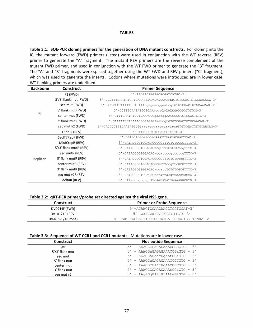
77
TABLES Table 3.1: SOE‐PCR cloning primers for the generation of DNA mutant constructs. For cloning into the IC, the mutant forward (FWD) primers (listed) were used in conjunction with the WT reverse (REV) primer to generate the “A” fragment. The mutant REV primers are the reverse complement of the mutant FWD primer, and used in conjunction with the WT FWD primer to generate the “B” fragment. The “A” and “B” fragments were spliced together using the WT FWD and REV primers (“C” fragment), which was used to generate the inserts. Codons where mutations were introduced are in lower case. WT flanking primers are underlined. Backbone Construct Primer Sequence
IC
F1 (FWD) 5’-AACGACAGGAGCACGATCATGC-3’
5’/3’ flank mut (FWD) 5’-GCCTTTCAATATGCTGAAAcgaGAGAGAAACcgaGTGTCGACTGTGCAACAGC-3’
seq mut (FWD) 5’-GCCTTTCAATATGCTGAAAcgagaacggaatcgtGTGTCGACTGTGCAACAG-3’
5’ flank mut (FWD) 5’-CCTTTCAATATGCTGAAAcgaGAGAGAAACCGCGTGTCG-3’
center mut (FWD) 5’-CTTTCAATATGCTGAAACGCgaacggAACCGCGTGTCGACTGTG-3’
3’ flank mut (FWD) 5’-CAATATGCTGAAACGCGAGAGAaatcgtGTGTCGACTGTGCAACAG-3’
seq mut v2 (FWD) 5’-CACGCCTTTCAATATGCTGaagagggaacgtaatagaGTGTCGACTGTGCAACAG-3’
ESphIR (REV) 5’-TTTCCGACTGCATGCTCTTC-3’
Replicon
SacIT7RepF (FWD) 5’-CGAGCTCGCGGCCGCAAATTTAATACGACTCAC-3’
MluICrepR (REV) 5’-CATACGCGTGGACACGCGGTTTCTCTCGCGTTTC-3’
5’/3’ flank mutR (REV) 5’-CATACGCGTGGACACtcgGTTTCTCTCtcgTTTC-3’
seq mutR (REV) 5’-CATACGCGTGGACACacgattccgttctcgTTTC-3’
5’ flank mutR (REV) 5’-CATACGCGTGGACACGCGGTTTCTCTCtcgTTTC-3’
center mutR (REV) 5’-CATACGCGTGGACACGCGGTTccgttcGCGTTTC-3’
3’ flank mutR (REV) 5’-CATACGCGTGGACACacgattTCTCTCGCGTTTC-3’
seq mut v2R (REV) 5’-CATACGCGTGGACACtctattacgttccctcttC-3’
deltaR (REV) 5’-CATacgcgtgcgtTTCAGCATATTGAAAGGCGTG-3’
Table 3.2: qRT‐PCR primer/probe set directed against the viral NS5 gene.
Construct Primer or Probe Sequence DV9944F (FWD) 5’-ACAAGTCGAACAACCTGGTCCAT-3’ DV10121R (REV) 5’-GCCGCACCATTGGTCTTCTC-3’
DV‐NS5‐F/T(Probe) 5’-FAM-TGGGATTTCCTCCCATGATTCCACTGG-TAMRA-3’
Table 3.3: Sequence of WT CCR1 and CCR1 mutants. Mutations are in lower case.
Construct Nucleotide Sequence WT 5’ – AAACGCGAGAGAAACCGCGTG - 3’
5’/3’ flank mut 5’ - AAACGaGAGAGAAACCGaGTG - 3’ seq mut 5’ - AAACGaGAacGgAAtCGtGTG - 3’
5’ flank mut 5’ - AAACGaGAGAGAAACCGCGTG - 3’ center mut 5’ - AAACGCGAacGgAACCGCGTG - 3’ 3’ flank mut 5’ - AAACGCGAGAGAAAtCGtGTG - 3’ seq mut v2 5’ - AAgaGgGAacGtAAtaGaGTG - 3’

78
REFERENCES 1. Alvarez, D. E., A. L. De Lella Ezcurra, S. Fucito, and A. V. Gamarnik. 2005. Role of RNA structures
present at the 3'UTR of dengue virus on translation, RNA synthesis, and viral replication. Virology 339:200‐212
2. Alvarez, D. E., M. F. Lodeiro, S. J. Ludueña, L. I. Pietrasanta, and A. G. Gamarnik. 2005. Long‐range RNA‐RNA interactions circularize the dengue virus genome. J Virol 79:6631‐6643.
3. Alvisi, G., V. Madan, and R. Bartenshlager. 2011. Hepatitis C virus and host cell lipids: an intimate connection. RNA Biol 8:258‐269.
4. Chadwick, D. R., and A. M. Lever. 2000. Antisense RNA sequences targeting the 5' leader packaging signal region of human immunodeficiency virus type‐1 inhibits viral replication at post‐transcriptional stages of the life cycle. Gene Ther 7:1362‐1368.
5. Chiu, W. W., R. M. Kinney, and T. W. Dreher. 2005. Control of translation by the 5'‐ and 3'‐terminal regions of the dengue virus genome. J Virol 79:8303‐8315.
6. Chua, J. J., M. M. Ng, and V. T. Chow. 2004. The non‐structural 3 (NS3) protein of dengue virus type 2 interacts with human nuclear receptor binding protein and is associated with alterations in membrane structure. Virus Res 102:151‐163.
7. Clyde, K., J. Barrera, and E. Harris. 2008. The capsid‐coding region hairpin element (cHP) is a critical determinant of dengue virus and West Nile virus RNA synthesis. . Virology 379:314‐323.
8. Clyde, K., and E. Harris. 2006. RNA secondary structure in the coding region of dengue virus type 2 directs translation start codon selection and is required for viral replication. J Virol 80:2170‐2182.
9. Deubel V., and D. J. P. 1981. Morphogenesis of yellow fever virus in Aedes aegypti cultured cells. I. Isolation of different cellular clones and the study of their susceptibility to infection with the virus. Am J Trop Med Hyg 30:1060‐1070.
10. Diamond, M. S., D. Edgil, T. G. Roberts, B. Lu, and E. Harris. 2000. Infection of human cells by dengue virus is modulated by different cell types and viral strains. J Virol 74:7814‐7823.
11. Diamond, M. S., and E. Harris. 2001. Interferon inhibits dengue virus infection by preventing translation of viral RNA through a PKR‐independent mechanism. Virology 289:297‐311.
12. Elshuber, S., and C. W. Mandl. 2005. Resuscitating mutations in a furin cleavage‐deficient mutant of the flavivirus tick‐borne encephalitis virus. J Virol 79:11813‐11823.
13. Fayzulin, R., and I. Frolov. 2004. Changes of the secondary structure of the 5' end of the Sindbis virus genome inhibit virus growth in mosquito cells and lead to accumulation of adaptive mutations. J Virol 78:4953‐4964.
14. Filomatori, C. V., M. F. Lodeiro, D. E. Alvarez, M. M. Samsa, L. Pietrasanta, and A. V. Gamarnik. 2006. A 5' RNA element promotes dengue virus RNA synthesis on a circular genome. Genes Dev 20:2238‐2249.
15. Friebe, P., and E. Harris. 2010. Interplay of RNA elements in the dengue virus 5' and 3' ends required for viral RNA replication. J Virol 84:6103‐6118.
16. Friebe, P., P. Y. Shi, and E. Harris. 2011. The 5' and 3' downstream AUG region elements are required for mosquito‐borne flavivirus RNA replication. J Virol 85:1900‐1905.
17. Frolov, I., R. Hardy, and C. M. Rice. 2001. Cis‐acting RNA elements at the 5' end of Sindbis virus genome RNA regulate minus‐ and plus‐strand RNA synthesis. RNA 7:1638‐1651.
18. Frolova, E., I. Frolov, and S. Schlesinger. 1997. Packaging signals in alphaviruses. J Virol 71:248‐258.
19. Gubler, D. J., and G. Kuno. 1997. Dengue and dengue hemorrhagic fever. CAB International.

79
20. Hase, T., P. L. Summers, K. H. Eckels, and W. B. Baze. 1987. An electron and immunoelectron microscopic study of dengue‐2 virus infection of cultured mosquito cells: maturation events. Arch Virol 92:273‐291.
21. Hase, T., P. L. Summers, K. H. Eckels, and W. B. Baze. 1987. Maturation process of Japanese encephalitis virus in cultured mosquito cells in vitro and mouse brain cells in vivo. Arch Virol 96:135‐151.
22. Heinz, F., G. Auer, K. Stiasny, H. Holzmann, C. Mandl, F. Guirakhoo, and C. Kunz. 1994. The interactions of the flavivirus envelope proteins: implications for virus entry and release. Arch Virol 9(S):339‐348.
23. Helt, A.‐M., and E. Harris. 2005. S‐phase‐dependent enhancement of dengue virus 2 replication in mosquito cells, but not in human cells. JVirol 79:7291‐7299.
24. Hofacker, I. L., M. Fekete, and P. F. Stadler. 2002. Secondary structure prediction for aligned RNA sequences. J Mol Biol 319:1059‐1066.
25. Hofacker, I. L., W. Fontana, P. F. Stadler, S. Bonhoeffer, M. Tacker, and P. Schuster. 1994. Fast folding and comparison of RNA secondary structures. Monatsh Chem 125:167‐188.
26. Holden, K. L., D. Stein, T. C. Pierson, A. Ahmed, K. Clyde, P. Iverson, and E. Harris. 2006. Inhibition of dengue virus translation and RNA synthesis by a morpholino oligomer to the top of the 3’ stem‐loop structure. Virology 344:439‐452.
27. Ishak, R., D. G. Tovey, and C. R. Howard. 1988. Morphogenesis of yellow fever virus 17D in infected cell cultures. J Gen Virol 69:325‐335.
28. Jones, C. T., C. G. Patkar, and R. J. Kuhn. 2005. Construction and application of yellow fever virus replicons. Virology 331:247‐259.
29. Khromykh, A. A., A. N. Varnavski, and E. G. Westaway. 1998. Encapsidation of the flavivirus kunjin replicon RNA by using a complementation system providing Kunjin virus structural proteins in trans. J Virol 72:5967‐5977.
30. Kinney, R. M., S. Butrapet, G. J. Chang, K. R. Tsuchiya, J. T. Roehrig, N. Bhamarapravati, and D. J. Gubler. 1997. Construction of infectious cDNA clones for dengue 2 virus strain 16681 and its attenuated vaccine derivative, strain PDK‐53. Virology 230:300‐308.
31. Ko, K. K., A. Igarashi, and K. Fukai. 1979. Electron microscopic observations on Aedes albopictus cells infected with dengue viruses. Arch Virol 62:41‐52.
32. Kuhn, R. J., W. Zhang, M. G. Rossmann, S. V. Pletnev, J. Corver, E. Lenches, C. T. Jones, S. Mukhopadhyay, P. R. Chipman, E. G. Strauss, T. S. Baker, and J. H. Strauss. 2002. Structure of dengue virus: implications for flavivirus organization, maturation, and fusion. Cell 108:717‐25.
33. Kuno, G. 1997. Factors influencing the tranmsission of dengue viruses. In D. J. Gubler and G. Kuno (ed.), Dengue and Dengue Hemorrhagic Fever. CAB International, New York.
34. Laue, T., P. Emmerich, and H. Schmitz. 1999. Detection of dengue virus RNA in patients after primary or secondary dengue infection by using the TaqMan automated amplification system. J Clin Microbiol 37:2543‐2547.
35. Leary, K., and C. D. Blair. 1980. Sequential events in the morphogenesis of japanese encephalitis virus. J Ultrastruct Res 72:123‐129.
36. Lindenbach, B. D., H. J. Thiel, and C. M. Rice. 2007. Flaviviridae: the viruses and their replication, p. 1101‐1152. In D. M. Knipe and P. M. Howley (ed.), Fields Virology, 5th ed. Lippincott‐Raven Publishers, Philadelphia.
37. Linger, B. R., L. Kunovska, R. J. Kuhn, and B. L. Golden. 2004. Sindbis virus nucleocapsid assembly: RNA folding promotes capsid protein dimerization. RNA 10:128‐138.
38. Liu, Y., E. Wimmer, and A. V. Paul. 2009. Cis‐acting RNA elements in human and animal plus‐strand RNA viruses. Biochim Biophys Acta 1789:495‐517.
80
39. Lo, M., M. Tilgner, K. A. Bernard, and P.‐Y. Shi. 2003. Functional analysis of mosquito‐borne flavivirus conserved sequence elements within 3’ untranslated region of West Nile Virus by use of a reporting replicon that differentiates between viral translation and RNA replication. J Virol 77:10004‐10014.
40. Matsumara, T., K. Shiraki, T. Sashikata, and S. Hotta. 1977. Morphogenesis of dengue‐1 virus in cultures of a human leukemic leukocyte line (J‐111). Microbiol Immunol 21:329‐334.
41. Miller, W. A., and K. A. White. 2006. Long‐distance RNA‐RNA interactions in plant virus gene expression and replication. Annu Rev Phytopathol 44:447‐467.
42. Murphy, F. A. 1980. Togavirus morphology and morphogenesis, p. 241‐316. In R. W. Schlesinger (ed.), The togaviruses: biology, structure, replication. New York: Academic.
43. Nelson, S., C. A. Jost, Q. Xu, J. Ess, J. E. Martin, T. Oliphant, S. S. Whitehead, A. P. Durbin, B. S. Graham, M. S. Diamond, and T. C. Pierson. 2008. Maturation of West Nile virus modulates sensitivity to antibody‐mediated neutralization. PLoS Pathog 4:e1000060.
44. Ng, M. L. 1987. Ultrastructural studies of Kunjin virus‐infected Aedes albopictus cells. J Gen Virol 68:577‐582.
45. Niesters, H. G., and J. H. Strauss. 1990. Mutagenesis of the conserved 51‐nucleotide region of Sindbis virus. J Virol 64:1639‐1647.
46. Nowak, T., P. M. Farber, G. Wengler, and G. Wengler. 1989. Analyses of the terminal sequences of West Nile virus structural proteins and of the in vitro translation of these proteins allow the proposal of a complete scheme of the proteolytic cleavages involved in their synthesis. Virology 169:365‐376.
47. Ohyama, A., T. Ito, E. Tanimura, S. C. Huang, and J. Hsue. 1977. Electron microscopic observation of the budding maturation of group B arboviruses. Microbiol Immunol 21:535‐538.
48. Payne, A. F., I. Binduga‐Gajewska, E. B. Kauffman, and L. D. Kramer. 2006. Quantitation of flaviviruses by fluorescent focus assay. J Virol Meth 134:183‐189.
49. Rosen, L., and D. Gubler. 1974. The use of mosquitoes to detect and propagate dengue viruses. Am J Trop Med Hyg 23:1153‐1160.
50. Rosen, L., L. E. Roseboom, D. J. Gubler, J. C. Lien, and B. N. Chaniotis. 1985. Comparative susceptibility of mosquito species and strains to oral and parenteral infection with dengue and Japanese encephalitis viruses. Am J Trop Med Hyg 34:603‐615.
51. Sriurairatna, S., and N. Bhamarapravati. 1977. Replication of dengue‐2 virus in Aedes albopictus mosquitoes. An electron microscopic study. Am J Trop Med Hyg 26:1199‐1205.
52. Sriurairatna, S., N. Bhamarapravati, and O. Phalavadhtana. 1973. Dengue virus infection of mice: Morphology and morphogenesis of dengue type 2 virus in suckling mouse neurons. Infect Immun 8:1017‐1029.
53. Stadler, K., S. L. Allison, J. Schalich, and F. X. Heinz. 1997. Proteolytic activation of tick‐borne encephalitis virus by furin. J Virol 71:8475–8481.
54. Thompson, J. D., T. J. Gibson, F. Plewniak, F. Jeanmougin, and D. G. Higgins. 1997. The CLUSTAL_X Windows interface: flexible strategies for multiple sequence alignment aided by quality analysis tools. Nucleic Acids Res 25:4876‐4882.
55. Thurner, C., C. Witwer, I. L. Hofacker, and P. F. Stadler. 2004. Conserved RNA secondary structures in Flaviviridae genomes. J Gen Virol 85:1113‐1124.
56. Tilgner, M., and P. Y. Shi. 2004. Structure and function of the 3' terminal six nucleotides of the West Nile virus genome in viral replication. J Virol 78:8159‐8171.
57. Weiss, B., U. Geigenmuller‐Gnirke, and S. Schlesinger. 1994. Interactions between Sindbis virus RNAs and a 68 amino acid derivative of the viral capsid protein further defines the capsid binding site. Nucleic Acids Res 22:780‐786.

81
58. Welsch, S., S. Miller, I. Romero‐Brey, A. Merz, C. K. Bleck, P. Walther, S. D. Fuller, C. Antony, J. Krijnse‐Locker, and R. Bartenschlager. 2009. Composition and three‐dimensional architecture of the dengue virus replication and assembly sites. Cell Host Microbe 5:365‐375.
59. Wengler, G., and G. Wengler. 1989. Cell‐associated West Nile flavivirus is covered with E+pre‐M protein heterodimers which are destroyed and reorganized by proteolytic cleavage during virus release. J Virol 63:2521‐2526.
60. Yu, I. M., H. A. Holdaway, P. R. Chipman, R. J. Kuhn, M. G. Rossmann, and J. Chen. 2009. Association of the pr peptides with dengue virus at acidic pH blocks membrane fusion. J Virol 83:12101‐12107.
61. Zeng, L., B. Falgout, and L. Markoff. 1998. Identification of specific nucleotide sequences within conserved 3'‐SL in the dengue type 2 virus genome required for replication. J Virol 72:7510‐7522.
62. Zhang, W., P. R. Chipman, J. Corver, P. R. Jonhnson, Y. Zhang, S. Mukhopadhyay, T. S. Baker, J. H. Strauss, M. G. Rossmann, and R. J. Kuhn. 2003. Visualization of membrane protein domains by cryo‐electron microscopy of dengue virus. Nat Struct Biol 10:907‐912.
63. Zhang, Y., J. Corver, P. R. Chipman, W. Zhang, S. V. Pletnev, D. Sedlak, T. S. Baker, J. H. Straus, R. J. Kuhn, and M. G. Rossmann. 2003. Structures of immature flavivirus particles. EMBO J 22:2604‐2613.
64. Zhang, Y., V. A. Kostyuchenko, and M. G. Rossmann. 2007. Structural analysis of viral nucleocapsids by subtraction of partial projections. J Struct Biol 157:356‐364.
65. Zuker, M. 2003. Mfold web server for nucleic acid folding and hybridization prediction. Nucleic Acids Res 31:3406‐3415.
66. Zybert, I. A., H. van der Ende‐Metselaar, J. Wilschut, and J. M. Smit. 2008. Functional importance of dengue virus maturation: infectious properties of immature virions. J Gen Virol 89:3047‐3051.

82
CHAPTER FOUR
“TYING UP LOOSE ENDS”

83
“TYING UP LOOSE ENDS” Identification of novel coding‐region RNA elements. The goal of this study was to identify additional RNA elements located in the DENV coding‐region that regulate the intracellular viral life cycle. Based on sequence and secondary structure conservation within the DENV serogroup, three candidate RNA structural elements (C‐1 C‐2 and NS5‐2) and three candidate RNA sequence elements (prMCR1, NS5CR1 and CCR1) were identified and characterized for their effect on viral replication (discussed in Chapter 2). The initial screens eliminated all but one candidate RNA sequence element, the conserved capsid‐coding region 1 (CCR1), which was shown to regulate the viral life cycle in both baby hamster kidney (BHK) and Aedes albopictus mosquito (C6/36) cells, though its effects are more dramatic in mosquito cells (discussed in Chapter 3). Altering the primary nucleotide sequence of CCR1 not only reduced viral titer in mosquito cells, but additionally, CCR1 mutant viruses did not replicate to wild‐type (WT) levels in Ae. aegypti mosquitoes. Decreased viral replication in the mosquito body cavity led to a reduction in viral dissemination to the salivary glands, which could have dramatic implications for viral transmission. Furthermore, CCR1 was shown to mediate post‐RNA replication events, possibly acting as a sequence‐dependent assembly signal, which has yet to be identified in DENV. C‐1 is a putative RNA structure element that was eliminated as a candidate during the initial screens when it displayed WT replication in BHK cells but had inconclusive results in infected C6/36 cells. The “C1sil mut” and “C1sil cmut” constructs had distinct structural changes to C‐1, but varied in their ability to replicate in infected C6/36 cells, implying that C‐1 might have a sequence‐dependent rather than a structure‐dependent effect on the viral life cycle in this cell line. However, C‐1 only has 44% sequence homology (~52% background, data not shown) in the DENV serogroup and 32% sequence homology (~22% background, data not shown) in the Japanese encephalitis virus (JEV) serogroup, which indicates that C‐1 is not likely to be a candidate RNA sequence element. However, given the preliminary evidence in infected C6/36 cells, additional silent point mutations could be introduced so as to disrupt the entire C‐1 nucleotide sequence irrespective of structural changes and the resulting variants screened for their effects on the viral life cycle in C6/36 cells. As evidenced in other RNA viruses (2), it is possible that additional regulatory RNA elements might exist within the DENV coding‐region. In the search for additional candidate RNA structural elements, the phylogenetically conserved structure predictions for the NS5‐coding sequence were re‐examined. In brief, the DENV, JEV and tick‐borne encephalitis virus (TBEV) serogroup sequence alignments that were generated using Clustal W2 (3) were modified so that they would include a large portion of the NS5‐coding region, which was subsequently analyzed using RNAalifold (1). In this investigation, two additional candidate RNA structure elements were identified in the NS5‐coding region, NS5‐1 and NS5‐2, which were shown to be conserved in the DENV (Fig. 4.1A), JEV (Fig. 4.1B) and TBEV (Fig. 4.1C) serogroups; however, NS5‐3 is not as well conserved as NS5‐1 in the JEV serogroup (Fig. 4.1B). Continuation of this study should include introducing silent point mutations that disrupt the predicted secondary structure of NS5‐1 and NS5‐3, and the resulting mutant constructs should be screened for viral infectivity in mammalian and mosquito cells. The identification of additional candidate RNA elements is an important first step in the characterization of the intracellular viral life cycle since there are still a number of stages that are not well understood. Sequencing vRNA from infected BHK cells. As discussed in Chapter 3, the infections in C6/36 cells confirmed that CCR1 has a more drastic phenotype in this cell line while the infections in BHK cells raised the possibility that revertant viruses were accumulating after serial passage. Briefly, to determine if CCR1 had an effect on early stages of the viral life cycle, virus‐containing supernatants were collected from transfected BHK cells, concentrated, and used to infect BHK and C6/36 cells at an MOI of 0.01 or with ~2.5 genome equivalents (G‐Eq) per cell. Virus‐containing supernatants from these infected cells

84
were then assessed for viral infectivity by plaque assay. When the infections were normalized by pfu, all the mutant viruses displayed a significantly lower viral titer than the WT at 24 hours post‐infection (hpi) in BHK cells, the “5’/3’ flank mut,” “5’ flank mut” and “seq mut v2” mutant viruses acquired WT levels by the end of the assay (Fig. 3.3A). Similar results were observed when the infections were normalized by G‐Eq, where at early timepoints post‐infection the “5’/3’ flank mut” and “center mut” constructs had significantly lower viral titers compared to the WT though by the end of the assay they attained WT levels (Fig. 3.3C). The fact that the mutant viruses accumulated to WT levels after serial passage in BHK cells regardless of whether the infections were normalized by pfu or G‐Eq could indicate the buildup of viral revertants. The likelihood that the mutant viruses undergo enough selective pressure to generate reversions in BHK cells is supported by the observation that the “center mut” mutant virus displays an intermediate plaque phenotype while in previous assays it demonstrated a small plaque phenotype (Fig. 3.2D). The possibility that viral revertants caused the accumulation of mutant viruses in infected BHK cells was confirmed with the observation that the mutant viruses also displayed a WT particle:pfu ratio in BHK cells at all timepoints tested (Fig. 3.6E and 3.6G). In an effort to determine whether viral revertants were the cause for the accumulation of mutant viruses in infected BHK cells, viral RNA (vRNA) was harvested from virus‐containing supernatants produced from infected BHK cells at 48 hpi. The vRNA was subjected to a reverse transcriptase (RT)‐PCR reaction using primers designed to amplify a cDNA fragment that contained the first 602 nucleotides of the DENV genome, which was then sequenced directly. Hence, each sequence in Table 4.1 represents a single sequencing reaction derived from the amplified cDNA fragments. Preliminary tests were conducted on the “CCR1orig mut” mutant viruses that were discussed in Chapter 2. The vast majority of the sequencing results were identical to the input virus (“CCR1orig mut”), though in one instance the sequence was that of the WT virus (“WT”) or half of the mutations reverted to the WT sequence (“CCR1orig mut like,” Table 4.1). In another instance, the sequence still contained the original mutations but two additional changes were found in the 3’ flanking sequence (“CCR1orig mut plus,” Table 4.1). The mixture of mutant and WT sequences in the cDNA amplicons implies that there is sufficient pressure in infected BHK cells to cause minor sequence changes in the CCR1 sequence as early as 48 hpi. However, this preliminary method of generating cDNA amplicons is not ideal given the incidence of sequences resembling other mutant constructs, which shows that common DNA surface contaminants can be amplified at random. An example of this can be observed in the three instances where the sequenced cDNA was shown to be identical or similar to the “seq mut v2” construct (“seq mut v2” and “seq mut v2 like”, Table 4.1), which happened to be in the process of being cloned while these experiments were being conducted. While one cDNA was shown to have the same sequence as the “5’ flank mut” construct, it is not likely that this is the result of a contamination since this construct had not yet been designed when these experiments were in progress. Interestingly, this result implies that this position in the CCR1 sequence has no impact on viral infectivity, which is consistent with the fact that the “5’ flank mut” construct had WT levels of replication in transfected and infected BHK cells (discussed in Chapter 3). In order to overcome the technical difficulties of this procedure, a collaboration with the Broad Institute (Cambridge MA) was established whereby the extracted vRNA samples would be sent for full genome single‐molecule sequencing. Full genome coverage would allow for the identification of reversions in CCR1 as well as in additional portions of the genome, which might indicate the presence of a secondary RNA element. Current studies are directed towards optomizing a high‐fidelity one‐step RT‐PCR protocol that produces four sets of overlapping amplicons that span the entire DENV genome, which will be sent to the Broad Institute for deep sequencing. While initial studies only examined the “CCR1orig mut” mutant virus at 48 hpi, the collaboration with the Broad Institute includes the investigation of vRNA isolated from the full panel of mutant viruses at early and late timepoints post‐

85
infection in both BHK and C6/36 cells, as well as sequencing the input virus that was used to infect both cell types.
FUTURE DIRECTIONS Determining the mechanism by which CCR1 regulates infectious particle production. As discussed in Chapter 3, CCR1 was implicated as an assembly signal for DENV that regulates the amount of infectious particle production in both BHK and C6/36 cells, though the precise mechanism by which this occurs is still under investigation. Virus particles within the virus‐containing supernatants from transfected BHK and C6/36 cells can be assessed by comparing the amount of vRNA to the amount of viral E or prM/M proteins present as determined by Western blot to calculate the vRNA:particle ratio. If there is no defect in viral assembly then the vRNA:particle ratio will be equivalent to that of the WT. An assembly defect might result in less mutant viral particles being detected in the supernatant as compared to the WT, and confirm the observation that the same amount of vRNA is being released from the cells (Fig.3.6A and 3.6B). Additionally, virus‐containing supernatants can be examined using sucrose gradient centrifugation and the amount of vRNA that is contained within assembled particles can be determined by comparing vRNA levels for both the WT and mutant viruses in E‐containing fractions. If assembly is modulated by CCR1, then mutant vRNA will be isolated from fractions that contain a disproportionate amount of the viral proteins capsid, prM/M or E while the WT vRNA will mostly associate with all three. The best approach to determining whether the accumulation of noninfectious virions is the result of a gross structural defect is to section BHK and C6/36 cells that have been infected with mutant CCR1 viruses and examine the samples using transmission electron microscopy (TEM). TEM would show any structural differences in the assembled particles produced by cells infected with the different mutant viruses as compared to the WT, which might explain how altering CCR1 can have different effects on infectious particle production depending on the cell line being tested. As discussed, virion maturation could also be affected as a result of a defect in viral assembly, leading to a decrease in the amount of infectious particles produced. To detect this, virus‐containing supernatants would be subjected to furin treatment in vitro, and the resulting viruses used to infect a fresh monolayer of BHK cells. Infection can be assayed at 24 hpi using immunoflourescence to determine if treatment with furin increases the number of cells that could become infected, which would imply that insufficient cleavage by furin is the cause for the production of noninfectious particles. While the manner in which CCR1 decreases the number of assembled particles that can undergo cell‐to‐cell spread is still under investigation, whether CCR1 regulation is dependent on RNA‐RNA and/or RNA‐protein interactions has yet to be determined. To ascertain whether CCR1 interacts with a protein‐binding partner, electrophoretic mobility shift assays (EMSAs) can be carried out using radiolabelled WT and mutant CCR1 RNAs that are incubated with infected cell lysates. When these reactions are analyzed by agarose gel electrophoresis, proteins that bind to the WT RNA will cause a shift to occur that should not be observed when the mutant CCR1 RNA is utilized. For these experiments, uninfected lysates would be used as a negative control, and no differences in the mobility shift pattern are expected when using uninfected lysates as compared to the infected cell lysates. If a difference in the mobility shift pattern is observed in the preliminary experiments, an RNA affinity column will be employed using either the WT or mutant CCR1 RNAs. Infected cell lysates will be passed over the column, and proteins bound with high affinity will be eluted using a high molar salt solution and analyzed using polyacrylamide gel electrophoresis (PAGE). Eluates that display differences by PAGE analysis will be examined using multidimensional protein identification technology (MudPIT), which can be used to identify candidate protein‐binding partners. Candidate proteins can be isolated while bound to the WT CCR1 sequence using RNA immunoprecipitation (RIP) assays, and specificity can

86
be assessed with quantitative real‐time (qRT)‐PCR targeting CCR1. If the candidate protein is commercially available, protein‐binding can also be assessed using EMSAs whereby either the WT or mutant CCR1 RNAs are radioactively labeled and incubated with increasing concentrations of the candidate protein before being analyzed by agarose gel electrophoresis. The specificity of the interaction can be determined by adding increasing amounts of the unlabelled WT sequence to compete away the protein bound to the labeled mutant CCR1 RNAs. An RNA protection assay will be used to determine the binding site of the candidate protein along the CCR1 sequence. Functional assays would then be needed to establish the biological relevance of the interaction. Cells will need to be treated with small interfering RNAs (siRNAs) directed against the candidate protein to knock down its expression. If viral infection kinetics are altered when these treated cells are infected with WT virus, this would indicate that CCR1 is involved in the proposed protein‐RNA interaction with the candidate protein. These studies will help elucidate whether there are candidate protein(s) that bind to CCR1 and modulate its role on infectious particle production as well as provide some indication of how specific that interaction is. If CCR1 functions through an RNA‐RNA interaction, this will be addressed by studying the incidence of viral revertants during vRNA sequencing of the virus‐containing supernatants derived from infected BHK and C6/36 cells. The preliminary sequencing results described earlier, give some indication that there is no alternate RNA element in the DENV genome since reversions have been observed in the region of interest though these results are not conclusive. Full‐genome sequencing of extracted vRNAs will help elucidate whether or not additional unknown RNA elements are implicated since individual cDNAs can be compared from a single sample rather than examining a consensus sequence derived from multiple cDNAs in the same sample. If vRNA sequencing yields information regarding whether or not there is an additional RNA element in the DENV genome, the interaction can be confirmed using EMSAs whereby CCR1 is radiolabelled and increasing concentrations of the secondary RNA element are added. If CCR1 is involved in an RNA‐RNA interaction then a mobility shift should be observed when the secondary RNA element is added and this shift will be abolished when either CCR1 or the secondary RNA element is mutated. Functional assays should include mutating the secondary RNA element to see if the same effect on noninfectious particle production is achieved as observed with the CCR1 mutants. Taken together, these assays would help determine a mechanism of action by which CCR1 modulates infectious viral particle production in both BHK and C6/36 cells.
CONCLUSIONS Overall, these studies are important for improving our understanding of cis‐acting determinants that differentially regulate the viral life cycle in the human host and mosquito vector. The goal of this study was to identify and characterize a cis‐acting coding‐region regulatory RNA element that modulates the viral life cycle since the predominant focus of research thus far has centered on those elements located in the UTRs. Using sequence conservation and secondary structure predictions, CCR1 has been identified as a novel coding‐region RNA sequence element that regulates infectious particle production in BHK and C6/36 cells, possibly serving as an assembly signal for DENV, which has not yet been identified. Furthermore, the critical role of CCR1 in C6/36 cells has been shown in vivo, whereby mutations to CCR1 reduce viral replication in Ae. aegypti mosquitoes and viral dissemination to the salivary glands, providing insight into viral replication strategies in the mosquito vector. Coding‐region RNA regulatory elements have been described in other RNA viruses and may serve as excellent targets for novel therapeutics since their evolution towards resistance is constrained by both their RNA regulatory function and amino acid coding capacity. Thus, studies that examine the regulatory mechanisms of coding‐region RNA elements are critical, because these determinants play important

87
roles in the viral life cycle and could potentially serve in the design of attenuated vaccine strains or act as candidate targets for antiviral agents.
MATERIALS AND METHODS Sequence conservation and secondary structure prediction algorithms. For conserved sequence prediction, sequences were aligned using the Clustal W2 web software (3) from the European Bioinformatics Institute. Portions of the Clustal W2 alignments were processed by the RNAalifold web server (1) from the Institute of Theoretical Chemistry at the University of Vienna in order to generate the phylogenetic conserved secondary structure predictions. RT‐PCR. vRNA in the virus‐containing supernatants was isolated using the ZR Viral RNA kit (Zymo Research). Viral cDNA was first produced using the One‐Step RT‐PCR System (Qiagen), purified using NucleoSpin Extract II (Macherey‐Nagel) and sequenced at the UC Berkeley Sequencing Facility. Subsequent analysis included producing viral cDNA using the SuperScript III One‐Step RT‐PCR System with Platinum Taq High Fidelity (Invitrogen), which was purified using NucleoSpin Extract II and will be sequenced at the Broad Institute (Cambridge MA). Primers sequences for the reverse transcriptase (RT)‐PCR are available in Table 4.2.

AFIGURES
5’3’
NS5‐3
NS5‐1
BB
5’3’
C
NS5‐1
NS5‐3C
5’3’
NS5‐1
NS5‐3
88

Figure 4.1: Putative RNA structural elements are identified in the NS5‐coding region based on structureconservation in the DENV, JEV and TBEV serogroups. Phylogenetic consensus structure based on a portion of, g p y g pthe NS5‐coding sequence from the (A) DENV serogroup (position 9647‐10159, numbering based on DENV2 Thaistrain 16681; accession codes DENV1 U88535, DENV2 NC_001474, DENV3 AY099336 and DENV4 GU289913),(B) JEV serogroup (position 9767‐10280, numbering based on JEV; accession codes JEV M18370, Murray Valleyencephalitis virus AF161266, WNV NC_001563, KUN AY274505, St. Louis encephalitis virus DQ525916 and YFVU17067) and (C) TBEV serogroups (position 9678‐10218, numbering based on TBEV; accession codes TBEVU27495, Powassan virus NC_003687 and Omsk hemorrhagic virus AY193805). Consensus structures werecomputed with RNAalifold (1). Covariance is depicted as a colored spectrum ranging from red, to dark pink,light pink, yellow and then green. Putative RNA structure elements NS5‐1 and NS5‐3 are indicated by circles orboxes.
89

90
TABLES Table 4.1: Sequencing results from BHK cells infected with the “CCR1orig mut” mutant virus at 48 hours post‐infection. CCR1 sequence is highlighted in yellow. Mutations are indicated by lower case. Capitalized nucleotides that have been bolded indicate reversion to the WT sequence whereas lower case nucleotides that have been bolded indicate mutations that were not engineered.
vRNA Sequencing Incidence Sequence Match 5’-CTGAAACGCGAGAGAAACCGCGTG-3’ 1 WT 5’-CTGAAgCGgGAGAGAAACCGCGTG-3’ 18 CCR1orig mut 5’-CTGAAgCGgGAGAGAAACCaCGcG-3’ 1 CCR1orig mut plus 5’-CcGAAgaGgGAacGtAAtaGaGTG-3’ 1 seq mut v2 like 5’-CTGAAACGaGAGAGAAACCGCGTG-3’ 1 5’ flank mut 5’-CTGAAgaGgGAacGtAAtaGaGTG-3’ 2 seq mut v2 5’-CTGAAACGgGAGAGAAACCGCGTG-3’ 1 CCR1orig mut like
Table 4.2: Sequencing primers used for the generation of cDNA amplicons using RT‐PCR. Sequencing Facility Primer Sequence
UC Berkeley F1 (FWD) 5’-AACGACAGGAGCACGATCATGC-3’ D2‐2 (REV) 5’-CAATCACGTACAAGTGTCCCC-3’
Broad Institute
broad_D2_F_11_b (FWD) 5’-CTACGTGGACCGACAAAGACAG-3’ broad_D2_R_3669 (REV) 5’-CCATRCCTATGTCATCYGTCAT-3’ broad_D2_F_2411 (FWD) 5’-ATGGTGCARGCYGATAGTGGTT-3’ broad_R_6183 (REV) 5’-TCTGTCTGCRTAGTTGATRCCTTC-3’
broad_D2_F_4919 (FWD) 5’-CTGGRACGTCAGGATCTCCAAT-3’ broad_D2b_R_8618 (REV) 5’-CGCTGTTGTCCRAAWGGAGT-3’ broad_D2_F_7293 (FWD) 5’-CTAGAWCCAATACCYTATGATCC-3’ broad_D2_R_10477 (REV) 5’-CCATGCGTACAGCTTCCATGGT-3’

91
REFERENCES 1. Hofacker, I. L., M. Fekete, and P. F. Stadler. 2002. Secondary structure prediction for aligned
RNA sequences. J Mol Biol 319:1059‐1066. 2. Liu, Y., E. Wimmer, and A. V. Paul. 2009. Cis‐acting RNA elements in human and animal plus‐
strand RNA viruses. Biochim Biophys Acta 1789:495‐517. 3. Thompson, J. D., T. J. Gibson, F. Plewniak, F. Jeanmougin, and D. G. Higgins. 1997. The
CLUSTAL_X Windows interface: flexible strategies for multiple sequence alignment aided by quality analysis tools. Nucleic Acids Res 25:4876‐4882.


![Dissertation Proposal[DRAFT]](https://img.pdfslide.us/doc/110x75/61a1e977b096c2291522df88/dissertation-proposaldraft.jpg)