Embed Size (px)
Citation preview
proteinsSTRUCTURE O FUNCTION O BIOINFORMATICS
SHORT COMMUNICATION
Dissection of the conformational cycle of themultidrug/lipidA ABC exporter MsbARupak Doshi, Barbara Woebking, and Hendrik W. van Veen*
Department of Pharmacology, University of Cambridge, Cambridge CB2 1PD, United Kingdom
INTRODUCTION
MsbA is an essential homodimeric ATP-binding cassette (ABC)
transporter in Escherichia coli and other gram-negative bacteria,
which mediates the transport of the Lipid A core moiety of LPS
from the cytoplasmic membrane to the outer membrane.1 It has
been shown to also transport many substrates of the human mul-
tidrug resistance P-glycoprotein ABCB1,2–4 and to exhibit an
overlapping drug specificity with the bacterial homologues LmrA
and Sav1866 from gram-positive Lactococcus lactis and Staphylococ-
cus aureus, respectively.4, 5 While much has been published
regarding ATP hydrolysis by MsbA in the past few years,2, 3, 6–9
recent focus has shifted to using this protein as a prototype ABC
exporter to study the conformational movements associated with
the catalytic cycle. This has primarily been done using X-ray crys-
tallization,10 cryo-electron microscopy (EM),11 and spectroscopic
techniques based on electron paramagnetic resonance (EPR) and
double electron–electron resonance (DEER)12–15 (Table I).
The first medium-resolution ABC exporter structure reported
was the ADP-bound outward-facing state for Sav1866 in which the
internal chamber between the membrane domains (MDs) faced the
periplasm/exterior of the cell, and in which the nucleotide-binding
domains (NBDs) were tightly dimerized.17 However, the physiologi-
cal relevance of this state was controversial due to contradictory bio-
chemical studies indicating that the absence of the g-Pi group desta-
bilizes the NBD dimer.18 As a result, Sav1866 was recrystallized with
50-adenylyl-b-g-imidodiphosphate (AMP-PNP) and because this
conformation was also overall outward-facing, the ADP-bound
structure was reinterpreted as the ATP-bound state19 (Table I). The
*Correspondence to: Hendrik W. van Veen, Department of Pharmacology, University of Cam-
bridge, Tennis Court Road, Cambridge CB2 1PD, United Kingdom.
E-mail: [email protected]
Received 7 May 2010; Revised 17 June 2010; Accepted 23 June 2010
Published online 6 July 2010 in Wiley Online Library (wileyonlinelibrary.com).
DOI: 10.1002/prot.22813
ABSTRACT
Recent crystal structures of the multidrug ATP-
binding cassette (ABC) exporters Sav1866 from
Staphylococcus aureus, MsbA from Escherichia coli,
Vibrio cholera, and Salmonella typhimurium, and
mouse ABCB1a suggest a common alternating
access mechanism for export. However, the molecu-
lar framework underlying this mechanism is crit-
ically dependent on assumed conformational rela-
tionships between nonidentical crystal structures
and therefore requires biochemical verification.
The structures of homodimeric MsbA reveal a pair
of glutamate residues (E208 and E2080) in the in-
tracellular domains of its two half-transporters,
close to the nucleotide-binding domains (NBDs),
which are in close proximity of each other in the
outward-facing state but not in the inward-facing
state. Using intermolecular cysteine crosslinking
between E208C and E208C0 in E. coli MsbA, we
demonstrate that the NBDs dissociate in nucleo-
tide-free conditions and come close on ATP bind-
ing and ADP�vanadate trapping. Interestingly, ADP
alone separates the half-transporters like a nucleo-
tide-free state, presumably for the following cata-
lytic cycle. Our data fill persistent gaps in current
studies on the conformational dynamics of a vari-
ety of ABC exporters. Based on a single biochemi-
cal method, the findings describe a conformational
cycle for a single ABC exporter at major check-
points of the ATPase reaction under experimental
conditions, where the exporter is transport active.
Proteins 2010; 78:2867–2872.VVC 2010 Wiley-Liss, Inc.
Key words: ABC exporter; conformational dynam-
ics; catalytic cycle; intermolecular cysteine cross-
linking; multidrug transport.
VVC 2010 WILEY-LISS, INC. PROTEINS 2867
authors further suggested that the outward-facing confor-
mation with ADP was probably a result of the experimen-
tal drawback of having the protein in detergent solution.19
Three MsbA structures were subsequently reported in dis-
tinct conformations, a nucleotide-free inward-facing (V-
shaped, open-apo) state with the NBDs widely separated
and predicted drug-binding sites in the MDs2 facing the
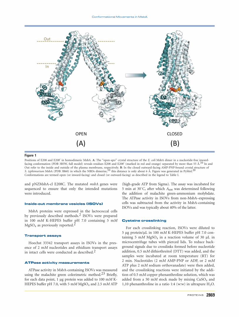
cytoplasm/interior of the cell for E. coli MsbA [Fig. 1(A)],
and AMP-PNP-bound and ADP�vanadate (Vi)-trapped
outward-facing states for Salmonella typhimurium MsbA10
[Fig. 1(B)]. The latter two were identical to those obtained
for Sav1866,17, 19 but the study did not report an ADP-
bound structure for MsbA. Recently, mouse multidrug re-
sistance P-glycoprotein ABCB1a was crystallized in a nu-
cleotide-free inward-facing state (Table I).21 It is evident
from Table I that no single ABC exporter has yet been crys-
tallized at all stages of the ATPase reaction to generate a
complete catalytic cycle.
Appreciating the importance of a physiological mem-
brane environment for the conformational movements of
transporters, EPR and DEER analyses on E. coli MsbA in
proteoliposomes revealed that the separation distances
between the NBDs in the nucleotide-free state and
ADP�Vi-trapped state were in accordance with the crystal
structures.12–15 However, interdomain distance measure-
ments of AMP-PNP- or ADP-bound forms of MsbA were
not reported (Table I). Furthermore, cryo-EM was used
on MsbA from S. typhimurium and Vibrio cholera recon-
stituted into proteoliposomes to study nucleotide-de-
pendent conformations.11 While the authors found
minor distinctions between the AMP-PNP-bound and
ADP�Vi-trapped structures in this work, they concluded
that the overall conformations were similar to the crystal
structures. In this case, nucleotide-free and ADP-bound
conformations were missing (Table I). Thus, although
major advancements have been made, till date no single
technique has been used to investigate the conformations
of a particular membrane-embedded ABC exporter at all
four known checkpoints of the ATPase cycle (namely, nu-
cleotide-free, ATP-bound, ADP�Pi-bound/hydrolysis in-
termediate, and ADP-bound) under physiological condi-
tions suitable for transport.
In a recent molecular dynamics study on S. typhimurium
MsbA, pairs of charged residues E208 and K212 in the
cytoplasmic extensions of transmembrane helices 4/40 ofhomodimeric MsbA, close to its NBDs, were implicated in
electrostatic and hydrogen bond interactions at the central
‘‘tetrahelix bundle’’ in the outward-facing conformation of
the transporter.22 We hypothesized that cysteine residues
at positions E208 and E2080 would be close enough to
crosslink in this state (distance �6 A) [Fig. 1(B)] but not
in the inward-facing conformation in which these residues
are predicted to be separated widely (distance �55 A)
[Fig. 1(A)]. Here, we have utilized E208C–E2080C disulfide
crosslinking in homodimeric E. coli MsbA in a physiologi-
cal phospholipid bilayer (plasma membrane vesicles) to
detect the nucleotide-dependent conformations that are
missing in current data sets.
MATERIALS AND METHODS
Bacterial strains and plasmids
L. lactis strain NZ9000 DlmrA DlmrCD,23 which is
devoid of the major endogenous multidrug transporters
LmrA and LmrCD, was used as a host for pNZ8048-
derived plasmids.24 E. coli strain XL1 Blue was a host for
the pGEM-5Zf(1) (Promega) cloning vector. Cells were
grown in M17 medium (Oxoid) and LB medium (For-
medium), respectively, as described previously.2
Site-directed mutagenesis
To create cysteine-less N-terminal His6-tagged MsbA
(MsbA-cl), endogenous C88 and C315 were each replaced
by S in the wildtype (Wt) E. coli msbA gene in vector
pGEMMsbA2 (generating pGEMMsbA-cl) by site-directed
mutagenesis using the QuickChange kit (Stratagene) with
the forward primer 50-CCA GCT ACT CTA TCT CCT
GGG TAT CAG G-30 and the reverse primer 50-CCA GGA
GAT AGA GTA GCT GGA GAC ATA G-30 for C88S, andthe forward primer 50-GCG GCT TCT CAG ACG CTG
TTT ACC ATT C-30 and the reverse primer 50-GCG TCT
GAG AAG CCG CCA TAC CGC GCT G-30 for C315S. TheE208C mutant was generated by similar methods in
pGEMMsbA-cl using the forward primer 50-CCA CCA
GCG CAT GCC AAA TGC TGA AG-30 and reverse primer
50-CTT CAG CAT TTG GCA TGC GCT GGT GG-30. ThemsbA-cl and msbA-cl E208C genes were subcloned as NcoI-
XbaI and NcoI-SacI fragments into pNZ8048 downstream
of the nisin A-inducible promoter, yielding pNZMsbA-cl
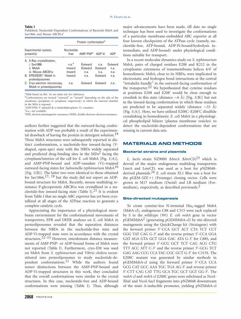
Table IPublished, Nucleotide-Dependent Conformations of Bacterial MsbA and
Sav1866, and Mouse ABCB1aa
Experimental system,protein(s)
Protein conformationb
Nucleotidefree AMP-PNPc ADP�Vi ADP
A. X-Ray crystallization,i. Sav1866 n.a.d Outward n.a. Outwardii. MsbA Inward Outward Outward n.a.iii. Mouse ABCB1a Inward n.a. n.a. n.a.
B. EPR/DEER,e MsbA inproteoliposomes
Inward n.a. Outward n.a.
C. Cryo-electron microscopy,MsbA in proteoliposomes
n.a. Outward Outward n.a.
aTable based on Ref. 16; see main text for references.bConformations are termed ‘‘outward’’ or ‘‘inward’’ depending on the side of the
membrane (periplasm or cytoplasm, respectively) to which the internal chamber
in the MDs is exposed.cAMP-PNP, 50-adenylyl-b-g-imidodiphosphate; Vi, vanadate.dn.a., not available.eEPR, electron paramagnetic resonance; DEER, double electron–electron resonance.
R. Doshi et al.
2868 PROTEINS
and pNZMsbA-cl E208C. The mutated msbA genes were
sequenced to ensure that only the intended mutations
were introduced.
Inside-out membrane vesicles (ISOVs)
MsbA proteins were expressed in the lactococcal cells
by previously described methods.2 ISOVs were prepared
in 100 mM K-HEPES buffer pH 7.0 containing 5 mM
MgSO4 as previously reported.2
Transport assays
Hoechst 33342 transport assays in ISOVs in the pres-
ence of 2 mM nucleotides and ethidium transport assays
in intact cells were conducted as described.2
ATPase activity measurements
ATPase activity in MsbA-containing ISOVs was measured
using the malachite green colorimetric method.23 Briefly,
for each data point, 1 lg protein was added to 100 mM K-
HEPES buffer pH 7.0, with 5 mM MgSO4 and 2.5 mM ATP
(high-grade ATP from Sigma). The assay was incubated for
5 min at 308C, after which A600 was determined following
the addition of malachite green-ammonium molybdate.
The ATPase activity in ISOVs from non-MsbA–expressing
cells was subtracted from the activity in MsbA-containing
ISOVs and was typically about 40% of the latter.
Cysteine crosslinking
For each crosslinking reaction, ISOVs were diluted to
5 lg protein/lL in 100 mM K-HEPES buffer pH 7.0 con-
taining 5 mM MgSO4 in a reaction volume of 30 lL in
microcentrifuge tubes with pierced lids. To reduce back-
ground signals due to crosslinks formed before nucleotide
addition, 0.5 mM dithiothreitol (DTT) was added, and the
samples were incubated at room temperature (RT) for
2 min. Nucleotides (2 mM AMP-PNP or ADP, or 2 mM
ATP plus 2 mM sodium orthovanadate) were then added,
and the crosslinking reactions were initiated by the addi-
tion of 0.5 mM copper phenanthroline solution, which was
added from a 50 mM stock made by mixing CuSO4 and
1,10 phenanthroline in a ratio 1:4 (w:w) in ultrapure H2O.
Figure 1Positions of E208 and E2080 in homodimeric MsbA. A: The ‘‘open-apo’’ crystal structure of the E. coli MsbA dimer in a nucleotide-free inward-
facing conformation (PDB 3B5W, full model) reveals residues E208 and E2080 (marked in red and orange) separated by more than 55 A.10 In and
Out refer to the inside and outside of the plasma membrane, respectively. B: In the closed outward-facing AMP-PNP-bound crystal structure of
S. typhimurium MsbA (PDB 3B60) in which the NBDs dimerize,10 this distance is only about 6 A. Figure was generated in PyMol.20
Conformations are termed open (or inward-facing) and closed (or outward-facing) as described in the legend to Table I.
Conformational Movements in MsbA
PROTEINS 2869
Some minor precipitates during the crosslink reactions
were redissolved by mixing gently, and the reaction was
allowed to occur for 5–7 min in a 308C shaker incubator.
The reactions were stopped by the addition of excess (10
mM) of the thiol alkylator N-ethylmaleimide and incuba-
tion at RT for 2 min. To avoid background crosslinking of
denatured proteins and prevent breaking of formed disul-
phide crosslinks, 15 lg ISOVs from the crosslinking reac-
tions were mixed with 53 SDS sample-loading buffer
devoid of DTT and separated on an SDS-PAGE without
any incubation. Gels were analyzed by Western blotting
using horseradish peroxidase-conjugated anti-pentaHis
secondary antibody and enhanced chemiluminescence
(Kibbutz Beit Haemek, Israel). Band intensities on Western
blot were compared by densitometric analyses using
ImageJ software version 1.43 (NIH).
RESULTS AND DISCUSSION
Functional properties of MsbA-cl E208C
The E208C mutant was generated in MsbA-cl. MsbA-cl
E208C was found to express at a similar level as MsbA-cl and
Wt E. coli MsbA in the plasma membrane of L. lactis, and to
mediate ethidium transport in intact cells and Hoechst 33342
transport in ISOVs (data not shown).2,3 The mutant also
exhibited ATPase activity in ISOVs [Fig. 2(A)]. Taken
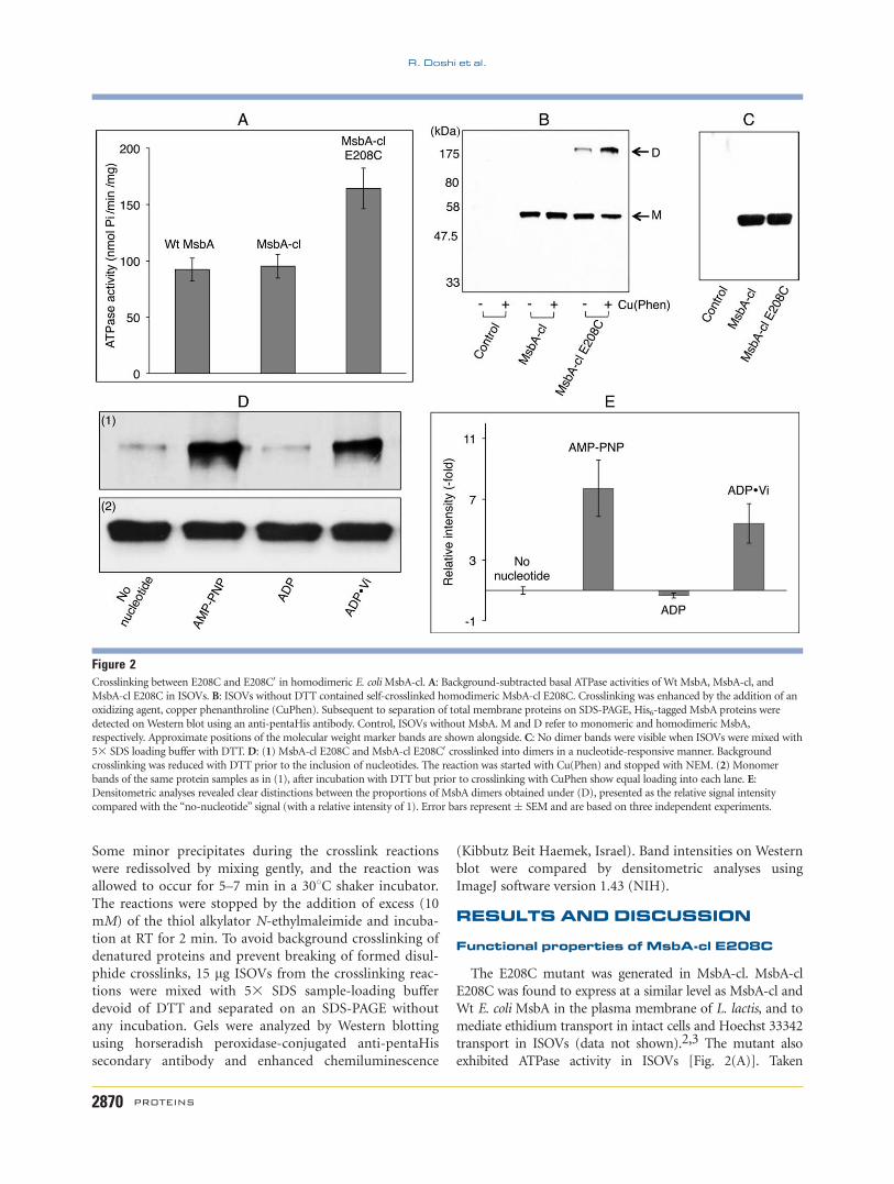
Figure 2Crosslinking between E208C and E208C0 in homodimeric E. coliMsbA-cl. A: Background-subtracted basal ATPase activities of Wt MsbA, MsbA-cl, and
MsbA-cl E208C in ISOVs. B: ISOVs without DTT contained self-crosslinked homodimeric MsbA-cl E208C. Crosslinking was enhanced by the addition of an
oxidizing agent, copper phenanthroline (CuPhen). Subsequent to separation of total membrane proteins on SDS-PAGE, His6-tagged MsbA proteins were
detected on Western blot using an anti-pentaHis antibody. Control, ISOVs without MsbA. M and D refer to monomeric and homodimeric MsbA,
respectively. Approximate positions of the molecular weight marker bands are shown alongside. C: No dimer bands were visible when ISOVs were mixed with
53 SDS loading buffer with DTT. D: (1) MsbA-cl E208C and MsbA-cl E208C0 crosslinked into dimers in a nucleotide-responsive manner. Background
crosslinking was reduced with DTT prior to the inclusion of nucleotides. The reaction was started with Cu(Phen) and stopped with NEM. (2) Monomer
bands of the same protein samples as in (1), after incubation with DTT but prior to crosslinking with CuPhen show equal loading into each lane. E:Densitometric analyses revealed clear distinctions between the proportions of MsbA dimers obtained under (D), presented as the relative signal intensity
compared with the ‘‘no-nucleotide’’ signal (with a relative intensity of 1). Error bars represent� SEM and are based on three independent experiments.
R. Doshi et al.
2870 PROTEINS
together, these observations demonstrate that the MsbA-cl
E208C mutant can be used to study catalytic cycle–depend-
ent conformational changes in MsbA.
MsbA-cl E208C undergoes self-crosslinking
When total membrane proteins obtained from ISOVs
from control cells without MsbA or cells expressing MsbA-
cl or MsbA-cl E208C were separated on an SDS-PAGE gel
(without DTT in the loading buffer) and subsequently ana-
lyzed by Western blotting, specific signals for the MsbA-cl
and MsbA-cl E208C monomers were obtained in addition
to a single band for the MsbA-cl E208C dimer [Fig. 2(B)].
The evidence that the dimer was due to intermonomer cys–
cys crosslinking came from the observation that the dimer
signal increased by exposure to oxidizing agent (copper phe-
nanthroline) whereas the signal of the monomer decreased
in the same sample [Fig. 2(B)]. Furthermore, the dimer
band for the E208C mutant was not observed when ISOVs
were diluted in sample-loading buffer containing DTT, prior
to separation on an SDS-PAGE gel [Fig. 2(C)]. We made
use of this observation to ‘‘reset’’ crosslinked MsbA dimers
that were formed during the preparation of membrane
vesicles, back to uncrosslinked dimers prior to examining
nucleotide dependence of the crosslinking reaction.
Formation of intermolecular E208Ccrosslinks in dimeric MsbA-cl is responsiveto nucleotides
Intermonomer crosslinking of MsbA-cl E208C mono-
mers was tested in the presence of various nucleotides
that represent intermediate steps of the ATPase cycle,
namely AMP-PNP (for ATP-bound, prehydrolysis),
ADP�Vi trapping (mimics the ADP�Pi transition interme-
diate) and ADP (posthydrolysis and Pi release). To initi-
ate crosslinking after nucleotide addition, copper phe-
nanthroline was added at a concentration equal to that
of the DTT. In comparison with nucleotide-free condi-
tions [Fig. 2(D)], we found a strong dimeric signal with
the nonhydrolyzable ATP analog AMP-PNP. This result
[Fig. 2(D)] and additional densitometric analyses of sim-
ilar signals in independent experiments [Fig. 2(E)] indi-
cated that a substantial proportion of MsbA dimers were
trapped in the outward-facing conformation, allowing
the engineered cysteines to come close enough for the
formation of crosslinks [Fig. 2(D)]. A similar result was
obtained with ADP�Vi trapping suggesting that MsbA
remained in a similar overall conformation as observed
with ATP binding [Fig. 2(D)]. However, we obtained a
major reduction in the dimer signal when ADP alone
was included in the crosslinking reaction [Fig. 2(D)].
Densitometry of signals in independent experiments indi-
cated that the signal intensity was below that obtained
for the no-nucleotide control [Fig. 2(E)], suggesting that
ADP binding favors an inward-facing open conforma-
tion. Although the use of DTT and copper phenanthro-
line enhanced signal strength of crosslinking compared
with background, a similar nucleotide dependence of
crosslinking was observed in their absence.
Our crosslinking data address three important issues.
First, the crosslinking data in the presence of AMP-PNP
are in accord with the AMP-PNP-bound outward-facing
crystal structure of Sav186619 and related structures for
MsbA.10 They support the idea that the outward-facing
crystal structure of Sav1866 in the presence of ADP
reflects either an ATP-bound or an ADP�Pi-boundstate.17 On the other hand, it might be possible that
Sav1866 responds to the nucleotides differently than
MsbA. Second, our signals in the presence of ADP sup-
port biochemical and structural evidence that the NBD
dimer is not formed with ADP alone.18 Third, our cross-
linking data in the presence of ATP and Vi are in agree-
ment with the notion that MsbA remains in an outward-
facing conformation during the transition from the ATP-
bound state to the catalytic transition (ADP�Pi bound)
intermediate, consistent with observations on MsbA by
cryo-EM11 and by EPR and DEER spectroscopy.12–15
Our findings point to a sustained dimerization of NBDs
during ATP binding and ADP�Vi trapping, which com-
plements EM analyses on human ABCB125 and MsbA.11
CONCLUSIONS
Ligand-driven conformational movements in ABC
transporters are difficult to predict and study, and static
crystal structures that hypothesize these conformational
movements do require physiological verification. Using
one and the same disulfide crosslinking technique, we
were able to study MsbA dimerization at the major
checkpoints of the ATPase reaction in plasma membrane
vesicles and under conditions in which the transporter is
transport active. This work provides a useful link
between fragmented observations on nucleotide-depend-
ent conformational movements in ABC exporters based
on different techniques (X-ray crystallography, EPR/
DEER spectroscopy, and EM).
ACKNOWLEDGMENTS
The authors thank Dr. Lisa Fagg, Dr. Daniel A. P. Gut-
mann and Ms. Wei Wang for technical support and dis-
cussions, and Dr. Markus Seeger for the kind gift of Pfu
polymerase. R. D. obtained an Overseas Research Stu-
dentship and Cambridge Commonwealth Trust Scholar-
ship, and received an American Alumni Award and Char-
ter Studentship from St. Edmund’s College, Cambridge.
REFERENCES
1. Raetz CRH, Guan Z, Ingram BO, Six DA, Song F, Wang X, Zhao J.
Discovery of new biosynthetic pathways: the lipid A story. J Lipid
Res 2009;50 (Supplement):S103–S108.
Conformational Movements in MsbA
PROTEINS 2871
2. Woebking B, Velamakanni S, Federici L, Seeger MA, Murakami S,
van Veen HW. Functional role of transmembrane helix 6 in drug
binding and transport by the ABC transporter MsbA. Biochemistry
2008;47:10904–10914.
3. Woebking B, Reuter G, Shilling RA, Velamakanni S, Shahi S, Venter
H, Balakrishnan L, van Veen HW. Drug-Lipid A interactions on the
Escherichia coli ABC transporter MsbA. J Bacteriol 2005;187:6363–
6369.
4. Reuter G, Janvilisri T, Venter H, Shahi S, Balakrishnan L, van Veen
HW. The ATP binding cassette multidrug transporter LmrA and
lipid transporter MsbA have overlapping substrate specificities. J
Biol Chem 2003;278:35193–35198.
5. Velamakanni S, Yao Y, Gutmann DAP, van Veen HW. Multidrug
transport by the ABC transporter Sav1866 from Staphylococcus aur-
eus. Biochemistry 2008;47:9300–9308.
6. Doerrler WT, Raetz CRH. ATPase activity of the MsbA lipid flip-
pase of Escherichia coli. J Biol Chem 2002;277:36697–36705.
7. Eckford PDW, Sharom FJ. Functional characterization of Escherichia
coli MsbA: interaction with nucleotides and substrates. J Biol Chem
2008;283:12840–12850.
8. Siarheyeva A, Sharom FJ. The ABC transporter MsbA interacts with
lipid A and amphipathic drugs at different sites. Biochem J
2009;419:317–328.
9. Westfahl KM, Merten JA, Buchaklian AH, Klug CS. Functionally
important ATP binding and hydrolysis sites in Escherichia coli
MsbA. Biochemistry 2008;47:13878–13886.
10. Ward A, Reyes CL, Yu J, Roth CB, Chang G. Flexibility in the ABC
transporter MsbA: alternating access with a twist. Proc Natl Acad
Sci USA 2007;104:19005–19010.
11. Ward A, Mulligan S, Carragher B, Chang G, Milligan RA. Nucleo-
tide dependent packing differences in helical crystals of the ABC
transporter MsbA. J Struct Biol 2009;165:169–175.
12. Zou P, McHaourab HS. Alternating access of the putative substrate-
binding chamber in the ABC transporter MsbA. J Mol Biol
2009;393:574–585.
13. Zou P, Bortolus M, McHaourab HS. Conformational cycle of the ABC
transporter MsbA in liposomes: detailed analysis using double elec-
tron–electron resonance spectroscopy. J Mol Biol 2009;393:586–597.
14. Borbat PP, Surendhran K, Bortolus M, Zou P, Freed JH,
McHaourab HS. Conformational motion of the ABC transporter
MsbA induced by ATP hydrolysis. PLoS Biol 2007;5:e271.
15. Zou P, Mchaourab HS. Increased sensitivity and extended range of
distance measurements in spin-labeled membrane proteins: Q-band
double electron-electron resonance and nanoscale bilayers. Biophys
J 2010;98:L18–L20.
16. Gutmann DAP, Ward A, Urbatsch IL, Chang G, van Veen HW.
Understanding polyspecificity of multidrug ABC transporters:
closing in on the gaps in ABCB1. Trends Biochem Sci 2010;
35:36–42.
17. Dawson RJP, Locher KP. Structure of a bacterial multidrug ABC
transporter. Nature 2006;443:180–185.
18. Smith PC, Karpowich N, Millen L, Moody JE, Rosen J, Thomas PJ,
Hunt JF. ATP binding to the motor domain from an ABC trans-
porter drives formation of a nucleotide sandwich dimer. Mol Cell
2002;10:139–149.
19. Dawson RJP, Locher KP. Structure of the multidrug ABC trans-
porter Sav1866 from Staphylococcus aureus in complex with AMP-
PNP. FEBS Lett 2007;581:935–938.
20. Delano WL. The PyMOL molecular graphics system, version 1.2rl.
Schrodinger: LLC; 2009.
21. Aller SG, Yu J, Ward A, Weng Y, Chittaboina S, Zhuo R, Harrell
PM, Trinh YT, Zhang Q, Urbatsch IL, Chang G. Structure of P-gly-
coprotein reveals a molecular basis for poly-specific drug binding.
Science 2009;323:1718–1722.
22. Weng J-W, Fan K-N, Wang W-N. The conformational transition
pathway of ATP binding cassette transporter MsbA revealed by at-
omistic simulations. J Biol Chem 2010;285:3053–3063.
23. Venter H, Velamakanni S, Balakrishnan L, van Veen HW. On the
energy-dependence of Hoechst 33342 transport by the ABC trans-
porter LmrA. Biochem Pharmacol 2008;75:866–874.
24. de Ruyter PG, Kuipers OP, de Vos WM. Controlled gene expression
systems for Lactococcus lactis with the food- grade inducer nisin.
Appl Environ Microbiol 1996;62:3662–3667.
25. Lee J-Y, Urbatsch IL, Senior AE, Wilkens S. Nucleotide-induced
structural changes in P-glycoprotein observed by electron micros-
copy. J Biol Chem 2008;283:5769–5779.
R. Doshi et al.
2872 PROTEINS