Embed Size (px)
Citation preview

Joana Isabel Silva Rodrigues
BSc Genetics and Biotechnology
Dissecting the role of alpha-synuclein
phosphorylation in Parkinson’s disease
Dissertation for obtaining a Master’s Degree in
Molecular Genetics and Biomedicine
Supervisor: Sandra Tenreiro, PhD, CEDOC – Chronic Diseases Research
Center
September 2017

II
III
Dissecting the role of alpha-synuclein phosphorylation in Parkinson’s Disease
Copyright © Joana Isabel Silva Rodrigues, Faculdade de Ciências e Tecnologia, Universidade Nova
de Lisboa
A Faculdade de Ciências e Tecnologia e a Universidade Nova de Lisboa têm o direito, perpétuo e
sem limites geográficos, de arquivar e publicar esta dissertação através de exemplares impressos
reproduzidos em papel ou de forma digital, ou por qualquer outro meio conhecido ou que venha a ser
inventado, e de a divulgar através de repositórios científicos e de admitir a sua cópia e distribuição
com objetivos educacionais ou de investigação, não comerciais, desde que seja dado crédito ao autor
e editor.

IV

Dissecting the role of alpha-synuclein phosphorylation in Parkinson’s Disease
V
Acknowledgments
The work presented here was only possible with the great support I received during this year from
the people here mentioned. Thereby, I would like to express my immense gratitude to the followings:
Firstly, I would like to thank Doctor Sandra Tenreiro, for all the support, encouragement, and
kindness. Thank you for mentoring me and for all the amazing opportunities you gave me, allowing me
to grow as a scientist and as person. To Professor Tiago Outeiro, for giving me the opportunity to work
in the Cellular and Molecular Neuroscience Unit and for providing all the necessary conditions to
develop my work. To Doctor Hugo Vicente Miranda, for his amazing critical spirit, incentive and
support.
I would like to thank CEDOC - NOVA Medical School, as institution and as a community, for
receiving me with open arms. Thank you to everyone who assisted me during my experiments. It was
a pleasure to work in such a motivational environment.
A very special thank you to “Morsas”, my girls. To Ana, our grandma, for all the amazing support,
all the critics and good advices, all the help and care. Thank you for putting up with us and for taking
care of us. Thank you for sharing your experiences, making me grow and helping me understand this
brave new world. The best advice I got from you was “Nos péis não”, and I will remember it for the rest
of my days. To Babita, our jester and my partner in crime. Hamilton brought us together and MCA kept
our flame alive. Thank you for always, and I mean ALWAYS, laughing with me and making me laugh.
Your puns will always make me happy. ChemiDoc will not be the same without you. You were the best
plate-shaking person during my yeast transformations and always will be, no one can ever replace
you. Thank you for the best roadtrips ever, I will never hear “Uptown Girl” and “It’s Not Unusual”
without you popping up in my head. May our childhood bands cheer you up forever. I hope we will be
together until we die and, when we die, we will be ghost friends and haunt Ana. To Gabigoo, our sun,
thank you for always being there right by my side, literal and figuratively. We shared so much during
this year, for the good and the bad, we were always there for each other and always will be. Our
friendship came from nothing and became one of the most precious things I have. Thank you for all
the laughs, all the good times and all the care. Despite the “All by Myself” jokes, I want you to know
that I will be there for you at any time. More recently, thank you for all the goodies you bring home; I
figured that your plan is to make me gain weight so the plane cannot take off. To the three of you,
thank you for making this year as amazing as it was. Thank you for your friendship and for being my
second family during this year, sometimes even my first family. I have not enough words to thank you
properly. May this friendship last a lifetime.
To all my friends, either from Torres Novas, UTAD or FCT, for always being there for me, and for
sharing my happiness. To Ana Silva, my Azorean friend, my roomate, for our friendship, which grew
exponenially over this year. For all the encouragement and for pushing me to do my best. I have a
special respect for you, I will be always pride of what you have accomplished. A gigantic thank you to
your cooking skills! See you in Azores! To “Gordos & Gorduchos” my family at UTAD, our friendship
will always be my most random friendship, our inside jokes, all the memes shared, all the pictures sent
and all the random conversations. May this never end, may this family be together for eternity. See

Dissecting the role of alpha-synuclein phosphorylation in Parkinson’s Disease
VI
you soon. A special thank you to Rita, Cláudia, João and Patrick. Each and every one of you
contributed to my happiness in your own peculiar ways, by being there with me or simply by always
putting a smile in my face, even when the distance between us was enormous and our schedules
were completely unsynchronized. It was a pleasure to have you with me during one more year and,
hopefully, in the years to come. I will have you in my heart wherever I go.
Last, but not least, I would like to thank my family, specially my parents, Susana and António, for
all the efforts they made and continue to make so I can pursuit this dream. Thank you for all the faith
you always had in me, for all the support and patience. The sacrifices made were gigantic, and I hope
that I have made you proud and that I will have the opportunity to give you as much as you gave me.
Without you, my dream would never become a reality. No matter how far I will go, I will always be
there with you. To ‘Vó Alice, there were so many times that I thought about you. I will always
remember the excitement with which you used to ask about my studies, and your smile every time I
explained something to you. I know that you would be proud today. We miss you every single day.
“There’s a million things I haven’t done. But just you wait”
Lin-Manuel Miranda

Dissecting the role of alpha-synuclein phosphorylation in Parkinson’s Disease
VII
Resumo
A doença de Parkinson é a segunda doença neurodegenerativa mais comum, caracterizada pela
agregação de alfa-sinucleína nos neurónios dopaminérgicos da substantia nigra pars compacta e,
consequentemente, pela morte neuronal. Os neurónios dopaminérgicos sobreviventes apresentam
depósitos de alfa-sinucleína, denominados Corpos de Lewy.
A origem da doença de Parkinson pode ser esporádica ou ter uma causa genética. Doença de
Parkinson associada ao SNCA, gene codificante da alfa-sinucleína, apresenta um padrão hereditário
autossómico dominante. Atualmente, seis mutações foram identificadas no gene SNCA. A alfa-
sinucleína é suscetível de sofrer diversas modificações pós-trasducionais, sendo que a fosforilação é
uma modificação de elevada importância, pois é a principal modificação encontrada nos agregados,
podendo ter um papel na regulação da estrutura, oligomerização e toxicidade da alfa-sinucleína.
Porém, ainda está a ser debatido se a fosforilação promove ou previne estas mesmas propriedades,
sendo que modelos celulares distintos apresentam resultados contraditórios.
Saccharomyces cerevisiae é um modelo celular estabelecido para o estudo da doença de
Parkinson, sendo que permite reproduzir diversas características, tais como agregação e toxicidade
celular. Neste estudo, plasmídeos multicópia contendo diferentes variações de alfa-sinucleína
fundidas com GFP sob a regulação do promotor GAL1 foram transformados em levedura. Os efeitos
da fosforilação de diversos resíduos na formação de inclusões de alfa-sinucleína e na sua toxicidade
foram avaliados. As seis mutações no gene SNCA (A30P, E46K, H50Q, G51D, A53E e A53T) foram
caracterizadas quanto à formação de inclusões, toxicidade, níveis proteicos, degradação, acidificação
vacuolar e produção de ROS.
A avaliação dos efeitos da fosforilação revelou que tanto o bloqueio como a mímica da
fosforilação da S129 superam os efeitos da fosforilação da S87, com a alfa-sinucleína S129A
promovendo um aumento da toxicidade, enquanto a alfa-sinucleína S129D apresenta um efeito
protetor. Em relação à fosforilação dos resíduos de tirosina, o bloqueio da fosforilação do resíduo
Y125 provocou uma diminuição na degradação proteica, resultando num aumento de toxicidade,
comprovado pelo ensaio de viabilidade celular de citometria de fluxo. As mutações familiares A30P,
G51D e A53E demonstraram uma toxicidade inferior à alfa-sinucleína WT, enquanto que as mutações
H50Q e A53T apresentaram efeitos semelhantes ao WT. O mutante E46K foi o único que exibiu uma
toxicidade mais elevada. Seria de esperar uma maior toxicidade por parte das alfa-sinucleínas
mutantes, tendo em conta que estas provocam um parkinsonismo mais severo e um aparecimento
precoce da doença. A divergência obtida poderá dever-se ao modelo celular usado, uma vez que as
leveduras têm um ambiente celular distinto dos neurónios dopaminérgicos afetados na doença de
Parkinson.
Palavras-chave: Doença de Parkinson, alfa-sinucleína, fosforilação, mutações familiares, S.
cerevisiae

Dissecting the role of alpha-synuclein phosphorylation in Parkinson’s Disease
VIII
Dissecting the role of alpha-synuclein phosphorylation in Parkinson’s Disease
IX
Abstract
Parkinson’s disease is the second most common neurodegenerative disorder, characterized by
an aggregation of alpha-synuclein in the dopaminergic neurons of the substantia nigra pars compacta
and, consequently, neuronal death. Surviving dopaminergic neurons present alpha-synuclein deposits,
known as Lewy Bodies.
Parkinson’s disease can have sporadic or genetic cause. Parkinson’s disease associated with
SNCA, the alpha-synuclein encoding gene, shows an autosomal dominant inheritance pattern. Until
now, six SNCA mutations were identified. Alpha-synuclein is prone to suffer several post-translational
modifications, critical for its function and structural properties. Phosphorylation is a modification of
major importance, since it it’s the modification most commonly found in aggregates; it may play an
important role in regulating alpha-synuclein structure, oligomerization and toxicity. However, whether
phosphorylation promotes or prevents these properties is still under debate, with distinct cellular
models presenting contradictory results.
Saccharomyces cerevisiae cells are an established model to study Parkinson’s disease, since it
can reproduce several features, such as protein aggregation and cellular toxicity. For this study,
multicopy plasmids containing different alpha-synuclein variants fused with GFP under the regulation
of a GAL1 promoter were transformed in yeast cells. Effects of phosphorylation at different residues in
alpha-synuclein inclusion formation and cell toxicity were evaluated. Furthermore, the six known
SNCA mutations (A30P, E46K, H50Q, G51D, A53E and A53T) were characterized regarding inclusion
formation, cell toxicity, protein levels, protein clearance, vacuole acidification and ROS production.
Evaluation of phosphorylation effects revealed that either blockage or mimic of S129
phosphorylation always overcome the S87 phosphorylation effect, with S129A increasing toxicity,
while S129D promotes protection. Regarding tyrosine residues phosphorylation, Y125 phosphorylation
blockage impaired protein clearance, increasing its toxicity. Concerning familial mutations, A30P,
G51D and A53E aSyn, point to a lower toxicity than WT aSyn, while the H50Q and A53T mutations
presented results very similar to WT, and E46K aSyn was the only one who presented higher toxicity.
This divergence is probably due to the cellular model used, since yeast cells present a distinct cellular
environment than the affected dopaminergic neurons in PD.
Keywords: Parkinson’s Disease, alpha-synuclein, phosphorylation, familial mutations, S.
cerevisiae

Dissecting the role of alpha-synuclein phosphorylation in Parkinson’s Disease
X

Dissecting the role of alpha-synuclein phosphorylation in Parkinson’s Disease
XI
Index
Acknowledgments ............................................................................................................................ V
Resumo .......................................................................................................................................... VII
Abstract ........................................................................................................................................... IX
Index ................................................................................................................................................ XI
Figure Index................................................................................................................................... XIII
Table Index ..................................................................................................................................... XV
List of abbreviations and acronyms .............................................................................................. XVII
1 Introduction ...............................................................................................................................1
1.1 Neurodegenerative diseases as proteinopathies ............................................................ 1
1.2 Synucleinopathies and Parkinson’s Disease ................................................................... 2
1.3 Alpha-synuclein and the synuclein family ........................................................................ 3
1.3.1 Alpha-synuclein – a multifunctional protein ................................................................. 5
1.3.2 SNCA Familial Mutations ............................................................................................. 6
1.3.3 Alpha-synuclein Post-Translational Modifications: Phosphorylation ........................... 6
1.3.4 Mechanisms of alpha-synuclein toxicity ...................................................................... 8
1.3.5 Alpha-synuclein clearance ......................................................................................... 11
1.4 CDNF - a non-conventional neurotrophic factor ............................................................ 15
1.5 Yeast as a cellular model to study Parkinson’s Disease ............................................... 16
1.6 Aims ............................................................................................................................... 18
2 Materials and Methods .......................................................................................................... 19
2.1 Yeast Strains and Plasmids ........................................................................................... 19
2.2 Escherichia coli manipulation ........................................................................................ 19
2.2.1 E. coli growth conditions ............................................................................................ 19
2.2.2 E. coli transformation ................................................................................................. 19
2.2.3 Plasmid DNA extraction ............................................................................................. 19
2.3 Saccharomyces cerevisiae manipulation ...................................................................... 20
2.3.1 S. cerevisiae growth conditions ................................................................................. 20
2.3.2 Yeast Transformation ................................................................................................ 21
2.4 Molecular biology: Plasmids construction ..................................................................... 22
2.4.1 Yeast plasmid constructions using site-directed mutagenesis .................................. 22

Dissecting the role of alpha-synuclein phosphorylation in Parkinson’s Disease
XII
2.4.2 CDNF plasmids construction by Gateway Recombination Cloning Technology ....... 23
2.5 Cell Viability Assays ...................................................................................................... 25
2.5.1 Spotting Assay ........................................................................................................... 25
2.5.2 Flow Cytometry .......................................................................................................... 25
2.6 Detection of superoxide radical production ................................................................... 25
2.7 Evaluation of vacuole acidification................................................................................. 26
2.8 Protein subcellular localization through fluorescence microscopy ................................ 26
2.9 Quantification of protein expression levels .................................................................... 27
2.9.1 Protein Extraction and Quantification ........................................................................ 27
2.9.2 Protein detection through Western Blot ..................................................................... 27
2.10 Statistical Analysis ......................................................................................................... 28
3 Results and Discussion ......................................................................................................... 29
3.1 Plasmid constructions .................................................................................................... 29
3.2 Evaluation of the phosphomutants effects in inclusion formation and cell viability ....... 29
3.3 Evaluation of aSyn familial mutations in aSyn inclusion formation and cell toxicity ...... 33
3.4 Phosphorylation levels of different aSyn variants .......................................................... 37
3.5 Protein Clearance .......................................................................................................... 39
3.5.1 Assessment of Rsp5 effect in inclusion clearance of each aSyn familial mutant ...... 41
3.6 Vacuole acidification evaluation .................................................................................... 42
3.7 Evaluation of ROS and superoxide production ............................................................. 44
3.8 CDNF effects on aSyn toxicity ....................................................................................... 45
3.8.1 Plasmid Construction through Gateway Recombination Technique ......................... 45
3.8.2 CDNF effects on cell viability, inclusion formation and aSyn expression levels ....... 45
4 Concluding remarks and future perspectives ........................................................................ 48
5 Bibliography ........................................................................................................................... 51
6 Supplementary Data .............................................................................................................. 65

Dissecting the role of alpha-synuclein phosphorylation in Parkinson’s Disease
XIII
Figure Index
Figure 1.1 Neurodegenerative diseases have in common the presence of misfolded and
aggregated proteins and, consequently, the presence of protein deposits. ............................................ 1
Figure 1.2 Parkinson’s disease motor and non-motor features. ...................................................... 3
Figure 1.3 SNCA can be divided into three regions. ........................................................................ 4
Figure 1.4 aSyn aggregation process culminates with the formation of Lewy Bodies. .................... 4
Figure 1.5 Schematic representation of the identified aSyn familial mutations. .............................. 6
Figure 1.6 Schematic representation of the aSyn residues prone to suffer phosphorylation. ......... 9
Figure 1.7 aSyn affects mitochondrial function by interacting with mitochondrial complex I. ........ 10
Figure 1.8 Dopamine synthesis is inhibited by aSyn. .................................................................... 12
Figure 1.9 Ubiquitin-proteasome system pathway. ........................................................................ 13
Figure 1.10 Vacuolar ATPase activity involved in lysosomal pH maintenance. ............................ 15
Figure 1.11 Basic schematic representation of the PI3K-Akt signalling pathway. ......................... 16
Figure 1.12 Effects of aSyn expression in yeast cells. ................................................................... 18
Figure 2.1 Schematic representation of the site-directed mutagenesis protocol. .......................... 23
Figure 2.2 Schematic representation of the Gateway Recombination Cloning Technique
procedure............................................................................................................................................... 24
Figure 3.1 Assessment of aSyn inclusion formation and respective clearance for each aSyn
phosphomutant through fluorescence microscopy. ............................................................................... 30
Figure 3.2 Evaluation of toxicity induced by each aSyn phosphomutant by spotting assay. ......... 32
Figure 3.3 Evaluation of toxicity induced by aSyn phosphomutants through flow cytometry using
PI. .......................................................................................................................................................... 33
Figure 3.4 Assessment of aSyn inclusion formation for each aSyn variant through fluorescence
microscopy. ........................................................................................................................................... 34
Figure 3.5 Evaluation of toxicity induced by each aSyn variant by spotting assay. ....................... 35
Figure 3.6 Evaluation of cytotoxicity induced by aSyn familial mutants through flow cytometry
using PI after 6 hours of protein induction. ............................................................................................ 36
Figure 3.7 Evaluation of cytotoxicity induced by aSyn familial mutants through flow cytometry
using PI after 6 hours of protein induction. ............................................................................................ 37
Figure 3.8 Total and phosphorylated aSyn levels after 6 hours of protein induction in W303.1A
yeast cells. ............................................................................................................................................. 38
Figure 3.9 Phosphorylated aSyn levels after 6 hours of protein induction in BY4741 yeast cells. 38
Figure 3.10 Assessment of aSyn inclusions clearance through fluorescence microscopy. .......... 39
Figure 3.11 Assessment of aSyn inclusions clearance through fluorescence microscopy after 13
hours of protein clearance. .................................................................................................................... 40
Figure 3.12 Clearance of total and phosphorylated aSyn is similar between distinct aSyn
mutations. .............................................................................................................................................. 41
Figure 3.13 Assessment of Rsp5 effect in aSyn inclusions clearance through fluorescence
microscopy after 3 and 6 hours of protein clearance. ........................................................................... 42

Dissecting the role of alpha-synuclein phosphorylation in Parkinson’s Disease
XIV
Figure 3.14 Evaluation of vacuole acidification in yeast cells transformed with each aSyn variant
after 6 and 13 hours of protein induction. .............................................................................................. 43
Figure 3.15 Evaluation of ROS production in yeast cells transformed with each aSyn variant after
6 and 13 hours of protein induction. ...................................................................................................... 44
Figure 3.16 Cell viability and inclusion formation assessment through spotting assay and
fluorescence microscopy, respectively. ................................................................................................. 46
Figure 3.17 Evaluation of total aSyn levels in the presence and absence of each CDNF plasmid.
............................................................................................................................................................... 47
Figure 6.1 Sequence alignments between aSyn WT sequence and the described aSyn mutants.
............................................................................................................................................................... 65
Figure 6.2 Sequence alignments between CDNF sequence and the described plasmids. ........... 66
Dissecting the role of alpha-synuclein phosphorylation in Parkinson’s Disease
XV
Table Index
Table 1.1 Description of aSyn familial mutations and respective phenotype observed. .................. 7
Table 2.1 Description of yeast strains used in this project ............................................................. 19
Table 2.2 Description of the yeast plasmids used in this project ................................................... 20
Table 2.3 Culture media used for yeast growth ............................................................................. 21
Table 2.4 Description of plasmids and primers used for site-directed mutagenesis...................... 22
Table 2.5 Description of the PCR reactions performed to amplify the CDNF sequence ............... 23
Table 2.6 PCR Reaction Mix used for CDNF amplification ............................................................ 23
Table 2.7 Positive and negative controls used for the membrane integrity assessment through
flow cytometry ........................................................................................................................................ 25
Table 2.8 Positive and negative controls used for the superoxide radical production assessment
through flow cytometry .......................................................................................................................... 26
Table 2.9 Description of the antibodies used in this project ........................................................... 28
Table 3.1 Phosphomutants used in this study. .............................................................................. 29
Table 4.1 Resume table of the results obtained in distinct assays for each aSyn familial mutant. 49

Dissecting the role of alpha-synuclein phosphorylation in Parkinson’s Disease
XVI

Dissecting the role of alpha-synuclein phosphorylation in Parkinson’s Disease
XVII
List of abbreviations and acronyms
AD Alzheimer’s Disease
Amp Ampicillin
aSyn Alpha-synuclein
Atg Autophagy-Related Protein
ATP Adenosine Triphosphate
bSyn Beta-synuclein
BAX Bcl-2-Like Protein X
BSA Bovine Serum Albumine
CDNF Cerebral Dopaminergic Neurotrophic Factor
CSM Complete Supplement Media
DA Dopamine
DDC DOPA Decarboxylase
DHE Dihydroethidium
E1 Ubiquitin-Activated Enzyme
E2 Ubiquitin Conjugating Enzyme
E3 Ubiquitin-Protein Ligase
E. coli Escherichia coli
EDTA Ethylenediamine Tetracetic Acid
ER Endoplasmic Reticulum
Gal Galactose
GFP Green Fluorescent Protein
Glu Glucose
gSyn Gama-synuclein
HECT Homologous to the E6-AP Carboxyl Terminus type
Kan Kanamycin
LB Lewy Body
LB media Luria Broth media
LiAc Lithium Acetate
Dissecting the role of alpha-synuclein phosphorylation in Parkinson’s Disease
XVIII
LN Lewy Neurites
L-DOPA L-3,4-dihydroxyphenylalanine
MAM Mitochondria-Associated ER Membranes
MANF Mesencephalic Astrocyte-Derived Neurotrophic Factor
MPTP 1-methyl-4-phenyl-1,2,3,6-tetrahydropyridine
mTOR Mammalian Target Of Rapamycin
NAC Non-Amyloid-beta Component
NSF N-Ethylmaleimide Sensitive Factor
NTF Neurotrophic Factor
OD Optical Density
OE Over-expression
pAG pAdvanced Gateway
PBS Phosphate Buffered Saline
PCR Polymerase Chain Reaction
PD Parkinson’s Disease
PEG Polyethylene Glycol
pH Potential of Hydrogen
PI Propidium Iodide
PIP3 Phosphatidylinositol 3,4,5-triphosphate
PI3K PI3 Kinase
PTEN Phosphatidylinositol 3-phosphatase
Raf Raffinose
RING Really Interesting New Gene
ROS Reactive Oxygen Species
RT Room Temperature
S Serine
S. cerevisiae Saccharomyces cerevisiae
SDS Sodium Dodecyl Sulfate
SDS-PAGE Sodium Dodecyl Sulfate Polyacrylamide Gel Electrophoresis
SNARE Soluble NSF Attachment Receptor

Dissecting the role of alpha-synuclein phosphorylation in Parkinson’s Disease
XIX
TBS Tris Buffered Saline
TBS-T Tris Buffered Saline – Tween 20
TE Tris – EDTA
TH Tyrosine Hydroxylase
UBC Ubiquitin Conjugating Enzyme
UPR Unfolded Protein Response
UPS Ubiquitin-Proteasome System
v-ATPase Vacuolar-ATPase
WT Wild-Type
Y Tyrosine
YNB Yeast Nitrogen Base
YPD Yeast Extract Peptone Dextrose
6-OHDA 6-hydroxydopamine

Dissecting the role of alpha-synuclein phosphorylation in Parkinson’s Disease
XX
Dissecting the role of alpha-synuclein phosphorylation in Parkinson’s Disease
1
1 Introduction
1.1 Neurodegenerative diseases as proteinopathies
The prevalence of chronic age-related diseases is growing due to the continuing demographic
shift of worldwide population towards an older society (Bourdenx et al., 2016). The neurodegeneration
process is long and involves the loss of brain and spinal cord cells, moreover, once the patient
experiences symptoms, the neurodegeneration is already advanced (Uversky, 2009).
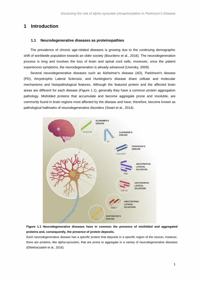
Several neurodegenerative diseases such as Alzheimer's disease (AD), Parkinson's disease
(PD), Amyotrophic Lateral Sclerosis, and Huntington's disease share cellular and molecular
mechanisms and histopathological features. Although the featured protein and the affected brain
areas are different for each disease (Figure 1.1), generally they have a common protein aggregation
pathology. Misfolded proteins that accumulate and become aggregate prone and insoluble, are
commonly found in brain regions most affected by the disease and have, therefore, become known as
pathological hallmarks of neurodegenerative disorders (Swart et al., 2014).
Figure 1.1 Neurodegenerative diseases have in common the presence of misfolded and aggregated
proteins and, consequently, the presence of protein deposits.
Each neurodegenerative disease has a specific protein that deposits in a specific region of the neuron, however,
there are proteins, like alpha-synuclein, that are prone to aggregate in a variety of neurodegenerative diseases
(Eftekharzadeh et al., 2016).
Dissecting the role of alpha-synuclein phosphorylation in Parkinson’s Disease
2
These aggregated proteins have a very ordered structure and often exhibit characteristics of
amyloid-like protein assemblies (Swart et al., 2014; Eftekharzadeh et al., 2016). In this state, proteins
develop elongated fibers with backbones consisting of many-stranded β-sheets (Eisenberg & Jucker,
2012). Both the structure and the biophysical properties of amyloid fibrils are similar across different
amyloid diseases, even though each disease involves the amyloidogenic aggregation of one or many
different proteins. Several studies reported that the formation of amyloid fibrils is associated with the
loss of protein function or a toxic gain of function (Eftekharzadeh et al., 2016).
Although each disease has specific protein aggregates as hallmarks, there is an overlapping
pattern e.g., PD shares the presence of alpha-synuclein (aSyn) inclusion bodies with several other
synucleinopathies (Uversky, 2009). Therefore, despite the diversity of proteins involved in each
disease, there are common disease mechanisms, indicating typical pathological patterns associated
with neuronal decline (Swart et al., 2014).
The main risk factor for all neurodegenerative diseases is still aging, suggesting that a decline in
protein quality control associated with aging may contribute to misfolding and, consequently,
aggregation of proteins in specific populations of brain cells (Chen et al., 2011).
1.2 Synucleinopathies and Parkinson’s Disease
Synucleinopathies exhibit, as hallmark, the presence of aggregated aSyn deposits in intracellular
inclusions, but display differences in symptomatology, brain areas and cell types affected, and
anatomical sites of onset (Dehay & Fernagut, 2016). Synucleinopathies include several diseases such
as PD, amyotrophic lateral sclerosis, neurodegeneration with brain iron accumulation type 1, pure
autonomic failure, multiple system atrophy and dementia with Lewy bodies (Uversky, 2009;
Tagliafierro, 2016).
PD is the second most prevalent neurodegenerative disorder, following AD, with a prevalence of
1% to 2% in people over 65 years of age (Lau & Breteler, 2006; Massano & Ferreira, 2016). PD is
considered a slowly progressive neurodegenerative disease which results from the interaction
between genetic and environmental factors wherein multiple neuroanatomical areas are implicated
leading to a range of features that begin years before diagnosis. Most PD cases are sporadic;
however, approximately 5% to 10% of PD patients have monogenic forms of the disease, exhibiting a
Mendelian type of inheritance (Kalia et al., 2015).
The pathological hallmark of PD is the progressive loss of dopaminergic neurons in the substantia
nigra pars compacta (SNpC) and the presence of Lewy Bodies (LB), as well as, Lewy Neurites (LN) in
the surviving neurons (Lau & Breteler, 2006; Massano & Ferreira, 2016). The resultant dopamine (DA)
deficiency leads to a movement disorder characterized by classical parkinsonian motor features,
described in figure 1.2 (Kalia et al., 2015).
The axons from the SNpC and other key dopaminergic areas in the brain project extensively to
form four main pathways namely, the mesocortical, mesolimbic, nigrostriatal and tuberoinfundibular
pathways, which are responsible for mediating several non-motor features of PD (Chaudhuri &
Schapira, 2009; Kalia et al. 2015). The motor features are identified relatively late in the pathological
process, when approximately 70% of the dopaminergic neurons are already dead (Uversky, 2009).

Dissecting the role of alpha-synuclein phosphorylation in Parkinson’s Disease
3
Figure 1.2 Parkinson’s disease motor and non-motor features.
The dopamine deficiency resultant from the loss of dopaminergic neurons in the SNpC leads to a series of motor
features. Moreover, four main pathways can also be affected leading to the development of non-motor features.
PD management involves primarily the symptomatic treatment with drugs that increase DA
concentration or stimulate DA receptors (Kalia et al., 2015). Despite these symptomatic treatment
efficiency, nowadays, there are no drugs that slow or change the disease course (Noyce et al., 2016).
The main PD risk factors are age superior than 50 years and male gender, being men 1.5 times
more likely to develop the disease (Wooten et al., 2004). Unlike the females, where prevalence tends
to stabilize over time, in males the incidence keeps rising after the age of 80 years (Hirsch & Steeves,
2016). In women, loss of estrogen production can also compromise protective effects and, some
evidences suggest that early menopause, ovary removal or hysterectomy increases risk in women,
similarly to that seen in men. Environmental factors and ethnicity are also considered risk factors for
the disease development. The prevalence of PD seems to be higher in Europe, North America and
South America (Dexter & Jenner, 2013; Kalia et al. 2015). Overall, age remains the major risk factor
for PD for the majority of the patients (Kalia et al., 2015).
1.3 Alpha-synuclein and the synuclein family
The synuclein family is comprised of aSyn, β-synuclein (bSyn) and γ-synuclein (gSyn), which are
small proteins highly conserved and highly expressed in neurons. (George, 2001; Sung & Eliezer,
2007). aSyn and bSyn are widely expressed in the central nervous system including neocortex,
striatum, hippocampus, thalamus, and cerebellum and are absent from peripheral tissues. In the other
hand, gSyn is mostly found in the peripheral nervous system but can also be expressed in other
tissues, such as in the brain and ovarian and breast cancer. (Lavedan, 1998; Sung & Eliezer, 2007).
SNCA, the encoding gene of aSyn, which is located on chromosome 4q22.1, has six exons
encoding a 140 amino-acid protein (Ozansoy & Ba, 2013). Genome wide association studies and
other gene-based approaches have implicated SNCA as a very significant genetic risk factor for some
synucleinopathies. Nevertheless, while coding missense mutations and multiplication of the SNCA
locus leads to familiar PD, the exact variants that contribute to sporadic PD remains unclear
(Tagliafierro, 2016).

Dissecting the role of alpha-synuclein phosphorylation in Parkinson’s Disease
4
aSyn is an intrinsically disordered protein that has a molecular weight of 14 kDa when unfolded
(Bourdenx et al., 2016). Its structure can be divided into three different regions as demonstrated in
Figure 1.3: (i) residues between 1 and 60, the N-terminal domain, code for amphipathic α-helices; (ii)
residues 61-95, the central region, contains the hydrophobic and highly amyloidogenic non-amyloid-
beta component (NAC) region and, at last, (iii) the residues 96-140 make up the C-terminal region rich
in acidic residues and prolines. The N-terminal and central regions comprise a membrane-binding
domain, while the C-terminal region is thought to be responsible for the protein-protein and protein-
small molecule interactions (Breydo et al., 2012). Additionally, the central domain is required for
oligomerization and fibrillation of the protein, since if this region is deleted, aSyn loses its ability to
form amyloid fibrils (Giasson et al., 2001).
Figure 1.3 aSyn is a small protein divided into three regions.
Firstly, a N-terminal region and a NAC domain, both comprising a membrane-binding domain and, lastly, a C-
terminal region putatively responsible for the protein interaction with proteins and small molecules.
In terms of structure, aSyn is naturally unfolded when in its monomeric form, however, under
certain conditions, it is prone to aggregate (Tyson et al., 2016). The process of aggregation begins
with the formation of relatively soluble oligomers that can self-assemble into insoluble amyloid fibrils,
resulting in the formation of deposits (Figure 1.4; Mukaetova-Ladinska & McKeith, 2006). These
deposits were first identified by Fritz Heinrich Lewy, which he named “Negrishen Körperchen” and are
currently known as LB. PD and DLB are defined by the existence of intracytoplasmic LB in surviving
neurons soma and LN in processes of degenerating nerve cells. (Dehay & Fernagut, 2016; Peelaerts
& Baekelandt, 2016).
Figure 1.4 aSyn aggregation process culminates with the formation of Lewy Bodies.
aSyn is prone to form intracellular aggregates under certain, yet not completely understood conditions. The
aggregation process starts with the formation of oligomeric structures, following fibril formation, and ending with
the formation of inclusion bodies, known as Lewy Bodies.
Since aSyn comprises an intrinsically disordered region, corresponding to the C-terminal region, it
is considered an intrinsically disordered protein (Bourdenx et al. 2016; Eftekharzadeh et al. 2016).
These proteins seem to be highly sensitive to misfolding and aggregation, probably because of their
unfolded nature (Eftekharzadeh et al., 2016). They do not fold into a specific secondary structure, as

Dissecting the role of alpha-synuclein phosphorylation in Parkinson’s Disease
5
well as tertiary structure when under normal conditions, staying in a partially folded or even unfolded
state (Oldfield & Dunker, 2014).
According with several studies, the major theory for aSyn toxicity is its tendency to aggregate.
However, it is still unclear whether the toxicity is due to the LB or to the transient oligomeric species
that are formed along the aggregation process (Volles & Lansbury Jr., 2003).
1.3.1 Alpha-synuclein – a multifunctional protein
The exact function of aSyn is still unknown, since it is involved in several cellular processes and
molecular interactions, especially at presynaptic sites. Although, it is thought to play a role in
maintaining a supply of synaptic vesicles, axonal transport and DA synthesis and metabolism (Sidhu
et al., 2004; Dunning et al., 2012).
aSyn is mostly present at presynaptic terminals and a role in synaptic plasticity has already been
envisioned. Indeed, aSyn may regulate synaptic vesicle mobilization at nerve terminals. Cabin et al.
found that cultured hippocampal neurons, where aSyn expression was knocked down, had fewer
synaptic vesicles when compared with the control neurons, particularly in the reserve pool (Cabin et
al., 2002). Similarly, the levels of synapsin, a protein essential for synaptic vesicle recycling,
decreased in cells where aSyn was depleted, suggesting a role of aSyn in genesis or maintenance of
the reserve pool of synaptic vesicles (Sidhu et al., 2004).
Regarding the aSyn effects on synapses, other studies demonstrated that aSyn has a role in the
assembly of the soluble N-ethylmaleimide sensitive factor (NSF) attachment receptors (SNARE)-
complex. This process is essential for several membrane fusion events, indicating a function of aSyn
in synaptic vesicle docking and fusion. The SNARE-complex is responsible for zipping the vesicles
onto the plasma membrane, which will then undergo fusion followed by synaptic stimulation
(Lautenschläger et al., 2017). However, there is still controversy on how aSyn acts on this complex. A
study developed by Burré et al. demonstrated that aSyn induces an increase in SNARE-complex
assembly (Burré et al., 2010), while others authors defend that aSyn has a negative regulatory
function (Darios et al., 2010; Thayanidhi et al., 2010).
Recently, aSyn was proposed to be a curvature-sensing and stabilizing protein, i.e., aSyn is able
to sense membrane curvature and stabilize it by inserting its amphipathic helix into the lipid surface
(Varkey et al. 2010; Middleton & Rhoades, 2010; Lautenschläger et al. 2017). Nuclear magnetic
resonance studies revealed that aSyn could bind to lipid membrane through its N-terminal and NAC
domain, possibly tethering two vesicles together or vesicles to the plasma membrane through a
double-anchor mechanism, facilitating exocytosis and endocytosis (Fusco et al., 2016). Regarding
neurotransmitters release, aSyn function is also debatable, with studies showing an inhibitory effect on
neurotransmitter release (Larsen et al., 2006; Gureviciene et al., 2007; Nemani et al., 2010; Scott et
al., 2010; Wu et al., 2010; Janezic et al., 2013) and others showing a neurotransmitter release
increase (Liu et al., 2004; Watson et al., 2009). Finally, aSyn has also been suggested to have an
effect in axonal transport of synaptic vesicles by interacting with numerous proteins that can bind or
are a part of the cytoskeleton (Sidhu et al., 2004).

Dissecting the role of alpha-synuclein phosphorylation in Parkinson’s Disease
6
1.3.2 SNCA Familial Mutations
PD associated with SNCA shows an autosomal dominant inheritance pattern with an early onset
and typically develops rapidly. Triplication of the genomic region containing SNCA has been described
as a cause of PD in several families; nevertheless, SNCA duplications have been reported as a more
common cause of familial and sporadic PD. These duplications and triplications of the SNCA locus
cause early-onset PD with the severity, as well as the age of onset, correlating with the number of
SNCA copies, suggesting a gene-dose effect (Hernandez et al., 2016).Until now, six mutations have
already been identified, being the A53T mutation the first acknowledged, followed by A30P and E46K.
On the other hand, the G51D, A53E and H50Q mutations were recently identified (Table 1.1; Pasanen
et al. 2014). The known mutations are present in the N-terminal region of the protein (Figure 1.5),
highlighting the importance of this domain in the pathological dysfunction of aSyn (Xu & Pu, 2016).
Figure 1.5 Schematic representation of the identified aSyn familial mutations.
Six SNCA mutations have already been identified, being all of them in the N-terminal region.
1.3.3 Alpha-synuclein Post-Translational Modifications: Phosphorylation
Post-translational modifications are critical for the function and the structural properties of
proteins. aSyn within LBs is subjected to several modifications including phosphorylation, glycation,
ubiquitination, cross-linking, truncations and nitration, facilitating misfolding and triggering
oligomerization and, consequently, fibrillation (Oueslati et al., 2010; Muntané et al., 2012). Evidences
from different models, and biochemical and biophysical studies suggest that phosphorylation of aSyn
may play an important role in the regulation of its own structure, membrane binding, oligomerization,
fibril formation, LB formation and neurotoxicity in vivo (Oueslati et al., 2010).

Dissecting the role of alpha-synuclein phosphorylation in Parkinson’s Disease
7
Table 1.1 Description of aSyn familial mutations and respective clinical features observed.
aSyn
Mutation
SNCA
Alteration Onset Clinical Features Reference
A30P G>C,
residue 88
60-80
years
Cognitive impairment was found in 2 of
the 4 patients carrying the mutation, with
no other non-motor features
Similar to typical PD
(Krüger et al. 1998;
Petrucci et al. 2016;
Kasten & Klein,
2013)
E46K G>A,
residue 188
60-70
years
Severe parkinsonism with dementia,
hallucinations, fluctuations of
consciousness and cognitive decline
(Zarranz et al., 2003)
H50Q T>G,
residue 150
55-60
years
Bilateral tremor at the age of 60, with
micrographia, slow movements and
problems turning
Later, presence of rigidity and
bradykinesia, apathy, anxiety and mild
cognitive impairment
(Appel-cresswell et
al., 2013; Proukakis
et al., 2013)
G51D G>A,
residue 152
20-60
years
Motor fluctuations, mild to moderate
response to L-3,4-dihydroxyphenylalanine
(L-DOPA) and a L-DOPA-induced
dyskinesia, psychiatric signs, pyramidal
tract involvement with a rapid progression
of the disease
(Lesage et al., 2013;
Petrucci et al., 2016)
A53E C>A,
residue 158
30-60
years
Numbness and occasional hypotensive
attacks
The phenotype progressed slowly to a
parkinsonian syndrome associated with
severe spasticity, myoclonic jerks and
psychiatric disturbances
(Pasanen et al.,
2014; Petrucci et al.,
2016)
A53T G>A,
residue 209
40-60
years
Rapid progression and variable
occurrence of psychiatric, cognitive, and
autonomic disorders
(Polymeropoulos et
al., 1997; Petrucci et
al., 2016)

Dissecting the role of alpha-synuclein phosphorylation in Parkinson’s Disease
8
Under normal conditions, only 4% of aSyn is constitutively phosphorylated at serine 129 (S129),
however, approximately 90% of the protein is phosphorylated at S129 in pathological inclusions in
post-mortem brain samples, being this phosphorylation the most abundant aSyn modification in LBs
(Anderson et al. 2006; Waxman & Giasson, 2011; Popova et al. 2015; Oueslati, 2016). These
numbers corroborate the hypothesis that aSyn phosphorylation may play a role in protein aggregation,
stability and toxicity (Samuel et al., 2016).
High levels of aSyn phosphorylated at serine 87 (S87) in synucleinopathies were also reported
(Paleologou et al., 2010). Following studies also revealed that aSyn can be phosphorylated at tyrosine
125, 133 and 136 (Y125, Y133 and Y136) and that phosphorylation at these sites suppresses
aggregation and toxicity induced by phosphorylated S129 (Oueslati et al., 2010). Human brains also
revealed that aSyn could be phosphorylated at tyrosine 39 (Y39) and that phosphorylation at this site,
as well as in Y125, had no correlation between increased levels of phosphorylation and the
pathological condition (Figure 1.6;Tenreiro et al. 2014).
Distinct in vitro phosphorylation experiments demonstrated different results on aSyn aggregation,
with some studies revealing increased fibril formation, while others showed a decrease (Fujiwara et
al., 2002; Paleologou et al., 2008; Schreurs et al., 2014). Additionally, the use of phosphomutants that
either promote or prevents phosphorylation in several in vivo models also presented contradictory
results (Samuel et al., 2016). Studies in transgenic mouse and Drosophila melanogaster revealed a
pathogenic role for aSyn phosphorylation, while studies in Caenorhabditis elegans and rats showed a
protective effect against neuronal dysfunction (Chen & Feany, 2005; Gorbatyuk et al. 2008; Kuwahara
et al. 2012). The function of phosphorylation is not the only subject needing elucidation, the stage
when this modification occurs is also a matter of debate. Some evidences suggest that aSyn
phosphorylation occurs after fibrillization, possibly to clear aSyn aggregates (Samuel et al., 2016).
In yeast, overexpression of S129A aSyn, a mutant that blocks phosphorylation at S129, caused
an increase in the growth defect provoked by wild-type (WT) aSyn; however, the S129E mutant, which
mimics phosphorylation at the same residue, had no effect in cell growth, suggesting that the lack of
phosphorylation of S129 enhances aSyn toxicity. Furthermore, the S129A mutation also increases the
percentage of cells with aSyn inclusions, suggesting that phosphorylation blockade leads to an
increase in trafficking defects caused by WT aSyn (Sancenon et al., 2012; Tenreiro et al., 2014a).
1.3.4 Mechanisms of alpha-synuclein toxicity
The precise cellular events that lead to the loss of dopaminergic neurons through aSyn action are
still poorly understood, however, the generation of reactive oxygen species (ROS) comprises a
leading hypothesis, since it can cause oxidative neuronal damage. A strong evidence of oxidative
stress in the nigra of PD patients was provided by post-mortem studies, revealing an increase in iron
content, mitochondrial dysfunction, oxidative damage to DNA, proteins, and lipids, among others
(Junn & Mouradian, 2002).

Dissecting the role of alpha-synuclein phosphorylation in Parkinson’s Disease
9
Figure 1.6 Schematic representation of the aSyn residues prone to suffer phosphorylation.
Almost every putative phosphorylation site is localized in the C-terminal region of the protein, except for Y39 and
S87
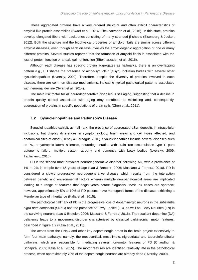
Mitochondria are crucial for the synthesis of adenosine triphosphate (ATP), reduction of oxidative
stress, Ca2+ storage, lipid metabolism and neuronal survival. Studies conducted by Parihar et al.
revealed that aggregated aSyn binds to mitochondria in a concentration-dependent manner (Parihar et
al., 2009). Moreover, other studies presented that aSyn, either WT or mutant, was able to interact with
membranes being crucial for aSyn cytotoxicity (Smith et al. 2005; Volles & Lansbury Jr, 2007).
Impairment of mitochondrial function has been associated with both WT and mutant aSyn
overexpression, but the exact interaction between aSyn and mitochondria is still under debate (Wales
et al., 2013). Numerous researches reported that mitochondrial function can be altered as a result of
aSyn binding to its inner membrane, where it can associate with mitochondrial complex I, decreasing
the mitochondrial activity (Liu et al., 2009; Winklhofer and Haass, 2010). Mitochondrial complex I is
responsible for catalyzing the first step in the electron transport chain which, in turn, is the main source
of ROS (Turrens, 2003). The binding between aSyn and complex I is followed by cytochrome c
release to cytosol, and increased in Ca2+ and ROS levels, culminating in cell death (Figure 1.7). The
precise mechanisms of how complex I inhibition can induce cell death are still poorly understood.
Nonetheless, three pathways have been proposed: (i) inhibition of complex I can induce mitochondrial
bioenergetics defects, decreasing ATP biosynthesis; (ii) complex I activity inhibition may increase
reductive stress, reducing molecular oxygen or activating redox sensitive signaling pathways, possibly
increasing cytochrome c intermembrane pool, activating mitochondrial-dependent apoptotic
machinery; (iii) impaired substrate oxidation at complex I can imbalance the NAD+/NADH ration,
resulting in inhibition of NAD+-dependent enzymes and consequent mitochondrial homeostasis
dysregulation (Perier et al., 2005; Imaizumi et al., 2015).
Besides, overexpression of the A53T aSyn mutant in neurons leads to an early time of onset of
these events (Devi et al., 2008; Parihar et al., 2008). Alterations in ATP production and differences in
mitochondrial membrane potential have also been reported (Parihar et al., 2009; Banerjee et al.,
2010).
Dissecting the role of alpha-synuclein phosphorylation in Parkinson’s Disease
10
Figure 1.7 aSyn affects mitochondrial function by interacting with mitochondrial complex I.
aSyn is believed to bind to the inner mitochondrial membrane, where it associates with mitochondrial
complex I. This assembly results in an increase of ROS production and Ca2+ levels and in the release of
cytochrome c, culminating with cell death.
Abnormalities in the balance between mitochondrial fusion and fission, vital for the maintenance
of mitochondrial activity, can also contribute to neuron dysfunction and cell death. Hence, mitophagy,
the delivering of damaged mitochondria to the lysosome, is required for the turnover and removal of
dysfunctional mitochondria, avoiding the generation and accumulation of ROS (Villacé et al., 2017).
Both WT and A53T aSyn promote an up-regulation of mitophagy leading to an immense mitochondrial
degradation, bioenergetic deficits and neuronal death (Chinta et al., 2011; Choubey et al., 2011).
The formation of aSyn aggregates, specially its mutant variations, is linked to an increase in ROS
production and, consequently, oxidative stress, which can be defined as a disequilibrium between the
levels of ROS produced and the system capability to detoxify the reactive intermediates, creating a
hazardous state and contributing to cellular damage (Dias et al., 2013). In vitro and in vivo studies
support the belief that an increased oxidative stress in the brain promotes aSyn aggregation, possibly
causing modifications in the nuclear membrane, and subsequent aSyn translocation to the nucleus
(Paxinou et al., 2001; Xu et al., 2006). Overall, ROS promotes aSyn aggregation, while
overexpression of aSyn leads to an increase in ROS levels, creating a cycle that leads to
neurodegeneration (Junn & Mouradian, 2002; Winklhofer & Haass, 2010). Recently, studies revealed
that aSyn is in fact not localized to mitochondria, but to a specific domain of the endoplasmic reticulum
(ER), called the mitochondria-associated ER membranes (MAM; Guardia-Laguarta et al. 2015;
Paillusson et al. 2017). Mitochondria communicates with the ER through MAM to regulate several
cellular processes, such as Ca2+ homeostasis, mitochondrial transport and biogenesis, mitochondrial
ATP production, lipid metabolism, ER stress, ubiquitin-proteasome system (UPS) and autophagy
(Paillusson et al., 2017). Alterations in the ER-mitochondria contact can cause deregulation of Ca2+
homeostasis, resulting in an inappropriate protein folding, metabolic alterations and apoptosis
(Guardia-Laguarta et al., 2015).
ER stress plays a crucial role in the development of several neurodegenerative diseases. ER
stress is usually caused by accumulation of misfolded proteins within the ER. To overcome ER stress,

Dissecting the role of alpha-synuclein phosphorylation in Parkinson’s Disease
11
cells activate an unfolded protein response (UPR), which protects cells from misfolded proteins
accumulation, by ceasing protein synthesis and activating mechanisms capable of decreasing protein
accumulation. In this case, proteins are translocated to the cytoplasm, where they undergo
degradation by the proteasome, through a process named ER associated degradation. However, if ER
stress persists, a cell death cascade is activated (Cooper et al. 2006; Doyle et al. 2011; Colla et al.
2012).
Misfolded aSyn could generate ER stress by, either interacting with ER chaperones or by
disturbing ER function, e.g., by affecting Ca2+ metabolism (Coune et al. 2012). Colla et al. proposed a
sequence of events connecting aSyn, ER stress and neurotoxicity i.e., low levels of aSyn in the ER
forms aSyn oligomers that assemble leading to the formation of insoluble aSyn aggregates. This
compromises the ER membranes integrity and exposes portions of the ER lumen to the cytosol,
resulting in chronic ER stress and, subsequently, cell death (Colla et al. 2012).
Moreover, in yeast, aSyn was reported to inhibit trafficking between ER and the Golgi complex,
strengthening cell toxicity and cell loss. Decrease of the ER-to-Golgi transport results in an
accumulation of protein in the ER, increasing ER stress (Cooper et al., 2006).
Originally, aSyn was described as a nuclear and pre-synaptic protein (Maroteaux et al. 1988).
aSyn mutations associated with PD, namely A30P, G51D and A53T, presented increased nuclear
localization in comparison to WT aSyn (Kontopoulos et al., 2006; Fares et al., 2014). Deregulation of
transcription is recognized as an important mechanism in neurodegeneration; indeed, the
overexpression of either WT or mutated aSyn leads to significant changes in the expression of genes
involved in neurotransmission, stress response, apoptosis, and transcription factors (Baptista et al.,
2003). Regulation of gene expression also depends on transcription factors distribution within the cell
and some studies revealed that transcription factors, as well as their regulatory kinases, were present
in aSyn aggregates, hence leading to transcription deregulation (Desplats et al., 2011; Wales et al.,
2013).
aSyn can also bind directly to histone-free and transcriptionally active DNA, changing its stability
and conformation (Hegde & Rao, 2003). Moreover, by increasing ROS levels within the nucleus,
glycated aSyn induces histone glycation and, subsequently, DNA damages (Padmaraju et al., 2011).
Regarding neurotransmission, it is known that the synthesis of DA comprises a rate-limiting step,
which is the conversion of tyrosine into L-DOPA by phosphorylated tyrosine hydroxylase (TH) before
being converted into DA by DOPA decarboxylase (DDC). TH is only activated when phosphorylated,
and this process of phosphorylation and dephosphorylation is highly important in the regulation of DA
biosynthesis (Kumer & Vrana, 1996). aSyn colocalizes with and binds to the dephosphorylated form of
TH, keeping it in its inactive form and, subsequently, causing a decrease in enzymatic activity and DA
synthesis (Figure 1.8, Perez et al. 2002).
1.3.5 Alpha-synuclein clearance
Unmodified aSyn is degraded via UPS and chaperone-mediated autophagy, whereas insoluble
and aggregated aSyn is degraded through macroautophagy (Cook & Petrucelli, 2010; Ebrahimi-
fakhari et al. 2011). The presence of aSyn, ubiquitin and proteasomal subunits within LBs reinforces a

Dissecting the role of alpha-synuclein phosphorylation in Parkinson’s Disease
12
relation between aSyn and proteasomal degradation dysfunction in PD pathogenesis (Betarbet et al.,
2005).
Figure 1.8 Dopamine synthesis is inhibited by aSyn.
TH phosphorylation is required for conversion of tyrosine into L-DOPA, a crucial step for DA synthesis. aSyn
binds to TH, hindering TH activation and, consequently, ceasing the conversion of tyrosine into L-DOPA (Perez et
al., 2002)
1.3.5.1 Alpha-synuclein degradation through ubiquitin-proteasome system and the
Rsp5 agent
The UPS is a multicomponent complex system capable of recognizing and degrading
unnecessary, misfolded, mutated and oxidatively damaged proteins (Betarbet et al., 2005; Wijayanti et
al., 2015).
Proteins to be degraded are first marked by covalent attachment of a polyubiquitin chain to a
lysine residue on the substrate. This process occurs through a series of enzyme-mediated reactions.
The ubiquitin-activated enzyme (E1) is responsible for ubiquitin activation, through an ATP-dependent
manner. The activated ubiquitin is transferred to a ubiquitin conjugating enzyme (UBC, also known as
E2) and, in the end, E2 associates with a ubiquitin-protein ligase (E3), that may or may not have the
substrate already bound. Ubiquitin is then transferred to a lysine residue of the substrate, which is
then degraded by a proteolytic complex – 26S proteasome (Figure 1.9; Weissman 2001; Betarbet et
al. 2005) The selectivity of ubiquitination and the recognition of substrates are mediated by E3s, either
alone or in combination with E2s. Additionally, the type of ubiquitin conjugation determines the destiny
of the ubiquitylated proteins, i.e., a single ubiquitin tag does not target a protein for degradation,
whereas a polyubiquitin chain does. A minimum of four ubiquitin molecules is required to target
proteins for degradation (Weissman, 2001).

Dissecting the role of alpha-synuclein phosphorylation in Parkinson’s Disease
13
Figure 1.9 Ubiquitin-proteasome system pathway.
The UPS pathway consists of a series of enzymatic reactions: E1 activates ubiquitin, which is then conjugated
with E2. E2 interacts with E3, which is bound to the substrate, transferring the ubiquitin to the substrate, which
marks the protein for degradation by the 26S proteasome. (u – ubiquitin)
Two distinct families of E3 ligases have been identified until now, namely the Really Interesting
New Gene (RING) and the Homologous to the E6-AP Carboxyl Terminus type (HECT), which differ
only in the way they transfer ubiquitin from E2 to the substrate (Weissman, 2001). For the present
study, the focus will be the HECT family. HECT is a domain of circa 350 amino-acids and is found at
the C-terminus of proteins. The majority of the HECT ligases contain protein-protein or protein-lipid
interaction domains located in the N-terminus of the protein and, based on this domain architecture,
the human HECTs can be divided in three groups: neuronal precursor cell-expressed developmentally
down-regulated gene 4 (Nedd4) family, HERC family and other HECTs. The Nedd4 family members
have an N-terminal C2 domain responsible for phospholipid binding, two to four WW domains that
recognize and bind to substrate proteins and a C-terminal HECT domain. (Rotin & Kumar, 2009;
Wijayanti et al. 2015). The Nedd4 family of enzymes binds specifically to substrates containing
proline-rich motifs, one of which was found in aSyn (Tofaris et al., 2011).
Rsp5 is the only Saccharomyces cerevisiae ortholog of NEDD4, and it is known to be involved in
numerous cellular functions, such as mitochondrial inheritance, chromatin remodeling, regulation of
transcription and endocytosis, sorting of numerous transmembrane proteins and of cargo into
multivesicular bodies from endocytic vesicles or from the Golgi apparatus to endosomes. Usually
Rsp5 modifies its protein target at the plasma membrane or Golgi apparatus with ubiquitin chains
linked to lysine 63, targeting the protein to being degraded by the endosomal-vacuolar pathway (Rotin
& Kumar, 2009). Tofaris et al. stated that Rsp5 promotes aSyn degradation through the endosomal-
vacuolar pathway, proving that Rsp5 ligase activity is essential for aSyn degradation and protects from
inclusion formation (Tofaris et al., 2011).

Dissecting the role of alpha-synuclein phosphorylation in Parkinson’s Disease
14
1.3.5.2 Autophagy and the importance of an acidic lysosomal environment
Autophagy is a self-degradative pathway, crucial for yeast survival during nutrient deprivation,
since it is responsible for macromolecules recycling, providing nutrients and energy. Autophagy
substrates comprise aggregate-prone cytoplasmic protein associated with neurodegenerative
diseases, such as aSyn (Rubinsztein et al., 2011). Three main forms of autophagy were described:
macroautophagy, microautophagy and chaperone mediated autophagy (Cuervo, 2004), differing from
each other’s in the mechanisms responsible for lysosomal delivery, whereas the final step of
lysosomal degradation is common to all forms (Wolfe et al., 2013).
Macroautophagy is the principal mechanism by which aSyn, and other long-lived proteins, are
degraded, as well as the only mechanism responsible for mitochondria recycling (Vogiatzi et al.,
2008). This process comprises the formation of autophagosomes that fuse with lysosomes to form
autophagolysosomes, which content is further degraded by acidic lysosomal hydrolases (Dehay et al.,
2010). The presence of aSyn aggregates in the PD patient’s brains suggests a deficiency in the
protein handling system, which can be confirmed through the evidence of autophagic vesicles
accumulation with a lysosomal depletion, as well as with the presence of key components of the
autophagosomes-lysosome pathway, such as the LC3 protein, as part of LBs (Dehay et al. 2010;
Bourdenx & Dehay, 2016). Moreover, changes in the morphology of the lysosomal system have been
observed in PD models (Stefanis et al., 2001). Bourdenx and Dehay proposed that LBs could be
originated from defective lysosomes or undegraded autophagic vesicles through the deposition of
other undegraded autophagic vesicles during the disease progression (Bourdenx & Dehay, 2016).
Defective lysosomal function is a major factor in neurodevelopmental diseases and has been
recognized as a key factor in the pathogenesis of some late-age onset disorders, including PD
(Colacurcio & Nixon, 2016). Lysosomes degradation occurs through the action of several hydrolases
with an optimum function in acidic conditions (pH 4-5) within lysosomes in order to activate the
mentioned enzymes (Wolfe et al., 2013). The lysosomal acidification is maintained by the vacuolar
ATPase (v-ATPase), which is an ATP dependent proton pump required to regulate pH homeostasis in
organelles (Diciccio & Steinberg, 2011). They are composed by 14 different subunits that are
organized into two domains: V1, a hydrolytic domain, and V0, a proton-translocation domain (Forgac,
2007). The v-ATPase actively transports H+ ions into the lysosome using energy from ATP hydrolysis,
making the lumen more acidic (Figure 1.10; Colacurcio & Nixon, 2016). Vacuolar acidification
mediated by v-ATPase has been identified as a positive regulator of mitochondria stability and lifespan
in yeast (Hughes & Gottschling, 2012). However, an increase in lysosomal pH was shown to decrease
lysosomal degradation of toxic proteins and lipids, trailed by downregulation of lysosomal enzyme
maturation and activity (Lee et al., 2013).

Dissecting the role of alpha-synuclein phosphorylation in Parkinson’s Disease
15
Figure 1.10 Vacuolar ATPase activity involved in lysosomal pH maintenance.
The v-ATPase uses metabolic energy originated from ATP hydrolysis to drive hydrogen proton into the lysosome,
maintaining the lysosomal pH.
1.4 CDNF - a non-conventional neurotrophic factor
Neurotrophic factors (NTFs) are secreted proteins that regulate the life and death of neurons
during development, responsible for neurons induction, specification, survival, and maturation, as well
as neurite growth and branching. Some NTFs can act in the adult brain, supporting, protecting, and
repairing mature neurons. Since PD is caused by the degeneration of dopaminergic neurons, many
NTFs have been explored for their neurotrophic and protective effects (Sullivan and Toulouse, 2011;
Voutilainen et al., 2015; Lindahl et al., 2017) .
Mesencephalic astrocyte-derived neurotrophic factor (MANF) and cerebral dopamine neurotrophic
factor (CDNF) comprise a conserved protein family with neurotrophic activities. MANF was first
identified by Petrova et al. from a culture medium of a rat astrocyte cell line as a novel neurotrophic
factor specific for dopaminergic neurons (Petrova et al., 2003). CDNF, a vertebrate specific paralogue
of MANF, was identified by bioinformatics and biochemical approaches (Lindholm et al., 2007). Both
neurotrophic factors are structurally different from the classical NTFs and its cytoprotective
mechanism of action is still unclear, in contrast with other known NTFs (Lindahl et al., 2017). Several
mechanisms of action have been proposed for CDNF, such as actions on mitochondrial complex I, ER
stress, oxidative stress, and anti-apoptotic effect. The analysis of CDNF and MANF structure revealed
that both proteins can have a double mechanism of action (Airavaara et al. 2012a).
The neurotrophic effects of both CDNF and MANF have been described in different animal
models of PD. CDNF had no effects in rat and mouse DA neurons in healthy rodents (Voutilainen et
al., 2015). However, when tested in rat 6-hydroxydopamine (6-OHDA) model of PD, CDNF was able
to protect and repair DA neurons (Lindholm et al., 2007). In mice treated with 1-methyl-4-phenyl-
1,2,3,6-tetrahydropyridine (MPTP), CDNF was shown to protect against MPTP toxicity and to restore
motor function (Airavaara et al. 2012b).
Both CDNF and MANF are mainly found in the ER suggesting that their most important function is
to regulate ER stress and UPR, however their mechanism of action is still unknown (Voutilainen et al.,
2015). Its effect in the ER is corroborated by the fact that extracellular CDNF only rescues neurons
degenerated via ER stress (Voutilainen et al., 2015). Also, a study performed by Zhou et al. revealed
Dissecting the role of alpha-synuclein phosphorylation in Parkinson’s Disease
16
that pre-treatment with CDNF was able to reduce the expression levels of proteins related to ER
stress (Zhou et al., 2016).
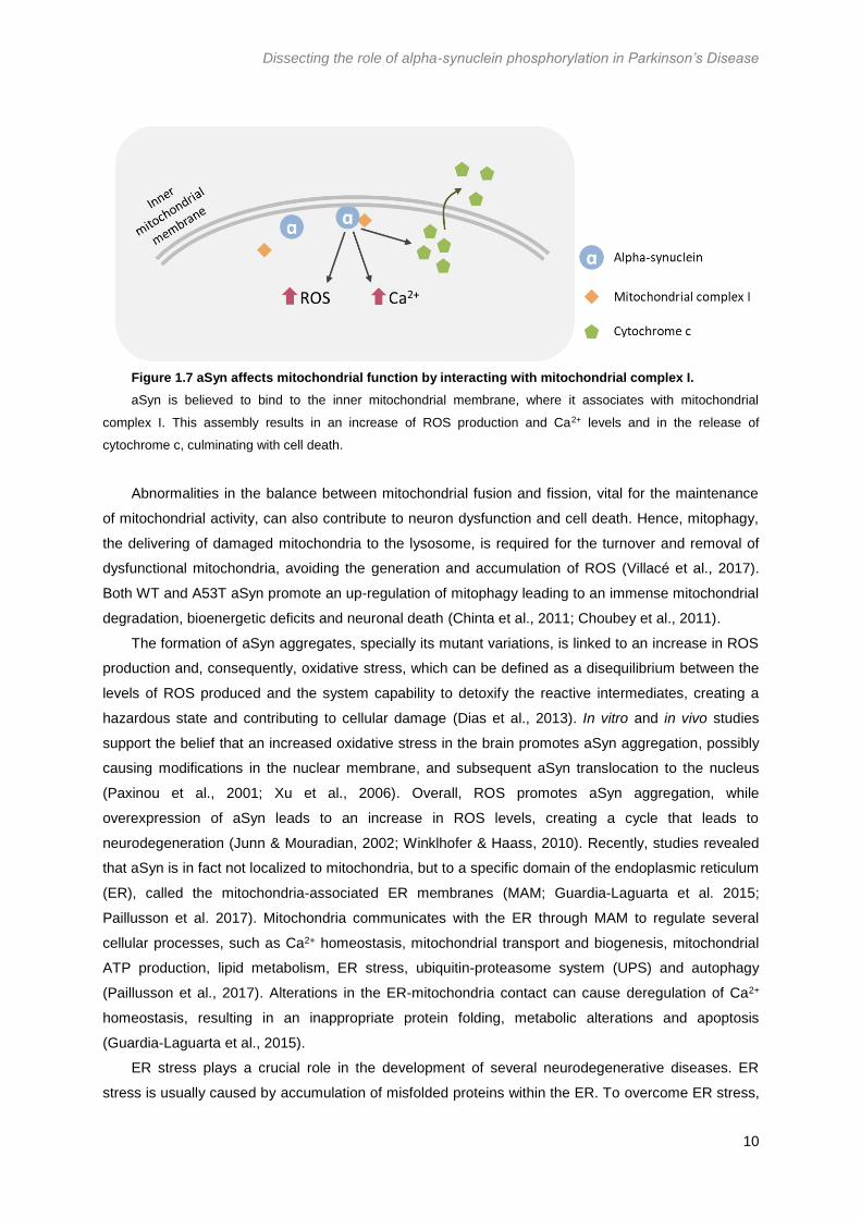
Recently, CDNF was shown to activate the survival promoting phosphatidylinositol 3 kinase
(PI3K)-Akt signaling pathway which is activated by neurotrophins and growth factors receptors.
(Figure 1.11; Voutilainen et al., 2017). In mammalian cells, phosphatidylinositol 3,4,5-triphosphate
(PIP3) generation at the plasma membrane is crucial for regulating proliferation and survival by PI3K-
Akt signaling pathway. PI3K generates PIP3, while phosphatidylinositol 3-phosphatase (PTEN)
degrades this lipid. PIP3 activates Akt which, in turn, activates the kinase mammalian target of
rapamycin (mTOR; Rodríguez-Escudeiro et al., 2005). This pathway promotes cell growth, survival
and differentiation, and is also capable of downregulating apoptotic signals. (Heras-Sandoval et al.,
2014).
Latge et al. found that CDNF prevents the toxic effects induced by aSyn oligomers in
dopaminergic neurons, however they did not find any evidences that CDNF interacts directly with
aSyn, either in monomeric or oligomeric form. In this sense, three different possible CDFN
mechanisms of actions were proposed: (i) CDNF binds to a transmembrane receptor, as other NTFs,
activating survival pathways that suppress aSyn toxic effects; (ii) CDNF stimulates cellular clearance
pathways and (iii) CDNF interacts with bcl-2-like protein X (BAX), inhibiting apoptosis (Latge et al.,
2015).
1.5 Yeast as a cellular model to study Parkinson’s Disease
Saccharomyces cerevisiae is a single-celled eukaryote, which contains membrane-bound
organelles, e.g., the nucleus, the endomembrane system and mitochondria (Duina et al., 2014). The
Figure 1.11 Basic schematic representation of the PI3K-Akt signalling pathway.
CDNF was found to activate the survival PI3K-Akt signalling pathway, leading to activation of mTOR which,
consequently, alters several cellular pathways.

Dissecting the role of alpha-synuclein phosphorylation in Parkinson’s Disease
17
unique features of S. cerevisiae granted its establishment as a robust model system in biology. These
features include its short generation time, easy handling, non-pathogenic nature, inexpensive culture
conditions and its amenability for genetic manipulation (Menezes et al., 2015). Its generation time is
about 90 minutes, under optimal laboratory conditions, through a budding process, in which small
daughter cells bud of the mother cell (Duina et al., 2014). Thus, sporulation of a particular diploid cell
generates different combinations of genotypes with desired genetic traits (Menezes et al., 2015).
Molecular genetics is supported by the high efficient homologous recombination pathway which allows
to insert, delete or mutate a genomic sequence up to the chromosome level easily (Sugiyama et al.,
2009).
Yeast cells have similarities to human cells, sharing fundamental aspects of eukaryotic cell
biology, namely the mechanisms of protein folding, quality control and degradation, the components
involved in the secretory pathway and vesicular trafficking, mitochondrial dysfunction and oxidative
stress and the mechanisms underlying cell death and survival. S. cerevisiae was the first eukaryote to
have its genome sequenced (Goffeau et al., 1996) and about 60% of the its genes display sequence
homology to a human orthologue and, of the human disease-related genes, over 25% have a
homologue in yeast (Franssens et al., 2013). Models can be based on the heterologous expression of
the human gene if the gene underlying the disease is absent in the yeast genome, or through the
study of the function and pathological role of the yeast corresponding gene, provided that it is present
in the yeast genome (Tenreiro & Outeiro, 2010).
Yeast models have been proven useful to understand the molecular mechanisms underlying PD,
as well as other synucleinopathies, since various features of PD can be reproduced in this model
(Tenreiro & Outeiro, 2010). The first yeast model of PD was based on the heterologous expression of
human aSyn, which lacks a yeast ortholog. In the yeast model developed by Outeiro and Lindquist,
yeast cells express WT aSyn or aSyn mutants fused with green fluorescent protein (GFP) under
regulation of the GAL1 promoter. This promoter is required for galactose catabolism and is only
activated in the presence of this sugar (Outeiro & Lindquist, 2003).
The increase in aSyn mediated toxicity is dose-dependent. Indeed, Petroi et al. demonstrated that
with only three integrated copies of WT aSyn, growth inhibition and inclusion formation was already
visible (Petroi et al., 2012). Microscopy analysis revealed that aSyn is localized in the plasma
membrane when one single copy is being expressed, while simply by doubling the number of aSyn
copies, aSyn localization shifts, forming cytoplasmic inclusions. Moreover, the number of aSyn copies
is also correlated with cytotoxicity, since with one single copy, there is no significant effect on
cytotoxicity, meanwhile, with only two copies, cytotoxicity increases drastically (Figure 1.12; Outeiro &
Lindquist, 2003; Gitler 2008) The formation of aSyn cytoplasmic inclusions in yeast cells is consistent
with previous studies in primary cortical neurons (Mclean et al., 2001). Expression of aSyn in yeast
cells is also known to induce oxidative stress and mitochondrial dysfunction and to affect vesicle
trafficking (Popova et al., 2015).

Dissecting the role of alpha-synuclein phosphorylation in Parkinson’s Disease
18
Figure 1.12 Effects of aSyn expression in yeast cells.
In the presence of a single aSyn copy, aSyn strongly localizes to the plasmatic membrane (A), whereas with only
two copies of aSyn, its cellular localization changes drastically, forming cytoplasmic inclusions (B). C) Cell viability
is not significantly affected in cells expressing only one copy of aSyn, meanwhile, when expressing two or more
copies, cell growth is completely inhibited (Gitler, 2008).
1.6 Aims
PD is characterized by aSyn aggregation, culminating with the formation of Lewy Bodies. aSyn
familial mutations, as well as duplications and triplications, are associated with familial forms of PD. It
is known that 90% of aSyn present in pathological inclusions is phosphorylated at S129, and several
other phosphorylation sites have already been identified. However, it is still unclear how
phosphorylation in different residues modulates distinct aSyn mechanisms of toxicity. In this context,
this study aims to clarify the interplay between phosphorylation in distinct aSyn residues using yeast
as a cellular model of PD. Moreover, this study also intends to characterize the mechanisms of toxicity
for each aSyn familial mutation.
Yeast strains expressing aSyn familial and phosphomutants will be characterized with respect to
aSyn cytotoxicity through evaluation of cell viability assessed by spotting assays and flow cytometry.
aSyn protein levels, aggregation and protein clearance will be evaluated for all mutants. Known
pathways of toxicity associated with aSyn will also be analysed, such as ROS production and
autophagy impairment.

Dissecting the role of alpha-synuclein phosphorylation in Parkinson’s Disease
19
2 Materials and Methods
2.1 Yeast Strains and Plasmids
The genotypes of the yeast strains used in this project are described in Table 2.1. All the plasmids
containing aSyn variations transformed in yeast cells, described in table 2.2 are under regulation of a
GAL1 promoter.
Table 2.1 Description of yeast strains used in this project
Yeast Strains Genotype Reference
W303.1A MATa can1Δ100 his3Δ11,15 leu2Δ3,112 trp1Δ1 ura3Δ1 ade2Δ1
(Thomas and Rothstein, 1989)
BY4741 MATa his3Δ1 leu2Δ0 met15Δ0 ura3Δ0 (Brachmann et al., 1998)
2.2 Escherichia coli manipulation
2.2.1 E. coli growth conditions
E. coli cells were cultivated at 37ºC, the optimum growth temperature, in Luria Broth (LB; Sigma-
Aldrich, St. Louis, MO, USA) media, supplemented with 2% agar or not, previously autoclaved. LB
selective medium were supplemented with 150mg/L of ampicillin (Amp) or 100mg/L of kanamycin
(Kan) after sterilization. E. coli cells were grown in the appropriated solid media at 37ºC, following
storage at 4ºC and regular picking to keep the cultures fresh. For longer periods of storage, glycerol
stocks were prepared. E. coli cells were inoculated in selective LB liquid media and grown over-night
at 37˚C with agitation (220rpm) and the glycerol stocks were made with a 1:1 proportion of bacterial
culture mixed with 50% (v/v) glycerol, and stored at -80˚C in cryovials.
2.2.2 E. coli transformation
DNA samples (50 to 100ng DNA) were added to 50µL of competent cells, which were then
incubated on ice for 30 min. A heat-shock was done by a 40 sec incubation at 42˚C followed by a 2
min incubation on ice. A volume of 0.9mL of LB liquid media was added to the cells that were then
incubated during 1 hour at 37˚C with agitation (220rpm). Cells were spun down during 1 min at
5000rpm, 850µL of media was removed, and the cells were plated on selective LB media, using glass
beads. The plates were incubated over-night at 37˚C.
2.2.3 Plasmid DNA extraction
For plasmid isolation from E. coli, individual clones were inoculated in 3mL of selective LB liquid
media and incubated over-night at 37˚C with agitation (220rpm). Using the NZYMiniprep kit (NZYTech,
Lisboa, Portugal), the DNA was extracted according to the protocol given by the manufacturer, with
the only alteration being the use of 35µL of purified water at 50ºC instead of elution buffer. DNA
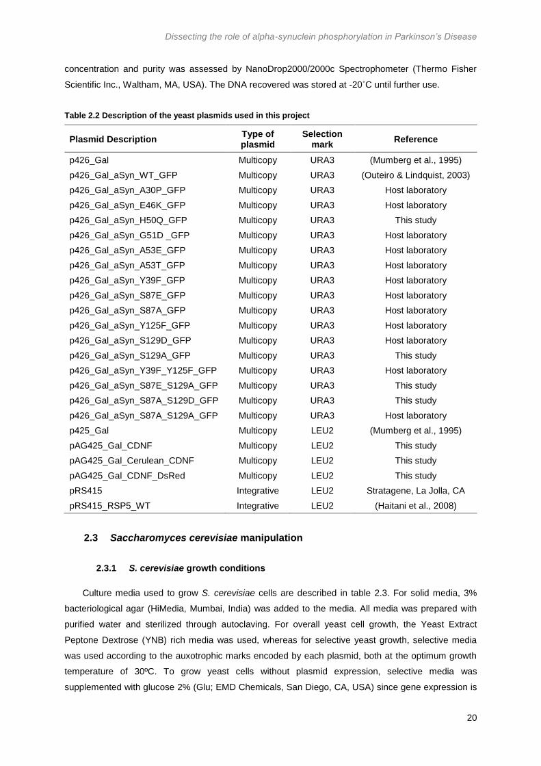
Dissecting the role of alpha-synuclein phosphorylation in Parkinson’s Disease
20
concentration and purity was assessed by NanoDrop2000/2000c Spectrophometer (Thermo Fisher
Scientific Inc., Waltham, MA, USA). The DNA recovered was stored at -20˚C until further use.
Table 2.2 Description of the yeast plasmids used in this project
Plasmid Description Type of plasmid
Selection mark
Reference
p426_Gal Multicopy URA3 (Mumberg et al., 1995)
p426_Gal_aSyn_WT_GFP Multicopy URA3 (Outeiro & Lindquist, 2003)
p426_Gal_aSyn_A30P_GFP Multicopy URA3 Host laboratory
p426_Gal_aSyn_E46K_GFP Multicopy URA3 Host laboratory
p426_Gal_aSyn_H50Q_GFP Multicopy URA3 This study
p426_Gal_aSyn_G51D _GFP Multicopy URA3 Host laboratory
p426_Gal_aSyn_A53E_GFP Multicopy URA3 Host laboratory
p426_Gal_aSyn_A53T_GFP Multicopy URA3 Host laboratory
p426_Gal_aSyn_Y39F_GFP Multicopy URA3 Host laboratory
p426_Gal_aSyn_S87E_GFP Multicopy URA3 Host laboratory
p426_Gal_aSyn_S87A_GFP Multicopy URA3 Host laboratory
p426_Gal_aSyn_Y125F_GFP Multicopy URA3 Host laboratory
p426_Gal_aSyn_S129D_GFP Multicopy URA3 Host laboratory
p426_Gal_aSyn_S129A_GFP Multicopy URA3 This study
p426_Gal_aSyn_Y39F_Y125F_GFP Multicopy URA3 Host laboratory
p426_Gal_aSyn_S87E_S129A_GFP Multicopy URA3 This study
p426_Gal_aSyn_S87A_S129D_GFP Multicopy URA3 This study
p426_Gal_aSyn_S87A_S129A_GFP Multicopy URA3 Host laboratory
p425_Gal Multicopy LEU2 (Mumberg et al., 1995)
pAG425_Gal_CDNF Multicopy LEU2 This study
pAG425_Gal_Cerulean_CDNF Multicopy LEU2 This study
pAG425_Gal_CDNF_DsRed Multicopy LEU2 This study
pRS415 Integrative LEU2 Stratagene, La Jolla, CA
pRS415_RSP5_WT Integrative LEU2 (Haitani et al., 2008)
2.3 Saccharomyces cerevisiae manipulation
2.3.1 S. cerevisiae growth conditions
Culture media used to grow S. cerevisiae cells are described in table 2.3. For solid media, 3%
bacteriological agar (HiMedia, Mumbai, India) was added to the media. All media was prepared with
purified water and sterilized through autoclaving. For overall yeast cell growth, the Yeast Extract
Peptone Dextrose (YNB) rich media was used, whereas for selective yeast growth, selective media
was used according to the auxotrophic marks encoded by each plasmid, both at the optimum growth
temperature of 30ºC. To grow yeast cells without plasmid expression, selective media was
supplemented with glucose 2% (Glu; EMD Chemicals, San Diego, CA, USA) since gene expression is
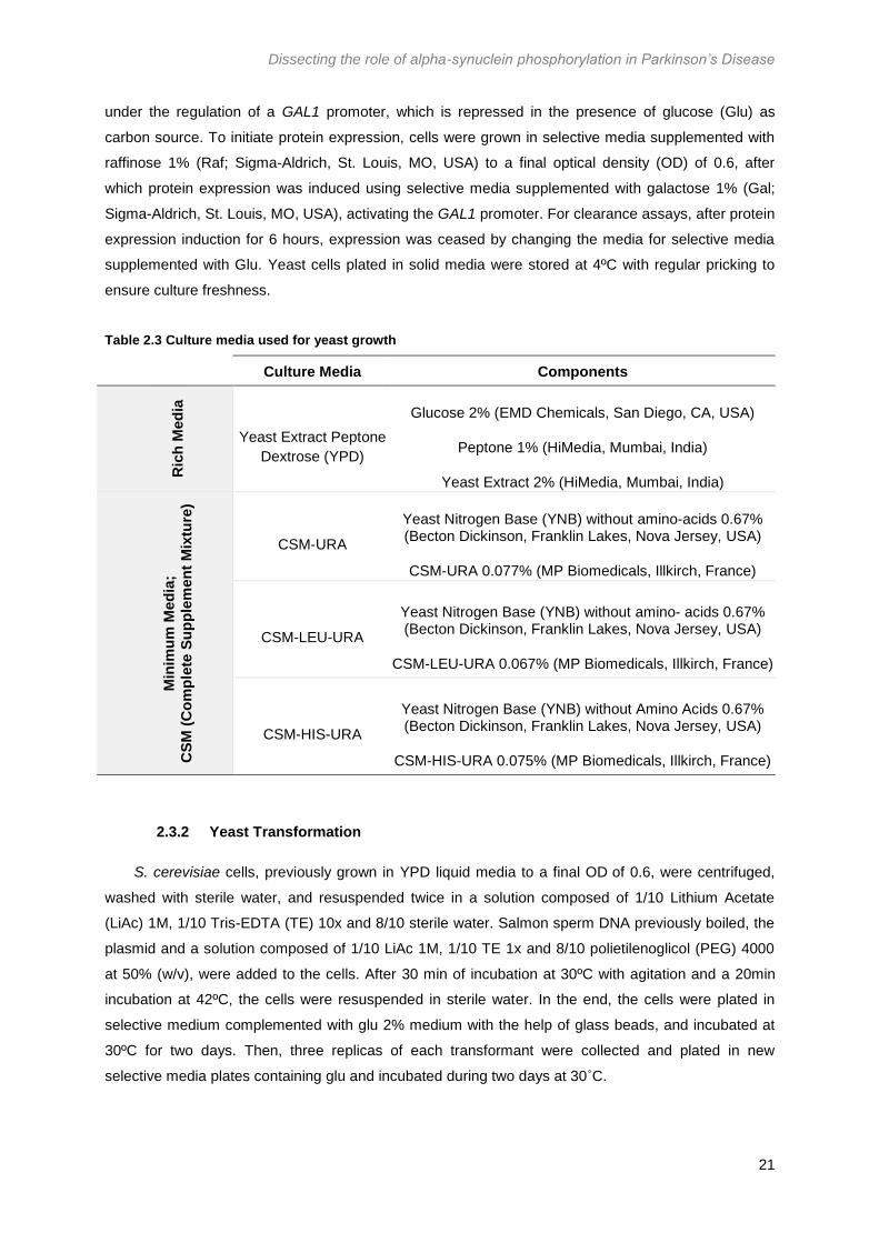
Dissecting the role of alpha-synuclein phosphorylation in Parkinson’s Disease
21
under the regulation of a GAL1 promoter, which is repressed in the presence of glucose (Glu) as
carbon source. To initiate protein expression, cells were grown in selective media supplemented with
raffinose 1% (Raf; Sigma-Aldrich, St. Louis, MO, USA) to a final optical density (OD) of 0.6, after
which protein expression was induced using selective media supplemented with galactose 1% (Gal;
Sigma-Aldrich, St. Louis, MO, USA), activating the GAL1 promoter. For clearance assays, after protein
expression induction for 6 hours, expression was ceased by changing the media for selective media
supplemented with Glu. Yeast cells plated in solid media were stored at 4ºC with regular pricking to
ensure culture freshness.
Table 2.3 Culture media used for yeast growth
Culture Media Components
Ric
h M
ed
ia
Yeast Extract Peptone
Dextrose (YPD)
Glucose 2% (EMD Chemicals, San Diego, CA, USA)
Peptone 1% (HiMedia, Mumbai, India)
Yeast Extract 2% (HiMedia, Mumbai, India)
Min
imu
m M
ed
ia;
CS
M (
Co
mp
lete
Su
pp
lem
en
t M
ixtu
re)
CSM-URA
Yeast Nitrogen Base (YNB) without amino-acids 0.67% (Becton Dickinson, Franklin Lakes, Nova Jersey, USA)
CSM-URA 0.077% (MP Biomedicals, Illkirch, France)
CSM-LEU-URA
Yeast Nitrogen Base (YNB) without amino- acids 0.67% (Becton Dickinson, Franklin Lakes, Nova Jersey, USA)
CSM-LEU-URA 0.067% (MP Biomedicals, Illkirch, France)
CSM-HIS-URA
Yeast Nitrogen Base (YNB) without Amino Acids 0.67% (Becton Dickinson, Franklin Lakes, Nova Jersey, USA)
CSM-HIS-URA 0.075% (MP Biomedicals, Illkirch, France)
2.3.2 Yeast Transformation
S. cerevisiae cells, previously grown in YPD liquid media to a final OD of 0.6, were centrifuged,
washed with sterile water, and resuspended twice in a solution composed of 1/10 Lithium Acetate
(LiAc) 1M, 1/10 Tris-EDTA (TE) 10x and 8/10 sterile water. Salmon sperm DNA previously boiled, the
plasmid and a solution composed of 1/10 LiAc 1M, 1/10 TE 1x and 8/10 polietilenoglicol (PEG) 4000
at 50% (w/v), were added to the cells. After 30 min of incubation at 30ºC with agitation and a 20min
incubation at 42ºC, the cells were resuspended in sterile water. In the end, the cells were plated in
selective medium complemented with glu 2% medium with the help of glass beads, and incubated at
30ºC for two days. Then, three replicas of each transformant were collected and plated in new
selective media plates containing glu and incubated during two days at 30˚C.

Dissecting the role of alpha-synuclein phosphorylation in Parkinson’s Disease
22
2.4 Molecular biology: Plasmids construction
2.4.1 Yeast plasmid constructions using site-directed mutagenesis
For site-directed mutagenesis, it was used a DNA template sample, and forward and reverse
primers specific for the mutations of interest, designed using the PrimerX bioinformatic tool (Table
2.4). The polymerase used was Pfu Turbo Polymerase (Agilent Technologies, Santa Clara, CA, USA)
and respective buffer, since this polymerase amplifies complex targets in higher yields than Taq
Polymerase and has proofreading properties.
Table 2.4 Description of plasmids and primers used for site-directed mutagenesis
Plasmid of interest Template plasmid Primer Forward Primer Reverse
p426_Gal_aSyn_H50Q_GFP p426_Gal_aSyn_WT_GFP 5’GGAGGGAGTGGTGCA
GGGTGTGGCAACAG 3’
5’CTGTTGCCACACCCT
GCACCACTCCCTCC 3’
p426_Gal_aSyn_S129A_GFP p426_Gal_aSyn_WT_GFP
5’CTTATGAAATGCCTG
CTGAGGAAGGGTATC
3’
5’GATACCCTTCCTCAG
CAGGCATTTCATAAG
3’
p426_Gal_aSyn_S87A_S129D_GFP p426_Gal_aSyn_S87A_GFP
5’GGCTTATGAAATGCCT
GATGAGGAAGGGTATC
AAG 3’
5’CTTGATACCCTTCCTC
ATCAGGCATTTCATAAG
CC 3’
p426_Gal_aSyn_S87E_S129A_GFP p426_Gal_aSyn_S87E_GFP 5’CTTATGAAATGCCTGC
TGAGGAAGGGTATC 3’
5’GATACCCTTCCTCAG
CAGGCATTTCATAAG 3’
The Polymerase Chain Reaction (PCR) conditions used for all amplifications was: initial
denaturation at 95ºC for 1 minute; 18 cycles of denaturation at 95ºC for 50 sec, annealing at 60ºC for
50 sec, following elongation at 68ºC during 8 min; a final elongation at 68ºC for 10 min. After
amplification, samples were treated with DpnI (Agilent Technologies, Santa Clara, CA, USA) during 7
hours at 37ºC, since this enzyme only cuts methylated plasmids, which happens only in naturally
amplified plasmids, allowing E. coli transformation with only newly-synthesized plasmids (Figure 2.1).

Dissecting the role of alpha-synuclein phosphorylation in Parkinson’s Disease
23
Figure 2.1 Schematic representation of the site-directed mutagenesis protocol.
2.4.2 CDNF plasmids construction by Gateway Recombination Cloning Technology
The CDNF plasmid available was a plasmid for expression on mammalian cells, for this reason, it
was necessary to generate a yeast-expressing plasmid containing the CDNF sequence. For that,
three different constructions were performed, one with WT CDNF alone, one with a N-terminal fusion
with Cerulean and, lastly, one with a DsRed fusion at the C-terminus. Three different pairs of primers
were used to amplify three distinct CDNF sequences (Table 2.5 and 2.6) in order to generate pDONR
vectors containing each CDNF sequence.
Table 2.5 Description of the PCR reactions performed to amplify the CDNF sequence
Fusion Primer Forward Primer Reverse
No fusion S_CDNF AS_CDNF
N-terminal fusion S_CDNF_N_fusion AS_CDNF
C-terminal fusion S_CDNF AS_CDNF_C_fusion
Table 2.6 PCR Reaction Mix used for CDNF amplification
The KAPA HiFi was added only after the first PCR cycle.
Component Volume (µL)
5X KAPA HiFi Buffer (KAPA Biosystems, Wilmington, MA, USA) 2.5
dNTPs (10mM) 0.7
Primer S (10µM) 1
Primer AS (10µM) 1
DNA template (15ng) 1
KAPA HiFi (KAPA Biosystems, Wilmington, MA, USA) 0.5
H2O 18.3
TOTAL 25

Dissecting the role of alpha-synuclein phosphorylation in Parkinson’s Disease
24
The PCR conditions used for all amplifications were: denaturation during 2 min at 92ºC; 3 cycles
of denaturation at 92ºC during 30 sec, annealing at 57ºC during 30 sec and an elongation step during
45 sec at 72ºC; followed by 25 similar cycles, where the annealing step was performed at 62ºC;
concluding with a final elongation step of 10 min at 72ºC.
After PCR, each sample was loaded into an 1% (w/v) agarose gel and ran at 90V to verify if the
PCR occurred as expected. The DNA was purified from the PCR amplifications using the Wizard SV
Gel and PCR Clean-Up System (Promega, Madison, WI, USA) and quantified using NanoDrop
2000/2000c Spectrophotometer (Thermo Fisher Scientific Inc., Waltham, MA, USA).
A BP reaction was performed to insert the amplified sequences into an intermediate plasmid, the
pDONR plasmid, using the Gateway BP Clonase Enzyme Mix (Invitrogen, Carlsbad, CA, USA). Each
mix was incubated at room temperature (RT) for 2 hours, after which were transformed in E. coli cells
and plated in LB media containing Kan, over-night. The pDONR plasmids containing the CDNF
sequences were extracted (see Section 2.1.5), followed by a LR reaction, where the CDNF sequence
was inserted into a series of pAdvanced Gateway (pAG) plasmids through recombination, using a
Gateway LR Clonase II Enzyme Mix (Invitrogen, Carlsbad, CA, USA). The mix was incubated for 2
hours at RT, following E. coli transformation (Section 2.2.2; Figure 2.2.). After growth, two colonies
from each transformation were collected, grown in LB liquid media and the plasmids were extracted as
described in Section 2.2.3. DNA samples were sent to sequencing by gold-standard Sanger
sequencing at STAB VIDA (Caparica, Almada, Portugal).
Figure 2.2 Schematic representation of the Gateway Recombination Cloning Technique procedure.

Dissecting the role of alpha-synuclein phosphorylation in Parkinson’s Disease
25
2.5 Cell Viability Assays
2.5.1 Spotting Assay
Cells were grown over-night at 30ºC in liquid media containing raffinose until they reached mid-log
phase. Initial volumes corresponding to an OD of 0.1 were deposited in the first lane of a 96-well plate
with sterile water to a final volume of 150µL. For the following lanes, a dilution of 1:3 was performed.
Using a previously sterilized stamp, the dilutions were plated in YPD media for growth control and SC
media complemented with gal to assess cell viability. The plates were incubated at optimal growth
temperature of 30ºC for 48 hours and 72 hours, respectively.
2.5.2 Flow Cytometry
Cell viability was also assessed through flow cytometry. After yeast cells growth and after 6 hours
of protein expression induction, a volume of cells correspondent to 1/OD was collected. Samples were
centrifuge for 3 min at 3000rpm and the pellet was resuspended in phosphate buffered saline (PBS)
1X. Propidium Iodide (PI) was added at 0.1mg/mL. Cells were incubated in the dark for 25 min at 30ºC
with agitation, followed by a spin down. Cells were washed twice with PBS 1X, transferred to flow
cytometry tubes and vortexed. Positive and negative controls were performed according to the table
2.7. A BD FACScantoTMII flow cytometer (Becton Dickinson, Franklin Lakes, Nova Jersey, USA),
equipped with a 20mW solid state 488nm laser was used for GFP and PI excitation (530/30nm and
485/42nm, respectively).
Table 2.7 Positive and negative controls used for the membrane integrity assessment through flow
cytometry
GFP PI Sample Treatment
- - Empty No PI
+ - aSyn_WT No PI
- + Empty Yeast dead cells (95ºC 10 min); stained
with PI
2.6 Detection of superoxide radical production
Flow cytometry was used to detect production of superoxide radical, using Dihydroethidium (DHE;
Life Technologies, Carlsbad, CA, USA). After 6 hours of protein expression induction, a volume of
cells correspondent to 1/OD was collected. Samples were centrifuge for 3 min at 3000rpm and the
pellet was resuspended in PBS 1X. DHE (3mM) was added, diluted in 500µL of PBS. Cells were
incubated in the dark for 15 min at 30ºC with constant agitation, followed by a spin down. Cells were
washed twice with PBS 1X, transferred to flow cytometry tubes and vortexed. Positive and negative
controls were performed according to the table 2.8. A BD FACScantoTMII flow cytometer (Becton

Dissecting the role of alpha-synuclein phosphorylation in Parkinson’s Disease
26
Dickinson, Franklin Lakes, Nova Jersey, USA), equipped with a 20mW solid state 488nm laser was
used for GFP and DHE excitation (530/30nm and 518/605nm, respectively).
Table 2.8 Positive and negative controls used for the superoxide radical production assessment through
flow cytometry
GFP DHE Sample Treatment
- - Empty No DHE
+ - aSyn_WT No DHE
- + Empty Yeast cells treated with H2O2 (0.2µM), 20 min with agitation; 30ºC; Addition of
DHE
2.7 Evaluation of vacuole acidification
Vacuole acidification was assessed through flow cytometry, using LysoTracker Red DND 99
(Thermo Fisher Scientific Inc., Waltham, MA, US), a dye highly specific for acidic organelles. After 6
hours of protein expression induction, a volume of cells correspondent to 1/OD was collected.
Samples were centrifuge for 3 min at 3000rpm and the pellet was resuspended in PBS 1X.
LysoTracker Red DND 99 (1mM) was added, diluted in 1000µL of PBS. Cells were incubated in the
dark for 30 min at 30ºC with constant agitation, followed by a spin down. Cells were washed twice with
PBS 1X, transferred to flow cytometry tubes and vortexed. Unstained and GFP positive controls were
performed. A BD FACSAriaTMII flow cytometer (Becton Dickinson, Franklin Lakes, Nova Jersey, USA)
with a 375 nm solid state laser, was used for GFP and mCherry excitation (530/30nm and 575/610nm,
respectively).
2.8 Protein subcellular localization through fluorescence microscopy
After 6 hours of protein expression, each sample was spun down and cells were retrieved from
the pellet to observe at the fluorescence microscope. Images were acquired using an Axio Imager 2
widefield fluorescent microscope (Carl Zeiss MicroImaging GmbH, Germany) and the Zeiss Zen
software (Carl Zeiss MicroImaging GmbH, Germany), with an amplification of 63X and with different
filters according to the tags observed. To see aSyn tagged with GFP, the Alexa Fluor 488 filter was
used, while to observe Cerulean and DsRed tag, the 4',6-diamidino-2-phenylindole (DAPI) and DsRed
filters were used, respectively. To see aSyn subcellular localization after protein clearance, protein
expression was ceased using liquid media complemented with glucose. Cultures were re-incubated for
6 hours or 13 hours, after which, cells were collected as before. The number of cells with inclusions
and the total cell number were counted using the ImageJ – Fiji image analysis software (Rasband,
1997).

Dissecting the role of alpha-synuclein phosphorylation in Parkinson’s Disease
27
2.9 Quantification of protein expression levels
2.9.1 Protein Extraction and Quantification
A culture volume correspondent to 2/OD was collected for yeast total protein extraction. The
culture was centrifuged and resuspended in MilliQ water, followed by a transference to a bead beater
vial. Cell were spun down and resuspended in tris-buffered saline (TBS) 1X with proteases inhibitors
(cOmplete™ ULTRA Tablets, Mini, EDTA-free, EASYpack Protease Inhibitor Cocktail by Roche,
Basel, Switzerland). For cell lysis, acid-washed glass beads were added to the sample and three
cycles of a 30 sec bead beater vortex followed by a 1 min ice incubation were done. Cells were
smoothly centrifuged (3 min, 700g, 4˚C) and the supernatant was collected. To quantify protein, the
Pierce BCA Protein Assay Kit (Thermo Fisher Scientific Inc., Waltham, MA, USA) was used according
to the manufacturer protocol.
2.9.2 Protein detection through Western Blot
Protein sample buffer (200mM Tris-HCl pH 6.8, 6% 2-mercaptoethanol, 8% sodium dodecyl
sulfate (SDS), 40% glycerol, 0.4% bromophenol blue) was added to each sample with the same total
protein quantity (8µg), following a 10 min denaturation at 100˚C before loading into a 12% acrylamide
gel. Protein samples were run in Sodium dodecyl sulfate polyacrylamide gel electrophoresis (SDS-
PAGE) during 20 min at 90V. After protein deposition between stacking and resolving gel, voltage was
changed to 120V. Protein was transferred to a nitrocellulose membrane using a Trans-Blot Turbo
transfer system (Bio-Rad, Hercules, CA, USA), according to the manufacturer’s protocol.
Membranes were blocked with 5% Bovine Serum Albumin (NZYTech, Lisboa, Portugal)/Tris-
buffered Saline – Tween 20 (5% BSA/TBS-T) (w/v), followed by an over-night incubation with a
primary antibody at 4ºC (Table 2.9). The antibodies were washed three times with TBS-T, followed by
a 1 hour and 30 min incubation with secondary antibodies, as depicted in table 2.9. Four washes with
TBS-T were performed after incubation with secondary antibodies, after which membranes were
revealed at ChemiDoc (Bio-Rad, Hercules, CA, USA) using Pierce™ ECL Western Blotting Substrate
(Thermo Fisher Scientific, Waltham, MA, USA). Images recovered from ChemiDoc were treated using
the Image Lab Software (Bio-Rad, Hercules, CA, USA).
To incubate a second antibody, membranes were stripped using a stripping solution (glycine, 10%
SDS, ddH2O; pH 2.0) during 45 min, followed by three washes with TBS 1X and three washes with
TBS-T. Membranes were again blocked and incubated with new antibodies, restarting the membrane
hybridization with new primary antibodies.

Dissecting the role of alpha-synuclein phosphorylation in Parkinson’s Disease
28
Table 2.9 Description of the antibodies used in this project
Antibody Manufacturer Description Secondary Antibody
Anti-aSyn BD Transduction Laboratories, San Jose, CA, USA
1:4000, mouse 1:10000, mouse
Anti-pS129 aSyn Wako Chemicals USA, Inc., Richmond, VA, USA
1:4000, mouse 1:10000, mouse
Anti-PGK Life Technologies, Grand Island, NY, USA 1:2000, mouse 1:5000, mouse
Anti-DsRed Clontech Laboratories, Inc., Montain View, CA, USA
1:2000, rabbit 1:5000, rabbit
Anti-GFP Neuromab, Davis, CA, USA 1:1000, mouse 1:5000, mouse
2.10 Statistical Analysis
To perform the statistical analysis, the Prism 6 (GraphPad Software Inc., La Jolla, CA, USA) was
used. Statistical analysis was performed using one-way ANOVA or two-way ANOVA, with Bonferroni’s
multiple comparison test, comparing the mean between distinct groups. A P value ≤ 0.01 was
considered statistically significant. Results are shown as mean ± standard deviation, for at least, three
independent experiments.

Dissecting the role of alpha-synuclein phosphorylation in Parkinson’s Disease
29
3 Results and Discussion
3.1 Plasmid constructions
Several plasmids used in this study were already available in the laboratory, nevertheless, some
plasmids had to be constructed, namely: aSyn_H50Q, aSyn_S129A, aSyn_S87E_S129A and
aSyn_S87A_S129D. To construct these plasmids, the site-directed mutagenesis technique was
performed using template plasmids and pairs of primers specific for each wanted mutation (see
Materials and Methods 2.4.1, Table 2.3). Plasmids were sequenced and aligned with the WT aSyn
sequence using the Clustal Omega bioinformatic tool, confirming the presence of the wanted
mutations (Supplementary Data, Figure 6.1).
3.2 Evaluation of the phosphomutants effects in inclusion formation and cell
viability
To unveil the role of aSyn phosphorylation, plasmids containing aSyn mutants where
phosphorylation-prone residues were mutated to mimic or block phosphorylation were transformed in
W303.1A yeast cells (Table 3.1). Since alanine (A) and phenylalanine (F) are phosphorylation-
resistant amino acids chemically similar to serine and tyrosine, respectively, they are used to block
phosphorylation in each residue; while glutamic acid (E) and aspartic acid (D) are non-phosphorylated
amino-acids chemically similar to phosphorylated serine amino acid, thus mimicking phosphorylation.
Table 3.1 Phosphomutants used in this study.
Every aSyn phosphomutant was expressed in a multicopy plasmid under the regulation of a GAL1 promoter.
Phosphomimics Phosphoblockers Double Mutants
S87E
S129D
Y39F
S87A
Y125F
S129A
Y39F_Y125F
S87E_S129E
S87E_S129A
S87A_S129E
S87A_S129A
Phosphomutants effects were evaluated regarding inclusion formation, clearance, and cell
toxicity, through spotting assay and flow cytometry. Inclusion formation was assessed after 3 and 6
hours of protein induction.
After 3 hours of protein induction (Figure 3.1B), WT aSyn presented a higher percentage of cells
with aSyn inclusions, suggesting a quicker aggregation process. Regarding Y phosphorylation,
phosphorylation blockade in each Y residue individually, it revealed a decrease in inclusion formation
when compared to WT aSyn. Additionally, when both Y residues (Y39 and Y125) are not
phosphorylated, inclusion formation decreases even further, pointing to a toxic effect of Y residues
phosphorylation and to an additive protective effect of Y phosphoblocking.

Dissecting the role of alpha-synuclein phosphorylation in Parkinson’s Disease
30
Figure 3.1 Assessment of aSyn inclusion formation and respective clearance for each aSyn
phosphomutant through fluorescence microscopy.
A) Intracellular localization of WT aSyn and each aSyn mutation after 3 and 6 hours of protein expression, and
after 6 hours of protein clearance in W303.1A yeast cells. B) Percentage of cells containing aSyn-GFP inclusions
after 3 hours of protein expression. C) Percentage of cells containing aSyn-GFP inclusions after 6 hours of protein
expression. D) Percentage of cells containing aSyn-GFP inclusions after 6 hours of clearance (6H Clearance),
normalized to 0H Clearance. Percentage values of each mutant were normalized to 0H Clearance. (***p<0.001,
****p<0.0001; one-way ANOVA with Bonferroni’s multiple comparison test; values represent mean ± SD from four
biological replicas).
Dissecting the role of alpha-synuclein phosphorylation in Parkinson’s Disease
31
Previous studies revealed that phosphorylation blocking of S129 (S129A) promoted aSyn
aggregation and cytotoxicity in different models (Gorbatyuk et al., 2008; Sancenon et al., 2012;
Tenreiro et al., 2014a; Azeredo da Silveira et al., 2017), and other studies exposed opposite results,
with S129A suppressing neuronal loss in Drosophila melanogaster (Chen and Feany, 2005). In yeast
cells, blockade of S129 phosphorylation was revealed to highly increase toxicity in yeast cells,
moreover, it also induced a significant increase in inclusion formation (Tenreiro et al., 2014a). Indeed,
probably due to its high toxicity, it was not possible to grow yeast cells containing the S129A aSyn
mutant expressed in a multicopy plasmid.
Nevertheless, S129A toxic effect can be supposed, since when expressing the double mutant
S87E_S129A aSyn, it is possible to see an increase in aSyn aggregation, when compared to S129D
aSyn, or even when compared to S87E and S87E_S129D mutants, indicating that the blocking of
S129 phosphorylation effects in aSyn inclusion formation overcomes the protective effect induced by
S87 phosphorylation. In contrast, blocking of S87 phosphorylation seems to induce a protective effect
over blockade of S129 phosphorylation (S87A_S129A), decreasing the percentage of cells with aSyn
inclusions. The protective effect of S87 phosphorylation blocking is supported by the results published
by Kim et. al, which stated that increased levels of S87 phosphorylation increased toxicity in H19-7
cells (Kim et al., 2006). Though, other study revealed that S87 phosphorylation inhibits aSyn fibrillation
and oligomerization (Paleologou et al., 2010).
With 6 hours of protein induction (Figure 3.1C), the percentage of cells containing aSyn inclusions
was similar between all aSyn variants, with exception for the double tyrosine mutants (Y39F_Y125F)
and the double mutant S87A_S129D, which presented a decrease in inclusion formation. These
results were in agreement with the results obtained only after 3 hours of protein expression. Once
again, blockage of S87 phosphorylation and blockage of both Y residues phosphorylation seem to
induce a protective effect. About the double tyrosine mutant, its reductive effect in aSyn inclusion
formation was also stated by Kleinknecht et. al in yeast cells (Kleinknecht et al., 2016).
Concerning inclusion clearance (Figure 3.1D), with only 6 hours of protein clearance, all
phosphomutants presented a similar inclusion clearance, with no significant differences amongst each
other. Clearance time could be increased to see more prominent differences between each
phosphomutant clearance, especially since aSyn S129 and Y39 phosphorylation were already found
to promote aSyn clearance in yeast cells (Mahul-Mellier et al., 2014; Tenreiro et al., 2014a).
Since a correlation between aSyn subcellular localization and cytotoxicity has been established by
several groups (Outeiro and Lindquist, 2003; Zabrocki et al., 2005; Gitler, 2008), spotting assay
(Figure 3.2) was performed to see the effects of each aSyn phosphomutant in cell viability. Cell
viability was also assessed through flow cytometry (Figure 3.3), using PI, which marks cells with a
compromised plasma membrane by binding to their nucleic acids. Flow cytometry allowed to see that
every aSyn mutant is being expressed at the same levels, since the percentage of GFP+ cells is
similar between samples (Figure 3.3B).
Regarding tyrosines phosphorylation, phosphorylation blockade at Y39 appears to have a
cytoprotective effect when compared with WT aSyn, however, phosphorylation blockade of Y125
decreases these protective effect, inducing a similar cell growth to cells transformed with WT aSyn.

Dissecting the role of alpha-synuclein phosphorylation in Parkinson’s Disease
32
When both Y phosphorylation is being blocked, toxicity is higher, suggesting that Y125F effect
overcomer the protective effect of Y39F. These results contradict the correlation between inclusion
formation and toxicity.
Regarding S phosphorylation, the toxic effect of S129 phosphoblocking can be deduced, since the
double mutant S87E_S129A present a decreased cell growth when compared to S87E_S129D and
S87E mutants, as well as when compared to WT aSyn. Corroborating the inclusion formation assay,
when phosphorylation of both S residues is being blocked, S87 phosphoblockade seem to induce a
protective effect over S129 phosphoblocking, since there is an increase in cell growth when compared
to S87E_S129A aSyn mutant. Moreover, similar to the obtained results, previous studies revealed that
S87E_S129D aSyn restored cell viability when compared to S87E aSyn, pointing to a protective effect
of S129 phosphorylation over S87 phosphorylation. Additionally, protein analysis revealed that yeast
cells expressing S87E aSyn present lower phosphorylated S129 levels, suggesting that S87
phosphorylation might be reducing S129 phosphorylation and, consequently, increasing toxicity
(Madaleno, 2017). However, when S87E is expressed with S129A, toxicity is increased, indicating that
S129A toxic effect adds to S87E toxicity, supporting the hypothesis that S87E toxicity is induced by
S129 phosphorylation levels decrease.
Figure 3.2 Evaluation of toxicity induced by each aSyn phosphomutant by spotting assay.
Spotting assay of the W303.1A yeast cells transformed with the indicated aSyn phosphomutants. Cell cultures
were adjusted to the same OD, serially diluted, and spotted into YPD and selective media complemented with Gal
as carbon source. (Image representative of four biological replicas)

Dissecting the role of alpha-synuclein phosphorylation in Parkinson’s Disease
33
In summary, blockage of S87 phosphorylation has a protective effect over S129 phosphoblocking
toxicity. Nevertheless, protein should be analyzed, to see phosphorylated S129 and S87 levels, as
well as total aSyn levels for each mutant, to understand how each residue phosphorylation is being
affected and its effect in cell viability.
3.3 Evaluation of aSyn familial mutations in aSyn inclusion formation and cell
toxicity
The subcellular localization of distinct aSyn variants was evaluated through fluorescence
microscopy. W303.1A yeast strain cells were transformed with plasmids containing distinct aSyn
variants (mutations) fused with GFP, under regulation of an inducible GAL1 promoter. Protein
expression was induced with selective media supplemented with Gal for 6 hours, after which cells
were collected for microscopy (Figure 3.4).
Concerning WT aSyn, about 60% of the cells present aSyn inclusions, whereas the remaining
cells do not present inclusions, presenting aSyn bound to the membrane, after 6 hours of expression
induction. Regarding the A30P mutant, microscopy revealed that it does not form aggregates when
overexpressed, keeping its cytoplasmic localization in a diffused manner, as described before (Outeiro
and Lindquist, 2003). A decrease in the percentage of cells containing aSyn inclusions was also
Figure 3.3 Evaluation of toxicity induced by aSyn phosphomutants through flow cytometry using PI.
A) Representative graphs of aSyn-GFP versus PI fluorescence assessed through flow cytometry in W303.1A
yeast cells transformed with each aSyn phosphomutant after 6 hours of protein expression. B) Frequency of GFP
positive cells in the indicated yeast cells. C) Frequency of PI positive cells in the indicated yeast cells. (*p<0.1,
one-way ANOVA with Bonferroni’s multiple comparison test using WT as control; values represent mean ± SD
from three biological replicas).

Dissecting the role of alpha-synuclein phosphorylation in Parkinson’s Disease
34
observed with the G51D mutation (7.74±1.06%), presenting a cytoplasmic localization in the majority
of cells, as described by Fares et. al and Rutherford et. al (Fares et al., 2014; Rutherford et al., 2014).
Figure 3.4 Assessment of aSyn inclusion formation for each aSyn variant through fluorescence
microscopy.
A) Intracellular localization of WT aSyn and each aSyn mutation after 6 hours of protein expression in W303.1A
yeast cells. B) Percentage of cells containing aSyn-GFP inclusions after 6 hours of protein expression
(***p<0.001, ****p<0.0001; one-way ANOVA with Bonferroni’s multiple comparison test; values represent mean ±
SD from three biological replicas)
The E46K and A53E aSyn mutants showed a slightly decrease in inclusion formation with
45.84±2.94% and 46.9±3.06% of cells with inclusions, respectively; A53E aSyn results were in
agreement with the previous studies (Ghosh et al., 2014; Rutherford and Giasson, 2016), whereas the
E46K aSyn aggregation decrease showed contrary results to previous studies in SH-SY5Y cells
(Pandey et al., 2006), corroborating the fact that aSyn ability to aggregate varies from different cellular
models. Regarding the H50Q mutant, the percentage of cells with aSyn inclusions was similar to WT
aSyn (60.91±0.64%). However, several studies support that the H50Q mutation increases aSyn
aggregation (Ghosh et al., 2013; Khalaf et al., 2014; Rutherford et al., 2014; Xiang et al., 2015). While
distinct studies revealed an increase in A53T aSyn aggregation when compared to WT aSyn (Narhi et
al., 1999; Li et al., 2001; Lázaro et al., 2014), this assay showed a similar percentage of cells with
aSyn inclusions (57.19±1.42%) when compared to WT aSyn. All of these differences in inclusion
formation might be due to different cellular models used in each study, indicating that the differences
in the cellular environment are sufficient to modulate aSyn ability to aggregate.
Toxicity induced by each aSyn familial mutant was evaluated through spotting assay (Figure 3.5).
The A30P and the G51D mutations seem to be non-toxic, since their growth is similar to the growth of
yeast cells transformed with an empty plasmid. The previous microscopy results showed that, in fact,
these mutations induce less inclusion formation. On the other hand, E46K, H50Q and A53T aSyn,
appear to be more toxic, presenting a similar decrease in cell growth as WT aSyn. The A53E mutation
is more toxic than A30P or G51D mutation, but appears to be less cytotoxic when compared with WT
aSyn and the remaining aSyn mutations.

Dissecting the role of alpha-synuclein phosphorylation in Parkinson’s Disease
35
The spotting assay and the inclusion formation assay were in agreement, since the aSyn variants
with higher percentage of cells with inclusions (WT, E46K, H50Q, A53E and A53T aSyn) were the
ones who presented higher toxicity.
To quantitatively evaluate cytotoxicity induced by each aSyn variant, flow cytometry was
performed after 6 hours of protein expression (Figure 3.6). Flow cytometry results revealed that all
aSyn mutants are being expressed at the same levels, since the percentage of cells expressing GFP
is similar between each culture (Figure 3.6B). PI staining revealed that the E46K aSyn mutant is the
most cytotoxic, when comparing to WT aSyn. The A30P mutant revealed to be the less toxic (Figure
3.3C).
However, with only 6 hours of protein induction, cell viability was still high, with low percentages
of dead cells. Despite not having significant cell death, cells can still be being affected in several
pathways, leading to a later cell death. To obtain a more prominent cell death, protein was expressed
for a longer period, i.e., 13 hours of protein expression, after which flow cytometry was performed
(Figure 3.7). After 13 hours of protein expression, aSyn was still being expressed at the same level
between different variants (Figure 3.7B). Moreover, cell viability after 13 hours of protein expression
decreased dramatically, with higher percentages of PI-positive cells. Nevertheless, A30P aSyn
presented a low percentage of PI-positive cells, consistent with the spotting assay results and the
absence of inclusions (Lázaro et al., 2014).
Figure 3.5 Evaluation of toxicity induced by each aSyn variant by spotting assay.
Spotting assay of the W303.1A yeast cells transformed with the indicated aSyn variants. Cell cultures were
adjusted to the same OD, serially diluted, and spotted into YPD and selective media complemented with Gal as
carbon source. (Image representative of three biological replicas)
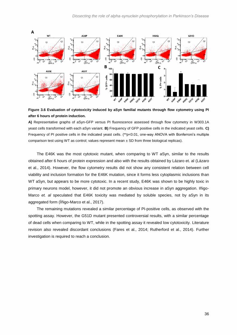
Dissecting the role of alpha-synuclein phosphorylation in Parkinson’s Disease
36
Figure 3.6 Evaluation of cytotoxicity induced by aSyn familial mutants through flow cytometry using PI
after 6 hours of protein induction.
A) Representative graphs of aSyn-GFP versus PI fluorescence assessed through flow cytometry in W303.1A
yeast cells transformed with each aSyn variant. B) Frequency of GFP positive cells in the indicated yeast cells. C)
Frequency of PI positive cells in the indicated yeast cells. (**p<0.01, one-way ANOVA with Bonferroni’s multiple
comparison test using WT as control; values represent mean ± SD from three biological replicas).
The E46K was the most cytotoxic mutant, when comparing to WT aSyn, similar to the results
obtained after 6 hours of protein expression and also with the results obtained by Lázaro et. al (Lázaro
et al., 2014). However, the flow cytometry results did not show any consistent relation between cell
viability and inclusion formation for the E46K mutation, since it forms less cytoplasmic inclusions than
WT aSyn, but appears to be more cytotoxic. In a recent study, E46K was shown to be highly toxic in
primary neurons model, however, it did not promote an obvious increase in aSyn aggregation. Iñigo-
Marco et. al speculated that E46K toxicity was mediated by soluble species, not by aSyn in its
aggregated form (Íñigo-Marco et al., 2017).
The remaining mutations revealed a similar percentage of PI-positive cells, as observed with the
spotting assay. However, the G51D mutant presented controversial results, with a similar percentage
of dead cells when comparing to WT, while in the spotting assay it revealed low cytotoxicity. Literature
revision also revealed discordant conclusions (Fares et al., 2014; Rutherford et al., 2014). Further
investigation is required to reach a conclusion.
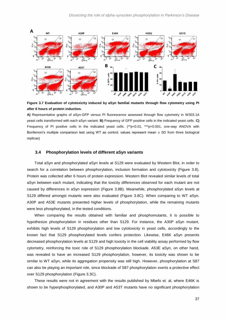
Dissecting the role of alpha-synuclein phosphorylation in Parkinson’s Disease
37
Figure 3.7 Evaluation of cytotoxicity induced by aSyn familial mutants through flow cytometry using PI
after 6 hours of protein induction.
A) Representative graphs of aSyn-GFP versus PI fluorescence assessed through flow cytometry in W303.1A
yeast cells transformed with each aSyn variant. B) Frequency of GFP positive cells in the indicated yeast cells. C)
Frequency of PI positive cells in the indicated yeast cells. (**p<0.01, ***p<0.001, one-way ANOVA with
Bonferroni’s multiple comparison test using WT as control; values represent mean ± SD from three biological
replicas)
3.4 Phosphorylation levels of different aSyn variants
Total aSyn and phosphorylated aSyn levels at S129 were evaluated by Western Blot, in order to
search for a correlation between phosphorylation, inclusion formation and cytotoxicity (Figure 3.8).
Protein was collected after 6 hours of protein expression. Western Blot revealed similar levels of total
aSyn between each mutant, indicating that the toxicity differences observed for each mutant are not
caused by differences in aSyn expression (Figure 3.8B). Meanwhile, phosphorylated aSyn levels at
S129 differed amongst mutants were also evaluated (Figure 3.8C). When comparing to WT aSyn,
A30P and A53E mutants presented higher levels of phosphorylation, while the remaining mutants
were less phosphorylated, in the tested conditions.
When comparing the results obtained with familiar and phosphomutants, it is possible to
hypothesize phosphorylation in residues other than S129. For instance, the A30P aSyn mutant,
exhibits high levels of S129 phosphorylation and low cytotoxicity in yeast cells, accordingly to the
known fact that S129 phosphorylated levels confers protection. Likewise, E46K aSyn presents
decreased phosphorylation levels at S129 and high toxicity in the cell viability assay performed by flow
cytometry, reinforcing the toxic role of S129 phosphorylation blockade. A53E aSyn, on other hand,
was revealed to have an increased S129 phosphorylation, however, its toxicity was shown to be
similar to WT aSyn, while its aggregation propensity was still high. However, phosphorylation at S87
can also be playing an important role, since blockade of S87 phosphorylation exerts a protective effect
over S129 phosphorylation (Figure 3.3C).
These results were not in agreement with the results published by Mbefo et. al, where E46K is
shown to be hyperphosphorylated, and A30P and A53T mutants have no significant phosphorylation
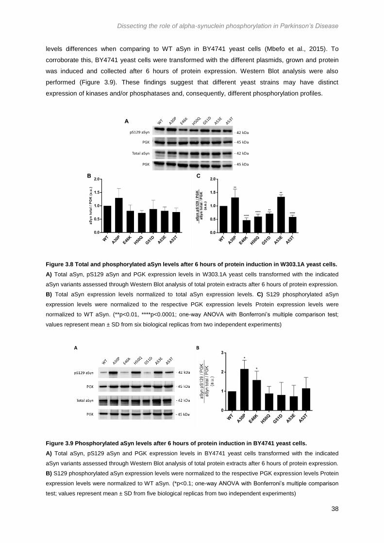
Dissecting the role of alpha-synuclein phosphorylation in Parkinson’s Disease
38
levels differences when comparing to WT aSyn in BY4741 yeast cells (Mbefo et al., 2015). To
corroborate this, BY4741 yeast cells were transformed with the different plasmids, grown and protein
was induced and collected after 6 hours of protein expression. Western Blot analysis were also
performed (Figure 3.9). These findings suggest that different yeast strains may have distinct
expression of kinases and/or phosphatases and, consequently, different phosphorylation profiles.
Figure 3.8 Total and phosphorylated aSyn levels after 6 hours of protein induction in W303.1A yeast cells.
A) Total aSyn, pS129 aSyn and PGK expression levels in W303.1A yeast cells transformed with the indicated
aSyn variants assessed through Western Blot analysis of total protein extracts after 6 hours of protein expression.
B) Total aSyn expression levels normalized to total aSyn expression levels. C) S129 phosphorylated aSyn
expression levels were normalized to the respective PGK expression levels Protein expression levels were
normalized to WT aSyn. (**p<0.01, ****p<0.0001; one-way ANOVA with Bonferroni’s multiple comparison test;
values represent mean ± SD from six biological replicas from two independent experiments)
Figure 3.9 Phosphorylated aSyn levels after 6 hours of protein induction in BY4741 yeast cells.
A) Total aSyn, pS129 aSyn and PGK expression levels in BY4741 yeast cells transformed with the indicated
aSyn variants assessed through Western Blot analysis of total protein extracts after 6 hours of protein expression.
B) S129 phosphorylated aSyn expression levels were normalized to the respective PGK expression levels Protein
expression levels were normalized to WT aSyn. (*p<0.1; one-way ANOVA with Bonferroni’s multiple comparison
test; values represent mean ± SD from five biological replicas from two independent experiments)
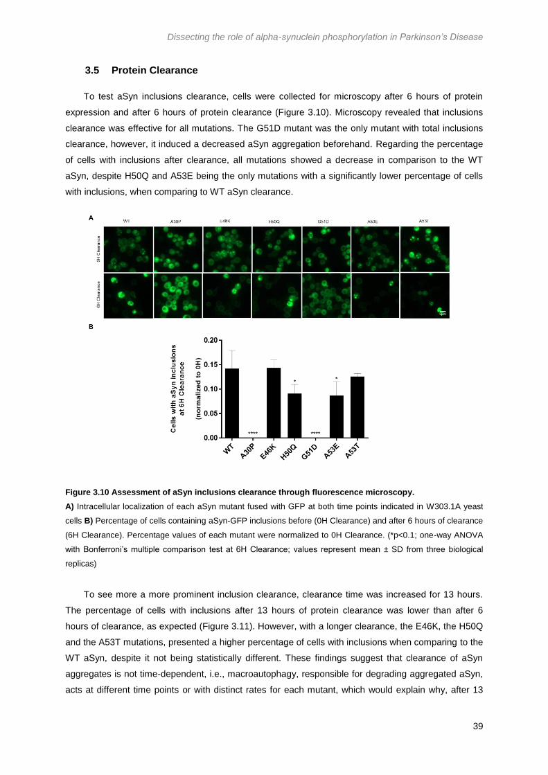
Dissecting the role of alpha-synuclein phosphorylation in Parkinson’s Disease
39
3.5 Protein Clearance
To test aSyn inclusions clearance, cells were collected for microscopy after 6 hours of protein
expression and after 6 hours of protein clearance (Figure 3.10). Microscopy revealed that inclusions
clearance was effective for all mutations. The G51D mutant was the only mutant with total inclusions
clearance, however, it induced a decreased aSyn aggregation beforehand. Regarding the percentage
of cells with inclusions after clearance, all mutations showed a decrease in comparison to the WT
aSyn, despite H50Q and A53E being the only mutations with a significantly lower percentage of cells
with inclusions, when comparing to WT aSyn clearance.
To see more a more prominent inclusion clearance, clearance time was increased for 13 hours.
The percentage of cells with inclusions after 13 hours of protein clearance was lower than after 6
hours of clearance, as expected (Figure 3.11). However, with a longer clearance, the E46K, the H50Q
and the A53T mutations, presented a higher percentage of cells with inclusions when comparing to the
WT aSyn, despite it not being statistically different. These findings suggest that clearance of aSyn
aggregates is not time-dependent, i.e., macroautophagy, responsible for degrading aggregated aSyn,
acts at different time points or with distinct rates for each mutant, which would explain why, after 13
Figure 3.10 Assessment of aSyn inclusions clearance through fluorescence microscopy.
A) Intracellular localization of each aSyn mutant fused with GFP at both time points indicated in W303.1A yeast
cells B) Percentage of cells containing aSyn-GFP inclusions before (0H Clearance) and after 6 hours of clearance
(6H Clearance). Percentage values of each mutant were normalized to 0H Clearance. (*p<0.1; one-way ANOVA
with Bonferroni’s multiple comparison test at 6H Clearance; values represent mean ± SD from three biological
replicas)

Dissecting the role of alpha-synuclein phosphorylation in Parkinson’s Disease
40
hours of protein clearance, WT aSyn presents a higher clearance when compared to the H50Q and
A53E mutants. The lack of statistically differences between each sample might be due to the low
percentages of all samples.
Figure 3.11 Assessment of aSyn inclusions clearance through fluorescence microscopy after 13 hours of
protein clearance.
A) Intracellular localization of each aSyn mutant fused with GFP at both time points indicated in W303.1A yeast
cells B) Percentage of cells containing aSyn-GFP inclusions before (0H Clearance) and after 13 hours of
clearance (13H Clearance). Percentage values at 0H of clearance were normalized to 100% and the values at
13H of clearance were normalized to the values at 0H. (*p<0.1; two-way repeated measures ANOVA with
Bonferroni’s multiple comparison test; values represent mean ±SD from three independent experiments)
Protein clearance was also evaluated by Western Blot. Protein levels of total and phosphorylated
aSyn at S129 were evaluated before and after 6 hours of protein clearance (Figure 3.12). Regarding
total levels of aSyn, the A30P mutant presented a higher protein clearance. Nonetheless, this high
clearance might be due to the high levels of total aSyn after 6 hours of protein induction (0H
Clearance). Meanwhile, concerning the phosphorylated aSyn, the E46K and G51D mutants show a
tendency to impair protein clearance, nevertheless there is no statistically significant differences
between each aSyn variant clearance. It would be interesting to assess which clearance mechanism is
being affected for each mutant, i.e., evaluate autophagy and proteasome function.

Dissecting the role of alpha-synuclein phosphorylation in Parkinson’s Disease
41
Figure 3.12 Clearance of total and phosphorylated aSyn is similar between distinct aSyn mutations.
A) Total aSyn, pS129 aSyn and PGK expression levels in W303.1A yeast cells transformed with the indicated
aSyn variants assessed through Western Blot analysis of total protein extracts after 6 hours of protein expression
(0H Clearance) and after 6 hours of protein clearance (6H Clearance). B) Total aSyn and pS129 aSyn expression
levels were normalized to the respective PGK expression levels. Protein levels at 6H Clearance were normalized
to levels at 0H Clearance. (two-way repeated measures ANOVA with Bonferroni’s multiple comparison test;
values represent mean ± SD from five biological replicas from two independent experiments)
3.5.1 Assessment of Rsp5 effect in inclusion clearance of each aSyn familial mutant
Rsp5 is a Nedd4 ortholog in S. cerevisiae, which was shown to promote aSyn degradation
through the endosomal-vacuolar pathway and to protect from inclusion formation. In this study, we co-
transformed yeast cells with a plasmid containing a Rsp5 sequence or a similar empty plasmid, with
each aSyn plasmid. Inclusion formation was assessed by fluorescence microscopy at 0, 3 and 6 hours
of clearance (Figure 3.13).
The results obtained were different than described by Tofaris et. al 2011. Rsp5 overexpression
decreased aSyn inclusion formation only in some aSyn mutants. Regarding WT aSyn, the aSyn
variant used by Tofaris et al, Rsp5 overexpression decreased inclusion formation after 3 hours of
protein clearance, however, after 6 hours of protein clearance, despite the absence of significant
differences, cells where Rsp5 was being overexpressed presented a higher inclusion formation than
cells transformed with the control plasmid. A decrease in inclusion formation was only visible in some
aSyn mutants, namely G51D, A53E and A53T. E46K aSyn mutant was the only mutant not showing
any protective effect for Rsp5; this could suggest that E46K aSyn is not mainly degraded by the
endosomal-vacuolar pathway or that it inhibits said pathway.
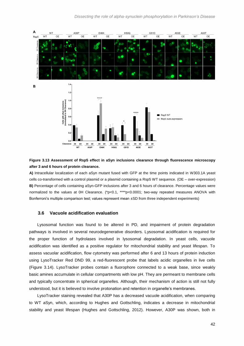
Dissecting the role of alpha-synuclein phosphorylation in Parkinson’s Disease
42
3.6 Vacuole acidification evaluation
Lysosomal function was found to be altered in PD, and impairment of protein degradation
pathways is involved in several neurodegenerative disorders. Lysosomal acidification is required for
the proper function of hydrolases involved in lysosomal degradation. In yeast cells, vacuole
acidification was identified as a positive regulator for mitochondrial stability and yeast lifespan. To
assess vacuolar acidification, flow cytometry was performed after 6 and 13 hours of protein induction
using LysoTracker Red DND 99, a red-fluorescent probe that labels acidic organelles in live cells
(Figure 3.14). LysoTracker probes contain a fluorophore connected to a weak base, since weakly
basic amines accumulate in cellular compartments with low pH. They are permeant to membrane cells
and typically concentrate in spherical organelles. Although, their mechanism of action is still not fully
understood, but it is believed to involve protonation and retention in organelle’s membranes.
LysoTracker staining revealed that A30P has a decreased vacuole acidification, when comparing
to WT aSyn, which, according to Hughes and Gottschling, indicates a decrease in mitochondrial
stability and yeast lifespan (Hughes and Gottschling, 2012). However, A30P was shown, both in
Figure 3.13 Assessment of Rsp5 effect in aSyn inclusions clearance through fluorescence microscopy
after 3 and 6 hours of protein clearance.
A) Intracellular localization of each aSyn mutant fused with GFP at the time points indicated in W303.1A yeast
cells co-transformed with a control plasmid or a plasmid containing a Rsp5 WT sequence. (OE – over-expression)
B) Percentage of cells containing aSyn-GFP inclusions after 3 and 6 hours of clearance. Percentage values were
normalized to the values at 0H Clearance. (*p<0.1, ****p<0.0001; two-way repeated measures ANOVA with
Bonferroni’s multiple comparison test; values represent mean ±SD from three independent experiments)

Dissecting the role of alpha-synuclein phosphorylation in Parkinson’s Disease
43
spotting assays and flow cytometry with PI, to be less cytotoxic when comparing to the remaining
aSyn variants. The observed decrease in Median Fluorescence Intensity for the A30P mutant can also
be affecting hydrolases activation, reducing A30P aSyn clearance. Once again, this is not in
agreement with the protein clearance assay, where A30P aSyn was shown to have a higher total aSyn
clearance. Besides, A30P was shown to be highly proteolytically degraded in the vacuole, after A30P
aSyn-containing vesicles merge with the vacuolar membrane with the support of Ypp1, a aSyn
protective protein (Flower et al., 2007). This decrease in LysoTracker fluorescence in the A30P
mutants can be a result of different vacuole morphology. Smaller vacuoles would internalize less
LysoTracker and, consequently, express less fluorescence. To test this hypothesis, vacuole
morphology could be assessed using probes specific for vacuoles, such as LysoTracker or FM 4-64,
and visualizing at the fluorescence microscope.
Figure 3.14 Evaluation of vacuole acidification in yeast cells transformed with each aSyn variant after 6
and 13 hours of protein induction.
A) Representative graphs of aSyn-GFP versus LysoTracker Red DND 99 fluorescence assessed through flow
cytometry in W303.1A yeast cells transformed with each aSyn variant after 6 and 13 hours of protein expression.
B) Median fluorescence intensity of each cell population after 6 hours of protein induction. C) Median
fluorescence intensity of each cell population after 13 hours of protein induction (**p<0.01; one-way ANOVA with
Bonferroni’s multiple comparison test using WT as control; values represent mean ± SD from three biological
replicas)
Regarding the remaining mutants, their vacuole acidification was similar to WT aSyn, indicating a
similar mitochondrial function, as well as a similar autophagy induction. This could also be assessed
by evaluating autophagy flux and induction, through Western Blot, by analyzing aSyn levels before
and after clearance in yeast strains where autophagy is inhibited, or using yeast cells co-transformed
with aSyn variants and plasmids expressing ATG8, which encodes for an autophagy related protein,

Dissecting the role of alpha-synuclein phosphorylation in Parkinson’s Disease
44
required for autophagosomal membranes formation, that is degraded in the vacuole. By quantifying
Atg8 protein levels, it would be possible to infer autophagy induction and flux (Tenreiro et al., 2014a).
3.7 Evaluation of ROS and superoxide production
The production of ROS was evaluated through flow cytometry at 6 and 13 hours of protein
induction, using DHE, a superoxide indicator (Figure 3.15). DHE usually displays blue fluorescence
when in the cytosol, but when oxidized by superoxides or by other sources of ROS, it intercalates with
DNA, staining the nucleus red. DHE fluorescence intensity was similar between all the aSyn mutants,
having no differences when comparing to WT aSyn, i.e., the ROS production is similar amongst all
aSyn variants. However, E46K and G51D seem to exhibit a more intense fluorescence, indicating a
higher production of ROS. Since a higher production of ROS would induce aSyn aggregation, and
vice-versa, these results are not in agreement with the inclusion formation results. However, increased
ROS levels also causes oxidative stress, which leads to cell death, which is in agreement with the
E46K cell viability results obtained with flow cytometry. This suggests that E46K toxicity might be
mainly due to an excessive ROS production, and not due to aSyn aggregation alone.
Mitochondrial dysfunction also provokes ROS accumulation, which means that E46K and G51D
could be impairing mitochondrial function, somehow, leading to ROS production and, eventually, cell
death.
Figure 3.15 Evaluation of ROS production in yeast cells transformed with each aSyn variant after 6 and 13
hours of protein induction.
A) Representative graphs of aSyn-GFP versus DHE fluorescence assessed through flow cytometry in W303.1A
yeast cells transformed with each aSyn variant after 6 and 13 hours of protein expression. B) Median
fluorescence intensity of each cell population after 6 hours of protein induction. C) Median fluorescence intensity
of each cell population after 13 hours of protein induction (values represent mean ± SD from three biological
replicas)

Dissecting the role of alpha-synuclein phosphorylation in Parkinson’s Disease
45
3.8 CDNF effects on aSyn toxicity
3.8.1 Plasmid Construction through Gateway Recombination Technique
CDNF sequence was only available in a mammalian plasmid, so to generate constructs in
yeast expression vectors with N-terminal and C-terminal fusions to fluorescent proteins and without
fusion, the Gateway Recombination Cloning Technique was performed according to Materials and
Methods 2.3.2. Three constructs were performed: (i) CDNF with no fusion; (ii) CDNF with an N-
terminal fusion with Cerulean and (iii) CDNF with a C-terminal fusion with DsRed. Plasmids were sent
to sequencing to confirm proper plasmid construction (Supplementary Data, Figure 6.2).
3.8.2 CDNF effects on cell viability, inclusion formation and aSyn expression levels
To evaluate whether the distinct CDNF constructions would affect CDNF effects, as well as to
assess the putative CDNF cytoprotective effect against aSyn induced toxicity in yeast cells. W303.1A
yeast cells were transformed with WT aSyn alone or co-transformed with each CDNF plasmid. Cell
viability was assessed through spotting assay (Figure 3.16A), which revealed a similar aSyn cytotoxic
effect in the presence of CDNF when compared to aSyn effect per si. Moreover, this assay allowed to
conclude that, by tagging CDNF at N-terminus or C-terminus, the CDNF effect produced is similar,
indicating that the tags do not affect CDNF function. The CDNF effect in the aSyn inclusions formation
was also assessed through fluorescence microscopy (Figure 3.16B) after 6 hours of protein induction.
CDNF tagging with both Cerulean and DsRed revealed a cytoplasmic localization. The percentage of
cells with aSyn inclusions were counted to evaluate the CDNF effect in inclusion formation as well as
the tags effect on CDNF function. The presence of CDNF did not affect inclusion formation, as well as
the presence of each tag (Figure 3.16C).
CDNF effects on total aSyn expression levels were also evaluated through Western Blot (Figure
3.17A). There were no significant effects for each CDNF construction in the total aSyn levels (Figure
3.17B). Overall, CDNF expression did not caused any significant changes in toxicity, inclusion
formation and total aSyn protein levels in yeast cells, despite the CDNF protective effect observed
against aSyn oligomers in mice mesencephalon neurons cultures (Latge et al., 2015).
CDNF mechanism is still unknown, nevertheless it is believed that CDNF is involved in ER stress
and UPR regulation (Voutilainen et al., 2015). Recently, it was proposed that CDNF activated the
survival promoting PI3K-Akt signaling pathway (Voutilainen et al., 2017). This pathway involves the
generation of PIP3 in the plasma membrane, which is responsible for activating Akt, culminating with
the regulation of mammalian cell proliferation and survival. However, in S. cerevisiae cells, PIP3
appears to have no role in its physiology (Wera et al., 2001), ceasing the survival pathway.
Accordingly, the results here obtained with no effect of CDNF expression on aSyn toxicity can be due
to the non-conservation of the CDNF function in the yeast model.

Dissecting the role of alpha-synuclein phosphorylation in Parkinson’s Disease
46
Figure 3.16 Cell viability and inclusion formation assessment through spotting assay and fluorescence
microscopy, respectively.
A) Spotting assay of the indicated W303.1A yeast cells. Cell cultures were adjusted to the same OD, serially
diluted, and spotted into YPD and selective media complemented with Gal as carbon source. B) Intracellular
localization of WT aSyn and CDNF without fusion or fused with Cerulean or DsRed at N- or C-terminus,
respectively, after 6 hours of protein expression. C) Percentage of cells containing aSyn-GFP inclusions after 6
hours of protein expression. (one-way ANOVA with Bonferroni’s multiple comparison test; values represent mean
± SD from three biological replicas).

Dissecting the role of alpha-synuclein phosphorylation in Parkinson’s Disease
47
Figure 3.17 Evaluation of total aSyn levels in the presence and absence of each CDNF plasmid.
A) Total aSyn and PGK expression levels in W303.1A yeast cells transformed with the indicated CDNF variants
and WT aSyn assessed by Western Blot analysis of total protein extracts after 6 hours of protein expression. B)
Total aSyn expression levels were normalized to the PGK expression levels. Protein levels were normalized to
WT aSyn. (one-way ANOVA with Bonferroni’s multiple comparison test; values represent mean ± SD from three
biological replicas)

Dissecting the role of alpha-synuclein phosphorylation in Parkinson’s Disease
48
4 Concluding remarks and future perspectives
PD, being the second most common neurodegenerative disease, needs, undoubtedly, a more
comprehensive study. It is known that the dopaminergic neurons in the SNpC present aSyn
aggregates, however, despite all the efforts, it is still unknown whether aggregation prevents or
induces aSyn toxicity. Likewise, it is known that aSyn is prone to suffer several PTMs, including
phosphorylation. However, it is still unclear whether phosphorylation prevents or promotes toxicity.
These uncertainties are due to distinct results obtained in different cellular models.
Coding missense mutations and multiplication of the SNCA locus leads to familiar PD. Until now,
six aSyn mutations were already identified, however, their exact effects in several cellular pathways is
still not completely understood.
In this work, S. cerevisiae cells, being a well-established model to study PD, were used to study
the effects of aSyn phosphorylation in different phosphorylation-prone residues. Similarly, toxicity of
each aSyn familial mutant was also characterized using the budding yeast. Multicopy plasmids were
used to express aSyn fused with GFP, under the regulation of a GAL1 promoter.
Overexpression of each phosphomutant in yeast cells, revealed that S129 phosphorylation plays
a major role, with its phosphoblockers and phosphomimics mutants presenting an effect capable of
overlapping S87 phosphorylation or its blockage. Blockage of S129 phosphorylation induces a toxic
effect, however, S87 phosphorylation blockade exerts a protective effect, even capable of protecting
against S129 phosphoblocking. A great limitation of this work was the lack of a proper antibody
specific for phosphorylated S87, making it impossible to evaluate S87 phosphorylation levels.
Regarding the Y residues, blockage of both residues revealed a protective effect in inclusion
formation; however, this effect was not observed in the cell viability assay by flow cytometry,
contradicting the theory that aSyn inclusion formation is related to its toxicity. The inclusion clearance
assay suggested that Y125 phosphorylation impairs protein clearance, even though the differences
are not significant. This could explain the observed toxicity, despite the decreased inclusion formation.
The effects of aSyn familial mutants were evaluated regarding inclusion formation, cell toxicity,
protein clearance, ROS production and vacuolar acidification. Results obtained for each familial
mutant, comparing to WT aSyn, are resumed in Table 4.1. In summary, A30P aSyn was revealed to
be a less toxic mutation; despite E46K showing a decrease in inclusion formation, it presented the
highest ROS production, suggesting that this is the mechanism by which E46K exerts its elevated
toxicity; H50Q and A53T mutations presented similar results regarding inclusion formation, cell toxicity
and ROS production in comparison to WT; G51D and A53E presented less inclusions, less toxicity
and higher clearance, suggesting to be less toxic than WT aSyn.
Familial mutations in the SNCA locus induce a more severe parkinsonism with an early onset,
however, the results obtained for several mutations, specially A30P, G51D and A53E aSyn, point to a
lower toxicity than WT aSyn in yeast cells, while the remaining mutations presented results very
similar to WT. This divergence can be due to the cellular model used. Indeed, a major limitation for the
study of PD has been the inexistence of a perfect model, since every model has its advantages and
limitations. For example, even in vivo studies in mice, were revealed to have a major limitation, since

Dissecting the role of alpha-synuclein phosphorylation in Parkinson’s Disease
49
mice are not able to reproduce the DA alterations observed in PD (Burbulla et al., 2017). Yeast cells
are able to reproduce several PD events; however, the yeast cellular environment is distinct than the
dopaminergic neurons cellular environment. This is corroborated by the existence of several studies
revealing dissimilar results in different cellular models. Indeed, it was already suggested that the
result’s discrepancy is due to distinct efficiencies or expression levels of distinct kinases or
phosphatases in the different cellular models (Oueslati et al., 2013; Schreurs et al., 2014). Besides,
the mechanisms by which each aSyn mutations is inducing toxicity might not be conserved in yeast
cells.
Table 4.1 Resume table of the results obtained in distinct assays for each aSyn familial mutant.
For this table, each aSyn familial mutant was compared to WT aSyn ( increased results; decreased results, ≈
similar results)
Inclusion Formation
Cell Toxicity pS129 Levels
Protein Clearance
Lysosomal Acidification
ROS production
A30P ≈
E46K ≈
H50Q ≈ ≈ ≈ ≈
G51D ≈ ≈
A53E ≈ ≈
A53T ≈ ≈ ≈ ≈
Despite the rarity of familial PD cases, their exhaustive study can bring light to aSyn function and
increase the understanding about this aging disorder. With this in mind, the study of each SNCA
familial mutation should be carried on in a mammalian cellular model, since these are more similar to
the affected neurons in the SNpC and different cellular models present contrasting results. Regarding
the studies in S. cerevisiae cells, aSyn degradation should be better elucidated, to understand which
degradation pathway is more prominent during aSyn clearance.

Dissecting the role of alpha-synuclein phosphorylation in Parkinson’s Disease
50

Dissecting the role of alpha-synuclein phosphorylation in Parkinson’s Disease
51
5 Bibliography
Airavaara, M.; Harvey, B. K.; Voutilainen, M. H.; Shen, H.; Chou, J.; Lindholm, P.; Lindahl, M.;
Tuominen, R. K.; Saarma, M.; Wang, Y.; et al. CDNF Protects the Nigrostriatal Dopamine System and
Promotes Recovery after MPTP Treatment in Mice. Cell Transpl. 2012a, 21 (6), 1213–1223.
Airavaara, M.; Voutilainen, M. H.; Wang, Y.; Hoffer, B. Neurorestoration. Park. Relat. Disord.
2012b, 1, 143–146.
Anderson, J. P.; Walker, D. E.; Goldstein, J. M.; Laat, R. De; Banducci, K.; Caccavello, R. J.;
Barbour, R.; Huang, J.; Kling, K.; Lee, M.; et al. Phosphorylation of Ser-129 Is the Dominant
Pathological Modification of a-Synuclein in Familial and Sporadic Lewy. J. Biol. Chem. 2006, 281 (40),
29739–29752.
Appel-cresswell, S.; Vilarino-guell, C.; Yu, I.; Shah, B.; Weir, D.; Thompson, C.; Szu-tu, C.; Trinh,
J.; Aasly, J. O.; Rajput, A.; et al. Alpha-Synuclein P . H50Q , a Novel Pathogenic Mutation for
Parkinson ’ S Disease. Mov. Disord. 2013, 28 (6), 811–813.
Azeredo da Silveira, S.; Schneider, B. L.; Cifuentes-diaz, C.; Sage, D.; Aebischer, P.
Phosphorylation Does Not Prompt , nor Prevent , the Formation of a -Synuclein Toxic Species in a Rat
Model of Parkinson ’ S Disease. Hum. Mol. Genet. 2017, 18 (5), 872–887.
Banerjee, K.; Sinha, M.; Le, C.; Pham, L.; Jana, S.; Chanda, D.; Cappai, R.; Chakrabarti, S. A -
Synuclein Induced Membrane Depolarization and Loss of Phosphorylation Capacity of Isolated Rat
Brain Mitochondria : Implications in Parkinson ’ S Disease. FEBS Lett. 2010, 584 (8), 1571–1576.
Baptista, M. J.; Farrell, C. O.; Daya, S.; Ahmad, R.; Miller, D. W.; Hardy, J.; Farrer, M. J.;
Cookson, M. R. Co-Ordinate Transcriptional Regulation of Dopamine Synthesis Genes by a -
Synuclein in Human Neuroblastoma Cell Lines. J. Neurochem. 2003, 85, 957–968.
Betarbet, R.; Sherer, T. B.; Greenamyre, J. T. Ubiquitin – Proteasome System and Parkinson ’ S
Diseases. Exp. Neurol. 2005, 191, S17–S27.
Bourdenx, M.; Dehay, B. What Lysosomes Actually Tell Us about Parkinson’s Disease? Ageing
Res. Rev. 2016.
Bourdenx, M.; Stavros, N.; Sanoudou, D.; Bezard, E.; Dehay, B.; Tsarbopoulos, A. Progress in
Neurobiology Protein Aggregation and Neurodegeneration in Prototypical Neurodegenerative
Diseases : Examples of Amyloidopathies , Tauopathies and Synucleinopathies. Prog. Neurobiol. 2016.
Brachmann, C.; Davies, A.; GJ, C.; Caputo, E.; Li, J.; Hieter, P.; JD, B. Designer Deletion Strains
Derived from Saccharomyces Cerevisiae S288C: A Useful Set of Strains and Plasmids for PCR-
Mediated Gene Disruption and Other Applications. Yeast 1998, 14 (2), 115–132.
Breydo, L.; Wu, J. W.; Uversky, V. N. α-Synuclein Misfolding and Parkinson ’ S Disease. BBA -
Mol. Basis Dis. 2012, 1822 (2), 261–285.
Dissecting the role of alpha-synuclein phosphorylation in Parkinson’s Disease
52
Burbulla, L. F.; Song, P.; Mazzulli, J. R.; Zampese, E.; Wong, Y. C.; Jeon, S.; Santos, D. P.;
Blanz, J.; Obermaier, C. D.; Strojny, C.; et al. Dopamine Oxidation Mediates Mitochondrial and
Lysosomal Dysfunction in Parkinson’s Disease. Science (80-. ). 2017, 9080 (September), 1–12.
Burré, J.; Sharma, M.; Tsetsenis, T.; Buchman, V.; Südhof, T. C. α -Synuclein Promotes SNARE-
Complex Assembly in Vivo and in Vitro. Science (80-. ). 2010, 329 (5999), 1663–1667.
Cabin, D. E.; Shimazu, K.; Murphy, D.; Cole, N. B.; Gottschalk, W.; Mcilwain, K. L.; Orrison, B.;
Chen, A.; Ellis, C. E.; Paylor, R.; et al. Synaptic Vesicle Depletion Correlates with Attenuated Synaptic
a-Synuclein. J. Neurosci. 2002, 22 (20), 8797–8807.
Chaudhuri, K. R.; Schapira, A. H. V. Non-Motor Symptoms of Parkinson ’ S Disease :
Dopaminergic Pathophysiology and Treatment. Lancet Neurol. 2009, 8 (5), 464–474.
Chen, B.; Retzlaff, M.; Roos, T.; Frydman, J. Cellular Strategies of Protein Quality Control. Cold
Spring Harb. Perspect. Biol. 2011, 3, 1–14.
Chen, L.; Feany, M. B. A -Synuclein Phosphorylation Controls Neurotoxicity and Inclusion
Formation in a Drosophila Model of Parkinson Disease. Nat. Neurosci. 2005, 8 (5), 657–663.
Chinta, S. J.; Mallajosyula, J. K.; Rane, A.; Andersen, J. K. Mitochondrial Alpha-Synuclein
Accumulation Impairs Complex I Function in Dopaminergic Neurons and Results in Increased
Mitophagy in Vivo. Neurosci Lett 2011, 486 (3), 235–239.
Choubey, V.; Safiulina, D.; Vaarmann, A.; Cagalinec, M.; Wareski, P.; Kuum, M.; Zharkovsky, A.;
Kaasik, A. Mutant A53T a -Synuclein Induces Neuronal Death by Increasing Mitochondrial Autophagy.
J. Biol. Chem. 2011, 286 (12), 10814–10824.
Colacurcio, D. J.; Nixon, R. A. Disorders of Lysosomal acidification—The Emerging Role of v-
ATPase in Aging and Neurodegenerative Disease. Ageing Res. Rev. 2016.
Colla, E.; Jensen, P. H.; Pletnikova, O.; Troncoso, J. C.; Glabe, C.; Lee, M. K. Accumulation of
Toxic a-Synuclein Oligomer within Endoplasmic Reticulum Occurs in a-Synucleinopathy In Vivo. J.
Neurosci. 2012a, 32 (10), 3301–3305.
Colla, E.; Coune, P.; Liu, Y.; Pletnikova, O.; Troncoso, J. C.; Schneider, B. L.; Lee, M. K.
Endoplasmic Reticulum Stress Is Important for the Manfestations of a-Synucleinopathy in Vivo. J
2012b, 32 (10), 3306–3320.
Cook, C.; Petrucelli, L. A Critical Evaluation of the Ubiquitin-Proteasome System. Biochim.
Biophys. Acta 2010, 1792 (7), 664–675.
Cooper, A. A.; Gitler, A. D.; Cashikar, A.; Haynes, C. M.; Hill, K. J.; Bhullar, B.; Liu, K.; Xu, K.;
Strathearn, K. E.; Liu, F.; et al. α-Synuclein Blocks ER-Golgi Traffic and Rab1 Rescues Neuron Loss
in Parkinson’s Models. Science (80-. ). 2006, 313 (5785), 324–328.
Cuervo, A. M. Autophagy : In Sickness and in Health. Trends Cell Biol. 2004, 14 (2), 70–77.
Dissecting the role of alpha-synuclein phosphorylation in Parkinson’s Disease
53
Darios, F.; Ruipérez, V.; López, I.; Villanueva, J.; Gutierrez, L. M.; Davletov, B. A-Synuclein
Sequesters Arachidonic Acid to Modulate SNARE-Mediated Exocytosis. EMBO Rep. 2010, 11 (7),
528–533.
Dehay, B.; Fernagut, P. Alpha-Synuclein-Based Models of Parkinson ’ S Disease. Rev. Neurol.
(Paris). 2016, 4–11.
Dehay, B.; Bove, J.; Rodríguez-muela, N.; Perier, C.; Recasens, A.; Boya, P.; Vila, M. Pathogenic
Lysosomal Depletion in Parkinson’ S Disease. J. Neurosci. 2010, 30 (37), 12535–12544.
Desplats, P.; Spencer, B.; Coffee, E.; Patel, P.; Michael, S.; Patrick, C.; Adame, A.; Rockenstein,
E.; Masliah, E. A-Synuclein Sequesters Dnmt1 from the Nucleus - A NOVEL MECHANISM FOR
EPIGENETIC ALTERATIONS IN LEWY BODY DISEASES. J. Biol. Chem. 2011, 286 (11), 9031–
9037.
Devi, L.; Raghavendran, V.; Prabhu, B. M.; Avadhani, N. G.; Anandatheerthavarada, H. K.
Mitochondrial Import and Accumulation of ␣ -Synuclein Impair Complex I in Human Dopaminergic
Neuronal Cultures and Parkinson Disease Brain * □. J. Biol. Chem. 2008, 283 (14), 9089–9100.
Dexter, D. T.; Jenner, P. Parkinson Disease : From Pathology to Molecular Disease Mechanisms.
Free Radic. Biol. Med. 2013, 62, 132–144.
Dias, V.; Junn, E.; Mouradian, M. M. The Role of Oxidative Stress in Parkinson’s Disease.
2Journal Park. Dis. 2013, 3 (4), 461–491.
Diciccio, J. E.; Steinberg, B. E. Lysosomal pH and Analysis of the Counter Ion Pathways That
Support Acidification. J. Gen. Physiol. 2011, 137 (4), 385–390.
Doyle, K. M.; Kennedy, D.; Gorman, A. M.; Gupta, S.; Healy, S. J. M.; Samali, A. Unfolded
Proteins and Endoplasmic Reticulum Stress in Neurodegenerative Disorders. J. Cell. Mol. Med. 2011,
15 (10), 2025–2039.
Duina, A. A.; Miller, M. E.; Keeney, J. B. Budding Yeast for Budding Geneticists : A Primer on the
Saccharomyces Cerevisiae Model System. Genetics 2014, 197 (May), 33–48.
Dunning, C. J. R.; Reyes, J. F.; Steiner, J. A.; Brundin, P. Progress in Neurobiology Can
Parkinson ’ S Disease Pathology Be Propagated from One Neuron to Another ? Prog. Neurobiol.
2012, 97 (2), 205–219.
Ebrahimi-fakhari, D.; Cantuti-castelvetri, I.; Fan, Z.; Masliah, E.; Hyman, B. T.; Mclean, P. J.;
Vivek, K. Distinct Roles in Vivo for the Ubiquitin-Proteasome System and the Autophagy-Lysosomal
Pathway in the Degradation of a-Synuclein. J. Neurosci. 2011, 31 (41), 14508–14520.
Eftekharzadeh, B.; Hyman, B. T.; Wegmann, S. Structural Studies on the Mechanism of Protein
Aggregation in Age Related Neurodegenerative Diseases. Mech. Ageing Dev. 2016, 156, 1–13.
Eisenberg, D.; Jucker, M. The Amyloid State of Proteins in Human Diseases. Cell 2012, 148 (6),

Dissecting the role of alpha-synuclein phosphorylation in Parkinson’s Disease
54
1188–1203.
Fares, M.; Ait-bouziad, N.; Dikiy, I.; Mbefo, M. K.; Jovic, A.; Kiely, A.; Holton, J. L.; Lee, S.; Gitler,
A. D.; Eliezer, D.; et al. The Novel Parkinson’s Disease Linked Mutation G51D Attenuates in Vitro
Aggregation and Membrane Binding of a-Synuclein , and Enhances Its Secretion and Nuclear
Localization in Cells. Hum. Mol. Genet. 2014, 23 (17), 4491–4509.
Flower, T. R.; Clark-dixon, C.; Metoyer, C.; Yang, H.; Shi, R.; Zhang, Z.; Witt, S. N. YGR198w
(YPP1) Targets A30P a-Synuclein to the Vacuole for Degradation. J. Cell Biol. 2007, 177 (6), 1091–
1104.
Forgac, M. Vacuolar ATPases : Rotary Proton Pumps in Physiology and Pathophysiology. Nat.
Rev. 2007, 8 (November), 917–929.
Franssens, V.; Bynens, T.; Brande, J. Van Den; Vandermeeren, K.; Verduyckt, M.; Winderickx, J.
The Benefits of Humanized Yeast Models to Study Parkinson ’ S Disease. Oxid. Med. Cell. Longev.
2013.
Fujiwara, H.; Hasegawa, M.; Dohmae, N.; Kawashima, A.; Masliah, E.; Goldberg, S.; Shen, J.;
Takio, K.; Iwatsubo, T. α -Synuclein Is Phosphorylated in Synucleinopathy Lesions. Nat. Cell Biol.
2002, 4 (February 2002), 160–164.
Fusco, G.; Pape, T.; Stephens, A. D.; Mahou, P.; Costa, A. R.; Kaminski, C. F.; Schierle, G. S. K.;
Vendruscolo, M.; Veglia, G.; Dobson, C. M.; et al. Structural Basis of Synaptic Vesicle Assembly
Promoted by a-Synuclein. Nat. Commun. 2016, 7 (12563), 1–11.
George, J. M. The Synucleins. Genome Biol. 2001, 3 (1), 1–6.
Ghosh, D.; Mondal, M.; Mohite, G. M.; Singh, P. K.; Ranjan, P.; Anoop, A.; Ghosh, S.; Jha, N. N.;
Kumar, A.; Maji, S. K. The Parkinson ’ S Disease-Associated H50Q Mutation Accelerates α-Synuclein
Aggregation in Vitro. Biochemistry 2013, 52, 6925–6927.
Ghosh, D.; Sahay, S.; Ranjan, P.; Salot, S.; Mohite, G. M.; Singh, P. K.; Dwivedi, S.; Carvalho, E.;
Banerjee, R.; Kumar, A.; et al. The Newly Discovered Parkinson ’ S Disease Associated Finnish
Mutation (A53E) Attenuates α-Synuclein Aggregation and Membrane Binding. Biochemistr 2014, 53
(41), 6419–6421.
Giasson, B. I.; Murray, I. V. J.; Trojanowski, J. Q.; Lee, V. M.-Y. A Hydrophobic Stretch of 12
Amino Acid Residues in the Middle of a-Synuclein Is Essential for Filament Assembly. J. Biol. Chem.
2001, 276 (4), 2380–2386.
Gitler, A. D. Beer and Bread to Brains and Beyond : Can Yeast Cells Teach Us about
Neurodegenerative Disease ? Neurosignals. 2008, 16, 52–62.
Goffeau, A.; Barrell, B. G.; Bussey, H.; Davis, R. W.; Dujon, B.; Feldmann, H.; Galibert, F.;
Hoheisel, J. D.; Jacq, C.; Johnston, M.; et al. Life with 6000 Genes. Science (80-. ). 1996, 274 (5287),
546 LP – 567.

Dissecting the role of alpha-synuclein phosphorylation in Parkinson’s Disease
55
Gorbatyuk, O. S.; Li, S.; Sullivan, L. F.; Chen, W.; Kondrikova, G.; Manfredsson, F. P.; Mandel, R.
J.; Muzyczka, N. The Phosphorylation State of Ser-129 in Human a-Synuclein Determines
Neurodegeneration in a Rat Model of Parkinson Disease. PNAS 2008, 105 (2), 763–768.
Guardia-Laguarta, C.; Area-gomez, E.; Schon, E. A.; Przedborski, S. A New Role for a -Synuclein
in Parkinson ’ S Disease : Alteration of ER – Mitochondrial Communication. Mov. Disord. 2015, 30 (8),
1026–1033.
Gureviciene, I.; Gurevicius, K.; Tanila, H. Role of α -Synuclein in Synaptic Glutamate Release.
Neurobiol. Dis. 2007, 28, 83–89.
Haitani, Y.; Nakata, M.; Sasaki, T.; Uchida, A.; Takagi, H. Engineering of the Yeast Ubiquitin
Ligase Rsp5 : Isolation of a New Variant That Induces Constitutive Inactivation of the General Amino
Acid Permease Gap1. FEMS Yeast Res 2008, 9, 73–86.
Hegde, M. L.; Rao, K. S. J. Challenges and Complexities of a -Synuclein Toxicity: New Postulates
in Unfolding the Mystery Associated with Parkinson’s Disease. Arch. Biochem. Biophys. 2003, 418,
169–178.
Heras-Sandoval, D.; Pérez-Rrojas, J. M.; Hernández-Damián, J.; Pedraza-Chaverri, J. The Role
of PI3K/AKT/mTOR Pathway in the Modulation of Autophagy and the Clearance of Protein Aggregates
in Neurodegeneration. Cell. Signal. 2014, 26 (12), 2694–2701.
Hernandez, D. G.; Reed, X.; Singleton, A. B. Genetics in Parkinson Disease: Mendelian versus
Non-Mendelian Inheritance. J. Neurochem. 2016, 41 (Special Issue on Parkinson’s Disease), 1–16.
Hirsch, L.; Steeves, T. The Incidence of Parkinson’s Disease : A Systematic Review and Meta-
Analysis. Neuroepidemiology 2016, 46, 292–300.
Hughes, A. L.; Gottschling, D. E. An Early-Age Increase in Vacuolar pH Limits Mitochondrial
Function and Lifespan in Yeast. Nature 2012, 492 (7428), 261–265.
Imaizumi, N.; Kwang, K.; Zhang, C.; Boelsterli, U. A. Mechanisms of Cell Death Pathway
Activation Following Drug-Induced Inhibition of Mitochondrial Complex I. Redox Biol. 2015, 4, 279–
288.
Íñigo-Marco, I.; Valencia, M.; Larrea, L.; Bugallo, R.; Martínez-goikoetxea, M. E46K α -Synuclein
Pathological Mutation Causes Cell-Autonomous Toxicity without Altering Protein Turnover or
Aggregation. PNAS 2017, 1–10.
Janezic, S.; Threlfell, S.; Dodson, P. D.; Dowie, M. J.; Taylor, T. N. De Ficits in Dopaminergic
Transmission Precede Neuron Loss and Dysfunction in a New Parkinson Model. PNAS 2013, 110
(42), E4016–E4025.
Junn, E.; Mouradian, M. M. Human a-Synuclein over-Expression Increases Intracellular Reactive
Oxygen Species Levels and Susceptibility to Dopamine. Neurosci. Lett. 2002, 320, 146–150.

Dissecting the role of alpha-synuclein phosphorylation in Parkinson’s Disease
56
Kalia, L. V; Lang, A. E.; Shulman, G. Parkinson ’ S Disease. Lancet 2015, 386 (9996), 896–912.
Kasten, M.; Klein, C. The Many Faces of Alpha-Synuclein Mutations. Mov. Disord. 2013, 28 (6),
697–701.
Khalaf, O.; Fauvet, B.; Oueslati, A.; Dikiy, I.; Ruggeri, F. S.; Mbefo, M. K.; Vercruysse, F.; Dietler,
G.; Lee, S.; Eliezer, D.; et al. The H50Q Mutation Enhances a-Synuclein Aggregation , Secretion , and
Toxicity. J. Biol. Chem. 2014, 289 (32), 21856–21876.
Kim, E. J.; Sung, J. Y.; Lee, H. J.; Rhim, H.; Hasegawa, M.; Iwatsubo, T.; Min, D. S.; Kim, J.; Paik,
S. R.; Chung, K. C. Dyrk1A Phosphorylates a-Synuclein and Enhances Intracellular Inclusion
Formation *. J. Biol. Chem. 2006, 281 (44), 33250–33257.
Kleinknecht, A.; Popova, B.; Lázaro, D. F.; Pinho, R. C-Terminal Tyrosine Residue Modifications
Modulate the Protective Phosphorylation of Serine 129 of α -Synuclein in a Yeast Model of Parkinson ’
S Disease. PLoS Genet. 2016, 12 (6), 1–39.
Kontopoulos, E.; Parvin, J. D.; Feany, M. B. A-Synuclein Acts in the Nucleus to Inhibit Histone
Acetylation and Promote Neurotoxicity. Hum. Mol. Genet. 2006, 15 (20), 3012–3023.
Krüger, R.; Kuhn, W.; Müller, T.; Woitalla, D.; Graeber, M.; Kösel, S.; Przuntek, H.; Epplen, J. T.;
Scöls, L.; Riess, O. Ala30Pro Mutation in the Gene Encoding a-Synuclein in Parkinson’s Disease. Nat.
Genet. 1998, 18 (2), 106–108.
Kumer, S. C.; Vrana, K. E. Intricate Regulation of Tyrosine Hydroxylase Activity and Gene
Expression. J. Neurochem. 1996, 67, 443–462.
Kuwahara, T.; Tonegawa, R.; Ito, G.; Mitani, S.; Iwatsubo, T. Phosphorylation of a-Synuclein
Protein at Ser-129 Reduces Neuronal Dysfunction by Lowering Its Membrane Binding Property in
Caenorhabditis Elegans. J. Biol. Chem. 2012, 287 (10), 7098–7109.
Larsen, K. E.; Schmitz, Y.; Troyer, M. D.; Mosharov, E.; Dietrich, P.; Quazi, A. Z.; Savalle, M.;
Nemani, V.; Chaudhry, F. A.; Edwards, R. H.; et al. A-Synuclein Overexpression in PC12 and
Chromaffin Cells Impairs Catecholamine Release by Interfering with a Late Step in Exocytosis. J.
Neurosci. 2006, 26 (46), 11915–11922.
Latge, C.; Cabral, K. M. S.; de Oliveira, G. A. P.; Raymundo, D. P.; Freitas, J. A.; Johanson, L.;
Romão, L. F.; Palhano, F. L.; Herrmann, T.; Almeida, M. S.; et al. The Solution Structure and
Dynamics of Full-Length Human Cerebral Dopamine Neurotrophic Factor and Its Neuroprotective Role
against a -Synuclein Oligomers. J. Biol. Chem. 2015, 290 (33), 20527–20540.
Lau, L. M. L. De; Breteler, M. M. B. Epidemiology of Parkinson ’ S Disease. 2006, 5 (June), 525–
535.
Lautenschläger, J.; Kaminski, C. F.; Kaminski Schierle, G. S. α-Synuclein – Regulator of
Exocytosis, Endocytosis, or Both? Trends Cell Biol. 2017, 27 (7), 468–479.
Dissecting the role of alpha-synuclein phosphorylation in Parkinson’s Disease
57
Lavedan, C. The Synuclein Family. Genome Res. 1998, 8, 871–880.
Lázaro, D. F.; Rodrigues, E. F.; Langohr, R.; Shahpasandzadeh, H.; Ribeiro, T.; Guerreiro, P.;
Gerhardt, E.; Kröhnert, K.; Klucken, J.; Pereira, M. D.; et al. Systematic Comparison of the Effects of
Alpha-Synuclein Mutations on Its Oligomerization and Aggregation. PLoS Genet. 2014, 10 (11).
Lee, J.; Yu, W. H.; Kumar, A.; Lee, S.; Mohan, P. S.; Peterhoff, C. M.; Wolfe, D. M.; Martinez-
vicente, M.; Massey, A. C.; Uchiyama, Y.; et al. Lysosomal Proteolysis and Autophagy Require
Presenilin 1 and Are Disrupted by Alzheimer-Related PS1 Mutations. Cell 2013, 141 (7), 1146–1158.
Lesage, S.; Anheim, M.; Letournel, F.; Bousset, L.; Pieri, L.; Madiona, K.; Alexandra, D.; Melki, R.;
Verny, C.; Brice, A. G51D a -Synuclein Mutation Causes a Novel Parkinsonian – Pyramidal
Syndrome. Ann. Neurol. 2013, 73, 459–471.
Li, J.; Uversky, V. N.; Fink, A. L. Effect of Familial Parkinson ’ S Disease Point Mutations A30P
and A53T on the Structural Properties , Aggregation , and Fibrillation of Human a-Synuclein †.
Biochemistryhemistr 2001, 40, 11604–11613.
Lindahl, M.; Saarma, M.; Lindholm, P. Unconventional Neurotrophic Factors CDNF and MANF :
Structure , Physiological Functions and Therapeutic Potential. Neurobiol. Dis. 2017, 97, 90–102.
Lindholm, P.; Voutilainen, M. H.; Laurén, J.; Peränen, J.; Leppänen, V.; Andressoo, J.-O.; Lindahl,
M.; Janhunen, S.; Kalkkinen, N.; Timmusk, T.; et al. Novel Neurotrophic Factor CDNF Protects and
Rescues Midbrain Dopamine Neurons in Vivo. Nature 2007, 448 (July), 73–78.
Liu, G.; Zhang, C.; Yin, J.; Li, X.; Cheng, F.; Li, Y.; Yang, H. A-Synuclein Is Differentially
Expressed in Mitochondria from Different Rat Brain Regions and Dose-Dependently down-Regulates
Complex I Activity. Neurosci. Lett. 2009, 454, 187–192.
Liu, S.; Ninan, I.; Antonova, I.; Battaglia, F.; Trinchese, F.; Narasanna, A.; Kolodilov, N.; Dauer,
W.; Hawkins, D.; Arancio, O. A -Synuclein Produces a Long-Lasting Increase in Neurotransmitter
Release. EMBO J. 2004, 23 (22), 4506–4516.
Madaleno, C. da S. Alpha-Synuclein Phosphorylation Role on Parkinson’s Disease, Universidade
de Lisboa, 2017.
Mahul-Mellier, A.-L.; Fauvet, B.; Gysbers, A.; Dikiy, I.; Oueslati, A.; Georgeon, S.; Lamontanara,
A. J.; Bisquertt, A.; Eliezer, D.; Masliah, E.; et al. C-Abl Phosphorylates a -Synuclein and Regulates Its
Degradation : Implication for a -Synuclein Clearance and Contribution to the Pathogenesis of
Parkinson ’ S Disease. Hum. Mol. Genet. 2014, 23 (11), 2858–2879.
Maroteaux, L.; Campanelli, J. T.; Scheller, R. H. Synuclein : A Neuron-Specific Presynaptic Nerve
Terminal Protein Localized to the Nucleus and Presynaptic Nerve Terminal. J. Neurosci. 1988, 8
(August), 2804–2815.
Massano, J.; Ferreira, M. An Updated Review of Parkinson ’ S Disease Genetics and
Clinicopathological Correlations. Acta Neurol. Scand. 2016, No. May, 1–12.

Dissecting the role of alpha-synuclein phosphorylation in Parkinson’s Disease
58
Mbefo, M. K.; Fares, M.; Paleologou, K.; Oueslati, A.; Yin, G.; Tenreiro, S.; Pinto, M.; Outeiro, T.;
Zweckstetter, M.; Masliah, E.; et al. Parkinson Disease Mutant E46K Enhances ␣ -Synuclein
Phosphorylation in Mammalian Cell Lines , in Yeast , and in Vivo *. 2015, 290 (15), 9412–9427.
Mclean, P. J.; Kawamata, H.; Hyman, B. T. A-Synuclein- Enhanced Green Fluorescent Protein
Fusion Proteins Form Proteasome Sensitive Inclusions in Primary Neurons. Neuroscience 2001, 104
(3), 901–912.
Menezes, R.; Tenreiro, S.; Macedo, D.; Santos, C. N.; Outeiro, T. F. From the Baker to the
Bedside : Yeast Models of Parkinson ’ S Disease. 2015, 2 (8), 262–279.
Middleton, E. R.; Rhoades, E. Effects of Curvature and Composition on a-Synuclein Binding to
Lipid Vesicles. Biophys. J. 2010, 99 (7), 2279–2288.
Mukaetova-Ladinska, E. B.; McKeith, I. G. Pathophysiology of Synuclein Aggregation in Lewy
Body Disease. Mech. Ageing Dev. 2006, 127, 188–202.
Mumberg, D.; Müller, R.; Funk, M. Yeast Vectors for the Controlled Expression of Heterologous
Proteins in Different Genetic Backgrounds. Gene 1995, 156, 119–122.
Muntané, G.; Ferrer, I.; Martinez-Vicente, M. Synuclein Phosphorilation and Truncation Are
Normal Events in the Adult Human Brain. Neuroscience 2012, 200, 106–119.
Narhi, L.; Wood, S. J.; Steavenson, S.; Jiang, Y.; Wu, G. M.; Anafi, D.; Kaufman, S. A.; Martin, F.;
Sitney, K.; Denis, P.; et al. Both Familial Parkinson’s Disease Mutations Accelerate a-Synuclein
Aggregation. J. Biol. Chem. 1999, 274 (14), 9843–9846.
Nemani, V. M.; Lu, W.; Berge, V.; Nakamura, K.; Onoa, B.; Lee, M. K.; Chaudhry, F. A.; Nicoll, R.
A.; Edwards, R. H. Increased Expression of a -Synuclein Reduces Neurotransmitter Release by
Inhibiting Synaptic Vesicle Reclustering after Endocytosis. Neuron 2010, 65 (1), 66–79.
Noyce, A. J.; Lees, A. J.; Schrag, A.; Weston, R. L. The Prediagnostic Phase of Parkinson ’ S
Disease. J Neurol Neurosurg Psychiatry 2016, 87, 871–878.
Oldfield, C. J.; Dunker, A. . Intrinsically Disordered Proteins and Intrisically Disorder Protein
Regions. Annu. Rev. Biochem 2014, 83, 553–584.
Oueslati, A. Implication of Alpha-Synuclein Phosphorylation at S129 in Synucleinopathies : What
Have We Learned in the Last Decade ? J. Parkinsons. Dis. 2016, 6, 39–51.
Oueslati, A.; Fournier, M.; Lashuel, H. A. Role of Post-Translational Modifications in Modulating
the Structure , Function and Toxicity of a-Synuclein : Implications for Parkinson ’ S Disease
Pathogenesis and Therapies; Elsevier B.V., 2010; Vol. 183.
Oueslati, A.; Schneider, B. L.; Aebischer, P.; Lashuel, H. A. Polo-like Kinase 2 Regulates
Selective Autophagic α -Synuclein Clearance and Suppresses Its Toxicity in Vivo. PNAS 2013, 110
(41), E3945–E3954.

Dissecting the role of alpha-synuclein phosphorylation in Parkinson’s Disease
59
Outeiro, T. F.; Lindquist, S. Yeast Cells Provide Insight into Alpha-Synuclein Biology and
Pathobiology. Science (80-. ). 2003, 302 (5651), 1772–1775.
Ozansoy, M.; Ba, A. N. The Central Theme of Parkinson ’ S Disease : α -Synuclein. 2013, No.
October 2012, 460–465.
Padmaraju, V.; Bhaskar, J. J.; Rao, U. J. S. P.; Salimath, P. V. Role of Advanced Glycation on
Aggregation and DNA Binding Properties of a-Synuclein. J. Alzheimer’s Dis. 2011, 24, 211–221.
Paillusson, S.; Gomez, P.; Radu, S.; Daniel, S.; Paul, L.; Devine, M. J.; Noble, W.; Hanger, D. P.;
Miller, C. C. J. α-Synuclein Binds to the ER – Mitochondria Tethering Protein VAPB to Disrupt Ca2+
Homeostasis and Mitochondrial ATP Production. Acta Neuropathol. 2017, 134 (1), 129–149.
Paleologou, K. E.; Schmid, A. W.; Rospigliosi, C. C.; Kim, H.; Lamberto, G. R.; Fredenburg, R. A.;
Lansbury, P. T.; Fernandez, C. O.; Eliezer, D.; Zweckstetter, M.; et al. Phosphorylation at Ser-129 but
Not the Phosphomimics S129E / D Inhibits the Fibrillation of a-Synuclein. J. Biol. Chem. 2008, 283
(24), 16895–16905.
Paleologou, K. E.; Oueslati, A.; Shakked, G.; Rospigliosi, C. C.; Kim, H.-Y.; Lamberto, G. R.;
Fernandez, C. O.; Schmid, A.; Chegini, F.; Gai, W. P.; et al. Phosphorylation at S87 Is Enhanced in
Synucleinopathies, Inhibits a-Synuclein Oligomerization and Influences Synuclein-Membrane
Interactions. J Neurosci 2010, 30 (9), 3184–3198.
Pandey, N.; Schmidt, R. E.; Galvin, J. E. The Alpha-Synuclein Mutation E46K Promotes
Aggregation in Cultured Cells. Exp. Neurol. 2006, 197, 515–520.
Parihar, M. S.; Parihar, A.; Fujita, M.; Hashimoto, M.; Ghafourifar, P. Mitochondrial Association of
Alpha-Synuclein Causes Oxidative Stress. Cell. Mol. Fife Sci. 2008, 65, 1272–1284.
Parihar, M. S.; Parihar, A.; Fujita, M.; Hashimoto, M.; Ghafourifar, P. Alpha-Synuclein
Overexpression and Aggregation Exacerbates Impairment of Mitochondrial Functions by Augmenting
Oxidative Stress in Human Neuroblastoma Cells. Int. J. Biochem. Cell Biol. 2009, 41, 2015–2024.
Pasanen, P.; Myllykangas, L.; Siitonen, M.; Raunio, A.; Kaakkola, S.; Lyytinen, J.; Tienari, P. J.;
Pöyhönen, M.; Paetau, A. A Novel a -Synuclein Mutation A53E Associated with Atypical Multiple
System Atrophy and Parkinson ’ S Disease-Type Pathology. Neurobiol. Aging 2014, 35 (9), 2180.e1–
e2180.e5.
Paxinou, E.; Chen, Q.; Weisse, M.; Giasson, B. I.; Norris, E. H.; Rueter, S. M.; Trojanowski, J. Q.;
Lee, V. M.; Ischiropoulos, H. Induction of a-Synuclein Aggregation by Intracellular Nitrative Insult. J.
Neurosci. 2001, 21 (20), 8053–8061.
Peelaerts, W.; Baekelandt, V. α-Synuclein Strains and the Variable Pathologies of
Synucleinopathies. J. Neurochem. 2016, 41 (Special Issue on Parkinson’s Disease), 1–19.
Perez, R. G.; Waymire, J. C.; Lin, E.; Liu, J. J.; Guo, F.; Zigmond, M. J. A Role for a-Synuclein in
the Regulation of Dopamine Biosynthesis. J. Neurosci. 2002, 22 (8), 3090–3099.

Dissecting the role of alpha-synuclein phosphorylation in Parkinson’s Disease
60
Perier, C.; Tieu, K.; Guégan, C.; Caspersen, C.; Jackson-lewis, V.; Carelli, V.; Martinuzzi, A.;
Hirano, M.; Przedborski, S.; Vila, M. Complex I Deficiency Primes Bax-Dependent Neuronal Apoptosis
through Mitochondrial Oxidative Damage. PNAS 2005, 102 (52), 19126–19131.
Petroi, D.; Popova, B.; Taheri-talesh, N.; Irniger, S.; Shahpasandzadeh, H. Aggregate Clearance
of a-Synuclein in Saccharomyces Cerevisiae Depends More on Autophagosome and Vacuole
Function Than on the Proteasome. J. Biol. Chem. 2012, 287 (33), 27567–27579.
Petrova, P. S.; Raibekas, A.; Pevsner, J.; Vigo, N.; Moore, M. K.; Peaire, A. E.; Shridhar, V.;
Smith, D. I.; Kelly, J.; Durocher, Y.; et al. MANF - A New Mesencephalic, Astrocyte-Derived
Neurotrophic Factor with Selectivety for Dopaminergic Neurons. J. Mol. Neurosci. 2003, 20, 173–187.
Petrucci, S.; Ginevrino, M.; Maria, E. Phenotypic Spectrum of Alpha-Synuclein Mutations : New
Insights from Patients and Cellular Models. Park. Relat. Disord. 2016, 22, S16–S20.
Polymeropoulos, M. H.; Lavedan, C.; Leroy, E.; Ide, S. E.; Dehejia, A.; Dutra, A.; Pike, B.; Root,
H.; Rubenstein, J.; Boyer, R.; et al. Mutation in the α -Synuclein Gene Identified in Families with
Parkinson ’ S Disease Mutation in the a-Synuclein Gene Identified in Families with Parkinson ’ S
Disease. Science (80-. ). 1997, 276, 2045–2047.
Popova, B.; Kleinknecht, A.; Braus, G. H. Posttranslational Modifications and Clearing of α-
Synuclein Aggregates in Yeast. Biomolecules 2015, 5, 617–634.
Proukakis, C.; Dudzik, C. G.; Brier, T.; Mackay, D. S.; Cooper, J. M.; Millhauser, G. L.; Houlden,
H.; Schapira, A. H. V. A Noval a-Synuclein Missense Mutation in Parkison’s Disease. Neurology 2013,
80, 1062–1064.
Rasband, W. S. ImageJ https://imagej.nih.gov/ij/.
Rodríguez-Escudeiro, I.; Roelants, F. M.; Thorner, J.; Nombela, C.; Molina, M.; Cid, V. J.
Reconstitution of the Mammalian PI3K / PTEN / Akt Pathway in Yeast. Biochem J 2005, 390, 613–
623.
Rotin, D.; Kumar, S. Physiological Functions of the HECT Family of Ubiquitin Ligases. Nat. Rev.
2009, 10 (June), 398–409.
Rubinsztein, D. C.; Marin, G.; Kroemer, G. Autophagy and Aging. Cell 2011, 146, 682–695.
Rutherford, N. J.; Giasson, B. I. The A53E a-Synuclein Pathological Mutation Demonstrates
Reduced Aggregation Propensity in Vitro and in Cell Culture. Neurosci. Lett. 2016, 597, 43–48.
Rutherford, N. J.; Moore, B. D.; Golde, T. E.; Giasson, B. I. Divergent Effects of the H50Q and
G51D SNCA Mutations on the Aggregation of a-Synuclein. J. Neurochem. 2014, 131, 859–867.
Samuel, F.; Flavin, W. P.; Iqbal, S.; Pacelli, C.; Sri Renganathan, S. D.; Trudeau, L.-E.; Campbell,
E. M.; Fraser, P. E.; Tandon, A. Effects of Serine 129 Phosphorylation in a-Synuclein Aggregation,
Membrane Association, and Internalization. J. Biol. Chem. 2016, 291, 4374–4385.
Dissecting the role of alpha-synuclein phosphorylation in Parkinson’s Disease
61
Sancenon, V.; Lee, S.; Patrick, C.; Griffith, J.; Paulino, A.; Outeiro, T. F.; Reggiori, F.; Masliah, E.;
Muchowski, P. J. Suppression of a -Synuclein Toxicity and Vesicle Trafficking Defects by
Phosphorylation at S129 in Yeast Depends on Genetic Context. 2012, 21 (11), 2432–2449.
Schreurs, S.; Gerard, M.; Derua, R.; Waelkens, E.; Taymans, J. In Vitro Phosphorylation Does
Not Influence the Aggregation Kinetics of WT α -Synuclein in Contrast to Its Phosphorylation Mutants.
Int. J. Mol. Sci. 2014, 15, 1040–1067.
Scott, D. A.; Tabarean, I.; Tang, Y.; Cartier, A.; Masliah, E.; Roy, S. A Pathologic Cascade
Leading to Synaptic Dysfunction in a-Synuclein-Induced Neurodegeneration. Neurobiol. Dis. 2010, 30
(24), 8083–8095.
Sidhu, A.; Wersinger, C.; Vernier, P. Does Alpha-Synuclein Modulate Dopaminergic Synaptic
Content and Tone at the Synapse? FASEB J. 2004, 18 (6), 637–647.
Smith, W. W.; Jiang, H.; Pei, Z.; Tanaka, Y.; Morita, H.; Sawa, A.; Dawson, V. L.; Dawson, T. M.;
Ross, C. A. Endoplasmic Reticulum Stress and Mitochondrial Cell Death Pathways Mediate A53T
Mutant Alpha-Synuclein-Induced Toxicity. Hum. Mol. Genet. 2005, 14 (24), 3801–3811.
Stefanis, L.; Larsen, K. E.; Rideout, H. J.; Sulzer, D.; Greene, L. A. Expression of A53T Mutant
But Not Wild-Type a-Synuclein in PC12 Cells Induces Alterations of the Ubiquitin-Dependent
Degradation System , Loss of Dopamine Release , and Autophagic Cell Death. J. Neurosci. 2001, 21
(24), 9549–9560.
Sugiyama, M.; Yamagishi, K.; Kim, Y.-H.; Kaneko, Y.; Nishizawa, M.; Harashima, S. Advances in
Molecular Methods to Alter Chromosomes and Genome in the Yeast Saccharomyces Cerevisiae. Appl
Microbiol Biotecnol 2009, 84, 1045–1052.
Sullivan, A. M.; Toulouse, A. Neurotrophic Factors for the Treatment of Parkinson ’ S Disease.
Cytokine Growth Factor Rev. 2011, 22, 157–165.
Sung, Y.; Eliezer, D. Residual Structure, Backbone Dynamics, and Interactions within the
Synuclein Family. J Mol Biol 2007, 372 (3), 689–707.
Swart, C.; Haylett, W.; Kinnear, C.; Johnson, G.; Bardien, S.; Loos, B. Neurodegenerative
Disorders : Dysregulation of a Carefully Maintained Balance ? Exp. Gerontol. 2014, 58, 279–291.
Tagliafierro, L. Up-Regulation of SNCA Gene Expression : Implications to Synucleinopathies.
Neurogenetics 2016, 17, 145–157.
Tenreiro, S.; Outeiro, T. F. Simple Is Good : Yeast Models of Neurodegeneration. 2010, 10, 970–
979.
Tenreiro, S.; Reimão-Pinto, M. M.; Antas, P.; Rino, J.; Wawrzycka, D.; Macedo, D.; Rosado-
Ramos, R.; Amen, T.; Waiss, M.; Magalhães, F.; et al. Phosphorylation Modulates Clearance of
Alpha- Synuclein Inclusions in a Yeast Model of Parkinson ’ S Disease. PLoS Genet. 2014a, 10 (5).
Dissecting the role of alpha-synuclein phosphorylation in Parkinson’s Disease
62
Tenreiro, S.; Eckermann, K.; Outeiro, T. F. Protein Phosphorylation in Neurodegeneration : Friend
or Foe ? Front. Mol. Neurosci. 2014b, 7 (May), 1–30.
Thayanidhi, N.; Helm, J. R.; Nycz, D. C.; Bentley, M.; Liang, Y.; Hay, J. C. A-Synuclein Delays
Endoplasmic Reticulum ( ER ) -to-Golgi Transport in Mammalian Cells by Antagonizing ER / Golgi
SNAREs. Mol. Biol. Cell 2010, 21, 1850–1863.
Thomas, B. J.; Rothstein, R. Elevated Recombination Rates in Transcriptionally Active DNA. Cell
Press 1989, 56, 619–630.
Tofaris, G. K.; Tae, H.; Hourez, R.; Jung, J.; Pyo, K.; Goldberg, A. L. Ubiquitin Ligase Nedd4
Promotes α -Synuclein Degradation by the Endosomal – Lysosomal Pathway. PNAS 2011, 108 (41),
17004–17009.
Turrens, J. F. Mitochondrial Formation of Reactive Oxygen Species. J. Physiol. 2003, 522 (2),
335–344.
Tyson, T.; Steiner, J. A.; Brundin, P. Sorting out Release, Uptake and Processing of Alpha-
Synuclein during Prion-like Spread of Pathology. J. Neurochem. 2016, 41 (Special Issue on
Parkinson’s Disease), 1–15.
Uversky, V. N. Intrinsic Disorder in Proteins Associated with Neurodegenerative Diseases. Front.
Biosci. 2009, 14, 5188–5238.
Varkey, J.; Isas, J. M.; Mizuno, N.; Jensen, M. B.; Bhatia, V. K.; Jao, C. C.; Petrlova, J.; Voss, J.
C.; Stamou, D. G.; Steven, A. C.; et al. Membrane Curvature Induction and Tubulation Are Common
Features of Synucleins and Apolipoproteins. J. Biol. Chem. 2010, 285 (42), 32486–32493.
Villacé, P.; Mella, R. M.; Kortazar, D. Mitochondria in the Context of Parkinson’s Disease. Neural
Regen. Res. 2017, 12 (2), 214–215.
Vogiatzi, T.; Xilouri, M.; Vekrellis, K.; Stefanis, L. Wild Type a-Synuclein Is Degraded by
Chaperone-Mediated Autophagy and Macroautophagy in Neuronal Cells. J. Biol. Chem. 2008, 283
(35), 23542–23556.
Volles, M. J.; Lansbury Jr, P. T. Zeroing in on the Pathogenic Form of a -Synuclein and Its
Mechanism of Neurotoxicity in Parkinson’s Disease. Biochemistry 2003, 42 (26).
Volles, M. J.; Lansbury Jr, P. T. Relationships between the Sequence of a-Synuclein and Its
Membrane Affinity, Fibrillization Propensity, and Yeast Toxicity. J Mol Biol 2007, 366 (5), 1510–1522.
Voutilainen, M. H.; Arumäe, U.; Airavaara, M.; Saarma, M. Therapeutic Potential of the
Endoplasmic Reticulum Located and Secreted CDNF / MANF Family of Neurotrophic Factors in
Parkinson ’ S Disease. FEBS Lett. 2015, 589 (24), 3739–3748.
Voutilainen, M. H.; Lorenzo, F. De; Stepanova, P.; Bäck, S.; Yu, L.; Lindholm, P.; Pörsti, E.;
Saarma, M.; Männistö, P. T.; Tuominen, R. K. Evidence for an Additive Neurorestorative Effect of

Dissecting the role of alpha-synuclein phosphorylation in Parkinson’s Disease
63
Simultaneously Administered CDNF and GDNF in Hemiparkinsonian Rats : Implications for Different
Mechanism of Action. eNeuro 2017, 4 (February), 1–14.
Wales, P.; Pinho, R.; Lázaro, D. F.; Outeiro, T. F. Limelight on Alpha-Synuclein : Pathological and
Mechanistic Implications in Neurodegeneration. J. Parkinsons. Dis. 2013, 3, 415–459.
Watson, J. B.; Hatami, A.; David, H.; Masliah, E.; Roberts, K.; Evans, C. E.; Levine, M. S.
Aterations in Corticostriatal Synaptic Plasticity in Mice Overexpressing Human a-Synuclein.
Neuroscience 2009, 159 (2), 501–513.
Waxman, E. A.; Giasson, B. I. Characterization of Kinases Involved in the Phosphorylation of
Aggregated a-Synuclein. J Neurosci Res 2011, 89 (2), 231–247.
Weissman, A. M. Themes and Variations on Ubiquitylation. Nat. Rev. 2001, 2 (March), 169–178.
Wera, S.; Bergsma, J. C. T.; Thevelein, J. M. Phosphoinositides in Yeast: Genetically Tractable
Signalling. FEMS Yeast Res 2001, 1, 9–13.
Wijayanti, I.; Watanabe, D.; Oshiro, S.; Takagi, H. Isolation and Functional Analysis of Yeast
Ubiquitin Ligase Rsp5 Variants That Alleviate the Toxicity of Human a -Synuclein. J. Biochem. 2015,
157 (4), 251–260.
Winklhofer, K. F.; Haass, C. Mitochondrial Dysfunction in Parkinson ’ S Disease. Biochim.
Biophys. Acta 2010, 1802 (1), 29–44.
Wolfe, D. M.; Lee, J.; Kumar, A.; Lee, S.; Orenstein, S. J.; Nixon, R. A. Autophagy Failure in
Alzheimer ’ S Disease and the Role of Defective Lysosomal Acidification. Eur. J. Neurol. 2013, 37
(January), 1949–1961.
Wong, Y. C.; Krainc, D. α -Synuclein Toxicity in Neurodegeneration : Mechanism and Therapeutic
Strategies. Nat. Publ. Gr. 2017, 23 (2), 1–13.
Wooten, G. F.; Currie, L. J.; Bovbjerg, V. E.; Lee, J. K.; Patrie, J. Are Men at Greater Risk for
Parkinson’s Disease than Women? J. Neurol. Neurosurg. Psychiatry 2004, 75, 637–640.
Wu, N.; Joshi, P. R.; Cepeda, C.; Masliah, E.; Levine, M. S. Alpha-Synuclein Overexpression in
Mice Alters Synaptic Communication in the Corticostriatal Pathway. J. Neurosci. Res. 2010, 88 (8),
1764–1776.
Xiang, W.; Menges, S.; Schlachetzki, J. C. M.; Meixner, H.; Hoffmann, A.; Schlötzer-schrehardt,
U.; Becker, C.; Winkler, J.; Klucken, J. Posttranslational Modification and Mutation of Histidine 50
Trigger Alpha Synuclein Aggregation and Toxicity. Mol. Neurodegener. 2015, 10 (8), 1–16.
Xu, L.; Pu, J. Alpha-Synuclein in Parkinson ’ S Disease : From Pathogenetic. Parkinsons. Dis.
2016, 2016.
Xu, S.; Zhou, M.; Yu, S.; Cai, Y.; Zhang, A.; Chan, P.; Ue, K. Oxidative Stress Induces Nuclear
Translocation of C-Terminus of. Biochem. Biophys. Res. Commun. 2006, 342, 330–335.

Dissecting the role of alpha-synuclein phosphorylation in Parkinson’s Disease
64
Zabrocki, P.; Pellens, K.; Vanhelmont, T.; Vandebroek, T.; Griffioen, G.; Wera, S.; Leuven, F.
Van; Winderickx, J. Characterization of a -Synuclein Aggregation and Synergistic Toxicity with Protein
Tau in Yeast. FEBS J. 2005, 272, 1386–1400.
Zarranz, J. J.; Alegre, J.; Go, J. C.; Lezcano, E.; Ros, R.; Ampuero, I.; Hoenicka, J.; Rodriguez,
O.; Ser, T.; Mun, D. G. The New Mutation , E46K , of a -Synuclein Causes Parkinson and Lewy Body
Dementia. Ann. Neurol. 2003, 55 (2), 164–173.
Zhou, W.; Chang, L.; Fang, Y.; Du, Z.; Li, Y.; Song, Y.; Hao, F.; Lv, L.; Wu, Y. Cerebral Dopamine
Neurotrophic Factor Alleviates A-Beta-25-35-Induced Endoplasmic Reticulum Stress and Early
Synaptotoxicity in Rat Hippocampal Cells. Neurosci. Lett. 2016, 633, 40–46.

Dissecting the role of alpha-synuclein phosphorylation in Parkinson’s Disease
65
6 Supplementary Data
Figure 6.1 Sequence alignments between aSyn WT sequence and the described aSyn mutants.
Plasmids were obtained through site-directed mutagenesis, according to Material and Methods 2.4.1.

Dissecting the role of alpha-synuclein phosphorylation in Parkinson’s Disease
66
Figure 6.2 Sequence alignments between CDNF sequence and the described plasmids.
Plasmids were obtained through Gateway Recombination Cloning Technique, according to Material and
Methods 2.3.2.

Dissecting the role of alpha-synuclein phosphorylation in Parkinson’s Disease
67