Embed Size (px)
Citation preview
Journal of Sea Research 63 (2010) 36–51
Contents lists available at ScienceDirect
Journal of Sea Research
j ourna l homepage: www.e lsev ie r.com/ locate /seares
Dispersal, survival and delayed growth of benthic foraminiferal propagules
Elisabeth Alve a,⁎, Susan T. Goldstein b
a Department of Geosciences, University of Oslo, P.O. Box 1047 Blindern, 0316 Oslo, Norwayb Department of Geology, University of Georgia, Athens, Georgia 30602, USA
⁎ Corresponding author. Tel.: +47 22857333; fax: +E-mail address: [email protected] (E. Alve).
1385-1101/$ – see front matter © 2009 Elsevier B.V. Adoi:10.1016/j.seares.2009.09.003
a b s t r a c t
a r t i c l e i n f oArticle history:Received 7 April 2009Received in revised form 8 September 2009Accepted 24 September 2009Available online 6 October 2009
Keywords:DispersalDormancyBiogeographyColonizationHidden Diversity
New data support our previously published propagule dispersal hypothesis and show that propagules ofsome benthic foraminiferal species can survive for two years before growth commences. Following exposureto simulated shallow-water conditions, shallow-water species of benthic foraminifera appeared and grew inlarge numbers (commonly >100 ind/12 ml sediment) in the <32 µm-size sediment fraction collected from320 m water depth in the Skagerrak basin (North Sea). None of the shallow-water species that grewabundantly (Planorbulina mediterranensis, Morulaeplecta bulbosa, Bolivina pseudoplicata, Cuneata arctica,Eggerelloides scaber, Gavelinopsis praegeri) seem to grow or reproduce at or in the vicinity of the samplingsite. Consequently, they must have been transported there as <32 µm-sized individuals. Their suddenappearance when exposed to shallow-water conditions suggests that they had been transported to thesampling site as propagules and that they could survive in the sediments until conditions became suitable forgrowth and, for some, reproduction. The lack of agglutination on the proloculi of the agglutinated taxa thatappeared in the growth-chambers may enhance their passive transport via currents and, thereby, dispersal.Of all the indigenous foraminiferal species that occur at the sampling site, only Textularia earlandi andBolivinellina pseudopunctata continued to grow and reproduce when transferred from bathyal (320 m) tosimulated shallow-water (0 m) conditions. The former is considered a highly opportunistic species.According to the literature, most of the morphospecies which grew in the experiments are cosmopolitan. Ourresults indicate substantial inter-specific differences in dispersal potential and support previous suggestionsthat among free-living species, some serial forms have the potential for long-distance dispersal. Still,oceanographic, physical and ecological boundaries and barriers constrain the distribution of most species. Inaddition to benthic foraminifera, Gromia spp. (rhizarian protists related to the foraminifera) grew in >60% ofthe experimental growth-chambers.
47 22854215.
ll rights reserved.
© 2009 Elsevier B.V. All rights reserved.
1. Introduction
Understanding dispersal mechanisms is important for understand-ing the historical development of biogeography and biodiversitypatterns. What processes drive and limit protist dispersal andcolonization? Are protistan biogeographic patterns characterizedfundamentally by cosmopolitan distributions as outlined in the“ubiquity model” (e.g., Fenchel, 2005; Finlay et al., 2006), or ratherdoes the “moderate endemicitymodel” prevail (Foissner, 2006;Weisse,2008)? Although a range of different organisms from terrestrial tomarine habitats have been considered, benthic foraminifera, whichcomprise one of the most common, diverse and widespread marinemicrofossil groups throughout the Phanerozoic, have received relativelylittle attention.
Based on a survey of all reported occurrences of living (stained)benthic foraminifera from the world's oceans today, Murray (2007)
concluded that most of the ~2140 known hard-shelledmorphospeciesare rare and endemic; very few (5% or less) are cosmopolitan. Ifindeed most are endemic, how can we explain the broad biogeo-graphic, bathymetric and environmental ranges that are recorded forsome species (e.g., Belasky 1996; Gooday et al., 2004, 2007;Pawlowski et al., 2007; Brandt et al., 2007; Hayward et al., 2007b;Pawlowski and Holzmann, 2008)? Are broad distributions a result oflong geological ranges (e.g., Pawlowski et al., 2007), or, alternatively,do these broad patterns stem from differential life history dynamics,propagule survival, and dispersal potential among different forami-niferal species? As for many other groups of organisms, taxonomicproblems, including misidentifications and synonomies, hamper thedelineation of biogeographic patterns. Indeed, Murray (2007) sus-pects that 10–25% of all live species names are synonyms. Further, ifmost species are rare, it is likely that many have not been reported asliving (i.e., under-sampling). In addition, the extent of cryptic speciesoccurrences in benthic foraminifera has yet to be systematicallyassessed.
Traditionally, biogeographic studies on benthic foraminifera havefocused on where different species are recorded, whereas the
37E. Alve, S.T. Goldstein / Journal of Sea Research 63 (2010) 36–51
mechanisms concerning how they got there, are seldom discussed.Plankton studies have shown that permanently benthic foraminifera(i.e., those lacking a meroplanktonic life stage), up to several hundredmicrons in size, may be present in the water masses (discussion inAlve, 1999). However, because dispersal is passive, the smaller the sizeof an individual, the greater the potential for long-distance dispersal,which in turn depends on survival time.
Experiments (Alve and Goldstein, 2002, 2003) have suggestedpassive transport of propagules (tiny juveniles) as an efficient meansof dispersal in some shallow-water species. Our data showed thatboth sexually- and asexually-produced propagules of inter- to shallowsubtidal species can rest and survive in a cryptic state for months.Following passive transport, they constitute a substantial bank ofindividuals in environments beyond the natural distribution ofconspecific adults, and may grow in sediments from these environ-ments when exposed to favourable conditions.
Here we address new questions regarding foraminiferal dispersalby propagules: Do shelf basin sediments contain propagules of“exotic” species which do not grow and reproduce in situ? If so, canthey survive for extended periods (here up to two years) beforegrowth and reproduction commence? To focus on the dispersalpotential of the smallest possible ontogenetic stages of benthicforaminifera (i.e., the lightest ones with the highest potential fortransport), we only used the <32 μm-sized fraction of sediments.Unless otherwise stated, our discussion on the biogeography ofparticular taxa concerns morphospecies.
2. Material and methods
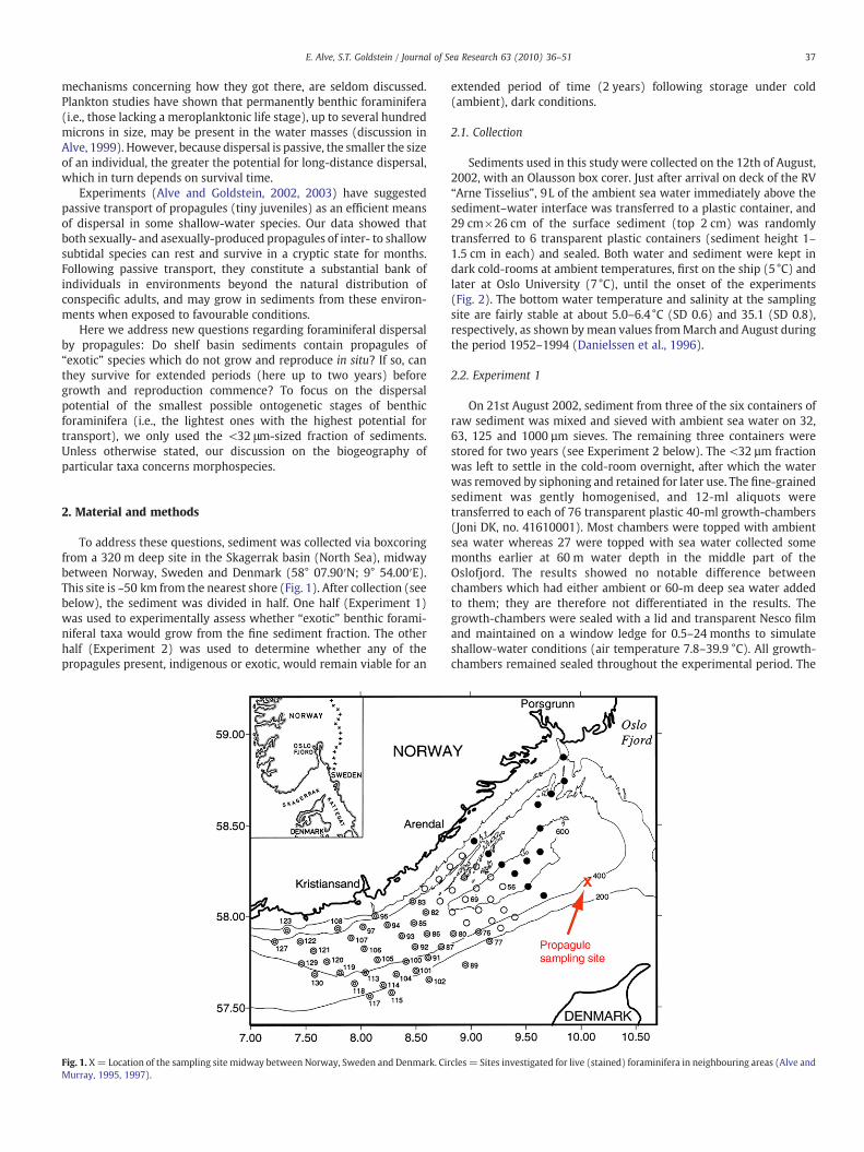
To address these questions, sediment was collected via boxcoringfrom a 320 m deep site in the Skagerrak basin (North Sea), midwaybetween Norway, Sweden and Denmark (58° 07.90′N; 9° 54.00′E).This site is ~50 km from the nearest shore (Fig. 1). After collection (seebelow), the sediment was divided in half. One half (Experiment 1)was used to experimentally assess whether “exotic” benthic forami-niferal taxa would grow from the fine sediment fraction. The otherhalf (Experiment 2) was used to determine whether any of thepropagules present, indigenous or exotic, would remain viable for an
Fig. 1. X= Location of the sampling site midway between Norway, Sweden and Denmark. CirMurray, 1995, 1997).
extended period of time (2 years) following storage under cold(ambient), dark conditions.
2.1. Collection
Sediments used in this study were collected on the 12th of August,2002, with an Olausson box corer. Just after arrival on deck of the RV“Arne Tisselius”, 9L of the ambient sea water immediately above thesediment–water interface was transferred to a plastic container, and29 cm×26 cm of the surface sediment (top 2 cm) was randomlytransferred to 6 transparent plastic containers (sediment height 1–1.5 cm in each) and sealed. Both water and sediment were kept indark cold-rooms at ambient temperatures, first on the ship (5°C) andlater at Oslo University (7°C), until the onset of the experiments(Fig. 2). The bottom water temperature and salinity at the samplingsite are fairly stable at about 5.0–6.4°C (SD 0.6) and 35.1 (SD 0.8),respectively, as shown by mean values fromMarch and August duringthe period 1952–1994 (Danielssen et al., 1996).
2.2. Experiment 1
On 21st August 2002, sediment from three of the six containers ofraw sediment was mixed and sieved with ambient sea water on 32,63, 125 and 1000 µm sieves. The remaining three containers werestored for two years (see Experiment 2 below). The <32 µm fractionwas left to settle in the cold-room overnight, after which the waterwas removed by siphoning and retained for later use. The fine-grainedsediment was gently homogenised, and 12-ml aliquots weretransferred to each of 76 transparent plastic 40-ml growth-chambers(Joni DK, no. 41610001). Most chambers were topped with ambientsea water whereas 27 were topped with sea water collected somemonths earlier at 60 m water depth in the middle part of theOslofjord. The results showed no notable difference betweenchambers which had either ambient or 60-m deep sea water addedto them; they are therefore not differentiated in the results. Thegrowth-chambers were sealed with a lid and transparent Nesco filmand maintained on a window ledge for 0.5–24 months to simulateshallow-water conditions (air temperature 7.8–39.9 °C). All growth-chambers remained sealed throughout the experimental period. The
cles = Sites investigated for live (stained) foraminifera in neighbouring areas (Alve and
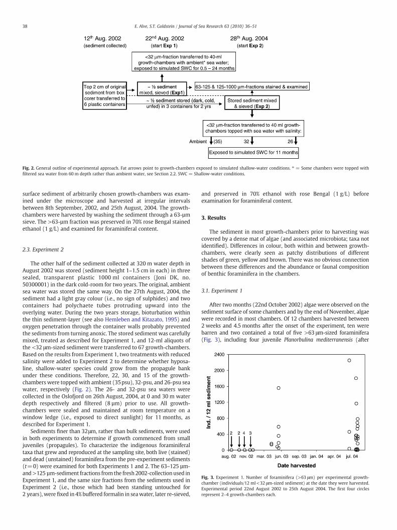
Fig. 2. General outline of experimental approach. Fat arrows point to growth-chambers exposed to simulated shallow-water conditions. ⁎ = Some chambers were topped withfiltered sea water from 60 m depth rather than ambient water, see Section 2.2. SWC = Shallow-water conditions.
38 E. Alve, S.T. Goldstein / Journal of Sea Research 63 (2010) 36–51
surface sediment of arbitrarily chosen growth-chambers was exam-ined under the microscope and harvested at irregular intervalsbetween 8th September, 2002, and 25th August, 2004. The growth-chambers were harvested by washing the sediment through a 63-µmsieve. The >63-µm fraction was preserved in 70% rose Bengal stainedethanol (1 g/L) and examined for foraminiferal content.
Fig. 3. Experiment 1. Number of foraminifera (>63 μm) per experimental growth-chamber (individuals/12 ml<32 μm-sized sediment) at the date they were harvested.Experimental period 22nd August 2002 to 25th August 2004. The first four circlesrepresent 2–4 growth-chambers each.
2.3. Experiment 2
The other half of the sediment collected at 320 m water depth inAugust 2002 was stored (sediment height 1–1.5 cm in each) in threesealed, transparent plastic 1000 ml containers (Joni DK, no.50300001) in the dark cold-room for two years. The original, ambientsea water was stored the same way. On the 27th August, 2004, thesediment had a light gray colour (i.e., no sign of sulphides) and twocontainers had polychaete tubes protruding upward into theoverlying water. During the two years storage, bioturbation withinthe thin sediment-layer (see also Hemleben and Kitazato, 1995) andoxygen penetration through the container walls probably preventedthe sediments from turning anoxic. The stored sediment was carefullymixed, treated as described for Experiment 1, and 12-ml aliquots ofthe <32 μm-sized sediment were transferred to 67 growth-chambers.Based on the results from Experiment 1, two treatments with reducedsalinity were added to Experiment 2 to determine whether hyposa-line, shallow-water species could grow from the propagule bankunder these conditions. Therefore, 22, 30, and 15 of the growth-chambers were toppedwith ambient (35psu), 32-psu, and 26-psu seawater, respectively (Fig. 2). The 26- and 32-psu sea waters werecollected in the Oslofjord on 26th August, 2004, at 0 and 30 m waterdepth respectively and filtered (8 µm) prior to use. All growth-chambers were sealed and maintained at room temperature on awindow ledge (i.e., exposed to direct sunlight) for 11 months, asdescribed for Experiment 1.
Sediments finer than 32μm, rather than bulk sediments, were usedin both experiments to determine if growth commenced from smalljuveniles (propagules). To characterize the indigenous foraminiferaltaxa that grew and reproduced at the sampling site, both live (stained)and dead (unstained) foraminifera from the pre-experiment sediments(t=0) were examined for both Experiments 1 and 2. The 63–125 µm-and>125 µm-sediment fractions from the fresh 2002-collectionused inExperiment 1, and the same size fractions from the sediments used inExperiment 2 (i.e., those which had been standing untouched for2 years), were fixed in 4%buffered formalin in seawater, later re-sieved,
and preserved in 70% ethanol with rose Bengal (1 g/L) beforeexamination for foraminiferal content.
3. Results
The sediment in most growth-chambers prior to harvesting wascovered by a dense mat of algae (and associated microbiota; taxa notidentified). Differences in colour, both within and between growth-chambers, were clearly seen as patchy distributions of differentshades of green, yellow and brown. There was no obvious connectionbetween these differences and the abundance or faunal compositionof benthic foraminifera in the chambers.
3.1. Experiment 1
After two months (22nd October 2002) algae were observed on thesediment surface of some chambers and by the end of November, algaewere recorded in most chambers. Of 12 chambers harvested between2 weeks and 4.5 months after the onset of the experiment, ten werebarren and two contained a total of five >63 μm-sized foraminifera(Fig. 3), including four juvenile Planorbulina mediterranensis (after
39E. Alve, S.T. Goldstein / Journal of Sea Research 63 (2010) 36–51
3 months), 2 of which were found in agglutinated “cysts”, and onedeformed (see below) Textularia earlandi. Between April 2003 andAugust 2004, an additional 46 growth-chambers were harvested. Ofthese, 4 were barren of foraminifera, 4 contained >63 μm-sizedindividuals of indigenous species, and 38 (83%) contained exoticshallow-water species in addition to the indigenous ones. All forami-nifera were counted in 27 growth-chambers (table 1 in Appendix A),whereas only the presence–absence was recorded in the remaining 19.Themaximum number of individuals/12 ml sedimentwas 2247 (Fig. 3).
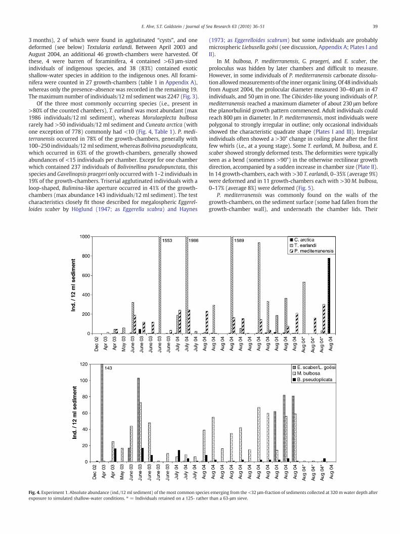
Of the three most commonly occurring species (i.e., present in>80% of the counted chambers), T. earlandi was most abundant (max1986 individuals/12 ml sediment), whereas Morulaeplecta bulbosararely had >50 individuals/12 ml sediment and Cuneata arctica (withone exception of 778) commonly had <10 (Fig. 4, Table 1). P. medi-terranensis occurred in 78% of the growth-chambers, generally with100–250 individuals/12 ml sediment,whereasBolivina pseuodoplicata,which occurred in 63% of the growth-chambers, generally showedabundances of <15 individuals per chamber. Except for one chamberwhich contained 237 individuals of Bolivinellina pseudopunctata, thisspecies andGavelinopsis praegeri only occurredwith 1–2 individuals in19% of the growth-chambers. Triserial agglutinated individuals with aloop-shaped, Bulimina-like aperture occurred in 41% of the growth-chambers (max abundance 143 individuals/12 ml sediment). The testcharacteristics closely fit those described for megalospheric Eggerel-loides scaber by Höglund (1947; as Eggerella scabra) and Haynes
Fig. 4. Experiment 1. Absolute abundance (ind./12 ml sediment) of the most common specieexposure to simulated shallow-water conditions. ⁎ = Individuals retained on a 125- rather
(1973; as Eggerelloides scabrum) but some individuals are probablymicrospheric Liebusella goësi (see discussion, Appendix A; Plates I andII).
In M. bulbosa, P. mediterranensis, G. praegeri, and E. scaber, theproloculus was hidden by later chambers and difficult to measure.However, in some individuals of P. mediterranensis carbonate dissolu-tion allowedmeasurements of the inner organic lining. Of 48 individualsfrom August 2004, the prolocular diameter measured 30–40 μm in 47individuals, and 50 μm in one. The Cibicides-like young individuals of P.mediterranensis reached a maximum diameter of about 230 μm beforethe planorbulinid growth pattern commenced. Adult individuals couldreach 800 μm in diameter. In P. mediterranensis, most individuals werepolygonal to strongly irregular in outline; only occasional individualsshowed the characteristic quadrate shape (Plates I and III). Irregularindividuals often showed a >30° change in coiling plane after the firstfew whirls (i.e., at a young stage). Some T. earlandi, M. bulbosa, and E.scaber showed strongly deformed tests. The deformities were typicallyseen as a bend (sometimes >90°) in the otherwise rectilinear growthdirection, accompanied by a sudden increase in chamber size (Plate II).In 14 growth-chambers, each with >30 T. earlandi, 0–35% (average 9%)were deformed and in 11 growth-chambers each with >30M. bulbosa,0–17% (average 8%) were deformed (Fig. 5).
P. mediterranensis was commonly found on the walls of thegrowth-chambers, on the sediment surface (some had fallen from thegrowth-chamber wall), and underneath the chamber lids. Their
s emerging from the <32 μm-fraction of sediments collected at 320 mwater depth afterthan a 63-μm sieve.
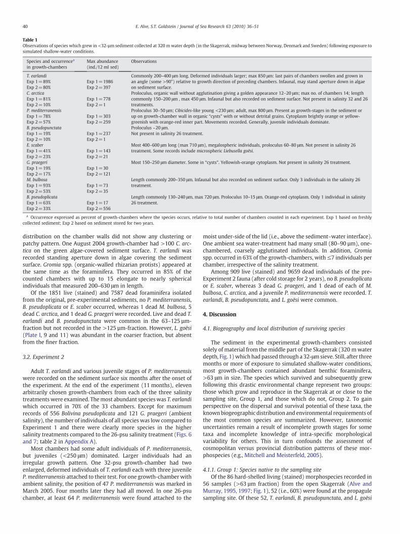
Table 1Observations of species which grew in <32-µm sediment collected at 320 mwater depth (in the Skagerrak, midway between Norway, Denmark and Sweden) following exposure tosimulated shallow-water conditions.
Species and occurrencea
in growth-chambersMax abundance(ind./12 ml sed)
Observations
T. earlandi Commonly 200–400 μm long. Deformed individuals larger; max 850 μm: last pairs of chambers swollen and grown inan angle (some >90°) relative to growth direction of preceding chambers. Infaunal, may stand aperture down in algaeon sediment surface.
Exp 1=89% Exp 1=1986Exp 2=80% Exp 2=397C. arctica Proloculus, organic wall without agglutination giving a golden appearance 12–20 μm; max no. of chambers 14; length
commonly 150–200 μm , max 450 μm. Infaunal but also recorded on sediment surface. Not present in salinity 32 and 26treatments.
Exp 1=81% Exp 1=778Exp 2=10% Exp 2=1P. mediterranensis Proloculus 30–50 μm; Cibicides-like young <230 μm; adult, max 800 μm. Present as growth-stages in the sediment or
up on growth-chamber wall in organic “cysts” with or without detrital grains. Cytoplasm brightly orange or yellow-greenish with orange-red inner part. Movements recorded. Generally, juvenile individuals dominate.
Exp 1=78% Exp 1=303Exp 2=57% Exp 2=259B. pseudopunctata Proloculus ~20 μm.Exp 1=19% Exp 1=237 Not present in salinity 26 treatment.Exp 2=10% Exp 2=1E. scaber Most 400–600 μm long (max 710 μm), megalospheric individuals, proloculus 60–80 μm. Not present in salinity 26
treatment. Some records include microspheric Liebusella goësi.Exp 1=41% Exp 1=143Exp 2=23% Exp 2=21G. praegeri Most 150–250 μm diameter. Some in “cysts”. Yellowish-orange cytoplasm. Not present in salinity 26 treatment.Exp 1=19% Exp 1=30Exp 2=17% Exp 2=121M. bulbosa Length commonly 200–350 μm. Infaunal but also recorded on sediment surface. Only 3 individuals in the salinity 26
treatment.Exp 1=93% Exp 1=73Exp 2=53% Exp 2=35B. pseudoplicata Length commonly 130–240 μm, max 720 μm. Proloculus 10–15 μm. Orange-red cytoplasm. Only 1 individual in salinity
26 treatment.Exp 1=63% Exp 1=17Exp 2=33% Exp 2=556
a Occurrence expressed as percent of growth-chambers where the species occurs, relative to total number of chambers counted in each experiment. Exp 1 based on freshlycollected sediment; Exp 2 based on sediment stored for two years.
40 E. Alve, S.T. Goldstein / Journal of Sea Research 63 (2010) 36–51
distribution on the chamber walls did not show any clustering orpatchy pattern. One August 2004 growth-chamber had >100 C. arc-tica on the green algae-covered sediment surface. T. earlandi wasrecorded standing aperture down in algae covering the sedimentsurface. Gromia spp. (organic-walled rhizarian protists) appeared atthe same time as the foraminifera. They occurred in 85% of thecounted chambers with up to 15 elongate to nearly sphericalindividuals that measured 200–630 μm in length.
Of the 1851 live (stained) and 7587 dead foraminifera isolatedfrom the original, pre-experimental sediments, no P. mediterranensis,B. pseudoplicata or E. scaber occurred, whereas 1 dead M. bulbosa, 5dead C. arctica, and 1 dead G. praegeri were recorded. Live and dead T.earlandi and B. pseudopunctata were common in the 63–125 µm-fraction but not recorded in the >125 µm-fraction. However, L. goësi(Plate I, 9 and 11) was abundant in the coarser fraction, but absentfrom the finer fraction.
3.2. Experiment 2
Adult T. earlandi and various juvenile stages of P. mediterranensiswere recorded on the sediment surface six months after the onset ofthe experiment. At the end of the experiment (11 months), elevenarbitrarily chosen growth-chambers from each of the three salinitytreatmentswere examined. Themost abundant specieswas T. earlandiwhich occurred in 70% of the 33 chambers. Except for maximumrecords of 556 Bolivina pseudoplicata and 121 G. praegeri (ambientsalinity), the number of individuals of all species was low compared toExperiment 1 and there were clearly more species in the highersalinity treatments compared to the 26-psu salinity treatment (Figs. 6and 7; table 2 in Appendix A).
Most chambers had some adult individuals of P. mediterranensis,but juveniles (<250 μm) dominated. Larger individuals had anirregular growth pattern. One 32-psu growth-chamber had twoenlarged, deformed individuals of T. earlandi each with three juvenileP. mediterranensis attached to their test. For one growth-chamber withambient salinity, the position of 47 P. mediterranensis was marked inMarch 2005. Four months later they had all moved. In one 26-psuchamber, at least 64 P. mediterranensis were found attached to the
moist under-side of the lid (i.e., above the sediment–water interface).One ambient sea water-treatment had many small (80–90 µm), one-chambered, coarsely agglutinated individuals. In addition, Gromiaspp. occurred in 63% of the growth-chambers, with≤7 individuals perchamber, irrespective of the salinity treatment.
Among 909 live (stained) and 9659 dead individuals of the pre-Experiment 2 fauna (after cold storage for 2 years), no B. pseudoplicataor E. scaber, whereas 3 dead G. praegeri, and 1 dead of each of M.bulbosa, C. arctica, and a juvenile P. mediterranensis were recorded. T.earlandi, B. pseudopunctata, and L. goësi were common.
4. Discussion
4.1. Biogeography and local distribution of surviving species
The sediment in the experimental growth-chambers consistedsolely of material from the middle part of the Skagerrak (320 mwaterdepth, Fig. 1)which had passed through a 32-μmsieve. Still, after threemonths or more of exposure to simulated shallow-water conditions,most growth-chambers contained abundant benthic foraminifera,>63 μm in size. The species which survived and subsequently grewfollowing this drastic environmental change represent two groups:those which grow and reproduce in the Skagerrak at or close to thesampling site, Group 1, and those which do not, Group 2. To gainperspective on the dispersal and survival potential of these taxa, theknownbiogeographic distribution and environmental requirements ofthe most common species are summarized. However, taxonomicuncertainties remain a result of incomplete growth stages for sometaxa and incomplete knowledge of intra-specific morphologicalvariability for others. This in turn confounds the assessment ofcosmopolitan versus provincial distribution patterns of these mor-phospecies (e.g., Mitchell and Meisterfeld, 2005).
4.1.1. Group 1: Species native to the sampling siteOf the 86 hard-shelled living (stained) morphospecies recorded in
56 samples (>63 µm fraction) from the open Skagerrak (Alve andMurray, 1995, 1997; Fig. 1), 52 (i.e., 60%) were found at the propagulesampling site. Of these 52, T. earlandi, B. pseudopunctata, and L. goësi
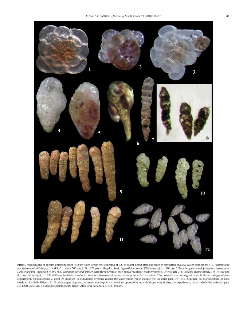
Plate I. Micrographs of species emerging from <32 μm-sized sediments collected at 320 m water depth after exposure to simulated shallow-water conditions: 1–3. Planorbulinamediterranensis d'Orbigny. 1 and 3; D=about 500 μm. 2; D=270 μm. 4. Megalospheric Eggerelloides scaber (Williamson). L=400 μm. 5. Rose Bengal stained, juvenile, microsphericLiebusella goësi Höglund. L=450 m. 6. Textularia earlandi Parker, with three juvenile, rose Bengal stained P. mediterranensis. L=500 μm. 7–8. Cuneata arctica (Brady). 7; L=390 μm.8; transmitted light, L=170–290 μm. Individuals reflect transitions between blunt and more pointed last chamber. The proloculi are not agglutinated. 9. Growth stages of pre-experiment, megalospheric L. goësi. As opposed to individuals growing during the experiment, these include the uniserial part. L=1030–2380 μm. 10. Morulaeplecta bulbosaHöglund. L=340–510 μm. 11. Growth stages of pre-experiment, microspheric L. goësi. As opposed to individuals growing during the experiment, these include the uniserial part.L=1270–2250 μm. 12. Bolivina pseudoplicata Heron-Allen and Earland. L=130–240 μm.
41E. Alve, S.T. Goldstein / Journal of Sea Research 63 (2010) 36–51
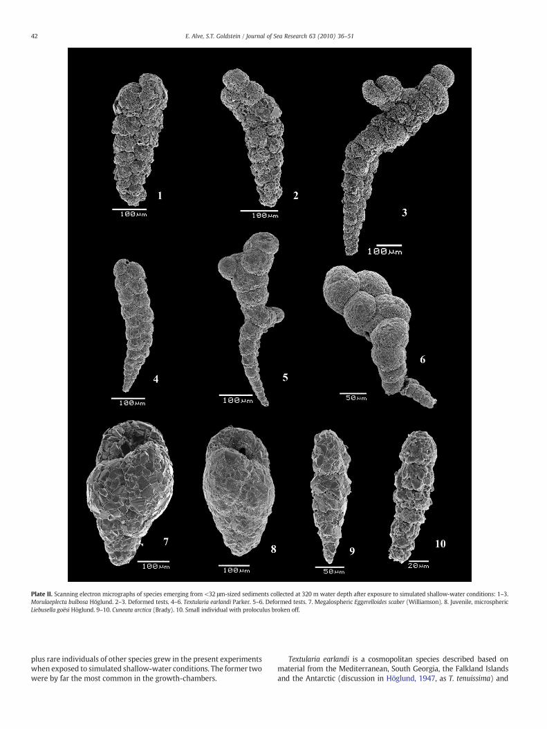
Plate II. Scanning electron micrographs of species emerging from <32 μm-sized sediments collected at 320 m water depth after exposure to simulated shallow-water conditions: 1–3.Morulaeplecta bulbosa Höglund. 2–3. Deformed tests. 4–6. Textularia earlandi Parker. 5–6. Deformed tests. 7. Megalospheric Eggerelloides scaber (Williamson). 8. Juvenile, microsphericLiebusella goësi Höglund. 9–10. Cuneata arctica (Brady). 10. Small individual with proloculus broken off.
42 E. Alve, S.T. Goldstein / Journal of Sea Research 63 (2010) 36–51
plus rare individuals of other species grew in the present experimentswhen exposed to simulated shallow-water conditions. The former twowere by far the most common in the growth-chambers.
Textularia earlandi is a cosmopolitan species described based onmaterial from the Mediterranean, South Georgia, the Falkland Islandsand the Antarctic (discussion in Höglund, 1947, as T. tenuissima) and
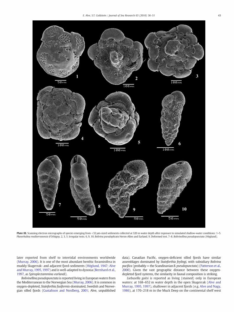
Plate III. Scanning electron micrographs of species emerging from <32 μm-sized sediments collected at 320 m water depth after exposure to simulated shallow-water conditions: 1–5.Planorbulina mediterranensis d'Orbigny. 2, 3, 5. Irregular tests. 6, 9, 10. Bolivina pseudoplicata Heron-Allen and Earland. 9. Deformed test. 7–8. Bolivinellina pseudopunctata (Höglund).
43E. Alve, S.T. Goldstein / Journal of Sea Research 63 (2010) 36–51
later reported from shelf to intertidal environments worldwide(Murray, 2006). It is one of the most abundant benthic foraminifera inmuddy Skagerrak- and adjacent fjord-sediments (Höglund, 1947; AlveandMurray, 1995, 1997) and iswell-adapted to dysoxia (Bernhard et al.,1997, as Spiroplectammina earlandi).
Bolivinellinapseudopunctata is reported living in Europeanwaters fromtheMediterranean to the Norwegian Sea (Murray, 2006). It is common inoxygen-depleted, Stainforthia fusiformis-dominated, Swedish and Norwe-gian silled fjords (Gustafsson and Nordberg, 2001; Alve, unpublished
data). Canadian Pacific, oxygen-deficient silled fjords have similarassemblages dominated by Stainforthia feylingi, with subsidiary Bolivinapacifica (probably=the ScandinavianB.pseudopunctata) (Patterson et al.,2000). Given the vast geographic distance between these oxygen-depleted fjord systems, the similarity in faunal composition is striking.
Liebusella goësi is reported as living (stained) only in Europeanwaters: at 168–652 m water depth in the open Skagerrak (Alve andMurray, 1995, 1997), shallower in adjacent fjords (e.g. Alve and Nagy,1986), at 170–218 m in the Muck Deep on the continental shelf west
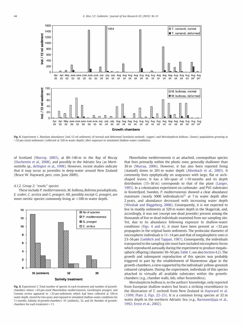
Fig. 5. Experiment 1. Absolute abundance (ind./12 ml sediment) of normal and deformed Textularia earlandi- (upper) and Morulaeplecta bulbosa- (lower) populations growing in<32 μm-sized sediments (collected at 320 m water depth) after exposure to simulated shallow-water conditions.
44 E. Alve, S.T. Goldstein / Journal of Sea Research 63 (2010) 36–51
of Scotland (Murray, 2003), at 80–140 m in the Bay of Biscay(Duchemin et al., 2008), and possibly in the Adriatic Sea (as Marti-nottiella sp., deStigter et al., 1998). However, recent studies indicatethat it may occur as juveniles in deep-water around New Zealand(Bruce W. Hayward, pers. com. June 2009).
4.1.2. Group 2: “exotic” speciesThese include P. mediterranensis,M. bulbosa, Bolivina pseudoplicata,
E. scaber, C. arctica and G. praegeri. All, possibly except G. praegeri, areinner-neritic species commonly living at <100 m water depth.
Fig. 6. Experiment 2. Total number of species in each treatment and number of growth-chambers where >63 μm-sized Planorbulina mediterranensis, Gavelinopsis praegeri, andCuneata arctica appeared in <32 μm-sediments which had been collected at 320 mwater depth, stored for two years and exposed to simulated shallow-water conditions for11 months. Salinity of growth-chambers: 35 (ambient), 32, and 26. Number of growth-chambers for each treatment=11.
Planorbulina mediterranensis is an attached, cosmopolitan speciesthat lives primarily within the photic zone, generally shallower than30 m (Murray, 2006). However, it has also been reported living(stained) down to 201 m water depth (Altenbach et al., 2003). Itcommonly lives epiphytically on seagrasses with large, flat or arch-shaped leaves. It has a life-span of >10 months and its depthdistribution (15–30 m) corresponds to that of the plant (Langer,1993). In a colonization experiment on carbonate- and PVC-substratesin Kosterfjord, Sweden, P. mediterranensis showed a clear abundancemaximum (nearly 5000 individuals/m2) at 7 m water depth after2 years, and abundance decreased with increasing water depth(Wisshak and Rüggeberg, 2006). Consequently, it is not expected tolive in muddy sediments at 320 m water depth in the Skagerrak, and,accordingly, it was not (except one dead juvenile) present among thethousands of live or dead individuals examined from our sampling site.Yet, due to its abundance following exposure to shallow-waterconditions (Figs. 4 and 6), it must have been present as <32 µmpropagules in the original basin sediments. The prolocular diameter ofmicrospheric individuals is 11–14 μmand that of megalospheric ones is23–56 μm (Loeblich and Tappan, 1987). Consequently, the individualstransported to the sampling sitemust have includedmicrospheric formswhich reproduced asexually during the experiment to producemegalo-spheric offspring (diameter 30–50 μm,Table 1; see also Section 4.2). Thegrowth and subsequent reproduction of this species was probablytriggered in part by the establishment of filamentous algae in thegrowth-chambers, a viewsupportedby the individuals' yellow-greenishcoloured cytoplasm. During the experiment, individuals of this speciesattached to virtually all available substrates within the growth-chambers (e.g., chamber walls, lids, other foraminifera).
Morulaeplecta bulbosa is, to the authors' knowledge, only reportedfrom European shallow-waters but bears a striking resemblance tothe illustration of T. earlandi from New Zealand in Hayward et al.(1999, Plate 2, Figs. 22–23). It is a common living species at 32 mwater depth in the northern Adriatic Sea (e.g., Barmawidjaja et al.,1992; Ernst et al., 2002).
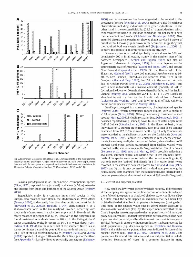
Fig. 7. Experiment 2. Absolute abundance (ind./12 ml sediment) of the most commonspecies (>63 μm) growing in <32 μm sediment collected at 320 m water depth, storeddark and cold for two years and exposed to simulated shallow-water conditions for11 months. Salinity of growth-chambers A) 35=ambient; B) 32; and C) 26.
45E. Alve, S.T. Goldstein / Journal of Sea Research 63 (2010) 36–51
Bolivina pseudoplicata is an inner-neritic, cosmopolitan species(Sliter, 1970), reported living (stained) in shallow (<50 m) estuariesand lagoons from Japan and both sides of the Atlantic Ocean (Murray,2006).
Eggerelloides scaber is a common inner shelf species in NWEurope, also recorded from Brazil, the Mediterranean, West Africa(Murray, 2006), and recently from the subantarctic southwest Pacific(Hayward et al., 2007a). Höglund (1947) characterised it as ashallow-water form in the Gullmarfjord, Sweden, occurring withthousands of specimens in each core sample at 15–20 m depth. Herarely recorded it deeper than 60 m. However, in the Skagerrak, hefound unstained individuals down to 204 m. In the Kattegat, the E.scaber assemblage typically occurs at 14–35 m water depth (Con-radsen et al., 1994). On the inner shelf of the southern North Sea, E.scaber dominates parts of the year at 52 mwater depth and canmakeup 1–10% of the live assemblage at 63 m (Murray, 1992), and Murray(2003) reported it living at 170 m in the Muck Deep, just off Scotland(see Appendix A). E. scaber lives epiphytically on seagrass (Debenay,
2000) and its occurrence has been suggested to be related to thepresence of Zostera (Mendes et al., 2004). Herbivory also fits with ourobservations including individuals with green cytoplasm. On theother hand, in thewestern Baltic Sea (23.5 m) organic detritus, whichtriggered reproduction in Elphidium excavatum, did not seem to havethe same effect on E. scaber (Schönfeld and Numberger, 2007). Also,an unfed disturbance experiment showed that it survived 3 weeks ofburial without moving up or down in the sediment, suggesting thatthe required food was evenly distributed (Duijnstee et al., 2003). Inconcert, this points to an omnivorous feeding strategy.
Cuneata arctica is recorded (probably dead) down to 100 andoccasionally 200 m in all oceans, mainly in the northern part of thenorthern hemisphere (Loeblich and Tappan, 1987), but also offArgentina (references in Haynes, 1973), in coastal lagoons on thesoutheastern coast of Australia (Yassini and Jones, 1995), and aroundNew Zealand (Hayward et al., 1999). On the Danish side of theSkagerrak, Höglund (1947) recorded unstained Reophax nana at 66–400 m. Live (stained) individuals are reported from 17 m in theOslofjord (Alve and Nagy, 1986), from 32 m in the northern AdriaticSea (as Acostata mariae, Ernst et al., 2002; Duijnstee et al., 2003), andwith a few individuals (as Clavulina obscura) generally at <80 m(occasionally down to 150 m) in the southernNorth Sea and the EnglishChannel (Murray, 2006, web tables WA 114, 117, 118). Live R. nana areabundant in salt marshes on the Atlantic side of North America(Goldstein and Watkins, 1998) and down to 40 m off Baja Californiaon the Pacific side (references in Murray, 2006).
Gavelinopsis praegeri is a cosmopolitan, clinging/attached species(Murray, 2006) which occasionally moves around with a speed of~1.24 μm/min (Gross, 2000). Although commonly considered a shelfspecies (Murray, 2006), includingestuaries (e.g.,Debenayet al., 2006), ithas been reported living (stained) down to 3736 m water depth in theGulf of Guinea (Altenbach et al., 2003). In the Skagerrak basin, livingindividuals of G. praegeri are extremely rare. In a total of 56 samplesexamined from 117 to 652 m water depth (Fig. 1), only 2 individualswere recorded at the shallowest station on the Danish side (Alve andMurray, 1995, 1997). Because it is epifaunal in high energy environ-ments, its test may readily be transported. Indeed, a few dead tests of G.praegeri (and other species transported from shallow-water) wererecorded on the southern slope of the Skagerrak basin, NW of Denmark(Bergsten et al., 1996; Alve and Murray, 1997) probably reflecting ashallower, southern source. Considering that 1) live (stained) indivi-duals of the species were not recorded at the present sampling site, 2)that only two live (stained) individuals (at 117 m water depth) wererecorded in the extensive data set reported by Alve and Murray (1995,1997), and 3) that it only occurred with 4 dead examples among thenearly 20,000 tests examined from the sampling site, it is inferred that itdoes not growand reproduce in soft sediment at 320 m in theSkagerrak.
4.2. Survival and dispersal potential
How could shallow-water specieswhich do not grow and reproduceat the sampling site appear in the fine-fraction of sediments collectedthere following exposure to simulated shallow-water conditions (Exp.1)? How could the same happen in sediments that had been keptisolated in the dark at ambient temperature for two years (duringwhichtime none of the shallow-water species grew) before exposure toshallow-water conditions (Exp. 2)? Our experiments show that the sixGroup 2-species were transported to the sampling site as <32 μm-sizedpropagules (juveniles), and that theymust be particularly resilient, havea good survival potential, and be able to remain dormant for two years.Survival for years in culturewithout reproduction has been reported foradult populations (e.g., deep-sea species in Hemleben and Kitazato,1995) and a high survival potential has been indicated for some of thepresent species (e.g., Ernst et al., 2002; Duijnstee et al., 2003). Thepresent results extend this resilience and survival potential to includejuveniles. Formation of “cysts” is a common feature in many
46 E. Alve, S.T. Goldstein / Journal of Sea Research 63 (2010) 36–51
foraminiferal species serving numerous functions such as feeding,reproduction, growth and protection (e.g., Myers, 1936; Heinz et al.,2005, and references therein). For P. mediterranensis and G. praegeri,residing in “cysts” on thewalls of the growth-chambers is in accordancewith Gross' (2000) observations, and it probably aided survival underotherwise stressful conditions.
Of all the indigenous foraminiferal species recorded at thesampling site, only T. earlandi and B. pseudopunctata grew andcontinued to flourish when transferred from bathyal (320 m) tosimulated shallow-water (0 m) conditions. The explosive reproduc-tion of the widely occurring T. earlandi probably reflects a particularlyopportunistic life strategy. The fact that it was recorded standingaperture down in algae indicates herbivory. Why didn't other shelfspecies grow in the present experiments? First, the environmentalconditions (e.g., sun light and UV radiation, temperature, lack ofappropriate food and biochemical exchange with surroundingenvironment) in the growth-chambers were dramatically differentfrom those at 320 m in the Skagerrak. Second, the experimentaldesign using <32 μm sediments only, excluded species with largerproloculi, including the megalospheric generation in some species(e.g., E. scaber and L. goësi; for proloculi sizes, see Appendix A). Third,the high abundance of T. earlandi may have suppressed growth ofother infaunal species. Fourth, life history dynamics or limitedsurvival capabilities of propagules of other shallow-water speciesmay have precluded their occurrence in basinal sediments of theSkagerrak.
Overall, the environmental conditions in the growth-chambersmust have been hostile to many species, implying that the ones whichdid grow are hardy species with a substantial survival potential, andall these morphospecies seem to be cosmopolitan. Their broaddistribution is probably in part due to a high survival and dispersalpotential in their propagules. For the agglutinated forms, the lack ofagglutination of the proloculi inM. bulbosa, T. earlandi, C. arctica (as R.nana), and microspheric E. scaber (Höglund, 1947) may increasebuoyancy, thus aiding dispersal of the juvenile stage compared to thesubsequent heavier agglutinated growth stages.
Kuhnt et al. (2005, p. 105) suggested that bolivinids and serialagglutinated species are “highly capable of survival, rapid dispersal,and rapid to explosive population increase”. Indeed, except for thetemporarily attached P. mediterranensis and G. praegeri, theforaminifera growing in our experiments are exactly bolivinidsand serial agglutinated species. Examples of resilient serial agglu-tinated species include survival and colonization of Textulariacushmani in the Red Sea during early Holocene (Almogi-Labinet al., 1996) and the sudden appearance of T. earlandi in an oil-treated mesocosm experiment (Ernst et al., 2006). The fact thatbolivinids are among the most commonly reported live benthicforaminifera in plankton tows (references in Alve, 1999), that abolivinid appeared and flourished in a sealed culture of isolatedindividuals of the protist Gromia oviformis (Alve and Goldstein,2002), and the indication that lower Miocene, planktic, biserialStreptochilus spp. evolved from benthic ancestors (Smart andThomas, 2007) point in the same direction.
Although attached forms may be transported, and therebydispersed, together with their substrate (e.g., on floating algae,Spindler, 1980), species of some, for instance Cibicides, are suggestedto disperse as “larvae” (Svavarsson and Davidsdottir, 1995) orpropagules (Beaulieu, 2001). Dispersal through propagule transportis probably essential for large-sized and attached foraminifera (Alveand Goldstein, 2003). The appearance of P. mediterranensis and G.praegeri in the present experiments supports this view.
Essential biological characteristics, such as modes of reproductionvary among benthic foraminifera, and their life cycle is more variedthan in virtually any other group of protists (Goldstein, 1999).Furthermore, lack of significant genetic differences between speci-mens collected at depths ranging from 1000 to 6300 m suggests that
Bathyallogromia weddellensis is adapted to conditions that span abroader bathymetric range than for most species (Gooday et al.,2004). Consequently, it is not unreasonable that other biologicalproperties, such as resilience, dispersal mechanisms and potentials,also vary within the group. On the contrary, field experiments haveshown that the rate of settlement and colonization of largerforaminifera onto new hard substrates vary between species (Fujita,2004), and the present experiments indicate substantial inter-specificdifferences in dispersal potential.
To our knowledge, a distinction between micro- and megalo-spheric populations of the exotic species growing in the experimentsis only known for P. mediterranensis (Loeblich and Tappan, 1987)and E. scaber (Höglund, 1947). In both species, the prolocular size ofthe megalospheric generation is larger than the sediment grain sizeused in the experiments. This is interesting because whereasmicrospheric forms of E. scaber commonly constitute only about5% of the natural Skagerrak coastal-populations (Höglund, 1947)they were transported to our sampling site and initially grew andreproduced asexually in the present experiments. Consequently, forP. mediterranensis and E. scaber, the dispersed propagules belongedto the diploid, sexually-produced microspheric generation. Thisimplies higher genetic variability in the populations (Hallock, 1985)and thereby enhances the possibility of speciation in recentlycolonized areas.
Transport of growth stages is well-known in benthic foraminifera(examples in Alve, 1999), but, as long as their survival potential isgood, small propagules are likely to travel further than largerindividuals. Our evidence for propagule dispersal supports Myers'(1936, p. 134) suggestion that “…juvenile Foraminifera are capable ofincreasing their flotation or surface resistance by extending numerousfilose pseudopodia. This would also tend to lower the specific gravityof the organism, since the extension of pseudopodia is accompaniedby the absorption of water.” Murray (2007, p. 172) posed thequestion: “If propagules aid dispersal of benthic foraminifera (……)why are so many species endemic?” As pointed out, biologicalcharacteristics vary between species and there is no reason whysurvival- and dispersal-properties should be different. However, dueto under-sampling and rarity, endemism may be less than so farappreciated (Finlay et al., 2004). An example is that six agglutinatedspecies not previously recorded from the Scottish shelf were foundthere for the first time by dissolving away all the calcareous taxaleaving behind (i.e., concentrating) the (originally rare) agglutinatedforms only (Murray, 2003).
Furthermore, survival and dispersal potential are not the only factorsthat influence a species' successful dispersal. For example, neriticbenthic macrofaunal biogeographic provincial boundaries are com-monly associated with oceanographic boundaries and agree with thoseof benthic foraminifera (e.g., Culver andBuzas, 1999). This indicates thateven though benthic foraminifera, as opposed to benthic macrofauna,lack a pelagic larval stage, similar factors limit their distribution/dispersal. Such boundaries probably dictate the distribution of manyspecies despite a high dispersal potential. Due to physical and ecologicalbarriers not even planktic foraminifera, which are capable of long-distance dispersal, are ubiquitously dispersed throughout the domainsto which they are adapted (Darling and Wade, 2008). Still, 53 of 878benthic species are currently occurring ubiquitously around North andCentral America. Further, these must have evolved recently anddispersed rapidly as they have no fossil record (Buzas and Culver,1991). Additionally, genetically verified species such as the shallow-water Ammonia type 1 and Psammophaga sp. are widely distributed(Pawlowski and Holzmann, 2008) and the genetic diversity in sometypically abyssal foraminiferal species is minimal on a global scale(Pawlowski et al., 2007). In concert, it seems that the biogeographicdistribution of benthic foraminifera depends on an interplay betweendispersal potential of different species and the position of environmen-tal boundaries. According to Vanormelingen et al. (2008, p. 393) “…the
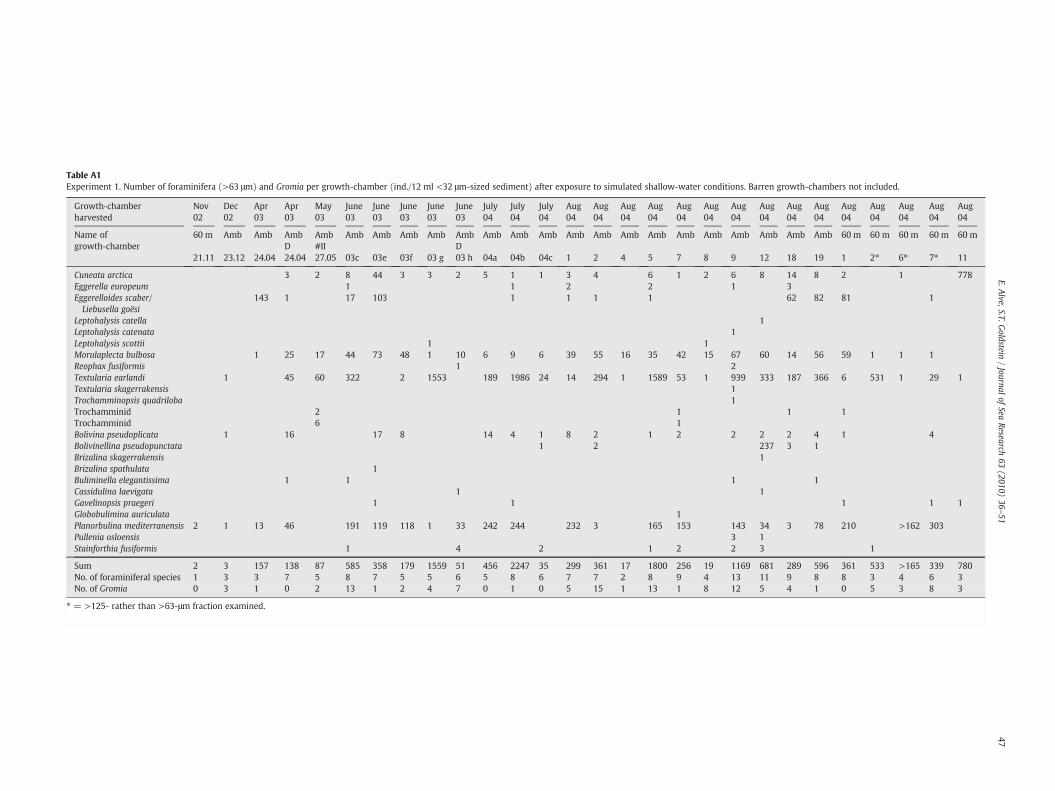
Table A1Experiment 1. Number of foraminifera (>63 μm) and Gromia per growth-chamber (ind./12 ml <32 μm-sized sediment) after exposure to simulated shallow-water conditions. Barren growth-chambers not included.
Growth-chamberharvested
Nov02
Dec02
Apr03
Apr03
May03
June03
June03
June03
June03
June03
July04
July04
July04
Aug04
Aug04
Aug04
Aug04
Aug04
Aug04
Aug04
Aug04
Aug04
Aug04
Aug04
Aug04
Aug04
Aug04
Aug04
Name ofgrowth-chamber
60 m Amb Amb AmbD
Amb#II
Amb Amb Amb Amb AmbD
Amb Amb Amb Amb Amb Amb Amb Amb Amb Amb Amb Amb Amb 60 m 60 m 60 m 60 m 60 m
21.11 23.12 24.04 24.04 27.05 03c 03e 03f 03 g 03 h 04a 04b 04c 1 2 4 5 7 8 9 12 18 19 1 2⁎ 6⁎ 7⁎ 11
Cuneata arctica 3 2 8 44 3 3 2 5 1 1 3 4 6 1 2 6 8 14 8 2 1 778Eggerella europeum 1 1 2 2 1 3Eggerelloides scaber/Liebusella goësi
143 1 17 103 1 1 1 1 62 82 81 1
Leptohalysis catella 1Leptohalysis catenata 1Leptohalysis scottii 1 1Morulaplecta bulbosa 1 25 17 44 73 48 1 10 6 9 6 39 55 16 35 42 15 67 60 14 56 59 1 1 1Reophax fusiformis 1 2Textularia earlandi 1 45 60 322 2 1553 189 1986 24 14 294 1 1589 53 1 939 333 187 366 6 531 1 29 1Textularia skagerrakensis 1Trochamminopsis quadriloba 1Trochamminid 2 1 1 1Trochamminid 6 1Bolivina pseudoplicata 1 16 17 8 14 4 1 8 2 1 2 2 2 2 4 1 4Bolivinellina pseudopunctata 1 2 237 3 1Brizalina skagerrakensis 1Brizalina spathulata 1Buliminella elegantissima 1 1 1 1Cassidulina laevigata 1 1Gavelinopsis praegeri 1 1 1 1 1Globobulimina auriculata 1Planorbulina mediterranensis 2 1 13 46 191 119 118 1 33 242 244 232 3 165 153 143 34 3 78 210 >162 303Pullenia osloensis 3 1Stainforthia fusiformis 1 4 2 1 2 2 3 1
Sum 2 3 157 138 87 585 358 179 1559 51 456 2247 35 299 361 17 1800 256 19 1169 681 289 596 361 533 >165 339 780No. of foraminiferal species 1 3 3 7 5 8 7 5 5 6 5 8 6 7 7 2 8 9 4 13 11 9 8 8 3 4 6 3No. of Gromia 0 3 1 0 2 13 1 2 4 7 0 1 0 5 15 1 13 1 8 12 5 4 1 0 5 3 8 3
⁎ = >125- rather than >63-μm fraction examined.
47E.A
lve,S.T.Goldstein
/Journal
ofSea
Research63
(2010)36
–51
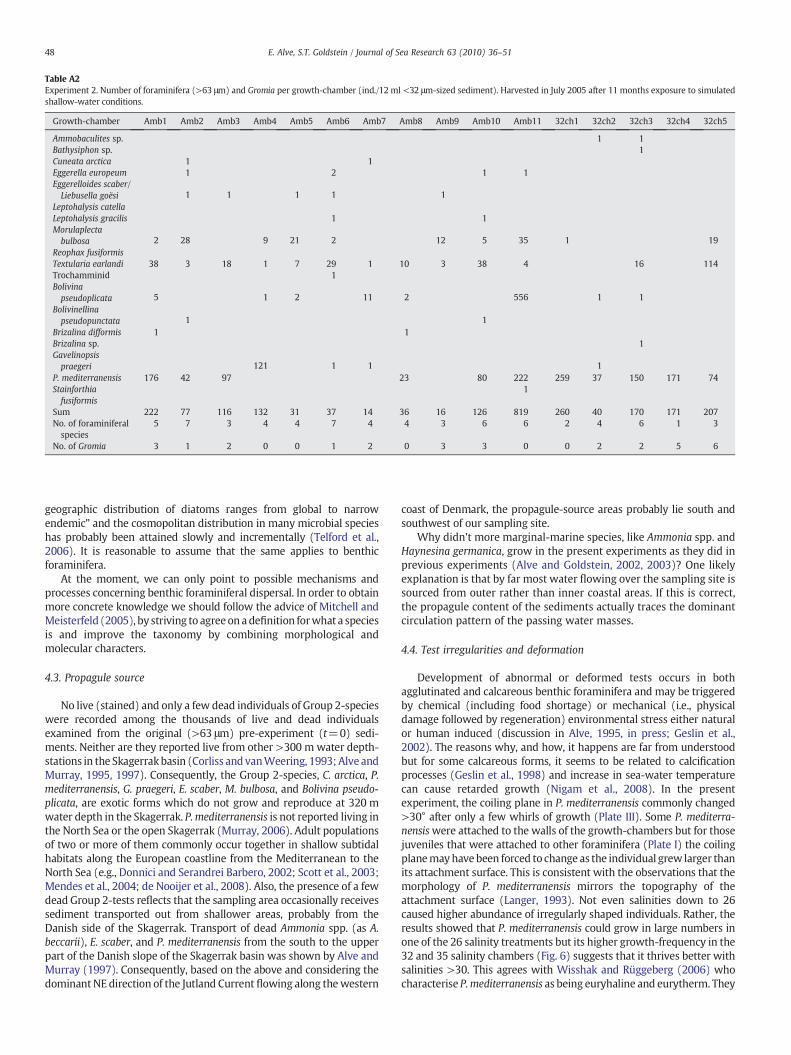
Table A2Experiment 2. Number of foraminifera (>63 μm) and Gromia per growth-chamber (ind./12 ml <32 μm-sized sediment). Harvested in July 2005 after 11 months exposure to simulatedshallow-water conditions.
Growth-chamber Amb1 Amb2 Amb3 Amb4 Amb5 Amb6 Amb7 Amb8 Amb9 Amb10 Amb11 32ch1 32ch2 32ch3 32ch4 32ch5
Ammobaculites sp. 1 1Bathysiphon sp. 1Cuneata arctica 1 1Eggerella europeum 1 2 1 1Eggerelloides scaber/Liebusella goësi 1 1 1 1 1
Leptohalysis catellaLeptohalysis gracilis 1 1Morulaplectabulbosa 2 28 9 21 2 12 5 35 1 19
Reophax fusiformisTextularia earlandi 38 3 18 1 7 29 1 10 3 38 4 16 114Trochamminid 1Bolivinapseudoplicata 5 1 2 11 2 556 1 1
Bolivinellinapseudopunctata 1 1
Brizalina difformis 1 1Brizalina sp. 1Gavelinopsispraegeri 121 1 1 1
P. mediterranensis 176 42 97 23 80 222 259 37 150 171 74Stainforthiafusiformis
1
Sum 222 77 116 132 31 37 14 36 16 126 819 260 40 170 171 207No. of foraminiferalspecies
5 7 3 4 4 7 4 4 3 6 6 2 4 6 1 3
No. of Gromia 3 1 2 0 0 1 2 0 3 3 0 0 2 2 5 6
48 E. Alve, S.T. Goldstein / Journal of Sea Research 63 (2010) 36–51
geographic distribution of diatoms ranges from global to narrowendemic” and the cosmopolitan distribution in many microbial specieshas probably been attained slowly and incrementally (Telford et al.,2006). It is reasonable to assume that the same applies to benthicforaminifera.
At the moment, we can only point to possible mechanisms andprocesses concerning benthic foraminiferal dispersal. In order to obtainmore concrete knowledge we should follow the advice of Mitchell andMeisterfeld (2005), by striving to agreeon adefinition forwhat a speciesis and improve the taxonomy by combining morphological andmolecular characters.
4.3. Propagule source
No live (stained) and only a few dead individuals of Group 2-specieswere recorded among the thousands of live and dead individualsexamined from the original (>63 μm) pre-experiment (t=0) sedi-ments. Neither are they reported live from other >300 m water depth-stations in the Skagerrakbasin (Corliss and vanWeering, 1993; Alve andMurray, 1995, 1997). Consequently, the Group 2-species, C. arctica, P.mediterranensis, G. praegeri, E. scaber, M. bulbosa, and Bolivina pseudo-plicata, are exotic forms which do not grow and reproduce at 320 mwater depth in the Skagerrak. P.mediterranensis is not reported living inthe North Sea or the open Skagerrak (Murray, 2006). Adult populationsof two or more of them commonly occur together in shallow subtidalhabitats along the European coastline from the Mediterranean to theNorth Sea (e.g., Donnici and Serandrei Barbero, 2002; Scott et al., 2003;Mendes et al., 2004; de Nooijer et al., 2008). Also, the presence of a fewdead Group 2-tests reflects that the sampling area occasionally receivessediment transported out from shallower areas, probably from theDanish side of the Skagerrak. Transport of dead Ammonia spp. (as A.beccarii), E. scaber, and P. mediterranensis from the south to the upperpart of the Danish slope of the Skagerrak basin was shown by Alve andMurray (1997). Consequently, based on the above and considering thedominant NE direction of the Jutland Current flowing along thewestern
coast of Denmark, the propagule-source areas probably lie south andsouthwest of our sampling site.
Why didn't more marginal-marine species, like Ammonia spp. andHaynesina germanica, grow in the present experiments as they did inprevious experiments (Alve and Goldstein, 2002, 2003)? One likelyexplanation is that by far most water flowing over the sampling site issourced from outer rather than inner coastal areas. If this is correct,the propagule content of the sediments actually traces the dominantcirculation pattern of the passing water masses.
4.4. Test irregularities and deformation
Development of abnormal or deformed tests occurs in bothagglutinated and calcareous benthic foraminifera and may be triggeredby chemical (including food shortage) or mechanical (i.e., physicaldamage followed by regeneration) environmental stress either naturalor human induced (discussion in Alve, 1995, in press; Geslin et al.,2002). The reasons why, and how, it happens are far from understoodbut for some calcareous forms, it seems to be related to calcificationprocesses (Geslin et al., 1998) and increase in sea-water temperaturecan cause retarded growth (Nigam et al., 2008). In the presentexperiment, the coiling plane in P. mediterranensis commonly changed>30° after only a few whirls of growth (Plate III). Some P. mediterra-nensiswere attached to the walls of the growth-chambers but for thosejuveniles that were attached to other foraminifera (Plate I) the coilingplanemayhave been forced to change as the individual grew larger thanits attachment surface. This is consistent with the observations that themorphology of P. mediterranensis mirrors the topography of theattachment surface (Langer, 1993). Not even salinities down to 26caused higher abundance of irregularly shaped individuals. Rather, theresults showed that P. mediterranensis could grow in large numbers inone of the 26 salinity treatments but its higher growth-frequency in the32 and 35 salinity chambers (Fig. 6) suggests that it thrives better withsalinities >30. This agrees with Wisshak and Rüggeberg (2006) whocharacterise P.mediterranensis as being euryhaline and eurytherm. They
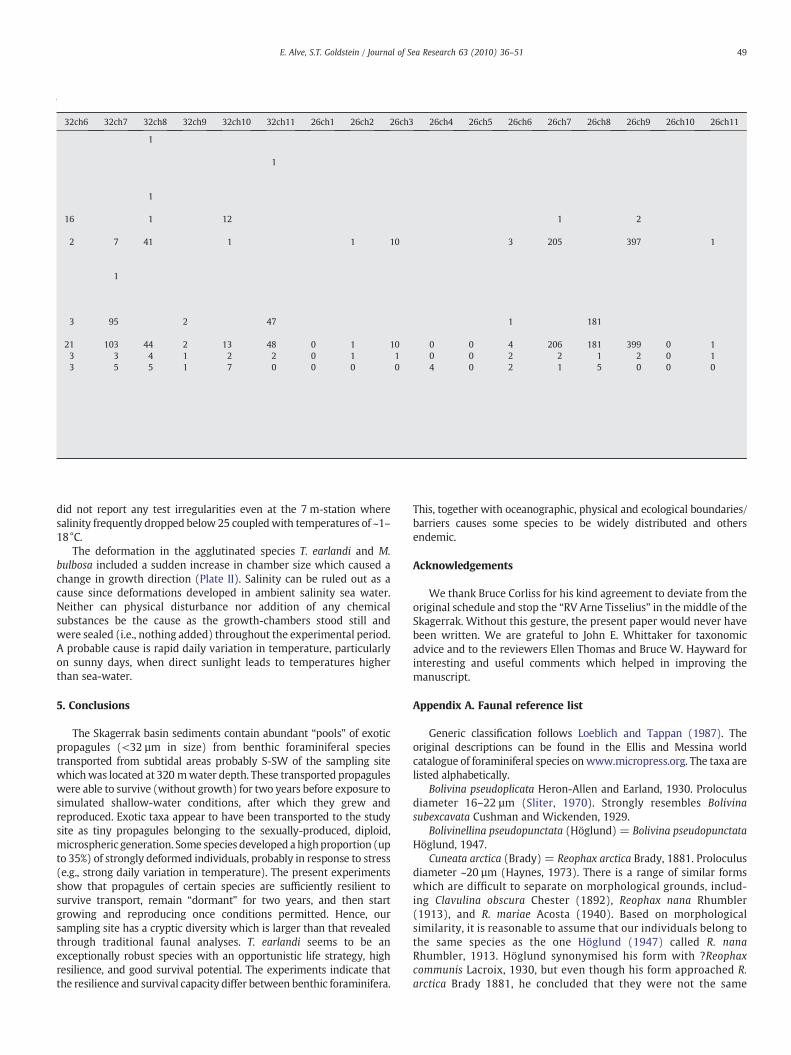
Table A2
32ch6 32ch7 32ch8 32ch9 32ch10 32ch11 26ch1 26ch2 26ch3 26ch4 26ch5 26ch6 26ch7 26ch8 26ch9 26ch10 26ch11
1
1
1
16 1 12 1 2
2 7 41 1 1 10 3 205 397 1
1
3 95 2 47 1 181
21 103 44 2 13 48 0 1 10 0 0 4 206 181 399 0 13 3 4 1 2 2 0 1 1 0 0 2 2 1 2 0 13 5 5 1 7 0 0 0 0 4 0 2 1 5 0 0 0
49E. Alve, S.T. Goldstein / Journal of Sea Research 63 (2010) 36–51
did not report any test irregularities even at the 7 m-station wheresalinity frequently dropped below 25 coupledwith temperatures of ~1–18°C.
The deformation in the agglutinated species T. earlandi and M.bulbosa included a sudden increase in chamber size which caused achange in growth direction (Plate II). Salinity can be ruled out as acause since deformations developed in ambient salinity sea water.Neither can physical disturbance nor addition of any chemicalsubstances be the cause as the growth-chambers stood still andwere sealed (i.e., nothing added) throughout the experimental period.A probable cause is rapid daily variation in temperature, particularlyon sunny days, when direct sunlight leads to temperatures higherthan sea-water.
5. Conclusions
The Skagerrak basin sediments contain abundant “pools” of exoticpropagules (<32 µm in size) from benthic foraminiferal speciestransported from subtidal areas probably S-SW of the sampling sitewhichwas located at 320 mwater depth. These transported propaguleswere able to survive (without growth) for two years before exposure tosimulated shallow-water conditions, after which they grew andreproduced. Exotic taxa appear to have been transported to the studysite as tiny propagules belonging to the sexually-produced, diploid,microspheric generation. Some species developed a highproportion (upto 35%) of strongly deformed individuals, probably in response to stress(e.g., strong daily variation in temperature). The present experimentsshow that propagules of certain species are sufficiently resilient tosurvive transport, remain “dormant” for two years, and then startgrowing and reproducing once conditions permitted. Hence, oursampling site has a cryptic diversity which is larger than that revealedthrough traditional faunal analyses. T. earlandi seems to be anexceptionally robust species with an opportunistic life strategy, highresilience, and good survival potential. The experiments indicate thatthe resilience and survival capacity differ between benthic foraminifera.
This, together with oceanographic, physical and ecological boundaries/barriers causes some species to be widely distributed and othersendemic.
Acknowledgements
We thank Bruce Corliss for his kind agreement to deviate from theoriginal schedule and stop the “RV Arne Tisselius” in the middle of theSkagerrak. Without this gesture, the present paper would never havebeen written. We are grateful to John E. Whittaker for taxonomicadvice and to the reviewers Ellen Thomas and Bruce W. Hayward forinteresting and useful comments which helped in improving themanuscript.
Appendix A. Faunal reference list
Generic classification follows Loeblich and Tappan (1987). Theoriginal descriptions can be found in the Ellis and Messina worldcatalogue of foraminiferal species on www.micropress.org. The taxa arelisted alphabetically.
Bolivina pseudoplicata Heron-Allen and Earland, 1930. Proloculusdiameter 16–22 µm (Sliter, 1970). Strongly resembles Bolivinasubexcavata Cushman and Wickenden, 1929.
Bolivinellina pseudopunctata (Höglund) = Bolivina pseudopunctataHöglund, 1947.
Cuneata arctica (Brady) = Reophax arctica Brady, 1881. Proloculusdiameter ~20 µm (Haynes, 1973). There is a range of similar formswhich are difficult to separate on morphological grounds, includ-ing Clavulina obscura Chester (1892), Reophax nana Rhumbler(1913), and R. mariae Acosta (1940). Based on morphologicalsimilarity, it is reasonable to assume that our individuals belong tothe same species as the one Höglund (1947) called R. nanaRhumbler, 1913. Höglund synonymised his form with ?Reophaxcommunis Lacroix, 1930, but even though his form approached R.arctica Brady 1881, he concluded that they were not the same
50 E. Alve, S.T. Goldstein / Journal of Sea Research 63 (2010) 36–51
because, according to Lacroix, R. arctica and R. communis, cannotbe confused. However, it is likely that Rhumbler, when describingR. nana, was not aware of Brady's 1881-description of R. arctica, ashe made no reference to Brady's work from the rather unknownAustro-Hungarian North-Polar Expedition (J.E. Whittaker, pers.com., 2008). Furthermore, there is a strong resemblance betweenour form and Reophax mariae Acosta, 1940. Brönnimann et al.(1992) described a new genus, Acostata, with R. mariae as typespecies and stated that the main difference between Acostata andCuneata is that the latter is compressed in transverse section,while the former is not. This feature is probably not a goodcriterion because Höglund's individuals of R. nana are “….usuallyround in transverse section, but it is not uncommon to find more orless compressed specimens, as Rhumbler also observed in hismaterial” (Höglund, 1947, p. 92–93). Except for the more or lesscompressed nature, it is not clear from Brönnimann et al.'s (1992)rather detailed descriptions how Acostata mariae differs from C.arctica. The same authors consider the species which Lutze(1974, pl. 1, fig. 16) called R. nana Rhumbler to be A. mariae butthey do not discuss R. nana. The published, morphologically-basedcriteria used to distinguish between the above-mentioned formsare too vague to draw firm conclusions as to whether or not theyare different morphospecies. Consequently, the oldest name isused here.
Eggerelloides scaber (Williamson) = Bulimina scabra Williamson,1858. Proloculum of microspheric forms (diameter 8–13 µm) are notagglutinated, whereas that of the megalospheric form (diameter 35–70 µm) is agglutinated (Höglund, 1947). The triserial individuals inthe present experiment showed a plastic morphology ranging fromtypical E. scaber to what looked like juvenile megalospheric Liebusellagoësi. However, of the hundreds of individuals examined, none haddeveloped the uniserial stage typical of L. goësi. Juvenile micro- andmegalosphaeric individuals of L. goësi from the original, pre-experiment assemblage (t=0) have a loop shaped Bulimina-likeaperture, bordered by a lip, extending from the basal suture of the lastchamber up the slightly excavated apertural face, just as in Eggerel-loides (Haynes, 1973). Due to their morphological resemblance, it islikely that deep shelf records of E. scaber are in fact microsphericjuveniles of L. goësi.
Epistominella vitrea Parker, 1953.Gavelinopsis praegeri (Heron-Allen and Earland) = Discorbina
praegeri Heron-Allen and Earland, 1913.Leptohalysis scottii (Chaster) = Reophax scottii Chaster, 1892.Liebusella goësi Höglund, 1947. Proloculum of microspheric forms
(diameter 9–13 µm) are not agglutinated, whereas that of themegalospheric form (diameter 90–190 µm) is agglutinated (Höglund,1947). Due to strong morphological similarity, it is likely thatmicrospheric juveniles of this species (i.e., individuals which havenot yet developed the uniserial part) have been mistaken as E. scaberby some authors.
Morulaeplecta bulbosa Höglund, 1947. Proloculus diameter 17–33 µm (Höglund, 1947). Considered by Duijnstee et al. (2003) to bethe same as Caronia silvestrii Brönnimann, Whittaker and Valleri,1992. Barmawidjaja et al. (1992, pl. 1, Figs. 5–7) report M. bulbosaliving in the same general shallow-water area (N Adriatic) as whereBrönnimann et al. (1992) collected their C. silvestrii. As Brönnimannet al. (1992) did not mention M. bulbosa and it is beyond the scope ofthe present paper to go into a taxonomic discussion, we use the oldername, M. bulbosa, for our present form.
Planorbulina mediterranensis d'Orbigny, 1826. Proloculus diameter11–14 (microspheric) and 23–56 µm (megalospheric) (Loeblich andTappan, 1987).
Stainforthia fusiformis (Williamson) = Bulimina pupoides d'Or-bigny var. fusiformis Williamson, 1858.
Textularia earlandi Parker, 1952 = Textularia tenuissima Earland,1933. The species is named Spiroplectammina earlandi (Parker) by some
authors (e.g., Haynes, 1973) due to the very small planospire consistingof 3 or 4 chambers closely coiled around the proloculus (for details, seeHöglund, 1947). Duchemin et al.'s (2008, pl. 1, Fig. 9) illustration ofTextularia porrecta (Brady), which dominates the foraminiferal assem-blage at 80 mwater depth in the Bay of Biscay, bears close resemblancewith our species.
Trochamminopsis quadriloba (Höglund) = Trochammina pusilusHöglund, 1947.
References
Almogi-Labin, A., Hemleben, C., Meischner, D., Erlenkeuser, H., 1996. Response of RedSea deep-water agglutinated foraminifera to water-mass changes during the LateQuaternary. Mar. Micropaleontol. 28, 283–297.
Altenbach, A.V., Lutze, G.F., Schiebel, R., Schönfeld, J., 2003. Impact of interrelated andinterdependent ecological controls on benthic foraminifera: an example from theGulf of Guinea. Palaeogeogr. Palaeoclimatol. Palaeoecol. 197, 213–238.
Alve, E., 1995.Benthic foraminiferal responses toestuarinepollution: a review. J. ForaminiferalRes. 25, 190–203.
Alve, E., 1999. Colonization of new habitats by benthic foraminifera: a review. Earth-Sci.Rev. 46, 167–185.
Alve, E., in press. A bisected Pelosina rejoined! J. Micropalaeontol.Alve, E., Goldstein, S.T., 2002. Resting stage inbenthic foraminiferal propagules: a key feature
for dispersal? Evidence from two shallow-water species. J. Micropalaeontol. 21, 95–96.Alve, E., Goldstein, S.T., 2003. Propagule transport as a key method of dispersal in
benthic foraminifera. Limnol. Oceanogr. 48, 2163–2170.Alve, E., Murray, J.W., 1995. Benthic foraminiferal distribution and abundance changes
in Skagerrak surface sediments: 1937 (Höglund) and 1992/1993 data compared.Mar. Micropaleontol. 25, 269–288.
Alve, E., Murray, J.W., 1997. High benthic fertility and taphonomy of foraminifera: a casestudy of the Skagerrak, North Sea. Mar. Micropaleontol. 31, 157–175.
Alve, E., Nagy, J., 1986. Estuarine foraminiferal distribution in Sandebukta, a branch ofthe Oslo Fjord. J. Foraminiferal Res. 16, 261–284.
Barmawidjaja, D.M., Jorissen, F.J., Puskaric, S., van der Zwaan, G.J., 1992. Microhabitatselection by benthic foraminifera in the northern Adriatic Sea. J. Foraminiferal Res.22, 297–317.
Beaulieu, S.E., 2001. Colonization of habitat islands in the deep sea: recruitment to glasssponge stalks. Deep-Sea Res. I 48, 1121–1137.
Belasky, P., 1996. Biogeography of Indo-Pacific larger foraminifera and scleractiniancorals: a probabilistic approach to estimating taxonomic diversity, faunalsimilarity, and sampling bias. Palaeogeogr. Palaeoclimatol. Palaeoecol. 122,119–141.
Bergsten, H., Nordberg, K., Malmgren, B., 1996. Recent benthic foraminifera as tracers ofwater masses along a transect in the Skagerrak, North-Eastern North Sea. J. Sea Res.35, 111–121.
Bernhard, J.M., Sen Gupta, B.K., Borne, P.F., 1997. Benthic foraminiferal proxy toestimate dysoxic bottom-water oxygen concentrations: Santa Barbara Basin, U.S.Pacific continental margin. J. Foraminiferal Res. 27, 301–310.
Brandt, A., Gooday, A.J., Brandao, S.N., Brix, S., Brokeland,W., Cedhagen, T., Choudhury, M.,Cornelius, N., Danis, B., DeMesel, I., Diaz, R.J., Gillan, D.C., Ebbe, B., Howe, J.A., Janussen,D., Kaiser, S., Linse, K., Malyutina, M., Pawlowski, J., Raupach, M., Vanreusel, A., 2007.First insights into the biodiversity and biogeography of the Southern Ocean deep sea.Nature 447, 307–311.
Brönnimann, P., Whittaker, J.E., Valleri, G., 1992. Agglutinated foraminifera from theLagoon of Venice, Italy. Rev. Paléobiol. 11, 97–109.
Buzas, M.A., Culver, S.J., 1991. Species diversity and dispersal of benthic foraminifera.BioScience 41, 483–489.
Conradsen, K., Bergsten, H., Knudsen, K.L., Nordberg, K., Seidenkrantz, M.-S., 1994.Recent benthic foraminiferal distribution in the Kattegat and Skagerrak, Scandi-navia. Cushman Fdn. Spec. Publ. 32, 53–68.
Corliss, B.H., van Weering, T.C.E., 1993. Living (stained) benthic foraminifera withinsurficial sediments of the Skagerrak. Mar. Geol. 111, 323–335.
Culver, S.J., Buzas, M.A., 1999. Biogeography of neritic benthic foraminifera. In: SenGupta, B.K. (Ed.), Modern Foraminifera. Kluwer, pp. 93–102.
Danielssen, D.S., Svendsen, E., Ostrowski, M., 1996. Long-term hydrographic variationin the Skagerrak based on the section Torungen–Hirtshals. ICES J. Mar. Sci. 53,917–925.
Darling, K.F., Wade, C.M., 2008. The genetic diversity of planktic foraminifera and theglobal distribution of ribosomal RNA genotypes. Mar. Micropaleontol. 67, 216–238.
De Nooijer, L.J., Duijnstee, I.A.P., Bergman, M.J.N., van der Zwaan, G.J., 2008. The ecologyof benthic foraminifera across the Frisian Front, southern North Sea. Est. Coast.Shelf Sci. 78, 715–726.
Debenay, J.-P., 2000. Foraminifers of tropical paralic environments. Micropaleontology46 (Suppl. 1), 153–160.
Debenay, J.-P., Bicchi, E., Goubert, E., du Châtelet, E.A., 2006. Spatio-temporaldistribution of benthic foraminifera in relation to estuarine dynamics (Vie estuary,Vendée, W France). Est. Coast. Shelf Sci. 67, 181–197.
deStigter, H.C., Jorissen, F.J., van der Zwaan, G.J., 1998. Bathymetric distribution andmicrohabitat partitioning of live (Rose Bengal stained) benthic foraminifera along ashelf to bathyal transect in the southern Adriatic Sea. J. Foraminiferal Res. 28,40–65.
Donnici, S., Serandrei Barbero, R., 2002. The benthic foraminiferal communities of thenorthern Adriatic continental shelf. Mar. Micropaleontol. 44, 93–123.
51E. Alve, S.T. Goldstein / Journal of Sea Research 63 (2010) 36–51
Duchemin, G., Jorissen, F.J., Le Loc'h, F., Andrieux-Loyer, F., Hilly, C., Thouzeau, G., 2008.Seasonal variability of living benthic foraminifera from the outer continental shelfof the Bay of Biscay. J. Sea Res. 59, 297–319.
Duijnstee, I.A.P., Ernst, S.R., van der Zwaan, G.J., 2003. Effect of anoxia on the verticalmigration of benthic foraminifera. Mar. Ecol., Prog. Ser. 246, 85–94.
Ernst, S., Duijnstee, I., van der Zwaan, B., 2002. The dynamics of the benthicforaminiferal microhabitat: recovery after experimental disturbance. Mar. Micro-paleontol. 46, 343–361.
Ernst, S.R., Morvan, J., Geslin, E., Le Bihan, A., Jorissen, F.J., 2006. Benthic foraminiferalresponse to experimentally induced Erika oil pollution. Mar. Micropaleontol. 61,76–93.
Fenchel, T., 2005. Cosmopolitan microbes and their “cryptic” species. Aquat. Microb.Ecol. 41, 49–54.
Finlay, B.J., Esteban, G.F., Fenchel, T., 2004. Protist diversity is different? Protist 155,15–22.
Finlay, B.J., Esteban, G.F., Brown, S., Fenchel, T., Hoef-Emden, K., 2006. Multiplecosmopolitan ecotypes within a microbial eukaryote morphospecies. Protist 157,377–390.
Foissner,W., 2006. Biogeography and dispersal ofmicro-organisms: a reviewemphasizingprotists. Acta Protozool. 45, 111–136.
Fujita, K., 2004. A field colonization experiment on small-scale distributions of algalsymbiont-bearing larger foraminifera on reef rubble. J. Foraminiferal Res. 34, 169–179.
Geslin, E.,Debenay, J.-P., Lesourd,M., 1998. Abnormalwall textures and test deformation inAmmonia (hyaline foraminifer). J. Foraminiferal Res. 28, 148–156.
Geslin, E., Debenay, J.P., Duleba, W., Bonetti, C., 2002. Morphological abnormalities offoraminiferal tests in Brazilian environments: comparison betweenpolluted and non-polluted areas. Mar. Micropaleontol 45, 151–168.
Goldstein, S.T., 1999. Foraminifera: a biological overview. In: Sen Gupta, B.K. (Ed.),Modern Foraminifera. Kluwer, pp. 37–55.
Goldstein, S.T., Watkins, G.T., 1998. Elevation and the distribution of salt-marshforaminifera, St. Catherines Island, Georgia: a taphonomic approach. Palaios 13,570–580.
Gooday, A.J., Holzmann, M., Guiard, J., Cornelius, N., Pawlowski, J., 2004. A newmonothalamous foraminiferan from 1000 to 6300 m water depth in the WeddellSea: morphological andmolecular characterisation. Deep-sea Res., Part 2, Top. Stud.Oceanogr. 51, 1603–1616.
Gooday, A.J., Cedhagen, T., Kamenskaya, O.E., Cornelius, N., 2007. The biodiversity andbiogeography of komokiaceans and other enigmatic foraminiferan-like protists in thedeep Southern Ocean. Deep-sea Res., Part 2, Top. Stud. Oceanogr. 54, 1691–1719.
Gross, O., 2000. Influence of temperature, oxygen and food availability on themigrationalactivity of bathyal benthic foraminifera: evidence by microcosm experiments.Hydrobiologia 426, 123–137.
Gustafsson, M., Nordberg, K., 2001. Living (stained) benthic foraminiferal response toprimary production and hydrography in the deepest part of the Gullmar Fjord,SwedishWest Coast, with comparisons to Höglund's 1927material. J. ForaminiferalRes. 31, 2–11.
Hallock, P., 1985. Why are larger foraminifera large? Paleobiology 11, 195–208.Haynes, J.R., 1973. Cardigan Bay recent foraminifera. Bull. Br. Mus. nat. Hist. (Zool.)
Suppl. 4, 1–245.Hayward, B.W., Grenfell, H.R., Reid, C.M., Hayward, K.A., 1999. Recent New Zealand
shallow-water benthic foraminifera: taxonomy, ecologic distribution, biogeogra-phy, and use in paleoenvironmental assessment. Inst. Geol. Nucl. Sci. Monogr. 21258 pp.
Hayward, B.W., Grenfell, H.R., Sabaa, A.T., Daymond-King, R., 2007a. Biogeography andecological distribution of shallow-water benthic foraminifera from the Auckland andCampbell Islands, subantarctic southwest Pacific. J. Microapalaeont. 26, 127–143.
Hayward, B.W., Grenfell, H.R., Sabaa, A.T., Neil, H.L., 2007b. Factors influencing thedistribution of subantarctic deep-sea benthic foraminifera, Campbell and BountyPlateaux, New Zealand. Mar. Micropaleontol. 62, 141–166.
Heinz, P., Geslin, E., Hemleben, C., 2005. Laboratory observations of benthic foraminiferalcysts. Mar. Biol. Res. 1, 149–159.
Hemleben, C., Kitazato, H., 1995. Deep-sea foraminifera under long-time observation inthe laboratory. Deep-Sea Res. I 42, 827–832.
Höglund, H., 1947. Foraminifera in the Gullmar Fjord and the Skagerak. Zool. BidragUppsala 26, 1–328.
Kuhnt,W., Hess, S., Holbourn, A., Paulsen, H., Salomon, B., 2005. The impact of the 1991Mt.Pinatubo eruption on deep-sea foraminiferal communities: a model for theCretaceous–Tertiary (K/T) boundary? Palaeogeogr. Palaeoclimatol. Palaeoecol. 224,83–107.
Langer, M.R., 1993. Epiphytic foraminifera. Mar. Micropaleontol. 20, 235–265.Loeblich, A.R., Tappan, H., 1987. Foraminiferal Genera and Their Classification. Von
Nostrand Reinhold Co., New York.Lutze, G.F., 1974. Benthische Foraminiferen in Oberflächen-Sedimenten des Persischen
Golfes. Teil 1-Arten. Meteor Forschungs Ergebnisse, Reihe C. Berlin 17, 1–66.Mendes, I., Gonzalez, R., Dias, J.M.A., Lobo, F., Martins, V., 2004. Factors influencing
recent benthic foraminifera distribution on the Guadiana shelf (SouthwesternIberia). Mar. Micropaleontol. 51, 171–192.
Mitchell, E.A.D., Meisterfeld, R., 2005. Taxonomic confusion blurs the debate oncosmopolitanism versus local endemism of free-living protists. Protist 156, 263–267.
Murray, J.W., 1992. Distribution and population dynamics of benthic foraminifera fromthe southern North Sea. J. Foraminiferal Res. 22, 114–128.
Murray, J.W., 2003. An illustrated guide to the benthic foraminifera of the Hebrideanshelf, west of Scotland, with notes on their mode of life. Palaeontologia Electronica,5, issue 2, art. 1, 31 pp., 1.4 Mb. http.//www-odp.tamu.edu/paleo/2002_2/guide/issue2_02.htm.
Murray, J.W., 2006. Ecology and Applications of Benthic Foraminifera. Cambridge,Cambridge University Press. 426 pp.
Murray, J.W., 2007. Biodiversity of living benthic foraminifera: how many species arethere? Mar. Micropaleontol. 64, 163–176.
Myers, E.H., 1936. The life-cycle of Spirillina vivipara Ehrenberg, with notes onmorphogenesis, systematics and distribution of the foraminifera. J. R. Micr. Soc. 56,120–146.
Nigam, R., Kurtarkar, S.R., Saraswat, R., Linshy, V.N., Rana, S.S., 2008. Response of benthicforaminifera Rosalina leei to different temperature and salinity, under laboratoryculture experiment. J. Mar. Biol. Assoc., U.K. 88, 699–704.
Patterson, R.T., Guilbault, J.P., Thomson, R.E., 2000. Oxygen level control onforaminiferal distribution in Effingham Inlet, Vancouver Island, British Columbia,Canada. J. Foraminiferal Res. 30, 321–335.
Pawlowski, J., Holzmann, M., 2008. Diversity and geographic distribution of benthicforaminifera: a molecular perspective. Biodivers. Conserv. 17, 317–328.
Pawlowski, J., Fahrni, J., Lecroq, B., Longet, D., Cornelius, N., Excoffier, L., Cedhagen, T.,Gooday, A.J., 2007. Bipolar gene flow in deep-sea benthic foraminifera. Mol. Ecol.16, 4089–4096.
Schönfeld, J., Numberger, L., 2007. Seasonal dynamics and decadal changes of benthicforaminiferal assemblages in the western Baltic Sea (NW Europe). J. Micropalaeontol.26, 47–60.
Scott, G.A., Scourse, J.D., Austin, W.E.N., 2003. The distribution of benthic foraminifera intheCeltic Sea: the significance of seasonal stratification. J. Foraminiferal Res. 33, 32–61.
Sliter, W.V., 1970. Inner-neritic Bolivinitidae from the eastern Pacific margin. Micropale-ontology 16, 155–174.
Smart, C.W., Thomas, E., 2007. Emendation of the genus Streptochilus Bronnimann andResig 1971 (Foraminifera) and new species from the lower Miocene of the Atlanticand Indian Oceans. Micropaleontology 53, 73–103.
Spindler, M., 1980. The pelagic gulf weed Sargassum natans as a habitat for the benthicforaminifera Planorbulina acervalis and Rosalina globularis. Neues Jahrb. Geol.PalaÉontol. Monatsh. 9, 569–580.
Svavarsson, J., Davidsdottir, B., 1995. Cibicides spp. (Protozoa, Foraminifera) as epizioteson the Arctic antenna-brooding Arcturus baffini (Crustacea, Isopda, Valvifera). PolarBiol. 15, 569–574.
Telford, R.J., Vandvik, V., Birks, H.J.B., 2006. Dispersal limitations matter for microbialmorphospecies. Science 312, 1015.
Vanormelingen, P., Verleyen, E., Vyverman, W., 2008. The diversity and distribution ofdiatoms: from cosmopolitanism to narrow endemism. Biodivers. Conserv. 17,393–405.
Weisse, T., 2008. Distribution and diversity of aquatic protists: an evolutionary andecological perspective. Biodivers. Conserv. 17, 243–259.
Wisshak, M., Rüggeberg, A., 2006. Colonisation and bioerosion of experimentalsubstrates by benthic foraminiferans from euphotic to aphotic depths (Kosterfjord,SW Sweden). Facies 52, 1–17.
Yassini, I., Jones, B.G., 1995. Foraminiferida and Ostracoda from estuarine and shelfenvironments on the southeastern coast of Australia. The University of WollongongPress, Wollongong, Australia, ISBN 0-86418-315, 484 pp.