Embed Size (px)
Citation preview
Disorders of Red Blood Cells
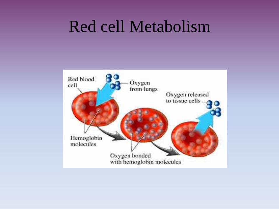
The function of RBC, facilitated by
hemoglobin molecule, is to transport
oxygen to the tissue.
Oxygen is poorly soluble in plasma, So
about 95%-98% is carried bound to
hemoglobin.
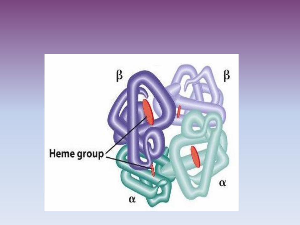
The Hb molecule is composed of two pairs of
structurally different α and β chains.
Each of four polypeptide chains consists of
globin (protein) and a haeme unite which
surrounds an atom of iron that binds oxygen.
Thus Hb can carry four molecules of Oxygen.
• The production of each type of globin chain is
controlled by individual structural genes with
five different gene loci.
• Mutations which occurs in anywhere in these
five loci resulted over 550 types of abnormal
Hb molecules.
Red cell Metabolism
• The RBC lacks mitochondria; it depends on glucose
and glycolytic pathway for its metabolic needs.
• The enzyme mediated anaerobic metabolism of
glucose generates the ATP need for normal membrane
function and ion transport.
• The depletion of glucose or the functional deficiency
of one the glycolytic enzymes leads to the premature
death of RBC.
An offshoot of the glycolytic pathway is the production
of 2,3-diphosphoglycerate (2,3-DPG), which binds to
the Hb molecule and reduces the affinity of Hb for
Oxygen.
This facilitates the release of Oxygen at the tissue level.
• An increase in the concentration of 2,3- DPG occurs in conditions:
Chronic hypoxia like in case anemia, chronic lung disease, and residence at high attitudes.
Hemoglobin Oxidation
• The combination of Hb with oxygen can be interrupted by certain chemicals e.g. nitrates and sulfates) and drugs that oxidize Hb to the inactive form.
• The nitrite ion reacts with Hb to produce methmoglobin, which has a low affinity for Oxygen.
• Large doses of nitrites can result in high levels of methmoglobin, causing pseudocyanosis and tissue hypoxia. For example, sodium nitrate, which is using curing meat, can produce methmoglobin when taken in large amounts.
Anemia
• Abnormally low number of circulating RBC or level of Hb or both, resulting in diminished oxygen-carrying capacity.
• Causing:
1). Excessive loss of RBC:
Hemorrhage
Hemolysis
2). Impaired RBC production result from:
Depression in bone marrow
Inhered
Nutritional
Bone marrow cancer
Hemolytic Anemia
• It is characterized by the premature destruction
of RBC , the retention in the body of iron and
the other products of Hb destruction and an
increase in erythropiosis.
• All most all types of hemolytic anemia are
distinguished by normocytic normochromic
RBC
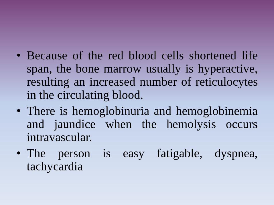
• Because of the red blood cells shortened lifespan, the bone marrow usually is hyperactive,resulting an increased number of reticulocytesin the circulating blood.
• There is hemoglobinuria and hemoglobinemiaand jaundice when the hemolysis occursintravascular.
• The person is easy fatigable, dyspnea,tachycardia
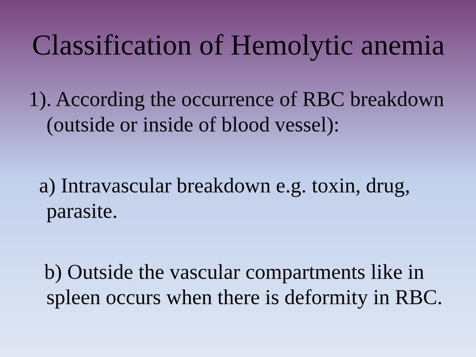
Classification of Hemolytic anemia
1). According the occurrence of RBC breakdown
(outside or inside of blood vessel):
a) Intravascular breakdown e.g. toxin, drug,
parasite.
b) Outside the vascular compartments like in
spleen occurs when there is deformity in RBC.
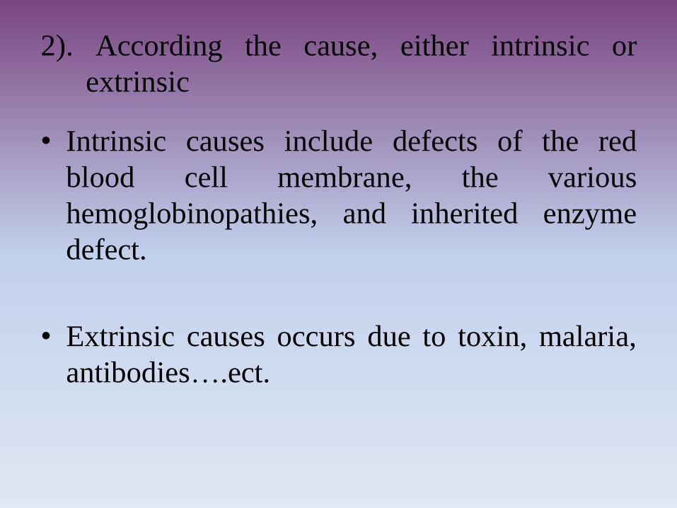
2). According the cause, either intrinsic or
extrinsic
• Intrinsic causes include defects of the red
blood cell membrane, the various
hemoglobinopathies, and inherited enzyme
defect.
• Extrinsic causes occurs due to toxin, malaria,
antibodies….ect.
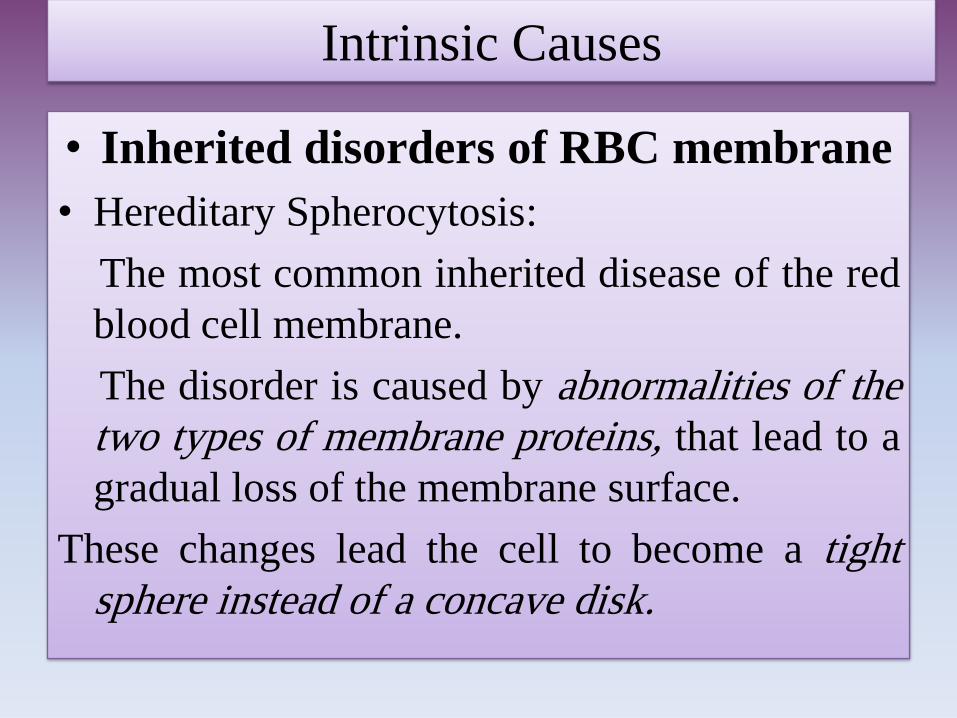
Intrinsic Causes
• Inherited disorders of RBC membrane
• Hereditary Spherocytosis:
The most common inherited disease of the red
blood cell membrane.
The disorder is caused by abnormalities of thetwo types of membrane proteins, that lead to a
gradual loss of the membrane surface.
These changes lead the cell to become a tight
sphere instead of a concave disk.
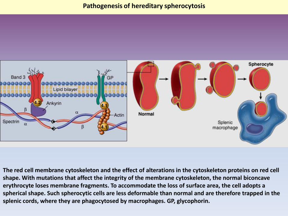
The red cell membrane cytoskeleton and the effect of alterations in the cytoskeleton proteins on red cell shape. With mutations that affect the integrity of the membrane cytoskeleton, the normal biconcave erythrocyte loses membrane fragments. To accommodate the loss of surface area, the cell adopts a spherical shape. Such spherocytic cells are less deformable than normal and are therefore trapped in the splenic cords, where they are phagocytosed by macrophages. GP, glycophorin.
Pathogenesis of hereditary spherocytosis
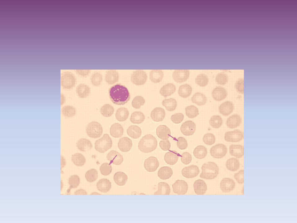
In this case the cell has ability to carry Oxygen but
membrane deformity makes the cell susceptible to
destruction as it pass through the spleen
• Clinical signs
• Mild hemolytic anemia, jaundice,
Splenomegaly, and gall stone.
• Treatment
Splenoctomy to reduce RBC destruction.
blood transfusion
2). Acanthosis
• Acanthosis refers to a group of anemias in
which the erythrocyte membrane shows
multiple irregular projections by defect in the
lipid bilayer.
• Occurs in liver diseases and inherited defect in
absorption and transport of lipid by intestin.
Abnormality in the HB
• The Thalassemias are the group of inherited disorders of Hb synthesis characterized by lack of or decreased synthesis of either α or β globin chains.
• In α- thalassemia, α globin chain synthesis reduced.
• In β-thalassemia, β chain synthesis is either absent or markedly deficient.
The Thalassemias
Two factors contribute to the anemia that occurs
in Thalassemias:
• 1). Low intracellular HB (hypochromic) is due
to the decrease synthesis of the affected chain.
• Continued production and accumulation of the
unaffected globin chain.
• The reduce Hb synthesis result in hypochromic,
microcytic anemia
• The accumulation of the unaffected chain refers
with normal red cell maturation and contributes
to membrane changes that lead to hemolysis and
anemia.
• α chain is insoluble so when accumulated in the
RBC form precipitate (Heinz bodies).
• The Heinz bodies impair DNA synthesis and
cause damage to the RBC.
• Accumulation of α chain leads to breakdown
RBC precursor in the bone marrow, and few of
those abnormal red cells leave bone marrow
will be destruct in spleen.
• β chain is soluble, so accumulation of β chain
produce less severity of thalassemias.
• The clinical manifestations of β thalassemia
are based on the severity of anemia.
• Person who carries one normal gene
(heterozygous) has minor thalassemia.
• Homozygous undergoes major thalassemia.
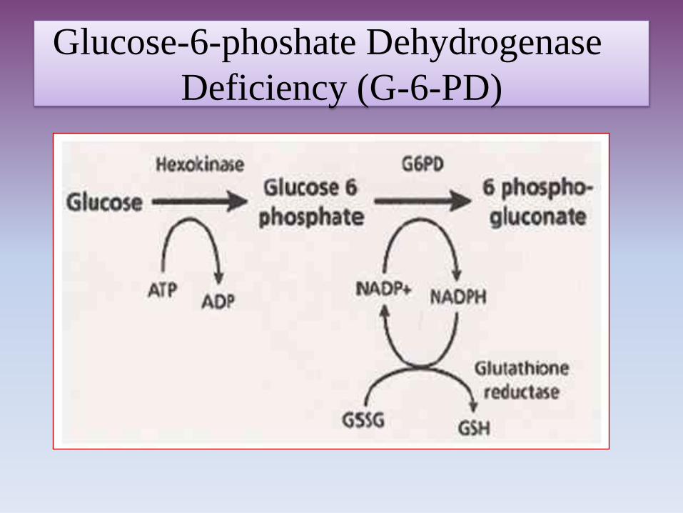
Glucose-6-phoshate Dehydrogenase
Deficiency (G-6-PD)
Glucose-6-phoshate Dehydrogenase
Deficiency (G-6-PD)
• Glucose-6-phosphate dehydrogenase is an X-linked recessive hereditary disease.
• characterized by abnormally low levels of glucose-6-phosphate dehydrogenase, a metabolic enzyme especially important in red blood cell metabolism.
• and the individuals with the disease may exhibit hemolytic anemia in response to infection and chemicals
The defect expressed only in the male and
homozygous females.
• The inclusion bodies makes red cells more
vulnerable to oxidants and causes direct
oxidation of Hb to methmoglobin.
• Methmoglobin cannot transport oxygen and
denaturating of the hemoglobin molecule to
form Heinz bodies.
Heinz Bodies
• These are inclusion bodies appeared in the red
blood cells due to damaging in Hb molecules.
• Many substances are potentially harmful into
people with G6PD deficiency such as:
• Antimalaria (primaquine, Pamaquine, and
chloroquine)
Other types of Anemias
• Decrease production of RBC:
• Occurs due to: Deficiency of nutrient for Hb synthesis like iron deficiency.
• Deficiency of nutrient for DNA synthesis like B12 (cobalamin) deficiency and folic acid deficiency.
• Vitamin B12 is essential for DNA synthesis and nuclear maturation.
• Vitamin B12 involved in reaction that prevents abnormal fatty acid from being incorporate into neuronal lipid.
• Vitamin B12 needs intrinsic factor for
absorption from intestine.
• When Vit. B12 is deficient the red cells that
produced are abnormally large because excess
cytoplasmic anemia. The cells have immature
nuclei and show evidence of cellular
destruction.
• The loss of red cells is a moderate to sever
anemia and mild jaundice.
Polycythemia
• Polycythemia or erythrocytosis is an increased
concentration of red blood cells.
• Such increase may be relative or absolute.
• Relative Polycythemia:
The red blood cell
mass increase because of loss of plasma.
This occurs due to water deprivation,
dehydration, vomiting, and excess use of
diuretics.
• Absolute Polycythemia is a rise in red blood
cells due to an increase in total red blood cell
mass and is classified as primary or secondary.
Primary Polycythemia
(Polycythemia Vera)
• Is a neoplastic disease of the pluripotential
cells of bone marrow characterized by an
absolute increase in total red blood cell mass
accompained by elevated white cell and
platelet counts. It is seen in men with median
age of 62 years but may occurs in many age.
MANIFESTATION OF
POLYCYTHEMIA VERA• Increase in red blood cells, Hb and blood
viscosity.
• Splenomegaly and depletion of iron store.
• Increase blood viscosity interferes with cardiac
output and blood flow.
• Hypertension is common and associated with
headache, dizziness, inability to concentrate
and some difficulty with hearing and vision
because of decrease cerebral blood flow
• Venous stasis, thromboembolism, and
hemorrhage due to defect in function of
platelet.
• Treatment:
1. Phlebotomy
2. Chemotherapy
Secondary Polycythemia
• Increase erythropoietin due to hypoxic conditions like chronic heart and lung diseases.
• Erythropoietin is secreted by kidney and on bone marrow to increase production of blood cells.