Embed Size (px)
Citation preview
Disorders of metabolism of proteins, lipids, carbohydrates, nucleid acid. Impaired metabolism of water. Impaired metabolism of calcium, iron and copper Crystals, concrements, pigments.
Disorders of metabolism of proteins
• Dystrophy – derrangement of cell metabolism
• Accumulation of metabolite (low-molecular – vacuolisation; high-molecular – hyaline droplets)
• In some cases – no accumulation of metabolite and only discrete changes of cell
• Revesible in majority of cases
• Structural changes: (a) enlargement of mitochondria, (b) vacuolar dystrophy, (c) hyalinne droplets,(d) mucous dystrophy, (e) fibrinoid dystrophy, (f) hyalinosis, (g) amyloidosis

• Enlargement of mitochondria: liver, kidney, myocardium, skeletal muscle; microscopicaly – enlargement of cells, granular cytoplasm (due to enlargement of mitochondria - see ELMI)
• Vacuolar dystrophy (hydropic degeneration): accumulation of electrolytes and water, vacuolisation of cytoplasm, oedema
• Hyalinne droplets (hyaline – astructural pink material in hematoxylin-eosin staining): hepatocytes in viral hepatitis (Councilmann bodies), renal tubuli in albuminuria, plasmocytes in chronic inflammation (Russell bodies)
• Mucous dystrophy: accumulation of mucopolysacharides (mucoviscidosis, alopecia mucinosa, mucopolysacharidoses – Hurler, Hunter, Sanfilippo, Morqui, Maroteaux-Lamy, beta-glukuronidase deficit, ganglion, myxedema, atherosclerosis, cystic medionecrosis
• Fibrinoid dystrophy/hyalinosis: colagen fibril changes (vessel wall), reticuline fibril changes (endocardium, joints, vessels, colagenoses, hyalinosis (polyserositis)

Mallory hyalin
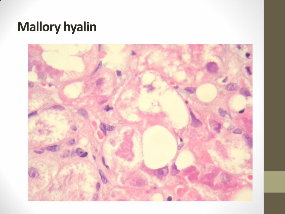
Mallory hyalin

Amyloidosis
Clinical classification Amyloid protein Localisation
Primary
(myeloma associated)
AL (Ig light chains) Kidney, spleen, heart, liver,
tongue
Secondary
(reactive – RA, chronic
infections, IBD, tumors)
AA (amyloid associated) Tongue, heart, liver, kidney,
spleen
Senile AS (prealbumin) Heart
Hemodialysis associated AH (b 2 mikroglobulin) Ledviny
Alzheimer associated A4 (b amyloid) Brain
Endocrine
(medulary thyroid carcinoma)
AE Thyroid

Disorders of metabolism of lipids

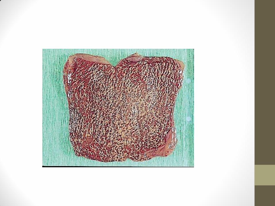

Sphingolipidosis - ELMI
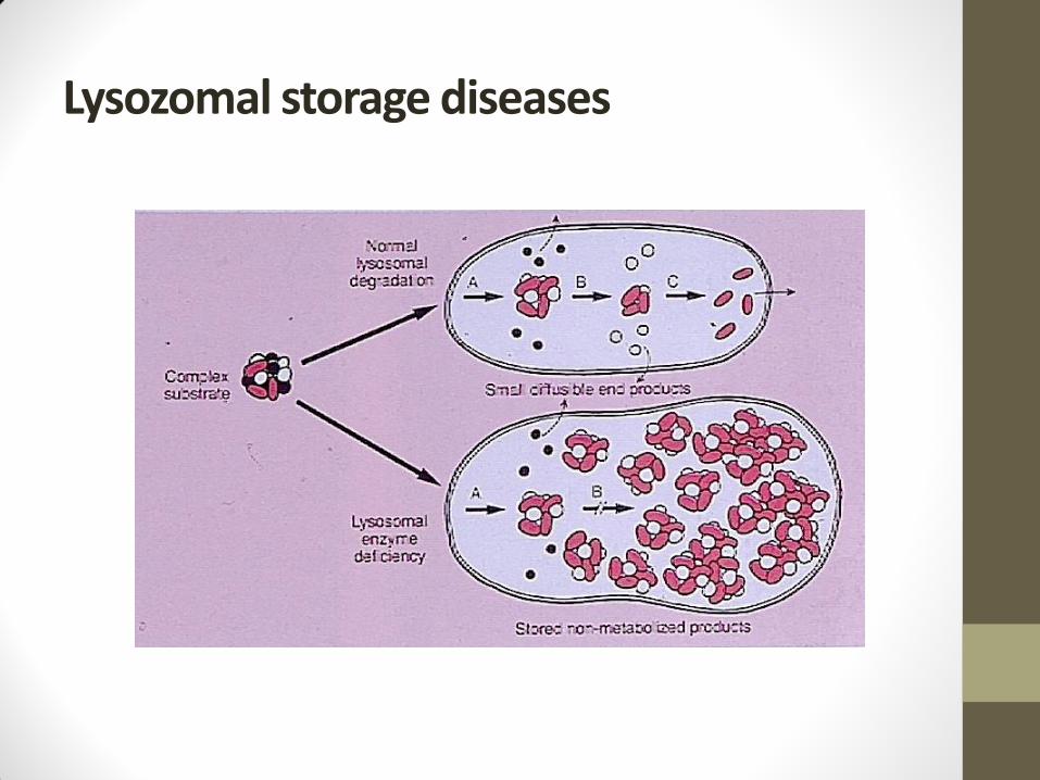
Lysozomal storage diseases

Disorders of metabolism of carbohydrates
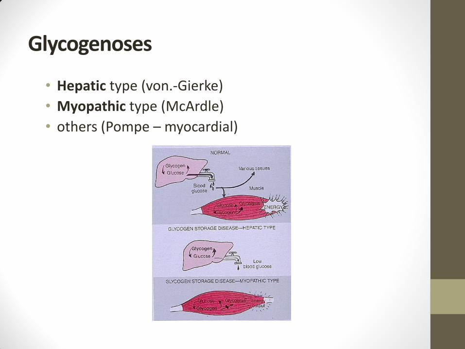
Glycogenoses
• Hepatic type (von.-Gierke)
• Myopathic type (McArdle)
• others (Pompe – myocardial)

Impaired metabolism of water.
• Related to distribution of electrolytes (K+phospates – intracellulary; Na +Cl+bicarbonates +Mg+sulphates – extracellulary)
• Intracellular accumulation of water - vacuolar dystrophy (sponge-like cytoplasm).
• Causes: hypoxia, starving, osmotic effects (osmotic nephrosis), hyperaldosteronism, viroses (herpes).
• Extracellular changes: a) dehydratation
b) hyperhydratation

Dehydratation
• Loss of water (hypertonic dehydratation) – diabetes insipidus
• Loss of water and Na (isotonic dehydratation) – vomiting, diarrhoea, burns
• Loss of Na (hypotonic dehydratation) – impaired resorbtion of Na in kidney, hypoaldosteronism

Hyperhydratation
Hypotonic
Isotonic
Accumulation of fluid: • Oedema
• Hydrops
• Anasarka
• Transsudation (hydrothorax, hydropericardium, ascites).

Oedema
• Venostatic (venous thrombosis, gravitation)
• Cardial (left/right ventricle failure)
• Hypoalbuminotic (low oncotic pressure)
• Lymphostatic (elephantiasis)
• Inflammatory (increased capillary permeability - exsudate)
• Angioneurotic (Quincke)
• Hormonal
• Others (intoxication, hypoxia, etc)

Impaired metabolism of calcium.
Level of Ca (9-11mg%) is in balance with phosphate ionts and is regulated by parathormone, calcitonin and vitamin D.
Impaired metabolism:
• Dystrophic calcification
• Metastatic calcification (hyperparathyroidism, hypovitaminosis D).
• Calcinosis – (skin, muscles = myositis ossificans progressiva, nerves; normální level of Ca).
• Calcifylaxis

Impaired metabolism of iron
• Fe presented in haemoglobin (73%), myoglobin (11%), feritin,enzymes and transferin.
• Absorption in small intestine regulated by apoferitin, efflux of Fe very limited
• Improper acculumation of Fe leads to:
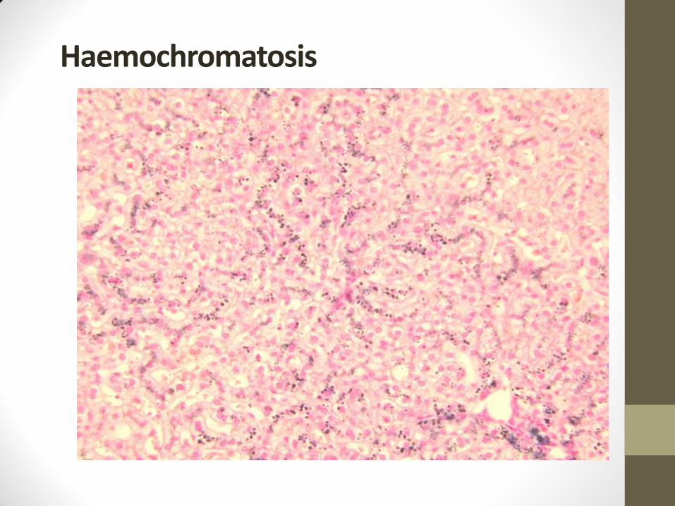
Haemochromatosis – geneticaly related impaired absorption of Fe (50-times increase). Deposition of haemosiderin in skin and pancreas (bronze diabetes), liver (pigmented cirrhosis), heart (failure), salivary glands, kidney, smooth muscle
Haemosiderosis – hemolysis, increased intake of Fe
Haemochromatosis

Haemochromatosis

Impaired metabolism of copper
Accumulation of copper due to insufficient production of ceruloplasmin : hepatolenticular degeneration - liver cirrhosis; destruction of cells in basal ganglia, damage of proximal tubuli in kidney, Kaiser-Fleischer ring in cornea.
2 clinical variants:
Wilson disease – damage of liver, extrapyramid symptoms, dementia, start since childhood.
Westfal-Strümpell pseudosclerosis – small neurological symptoms after puberty

Crystals
• Uric acid – arthritis uratica (gout) – impaired metabolism of purines
• Oxalates – colourless crystals in renal tubuli or myocardium in oxalosis
• Cholesterol – atherosclerosis, postinflammatory
• Paraprotein – renal tubuli in plasmocytoma
• Cystine – cystinosis (Lignac-Fanconi disease) – spleen, lymph nodes, kidnely, cornea
• Charcot-Leyden crystals – destruction of eosinophils

Gout


Concrements
Various tissues :
Gallbladder, bile ducts, uropoietic system, salivary glands, pancreas, prostate
Three main factors modulating concrement development:
• Increases concentration of substance
• Changes in colloid millieu (inflammation)
• Changes of pH (urine)
• Clinical consequences: obstruction of duct
Pigments
Autogeneous
• melanin (Addison disease, freckle, naevus, malignant melanoma / albinisms, vitiligo, leukoderma)
• lipofuscin
• haemosiderin
• bilirubin
Exogeneous
• pigmentation by respiratory tract, trauma, gastrointestinal tract, blood
• pneumokoniosis, koniofibrosis
• argyrosis – Ag
• chrysocyanosis – Au