Embed Size (px)
Citation preview

Disorders of Hemostasis
Dr. Batizy
11/2/06

Primary Hemostasis
• The platelet contains lysosomes, granules, and trilaminar plasma membrane, microtubules.
• Granules are key in primary hemostasis and contain ADP, Thromboxane, platelet factor 4, adhesive and aggregation glycoproteins, coagulation factors, and fibrinolytic inhibitors

Primary Hemostasis
• Dependent on Platelets and Von Willebrand Factor (vWF)
• Platelets gather and attach to vWF
• Platelets degranulate after attachment and release ADP and Thromboxane which attracts more platelets
• Forms a platelet plug
• Requires endothelial damage to adhere

Secondary Hemostasis
• Platelet aggregation initiates secondary hemostasis through the coagulation cascade
• Coagulation cascade is initiated by the intrinsic or extrinsic pathway
• The final cascade results in fibrin deposition cross-linking platelets and clot formation

The Coagulation Cascade
Common Pathway

A word on clotting factors
• Vitamin K Dependent Factors– Intrinsic Pathway : IX, X– Common Pathway: II– Extrinsic Pathway: VII
• All clotting Factors are produced in liver except vWF/VIII
• VIII produced by the vascular endothelium • Sites of heparin activity
– IIa, IXa, Xa ( major site), XIa, Platelet factor 3

A word on clotting factors
• Factor VIII – A factor by any other name?– Same factor: 3 different activities– VIII:C – antihemophilic or coagulation activity– vWF – supports platelet adhesion and carries
VIII in the blood– VIII:Ag – reacts with rabbit antibodies, relates
to measured plasma level rather than activity
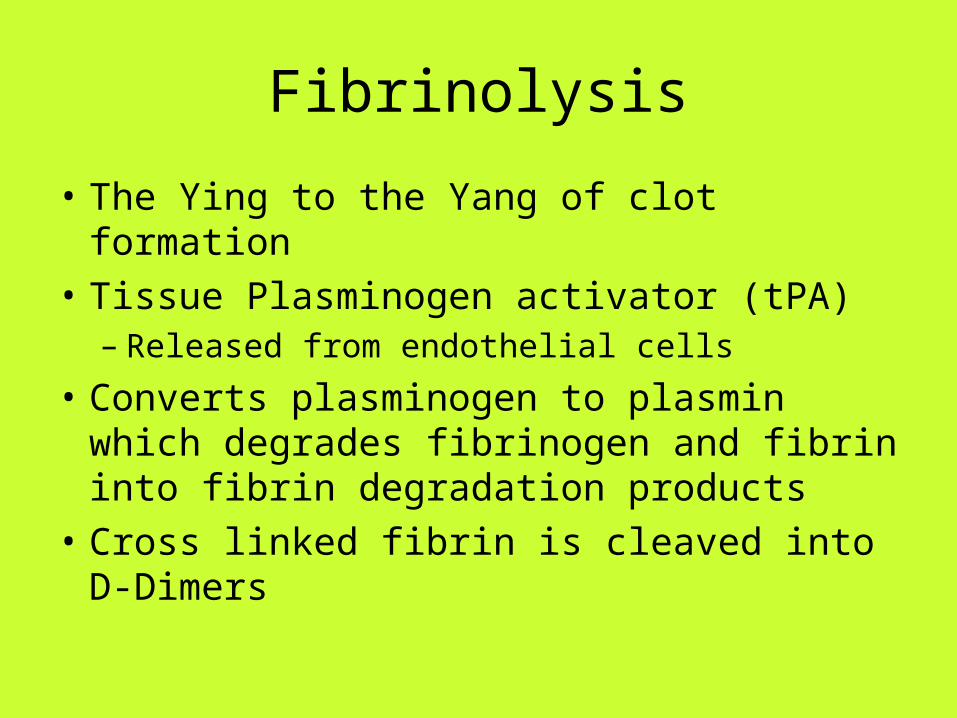
Fibrinolysis
• The Ying to the Yang of clot formation
• Tissue Plasminogen activator (tPA) – Released from endothelial cells
• Converts plasminogen to plasmin which degrades fibrinogen and fibrin into fibrin degradation products
• Cross linked fibrin is cleaved into D-Dimers

Testing the hemostatic system
• CBC– H/H drops often lag behind actual RBC loss due to
slow equilibration
• Blood smear– Schistocytes and fragemented RBC- DIC– Teardrop-shaped or nucleated RBC – Myelophthisic
disease– Characteristic WBC morphologies seen in
thrombocytopenia in infectious mononucleosus, folate, B12 deficiency, or leukemia

Testing the hemostatic system
• Platelet count– Thrombocytopenia : Less that 100,000/mL– Spontaneous bleeding possible: Less than
20,000/mL– Count does not have anything to do with
functionality of platelet

Testing the hemostatic system
• Bleeding time– Tests vascular integrity and platelet function– Incision on volar aspect of the forearm 1mm
deep and 1 cm long– BP cuff inflated to 40 mmHg– Normal < 8 minutes– Borderline 8-10 minutes– Abnormal 10 + minutes– Affected by ASA (permanent) and NSAIDs
Testing the hemostatic system
• Bleeding time– Prolonged with platelet counts below 100,000– When prolonged with platelet count over
100,000 suggests platelet dysfunction

Testing the hemostatic system
• Prothrombin Time– Test of extrinsic and common pathways– International Normalized Ratio used to
compensate for differences in thromboplastin reagents
– Used for coumadin– Elevated in patients with liver disease and
abnormalities in vitamin K sensitive factors

Testing the hemostatic system
• Partial Thromboplastin Time (PTT)– Tests intrinsic and common pathway– Average normal 25-29– Factor levels usually less than 40% to be
affected– Affected by heparin– Can be effected by coumadin at supra-
therapeutic levels due to effects on the common pathway

History and Physical
• Platelet Disorders– More common in
Women– Petechiae, Purpura,
mucosal bleeding
– More commonly acquired
• Coagulation Disorder– More common in Men
– Delayed deep muscle bleeding, hemarthrosis, hematuria
– More commonly congenital

Thrombocytopenia
• Usually mucosal bleeding
• Epistaxis, menorrhagia, and GI bleeding is common
• Trauma does not usually cause bleeding

Thrombocytopenia
• Three mechanisms of Thrombocytopenia– Decreased production
• Usually chemotherapy, myelophthisic disease, or BM effects of alcohol or thiazides
– Splenic Sequesteration• Rare• Results from malignancy, portal hypertension, or
increased Splenic RBC destruction ( hereditary spherocytosis, autoimmune hemolytic anemia)
– Increased Destruction

Thrombocytopenia
• Immune thrombocytopenia– Multiple causes including drugs, lymphoma, leukemia,
collagen vascular disease– Drugs Include
• Digitoxin, sulfonamindes, phenytoin, heparin, ASA, cocaine, Quinine, quinidine, glycoprotein IIb-IIa antagonists
– After stopping drugs platelet counts usually improve over 3 to 7 days
– Prednisone (1mg/kg) with rapid taper can shorten course

Thrombocytopenia
• HIT– Important Immunologic Thrombocytopenia– Usually within 5-7 days of Initiation of Heparin
Therapy but late onset cases are 14-40 days– Occurrence 1-5% with unfractionated heparin
and less than 1% with low molecular-weight heparin
– Thrombotic complications in up to 50% of HIT with loss of limb in 20% and mortality up to 30%

ITP
• Diagnosis of exclusion
• Associated with IgG anti-platelet antibody
• Platelet count falls to less that 20,000

ITP
• Acute Form– Most common in children 2 to 6 years– Viral Prodrome common in the 3 weeks prior– Self Limited and > 90% remission rate– Supportive Treatment– Steroids are not helpful
ITP
• Chronic Form– Adult disease primarily– Women more often than men– Insidious onset with no prodrome– Symptoms include: easy bruising, prolonged
menses, mucosal bleeding– Bleeding complications are unpredictable– Mortality is 1%– Spontaneous remission is rare

ITP
• Chronic Form– Hospitalization common because of a
complex differential diagnosis– Multiple treatments– Platelet transfusions are used only for life
threatening bleeding– Life threatening bleeding is treated with IV
Immune globulin (1g/kg)

TTPHUS
• Exist on a continuum and are likely the same disease
• Diagnosed by a common pentad– Microangiopathic Hemolytic Anemia: Schistocytes
membranes are sheared passing through microthrombi
– Thrombocytopenia: More sever in TTP– Fever– Renal Abnormalities: More prominent in HUS: include
Renal insufficiency, azotemia, proteinuria, hematuria, and renal failure
– Neurologic Abnormalities: hallmark of TTP 1/3 of HUS: Sx of HA, confusion, CN palsies, seizure,coma

TTPHUS
• Labs– PT, PTT, and fibrinogen are within reference
range– Helmet Cells (Shistocytes) are common

TTPHUS
• HUS– Most common in infants and children 6mo - 4
years– Often associated with a prodromal diarrhea– Strongest association to E. coli O157:H7 but
also associated with SSYC as well as multiple virus
– Prognosis• Mortality 5-15%• Younger patients do better

TTPHUS
• HUS– Treatment
• Mostly supportive• Plasma exchange reserved for sever cases• Treat hyperkalemia• Avoid antibiotics with Ecoli
– May actually increase verotoxin production with TMP-SMX
– May be helpful with cases of Shigella dysenteriae

TTPHUS
• TTP– More common in adults– Untreated mortality rate of 80% 1 to 3 months
after diagnosis– Aggressive plasma exchange has dropped
the mortality to 17%– Splenectomy, immune globulin, vincristine all
play a role in therapy

TTPHUS
• AVOID PLATELET TRANSFUSION– May lead to additional microthrombi in
circulation– Transfuse only with life threatening bleeding

Dilutional Thrombocytopenia
• PRBC are platelet poor
• Monitor platelet count with every 10 u PRBC
• Transfuse when count below 50,000
• Get them upstairs before you transfuse 10 units PRBC

DIC
• A few harmless snowflakes working together can create an avalanche of Destruction.

DIC
• Early recognition important secondary to potentially devastating sequelae and effective therapy
• DIC Sequence Platelets and coagulation factors consumed Thrombin directly activates fibrinogen Fibrin deposition Fibrinolysis Inhibition of platelets and fibrin polymerization Decrease in inhibition levels
• Entire process leads to a massive consumption of coagulation factors

DIC
• Life threatening combination of bleeding diathesis with small vessel ischemia
• There are varying levels of acuity • Recommended testing
– Peripheral Smear: Low platelets, schistocytes– Platelet count: Low (<100,000)– Pt, PTT, Thrombin Time: Prolonged– Fibrinogen: Low– Fibrin degredation products: zero to large

DIC
• Treatment– Dependent on whether bleeding or ischemia
predominate– If bleeding
• Platelets, FFP or Cryoprecipitate, and blood recommended
– With Ischemia• Heparin has a place in treatment• Examples include Retained fetus, purpura
fulminans, giant hemangioma, and acute promyelocytic leukemia

DIC
• Treatment– Goal in ER is suspicion, aggressive pursuit of
diagnosis, understanding complications, and rarely initiation of therapy

Coagulation Pathway Defects
• Hemophilia A
• Von Willebrand’s Disease
• Hemophilia B ( Christmas Disease)
Hemophilia A

Hemophilia A
• Variant form of Factor VIII
• 60 to 80 persons per million
• 70% Sex linked recessive
• Severity linked to level of VIII:C activity– 1% Severe– 1%-5% Moderate– 5-10% mild ( little risk of spontaneous
bleeding)

Hemophilia A
• Bleeding can occur anywhere – Deep muscles – Joints– Urinary Tract– Intracranial
• Recurrent Hemarthrosis and progressive join destruction are major cause of morbidity
• Intracranial bleed is major cause of death in all hemophiliacs

Hemophilia A
• Mucosal bleeding is rare unless associated with von Willebrands or Platelet inhibition
• Unlike platelet defects Trauma initiates bleeding
• Bleeding can occur usually by 8 hours but as late as 1 to 3 days after trauma

Hemophilia A
• Management:– Home therapy is increasingly common and
most report to ER only with complicated problems or Trauma
– Hospitals should have files of known hemophiliacs in the area
– Accepted therapy is with Factor VIII replacement or VIII:C
– Newer preparation carry lower risk for Hep B and Hep C transmission

Hemophilia A
• Management:– Multiple guidelines for therapy institution– Most important physician should believe a
patient saying they are bleeding and institute early therapy

Hemophilia A
• Prophylaxis– May require admission for anticipation of
delayed bleeding– Candidates:
• Deep lacerations• Soft tissue injury where hematoma could be
destructive ie: eye, mouth, neck, back, and spinal column

Hemophilia A
• Treatment of haemophilic synovitis– COX-2 important in Hemophiliacs because of
anti=inflammatory,and analgesic properties but they do not affect the platelet fuction
– With withdrawl of rofecoxib from the market celecoxib had become popular
– Study has shown that Celecoxib gives good relief of synovitis without serious adverse effects

Von Willebrand’s Disease
• Most common inherited bleeding disorder
• Without vWF the ability of platelets to adhere is diminished
• VIII:C has diminished activity
• Bleeding sites are primarily mucosal
• Hemarthrosis is rare
• Menorrhagia and GI bleed are common

Von Willebrand’s Disease
• Factor VIII replacement is treatment of choice
• FFP may be given in extreme circumstances
• Desmopressin is only useful for specific types of vWD and should only be give with advice from hematologist

Hemophilia B (Christmas Disease)

Hemophilia B (Christmas Disease)
• Clinically indistinguishable from hemophilia A
• Deficiency of factor IX
• Factor IX preparation used in treatment
• FFP and plasma prothrombin complex are also useful
• Gene manipulation in animals shows promising results for the future

Take home message
• All Bleeding stops…. Eventually

References
• Rosen’s
• Emedicine
• Celecoxib in the treatment of haemophilic synovitis, target joints, and pain in adults and children with haemophilia, Haemophilia (2006), 12, 514-517